
Assessing the Radiological Risks Associated with High Natural Radioactivity of Microgranitic Rocks: A Case Study in a Northeastern Desert of Egypt


 , and
, and
Abstract
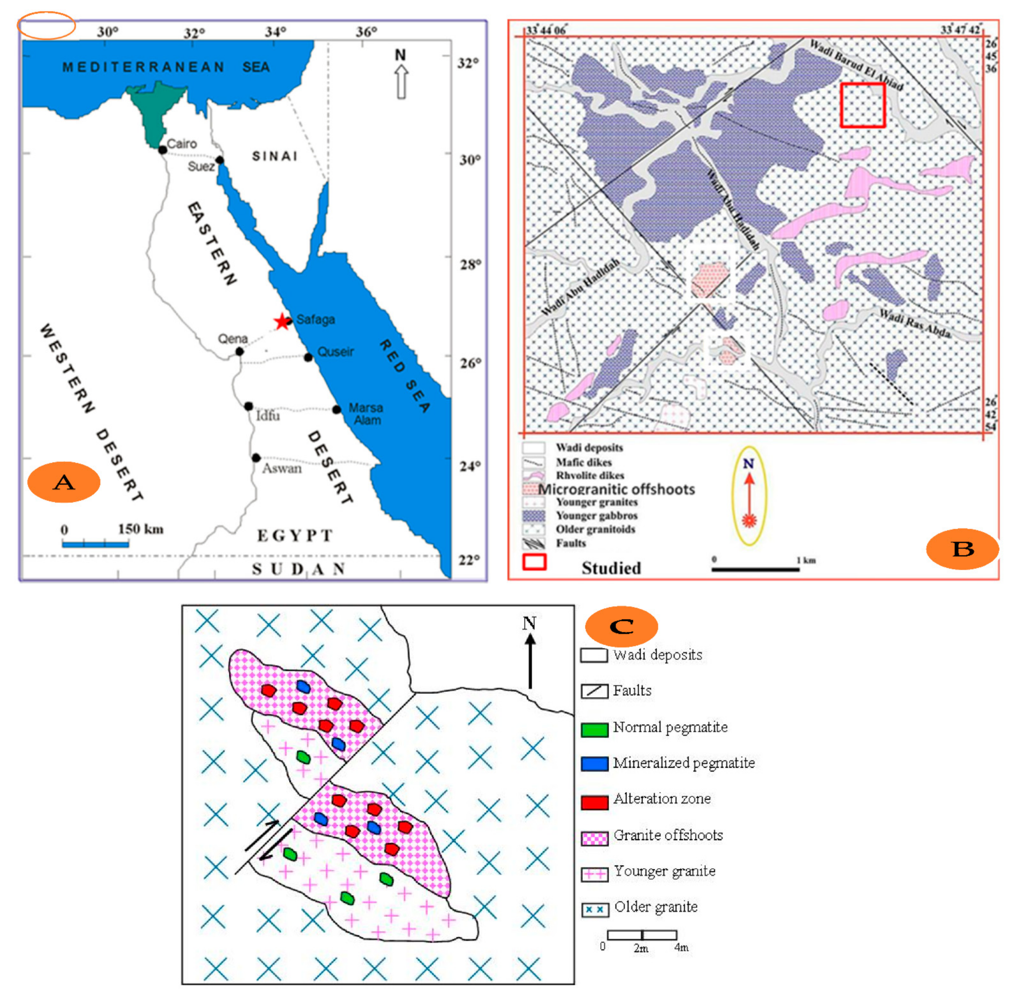
:1. Introduction
2. Methodology
2.1. Samples Preparation and Measurement
- -
- Calibration was started with the 137Cs source (gain adjustment) and then with 57Co source (zero adjustment).
- -
- The 137Cs source was repeated using a minimum procedure.
- (a)
- Equipment was setup using reference gamma-emitting sources (137Cs and 57Co) for energy calibration.
- (b)
- Assaying of the samples using a long period net count, 1000 s for each, in the shielded environment and determination of the gross counts for U, Th, eU (Ra), and K at their selected energy regions as well.
- (c)
- Determination of the background count rates in the selected energy regions (ROIs) for the laboratory with the detector.
- (d)
- The registered spectrometric data (gross counts for eU, eTh, Ra, and K) for each sample were processed by using the computer program “ANALYSIS” with the use of background count rates, sample weight, time of measurement, and the preliminary sensitivity constants to determine the U, Th, and Ra concentrations in parts per million (ppm) and the K concentration in wt%.
2.2. Geochemical Analysis
3. Data Treatment
3.1. Mobilization Type and Quantity
- (a)
- The paleo-uranium background:where U0 = 269.46, and = the average content and = the average regional ratio.
- (b)
- The mobilized amount of uranium (Um): Um = Up − U0, where Um = 26.84 and Up is the average content of uranium.If Um > 0, the uranium migrated into the geological body during late evolution. If Um > 2, a significant amount of uranium migrated in.
- (c)
- The mobilized uranium migration rate: P = × 100%, while P = 9.1%, As Um > 0, the uranium migrated in during the late evolution at a 9.1% migration rate. It was redistributed during or after alteration.
3.2. Radium Equivalent Activity
3.3. Absorbed Dose Rate in the Air ()
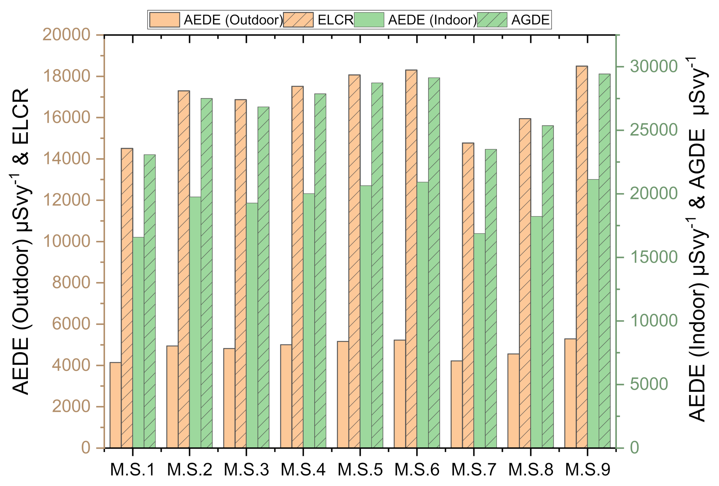
3.4. Annual Effective Dose Equivalent (AEDE)
3.5. External and Internal Hazard Index (Hex)
3.6. Gamma Activity Concentration Index (Iγ)
3.7. Annual Gonadal Dose Equivalent (AGDE)
3.8. Excess Lifetime Cancer Risk (ELCR)
3.9. Effective Dose Rate—Dorgan
4. Results
4.1. Radioactivity
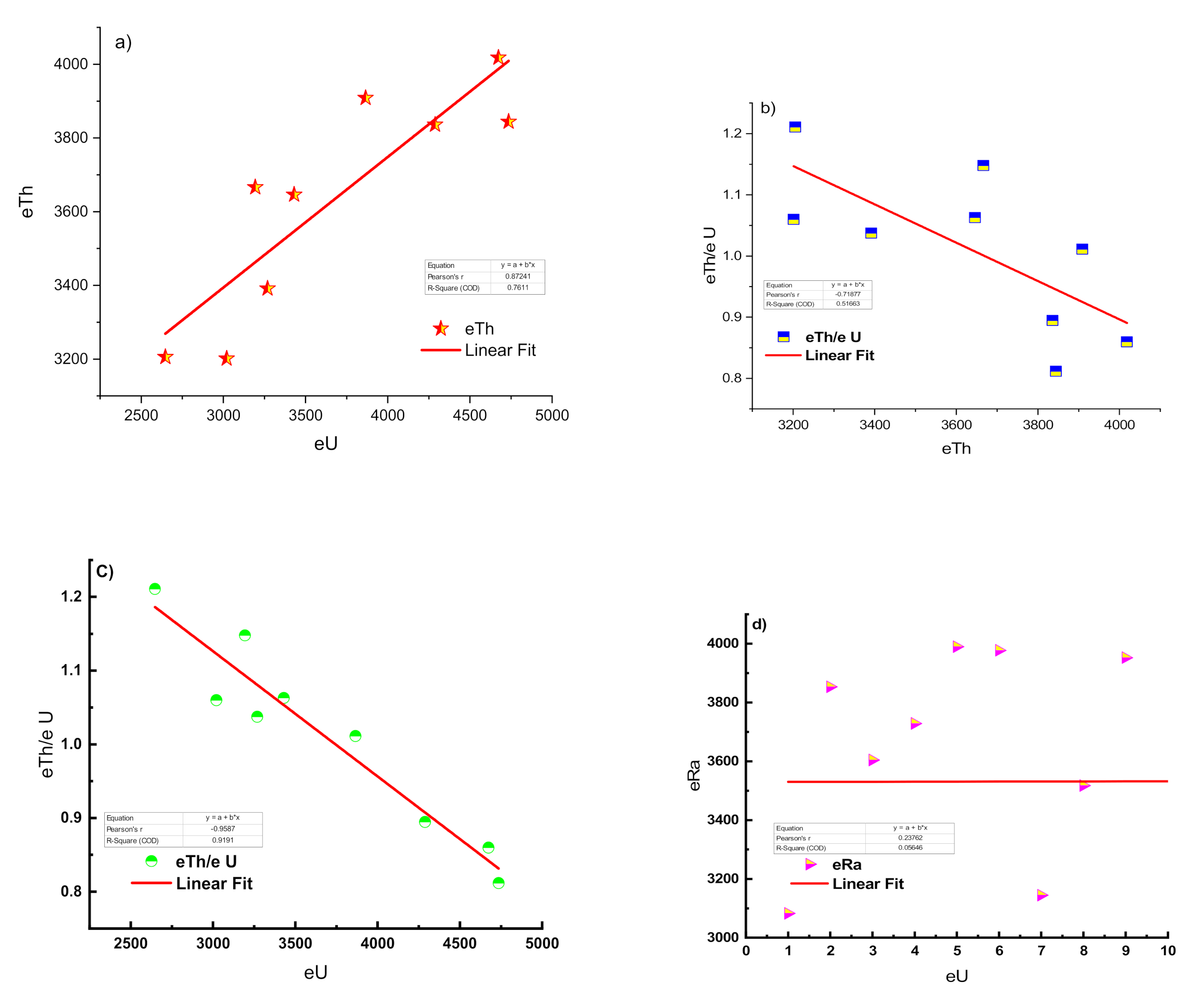
4.2. Radioactive Equilibrium
4.3. Uranium Mobilization and Migration
5. Discussion
6. Conclusions
Author Contributions
Funding
Institutional Review Board Statement
Informed Consent Statement
Data Availability Statement
Acknowledgments
Conflicts of Interest
References
- Rogers, J.J.W.; Adams, J.S.S. Uranium. In Handbook of Geochemistry; Wedepohl, K.H., Ed.; Springer: New York, NY, USA, 1969. [Google Scholar]
- Bowie, S.H.U.; Plant, J. Natural radioactivity in the environment. In Applied Environmental Geochemistry; Institute of Geological Sciences: London, UK, 1983. [Google Scholar]
- Shurmann, H.M.E. The Precambrian along the Gulf of Suez and the Northern Part of the Red Sea; EJ Brill: Leiden, The Netherlands, 1966; pp. 122–142. [Google Scholar]
- Awad, H.A.M.; Zakaly, H.M.H.; Nastavkin, A.V.; El-Taher, A. Radioactive content and radiological implication in granitic rocks by geochemical data and radiophysical factors, Central Eastern Desert, Egypt. Int. J. Environ. Anal. Chem. 2020. [Google Scholar] [CrossRef]
- Sabet, A.; AH, S. Problems of geological and economic evaluation of tantalium deposits in apogranites during stages of prospection. Ann. Geol. Surv. 1973, 3, 87–107. [Google Scholar]
- Omran, A.A. Geology, mineralogy and radioelements potentialiyy of microgranite dikes to the south of wadi Abu Hadieda area, Northern Eastern Desert, Egypt. Al-Azhar Bull. Sci. 2014, 25, 47–62. [Google Scholar] [CrossRef]
- Abed, N.S.; Monsif, M.A.; Zakaly, H.M.H.; Awad, H.A. Geological and Mineralogical Investigations of Microgranites at the Southeastern Part of Wadi Baroud, North Eastern Desert, Egypt. J. Rad. Nucl. Appl. 2021, 6, 135–149. [Google Scholar] [CrossRef]
- Abdel Hamid, A.A.A.; El Sundoly, H.I.; Abu Steet, A.A. Hydrothermal alteration and evolution of Zr-Th-U-REE mineralization in the microgranite of Wadi Ras Abda, North Eastern Desert, Egypt. Arab. J. Geosci. 2018, 11, 273. [Google Scholar] [CrossRef]
- Abd El Monsif, M. Comparason between Ras Baroud and Abu Hadeida Granites, Northern Eastern Desert, Egypt. To Determine the More Evolved One. Int. J. Min. Sci. 2020, 6, 30–45. [Google Scholar] [CrossRef]
- Monsif, M.A. Geochemistry and Radioactive Mineralization Inspections of the Pegmatite Bodies Associating Variable Granitic Environs, Abu Hadeida Area, North Eastern Desert, Egypt. J. Basic Environ. Sci. 2021, 8, 1–14. [Google Scholar]
- Beus, A.A. Metallogeny of Precambrian Rare-Metal Granitoids. Rev. Bras. Geociências 1982, 12, 410–413. [Google Scholar]
- Awad, H.A.; Zakaly, H.M.H.; Nastavkin, A.V.; El-Taher, A. Radioactive content in the investigated granites by geochemical analyses and radiophysical methods around Um Taghir, Central Eastern Desert, Egypt. J. Phys. Conf. Ser. 2020, 1582, 12007. [Google Scholar] [CrossRef]
- Omran, A.A. Geological, petrochemical studies and potentiality of uranium-thorium occurrences in Gabal Um Taghir El-Tahtani area with emphasis on the granitic rocks, Central Eastern Desert, Egypt. Ph.D. Thesis, Ain Shams University, Cairo, Egypt, 2005. [Google Scholar]
- EL Hadary, A.; Soliman, A.; Omran, A. Contributions to the Geology and Mineralogy of wadi Ras Abda area, North Eastern desert, Egypt. Nucl. Sci. Sci. J. 2015, 4, 47–59. [Google Scholar] [CrossRef]
- Matolin, M. Construction and use of spectrometric calibration pads laboratory γ-ray spectrometry, NMA, Egypt. In A Report to the Government of the Arab Republic of Egypt; IAEA: Vienna, Austria, 1991. [Google Scholar]
- El-Arabi, A.M.; Abbady, A.G.; Khalifa, I.H. Radioactive and Geochemistry Characteristics of the Garnetiferous Granite of Um Sleimat Area, Egypt. Online J. Earth Sci. 2007, 1, 9–20. [Google Scholar]
- Tufail, M.; Ahmad, N.; Mirza, S.M.; Mirza, N.M.; Khan, H.A. Natural Radioactivity from the Building Materials Used in Islamabad and Rawalpindi, Pakistan; Elsevier: Amsterdam, The Netherlands, 1992; Volume 121, pp. 283–291. [Google Scholar]
- Zakaly, H.M.; Uosif, M.A.; Madkour, H.; Tammam, M.; Issa, S.; Elsaman, R.; El-Taher, A. Assessment of natural radionuclides and heavy metal concentrations in marine sediments in view of tourism activities in Hurghada city, northern Red Sea, Egypt. J. Phys. Sci. 2019, 30, 21–47. [Google Scholar] [CrossRef]
- Örgün, Y.; Altinsoy, N.; Şahin, S.Y.; Güngör, Y.; Gültekin, A.H.; Karahan, G.; Karacik, Z. Natural and anthropogenic radionuclides in rocks and beach sands from Ezine region (Çanakkale), Western Anatolia, Turkey. Appl. Radiat. Isot. 2007, 65, 739–747. [Google Scholar] [CrossRef]
- UNSCEAR. Sources and Effects of Ionizing Radiation; United Nations Scientific Committee on the Effects of Atomic Radiation (UNSCEAR) 2000 Report; UNSCEAR: Vienna, Austria, 2010; Volume 1, ISBN 9789211422740. [Google Scholar]
- Uosif, M.A.M.; Hashim, M.; Issa, S.; Tamam, M.; Zakaly, H.M. Natural Radionuclides and Heavy Metals Concentration of Marine Sediments in Quseir City and Surrounding Areas, Red Sea Coast-Egypt. Int. J. Adv. Sci. Technol. 2016, 86, 9–30. [Google Scholar] [CrossRef]
- Kalaitzis, A.; Stoulos, S.; Melfos, V.; Kantiranis, N.; Filippidis, A. Application of zeolitic rocks in the environment: Assessment of radiation due to natural radioactivity. J. Radioanal. Nucl. Chem. 2019, 319, 975–985. [Google Scholar] [CrossRef]
- Abbasi, A.; Zakaly, H.M.H.; Mirekhtiary, F. Baseline levels of natural radionuclides concentration in sediments East coastline of North Cyprus. Mar. Pollut. Bull. 2020, 161, 111793. [Google Scholar] [CrossRef]
- Saito, K.; Jacob, P. Gamma Ray Fields in the Air Due to Sources in the Ground. Radiat. Prot. Dosim. 1995, 58, 29–45. [Google Scholar] [CrossRef]
- Nada, A. Evaluation of Natural Radionuclides at Um-Greifat Area, Eastern Desert of Egypt; Elsevier: Amsterdam, The Netherlands, 2003; Volume 58. [Google Scholar]
- Baykara, O.; Karatepe, Ş.; Doǧru, M. Assessments of natural radioactivity and radiological hazards in construction materials used in Elazig, Turkey. Radiat. Meas. 2011, 46, 153–158. [Google Scholar] [CrossRef]
- Lakehal, C.; Ramdhane, M.; Boucenna, A. Natural radionuclide concentrations in two phosphate ores of east Algeria. J. Environ. Radioact. 2010, 101, 377–379. [Google Scholar] [CrossRef] [PubMed]
- Nuclear Energy Agency. Exposure to Radiation from the Natural Radioactivity in Building Materials: Report; NEA Group of Experts OECD: Paris, France, 1979. [Google Scholar]
- Abbady, A.G.E.; Uosif, M.A.M.; El-Taher, A. Natural radioactivity and dose assessment for phosphate rocks from Wadi El-Mashash and El-Mahamid Mines, Egypt. J. Environ. Radioact. 2005, 84, 65–78. [Google Scholar] [CrossRef]
- El Galy, M.M.; El Mezayn, A.M.; Said, A.F.; El Mowafy, A.A.; Mohamed, M.S. Distribution and environmental impacts of some radionuclides in sedimentary rocks at Wadi Naseib area, southwest Sinai, Egypt. J. Environ. Radioact. 2008, 99, 1075–1082. [Google Scholar] [CrossRef]
- El Aassy, I.E.; Nada, A.A.; El Galy, M.M.; El Feky, M.G.; Abd El Maksoud, T.M.; Talaat, S.M.; Ibrahim, E.M. Behavior and environmental impacts of radionuclides during the hydrometallurgy of calcareous and argillaceous rocks, southwestern Sinai, Egypt. Appl. Radiat. Isot. 2012, 70, 1024–1033. [Google Scholar] [CrossRef] [PubMed]
- Arafa, W. Specific activity and hazards of granite samples collected from the Eastern Desert of Egypt. J. Environ. Radioact. 2004, 75, 315–327. [Google Scholar] [CrossRef] [PubMed]
- O’brien, R.; Sanna, R. The distribution of absorbed dose-rates in humans from exposure to environmental gamma rays. Health Phys. 1976, 30, 71–78. [Google Scholar] [CrossRef]
- Awad, H.A.; Zakaly, H.M.H.; Nastavkin, A.V.; El Tohamy, A.M.; El-Taher, A. Radioactive mineralizations on granitic rocks and silica veins on shear zone of El-Missikat area, Central Eastern Desert, Egypt. Appl. Radiat. Isot. 2021, 168, 109493. [Google Scholar] [CrossRef]
- Zakaly, H.M.H.; Uosif, M.A.M.; Issa, S.A.M.; Tekin, H.O.; Madkour, H.; Tammam, M.; El-Taher, A.; Alharshan, G.A.; Mostafa, M.Y.A. An extended assessment of natural radioactivity in the sediments of the mid-region of the Egyptian Red Sea coast. Mar. Pollut. Bull. 2021, 171, 112658. [Google Scholar] [CrossRef] [PubMed]
- Hussein, A.H. Lecture Course in Nuclear Geology; NMA: Cairo, Egypt, 1978; 101p. [Google Scholar]
- El-Galy, M.M. Geology, Radioactivity, Geochemistry and Tectonic Setting of Selected Granitic Rocks, West Gulf of Aqaba, Sinai, Egypt. Ph.D. Thesis, Tanta University, Tanta, Egypt, 1998. [Google Scholar]
- Hansink, J.D. Equilibrium Analysis of a Sandstone Roll-Front Uranium Deposit; INIS IAEA Organization; Rocky Mountain Energy Co.: Denver, CO, USA, 1976; pp. 683–693. [Google Scholar]
- Charbonneau, B.W. Radiometric study of three radioactive granites in the Canadian Shield: Elliot Lake, Ontario; Fort Smith, and Fury and Hecla, NWT. Uranium Granites 1982, 15, 91–99. [Google Scholar]
- El Galy, M.M.; El Feky, M.G.; Roz, M.E.; Nada, A. Disequilibrium in U-Series at G-II Occurrence of Uranium Mineralization at Gabal Gattar Area, North Eastern Desert, Egypt; A Comparative Study using HP Ge and Nal-Detectors. Arab J. Nucl. Sci. Appl. 2007, 40, 15–26. [Google Scholar]
- Awad, H.A.M.; Zakaly, H.M.H.; Nastavkin, A.V.; El-Taher, A. Radiological implication of the granitoid rocks and their associated jasperoid veins, El-Missikat area, Central Eastern Desert, Egypt. Int. J. Environ. Anal. Chem. 2020. [Google Scholar] [CrossRef]
- Deymar, S.; Yazdi, M.; Rezvanianzadeh, M.R.; Behzadi, M. Alkali metasomatism as a process for Ti–REE–Y–U–Th mineralization in the Saghand Anomaly 5, Central Iran: Insights from geochemical, mineralogical, and stable isotope data. Ore Geol. Rev. 2018, 93, 308–336. [Google Scholar] [CrossRef]
- Saunders, A.D.; Norry, M.J.; Tarney, J. Fluid influence on the trace element compositions of subduction zone magmas. Philos. Trans. R. Soc. Lond. Ser. A Phys. Eng. Sci. 1991, 335, 377–392. [Google Scholar] [CrossRef]
- Bailey, E.H.; Vala Ragnarsdottir, K. Uranium and thorium solubilities in subduction zone fluids. Earth Planet. Sci. Lett. 1994, 124, 119–129. [Google Scholar] [CrossRef]
- Tawfic, A.F.; Zakaly, H.M.H.; Awad, H.A.; Tantawy, H.R.; Abbasi, A.; Abed, N.S.; Mostafa, M. Natural radioactivity levels and radiological implications in the high natural radiation area of Wadi El Reddah, Egypt. J. Radioanal. Nucl. Chem. 2021, 327, 643–652. [Google Scholar] [CrossRef]
- Oyeyemi, K.D.; Usikalu, M.R.; Aizebeokhai, A.P.; Achuka, J.A.; Jonathan, O. Measurements of radioactivity levels in part of Ota Southwestern Nigeria: Implications for radiological hazards indices and excess lifetime cancer-risks. In J. Phys. Conf. Ser.; Institute of Physics Publishing: Philadelphia, PA, USA, 2017; Volume 852. [Google Scholar]
- Powell, B.A.; Hughes, L.D.; Soreefan, A.M.; Falta, D.; Wall, M.; DeVol, T.A. Elevated concentrations of primordial radionuclides in sediments from the Reedy River and surrounding creeks in Simpsonville, South Carolina. J. Environ. Radioact. 2007, 94, 121–128. [Google Scholar] [CrossRef] [PubMed] [Green Version]
- Alfonso, J.A.; Pérez, K.; Palacios, D.; Handt, H.; LaBrecque, J.J.; Mora, A.; Vásquez, Y. Distribution and environmental impact of radionuclides in marine sediments along the Venezuelan coast. J. Radioanal. Nucl. Chem. 2014, 300, 219–224. [Google Scholar] [CrossRef]
- Manigandan, P.K.; Chandar Shekar, B. Evaluation of radionuclides in the terrestrial environment of Western Ghats. J. Radiat. Res. Appl. Sci. 2014, 7, 310–316. [Google Scholar] [CrossRef] [Green Version]
- Qureshi, A.A.; Tariq, S.; Din, K.U.; Manzoor, S.; Calligaris, C.; Waheed, A. Evaluation of excessive lifetime cancer risk due to natural radioactivity in the rivers sediments of Northern Pakistan. J. Radiat. Res. Appl. Sci. 2014, 7, 438–447. [Google Scholar] [CrossRef] [Green Version]
- Lee, K.Y.; Yoon, Y.Y.; Cho, S.Y.; Ko, K.S.; Kim, Y. Regional characteristics of naturally occurring radionuclides in surface sediments of Chinese deserts and the Keum River area of Korea. J. Radioanal. Nucl. Chem. 2009, 281, 287–290. [Google Scholar] [CrossRef]
- El-Taher, A.; Al-Zahrani, J.H. Radioactivity measurements and radiation dose assessments in soil of Al-Qassim region, Saudi Arabia. Indian J. Pure Appl. Phys. 2014, 52, 147–154. [Google Scholar]
- Kapdan, E.; Karahan, A. Radioactivity Levels and Health Risks due to Radionuclides in the Soil of Yalova, Northwestern Turkey. Int. J. Environ. Res. 2011, 5, 837–846. [Google Scholar]
- Asgharizadeh, F.; Ghannadi, M.; Samani, A.B.; Meftahi, M.; Shalibayk, M.; Sahafipour, S.A.; Gooya, E.S. Natural radioactivity in surface soil samples from dwelling areas in Tehran city, Iran. Radiat. Prot. Dosim. 2013, 156, 376–382. [Google Scholar] [CrossRef]
- Wang, Z.; He, J.; Du, Y.; He, Y.; Li, Z.; Chen, Z.; Yang, C. Natural and artificial radionuclide measurements and radioactivity assessment of soil samples in eastern Sichuan province (China). Radiat. Prot. Dosim. 2012, 150, 391–397. [Google Scholar] [CrossRef]
- Agbalagba, E.O.; Onoja, R.A. Evaluation of natural radioactivity in soil, sediment and water samples of Niger Delta (Biseni) flood plain lakes, Nigeria. J. Environ. Radioact. 2011, 102, 667–671. [Google Scholar] [CrossRef]
- Dabayneh, K.M.; Mashal, L.A.; Hasan, F.I. Radioactivity concentration in soil samples in the southern part of the West Bank, Palestine. Radiat. Prot. Dosim. 2008, 131, 265–271. [Google Scholar] [CrossRef] [PubMed]
- Kolo, M.T.; Aziz, S.A.B.A.; Khandaker, M.U.; Asaduzzaman, K.; Amin, Y.M. Evaluation of radiological risks due to natural radioactivity around Lynas Advanced Material Plant environment, Kuantan, Pahang, Malaysia. Environ. Sci. Pollut. Res. 2015, 22, 13127–13136. [Google Scholar] [CrossRef] [PubMed]
- UNSCEAR. Effects of Ionizing Radiation: Report to the General Assembly, with Scientific Annexes World 2008; United Nations Scientific Committee on the Effects of Atomic Radiation: New York, NY, USA, 2010. [Google Scholar]
- Ghoneim, M.M.; Panova, E.G.; Abdel Gawad, A.E.; Awad, H.A.; Zakaly, H.M.H.; El-Taher, A. Analytical methodology for geochemical features and radioactive elements for intrusive rocks in El Sela area, Eastern Desert, Egypt. Int. J. Environ. Anal. Chem. 2021, in press. [Google Scholar] [CrossRef]
- Adams, J.A.S.; Osmond, J.K.; Rogers, J.J.W. The geochemistry of thorium and uranium. Phys. Chem. Earth 1959, 3, 298–348. [Google Scholar] [CrossRef]
- Turekirn, K.K.; Wedepohe, K.H. Distribution of the elements in some major of the Earth’s crust. Bull. Geol. Soc. Am. Bull. 1961, 72, 175–192. [Google Scholar] [CrossRef]
- El-Taher, A.; Zakaly, H.M.H.; Elsaman, R. Environmental implications and spatial distribution of natural radionuclides and heavy metals in sediments from four harbours in the Egyptian Red Sea coast. Appl. Radiat. Isot. 2018, 131, 13–22. [Google Scholar] [CrossRef]
- Gezer, F.; Turhan, Ş.; Kurnaz, A.; Ufuktepe, Y. Radiometric characterization of zeolite minerals used in many industries and assessment of radiological risks. Appl. Radiat. Isot. 2019, 152, 57–63. [Google Scholar] [CrossRef]


| Activity | 238U | 226Ra | 232Th | 40K | U Chem. (Bq/kg) | Uc/Ur | eU/Ra | eU/eTh | eTh/eU |
|---|---|---|---|---|---|---|---|---|---|
| Sample No: | |||||||||
| M.S.1 | 3020 | 3082 | 3201 | 566 | - | - | 0.98 | 0.94 | 1.06 |
| M.S.2 | 3430 | 3853 | 3646 | 1162 | 9943 | 2.90 | 0.89 | 0.94 | 1.06 |
| M.S.3 | 3194 | 3604 | 3666 | 1203 | 7457 | 2.33 | 0.89 | 0.87 | 1.15 |
| M.S.4 | 4288 | 3729 | 3836 | 1028 | 9943 | 2.32 | 1.15 | 1.12 | 0.89 |
| M.S.5 | 4735 | 3990 | 3844 | 1082 | 14,915 | 3.15 | 1.19 | 1.23 | 0.81 |
| M.S.6 | 3865 | 3977 | 3909 | 1619 | 10,689 | 2.77 | 0.97 | 0.99 | 1.01 |
| M.S.7 | 2647 | 3145 | 3205 | 1255 | 12,429 | 4.69 | 0.84 | 0.83 | 1.21 |
| M.S.8 | 3269 | 3517 | 3391 | 1038 | 24,858 | 7.60 | 0.93 | 0.96 | 1.04 |
| M.S.9 | 4673 | 3952 | 4018 | 1379 | 9943 | 2.13 | 1.18 | 1.16 | 0.86 |
| Mean | 3680 | 3650 | 3635 | 1148 | - | - | - | - | - |
| Mean | SD | Skewness | Kurtosis | Minimum | Maximum | |
|---|---|---|---|---|---|---|
| 238U (Bq/kg) | 3680 | 748 | 0.31 | −1.35 | 2647 | 4735 |
| 226Ra (Bq/kg) | 3650 | 346 | −0.79 | −0.76 | 3082 | 3990 |
| 232Th (Bq/kg) | 3635 | 304 | −0.47 | −1.31 | 3201 | 4018 |
| 40K (Bq/kg) | 1148 | 287 | −0.56 | 2.03 | 566 | 1619 |
| Radium equivalent (Raeq (Bq/kg) | 8937 | 782 | −0.66 | −1.02 | 7704 | 9804 |
| Absorbed dose rate (DR; nGy/h) | 3929 | 345 | −0.67 | −1.00 | 3381 | 4309 |
| Gamma index (Iγ) | 30.6 | 2.68 | −0.66 | −1.03 | 26.4 | 33.6 |
| Activity utilization index (I) | 148 | 13.2 | −0.69 | −0.94 | 127 | 162 |
| Excess lifetime cancer risk (ELCR) | 16,865 | 1482 | −0.67 | −1.00 | 14,512 | 18,497 |
| Annual gonadal equivalent (AGDE) | 26,834 | 2359 | −0.67 | −1.00 | 23,084 | 29,441 |
| AEDEindoor (µSv/y) | 19,274 | 1694 | −0.67 | −1.00 | 16,585 | 21,140 |
| AEDEoutdoor (µSv/y) | 4819 | 424 | −0.67 | −1.00 | 4146 | 5285 |
| Hex | 24.1 | 2.11 | −0.66 | −1.02 | 20.8 | 26.5 |
| Hin | 34.0 | 3.04 | −0.72 | −0.92 | 29.1 | 37.2 |
| Clark ratio (232Th/238U) | 1.00 | 0.03 | −0.37 | −1.65 | 0.95 | 1.04 |
| Country | Region | 238U (Bq/kg) | 232Th (Bq/kg) | 40K (Bq/kg) | Raeq (Bq/kg) | DR (nGy/h) | AEDEindoor (mSv/y) | AEDEoutdoor (mSv/y) | Type of Samples | Reference |
|---|---|---|---|---|---|---|---|---|---|---|
| USA | South Carolina | 37.8 | 45.3 | 609 | NA | NA | NA | NA | River sediments | Powell et al., (2007) [47] |
| Venezuela | Venezuelan coast | 11.4 | 14.5 | 153 | 44.3 | 20.6 | NA | NA | Marine sediments | Alfonso et al., (2014) [48] |
| India | Western Ghats | 36.3 | 108 | 232 | 208 | 133 | 0.449 | 0.112 | Soil samples | Manigandan and Chandar Shekar (2014) [49] |
| Egypt | El-Missikat area | 162 | 72.6 | 702 | 320 | 147.5 | 0.723 | 0.181 | Granitic rocks | Awad et al., (2020, 2021) [34,41] |
| Pakistan | Northern Pakistan | 50.7 | 70.2 | 514 | 191 | 87.5 | 0.810 | 0.110 | River’s sediments | Qureshi et al., (2014) [50] |
| Korea | Keum River area | 65.7 | 91.1 | 1005 | NA | NA | NA | NA | Stream sediments | Lee et al., (2009) [51] |
| Saudi Arabia | Qassim | 9.30 | 12.3 | 535 | 68.1 | 35.2 | 0.173 | 0.040 | Soil | El-Taher and Al-Zahrani (2014) [52] |
| Turkey | Northwestern Turkey | 22.3 | 26.8 | 419 | NA | 84.3 | 0.104 | NA | Soil samples | Kapdan and Karahan (2011) [53] |
| Iran | Tehran city | 38.8 | 43.4 | 555 | 144 | 69.1 | NA | 0.080 | Soil samples | Asgharizadeh et al., (2013) [54] |
| China | Eastern Sichuan | 26.0 | 49 | 440 | 130 | 60.0 | 0.074 | NA | Soil samples | Wang et al., (2012) [55] |
| Nigeria | Niger Delta | 18.0 | 22 | 210 | 76 | 30.0 | NA | 0.037 | Soil, sediment and water samples | Agbalagba and Onoja (2011) [56] |
| Palestine | West Bank | 68.7 | 48 | 630 | 186 | 88.2 | 0.61 | 0.110 | Soil samples | Dabayneh et al., (2008) [57] |
| Malaysia | Kuantan | 6.57 | 10.6 | 41.0 | 24.9 | 11.2 | NA | 0.010 | Soil samples | Kolo et al., (2015) [58] |
| Egypt | South Baroud | 3680 | 3635 | 1148 | 8937 | 3929 | 19.3 | 4.819 | Granitic rocks | Present study |
| Worldwide average | 33.0 | 45.0 | 412 | 370 | 58.0 | 0.41 | 0.070 | - | UNSCEAR (2008) [59] | |
Publisher’s Note: MDPI stays neutral with regard to jurisdictional claims in published maps and institutional affiliations. |
© 2022 by the authors. Licensee MDPI, Basel, Switzerland. This article is an open access article distributed under the terms and conditions of the Creative Commons Attribution (CC BY) license (https://creativecommons.org/licenses/by/4.0/).
Share and Cite
Abed, N.S.; Monsif, M.A.; Zakaly, H.M.H.; Awad, H.A.; Hessien, M.M.; Yap, C.K. Assessing the Radiological Risks Associated with High Natural Radioactivity of Microgranitic Rocks: A Case Study in a Northeastern Desert of Egypt. Int. J. Environ. Res. Public Health 2022, 19, 473. https://doi.org/10.3390/ijerph19010473
Abed NS, Monsif MA, Zakaly HMH, Awad HA, Hessien MM, Yap CK. Assessing the Radiological Risks Associated with High Natural Radioactivity of Microgranitic Rocks: A Case Study in a Northeastern Desert of Egypt. International Journal of Environmental Research and Public Health. 2022; 19(1):473. https://doi.org/10.3390/ijerph19010473
Chicago/Turabian StyleAbed, Neveen S., Mohamed Abdel Monsif, Hesham M. H. Zakaly, Hamdy A. Awad, Mahmoud M. Hessien, and Chee Kong Yap. 2022. "Assessing the Radiological Risks Associated with High Natural Radioactivity of Microgranitic Rocks: A Case Study in a Northeastern Desert of Egypt" International Journal of Environmental Research and Public Health 19, no. 1: 473. https://doi.org/10.3390/ijerph19010473