Article

Molecular Analysis of Pathogenicity, Adhesive Matrix Molecules (MSCRAMMs) and Biofilm Genes of Coagulase-Negative Staphylococci Isolated from Ready-to-Eat Food

by
Wioleta Chajęcka-Wierzchowska
1,*,
Joanna Gajewska
1,
Arkadiusz Józef Zakrzewski
1,
Cinzia Caggia
2 and
Anna Zadernowska
1
1
Department of Industrial and Food Microbiology, Faculty of Food Science, University of Warmia and Mazury in Olsztyn, 10-693 Olsztyn, Poland
2
Department of Agriculture, Food and Environment (Di3A), University of Catania, Via Santa Sofia 100, 95123 Catania, Italy
*
Author to whom correspondence should be addressed.
Int. J. Environ. Res. Public Health 2023, 20(2), 1375; https://doi.org/10.3390/ijerph20021375
Submission received: 24 October 2022 / Revised: 3 January 2023 / Accepted: 9 January 2023 / Published: 12 January 2023
(This article belongs to the Special Issue Food Safety and Human Health)

Abstract

:
This paper provides a snapshot on the pathogenic traits within CoNS isolated from ready-to-eat (RTE) food. Eighty-five strains were subjected to biofilm and slime production, as well as biofilm-associated genes (icaA, icaD, icaB, icaC, eno, bap, bhp, aap, fbe, embP and atlE), the insertion sequence elements IS256 and IS257 and hemolytic genes. The results showed that the most prevalent determinants responsible for the primary adherence were eno (57.6%) and aap (56.5%) genes. The icaADBC operon was detected in 45.9% of the tested strains and was correlated to slime production. Moreover, most strains carrying the icaADBC operon simultaneously carried the IS257 insertion sequence element. Among the genes encoding for surface proteins involved in the adhesion to abiotic surfaces process, atlE was the most commonly (31.8%) followed by bap (4.7%) and bhp (1.2%). The MSCRAMMs, including fbe and embp were detected in the 11.8% and 28.2% of strains, respectively. A high occurrence of genes involved in the hemolytic toxin production were detected, such as hla_yiD (50.6%), hlb (48.2%), hld (41.2%) and hla_haem (34.1%). The results of the present study revealed an unexpected occurrence of the genes involved in biofilm production and the high hemolytic activity among the CoNS strains, isolated from RTE food, highlighting that this group seems to be acquiring pathogenic traits similar to those of S. aureus, suggesting the need to be included in the routine microbiological analyses of food.

1. Introduction

Staphylococci are a heterogeneous group of microorganisms that inhabit many environments due to their tolerance to unfavorable conditions. They constitute the natural microbiota of animals and humans, inhabiting the digestive tract and respiratory system, and occur as physiological fauna and flora on the skin and mucous membranes. As a result, they can easily spread from food handlers, surfaces in contact with hands and surfaces that come into contact with food during processing and packaging [1]. Due to this fact and high tolerance to environmental factors, they are a frequent microflora in ready-to-eat food (RTE) [2,3].
The pathogenicity of CoNS has been less considered than CoPS (e.g., S. aureus), which have been regarded as commensal microorganisms for years. Nevertheless, the continuous discoveries and updates on the species and subspecies have revealed a heterogeneous group, from non-pathogenic to facultatively pathogenic species, with varying levels of potential virulence. For now, coagulase-negative staphylococci (CoNS), are increasingly mentioned as a causative factor behind infections in individuals with a compromised immunity and they are considered as opportunistic pathogens, as have been reported by clinicians and microbiologists [4,5,6]. This has encouraged scientists to conduct more in-depth studies of the bacteria’s pathogenicity [7]. In general, CoNS isolates are poorer in the virulence determinants responsible for aggression. Nevertheless, the factors involved in the colonization may successfully support the bacterial-host interaction, a phenomenon that may be at least partly based on the multifunctional nature of various staphylococcal virulence factors known to exhibit redundant and overlapping functions. Despite the intensified research into clinical staphylococci, very few reports have focused on CoNS isolated from food, especially from ready-to-eat (RTE) products. The identification of the virulence factors, such as enterotoxins, has been described in our previous studies [8]. We have demonstrated that CoNS isolated from RTE food have a large repertoire of genes encoding superantigens. We have also highlighted that strains belonging to S. epidermidis have similar regulatory systems as the pathogenic S. aureus [9]. Moreover, the pathogenicity of CoNS is also associated with adhesion factors, the production of biofilms and hemolysins.
Biofilm formation is part of a normal staphylococcus life cycle in the environment [9], thanks to which planktonic cells stick to abiotic (e.g., polyethylene, steel, rubber, glass) or biotic (live tissue or abiotic surfaces covered with proteins) surfaces and proliferate and accumulate in multilayer cell clusters, embedded in special three-dimensional structures as mushrooms or towers, separated by fluid-filled channels [10]. There is much evidence to indicate that by adopting this lifestyle, bacteria have an advantage over planktonic cells. Indeed, biofilms protect microorganisms against disinfectants, proteases secreted by the host’s defense cells and environmental stressors [11]. This protection can contribute to the survival of staphylococci in the food processing environment, increasing the risk of cross-contamination [12,13]. Therefore, in order to develop new biofilm contamination-related strategies, it is necessary to better understand the CoNS biofilm growth at the molecular level.
Biofilm development is a several-stage process, in which bacteria first adhere to the surface to be colonized (primary adherence), and subsequently they gather in a multi-layer cellular architecture (accumulation phase) [14]. Many specific agents, such as autolysin AtlE [15], fibrinogen-binding protein—Fbe [16], fibronectin-binding protein—Embp [17] and autolysin Aae [18], take part in this process. These microbial surface components recognize the adhesive matrix molecules (MSCRAMMs—microbial surface components recognizing adhesive matrix molecules), which have been shown to act as extracellular matrix components at the early stages of biofilm formation. Since the ability to adhere to extracellular matrix or plasma proteins is closely associated with the host cell invasion, the biofilm formation has been related to the staphylococcus pathogenicity [19,20]. Most of the bacteria have no contact with the surface after the accumulation phase, but they remain in the biofilm owing to the inter alia polysaccharide intercellular adhesin (PIA), a homoglycan composed of β-1,6-linked N-acetylglucosamine residues [21,22]. The PIA is induced by the co-expression of the intercellular adhesin locus icaADBC [23]. Data indicate that CoNS can form a biofilm also independently of the PIA. A significant role in the biofilm formation, in this case, is played by the extracellular matrix proteins, i.e., Aap [24].
Bacterial proteases become essential after adhesion. Their action involves cleaving the host’s proteins and enabling the microorganisms’ transition from an adhesive to an invasive phenotype [25,26]. The host’s cells can be invaded by means of a range of enzymes and cytolysins, which include hemolysins, that are classified into four types: alpha (α), beta (β), gamma (γ) and delta (δ) [27]. Toxin α is encoded by hla, and it acts as a cytotoxin against a wide range of human cells. The pathogenicity of the toxin is associated with hemolytic, dermal and neurotoxic effects [28]. Beta-hemolysin is a sphingomyelinase encoded by the hlb gene, and it is referred to as a “hot and cold” hemolysin, as incubation at temperatures under 10 °C boosts its cytolytic activity [29]. Toxin δ, encoded by the hld gene, is an exotoxin with a lysis capability for multiple cell types, including erythrocyte degradation [30].
As long as it is not clear whether all MSCRAMMs play a significant role in the biofilm formation and it is difficult to identify the differences between the invasive and commensal strains, as the virulence factors can be present in both. Recently, the sequential elements IS256 have been found to be useful in distinguishing between the isolates [31].
Therefore, this present study was designed to detect the biofilm-forming capability, to determine the individual virulence markers involved in the process (icaA, icaD, icaB, icaC, eno, bap, bhp, aap, fbe, embP, atlE) and to explore the relationship between the biofilm-forming capability and the presence of IS256/IS257 in the strains isolated from RTE food. Moreover, a correlation between the biofilm-forming capability and the hemolytic activity and the species was also performed.

2. Materials and Methods

2.1. Staphylococci Strains

Eighty-five strains of coagulase-negative staphylococci isolated from 198 ready-to-eat food samples, as previously described, were investigated [8]. Briefly, the strains were isolated from sushi, salads, fresh squeezed juices, hamburgers, beef tartar and salmon tartar obtained from 11 randomly selected bars and restaurants in Olsztyn, Poland. The isolation of the strains was performed using standard microbiological methods, the identification was performed using VITEK® MS (bioMérieux, Marcy l’Étoile, France), as previously described [8,32], and confirmed by the tuf gene sequencing, according to Li et al., 2012 [33].
The PCR products were resolved by electrophoresis and purified using the Clean-Up purification kit (A&A Biotechnology, Gdynia, Poland). The concentration and purity were measured using a DeNovix DS-11 spectrophotometer (DeNovix Inc., Wilmington, DE, USA). The PCR products were sequenced through the Sanger method at Genomed S.A. (Warsaw, Poland), using the same primers as those used for the PCR (Table S1).

2.2. Detection of the Ability of the Slime Production by the Congo Red Agar (CRA) Method

The ability to produce slime was determined using the Congo Red Agar method, according to Mathur et al. (2006) [34], as previously described [35]. A black colony was considered as a slime producer whereas Bordeaux and red colonies were considered as non-producing strains. All the tests were performed in triplicate.

2.3. Biofilm Forming Ability Detection by the Microtiter Plate Method (MTP)

The ability to produce a biofilm was tested according to Stepanović et al. (2007) [36], as previously described [35,37]. The absorbance at the 570 nm wavelength was measured with a spectrophotometric microplate reader Varioscan LUX (Thermo Scientific, Waltham, MA, USA). The following criteria were used for the biofilm gradation in the staphylococcal strains: the non-biofilm producers (OD ≤ ODc); the weak biofilm producers (ODc < OD ≤ 2 × ODc); the moderate biofilm producers (2 × ODc < OD ≤ 4 × ODc); the strong biofilm producers (4 × ODc < OD). All the tests were performed in triplicate.

2.4. Detection of the Biofilm-Associated Genes

The following biofilm-associated genes: icaA, icaD, icaB, icaC, eno, bap, bhp, aap, fbe, embP, atlE were amplified by PCR using specific primers (Table S1). The PCR products were visualized by electrophoresis on 1.5% agarose gels in a 1×TBE (Tris-borate-EDTA) buffer stained by 0.5 μg/mL of ethidium bromide (0.5 mg/mL; Sigma-Aldrich Corp., St. Louis, MO, USA) and visualized using the G-BOX F3 system (Syngene, Cambridge, UK). S. aureus ATCC 25923 (icaA and icaD genes), S. epidermidis ATCC 14990 (atlE, fbe) and S. epidermidis ATCC 35984 (aap, embP, bhp, icaB, icaC) were used as the control strains.

2.5. Detection of the Hemolysin Genes

The identification of the hemolysin encoding genes was performed by Multiplex PCR (hla_haem, hla/yidD and hlb) and single PCR (hld) using specific primers (Table S1). The PCR conditions were as previously described by Nasaj et al. 2020. [38] and S. heamolyticus ATCC 29970 (hla_haem), S. epidermidis ATCC 12228 (hla/yidD, hlb) and S. aureus N315 (hld) were used as positive controls.

2.6. Detection of IS256/IS257

The IS256/IS257 genes were investigated by the PCR amplification using specific primer sequences (Table S1). Each 25 μL of the PCR reaction mixture contained: 2 μL template DNA, 1 μL of each forward and reverse primers, 9 μL of sterile distilled water, and 12.5 μL of 2× Taq DreamTaq Green PCR Master Mix (2×) (ThermoFisher Scientific, Waltham, Massachusetts, USA) [39]. S. epidermidis ATCC 35983 and S. epidermidis ATCC 12228 were used as positive and controls, respectively.

2.7. Statistical Analysis

All statistical analyses were performed using GraphPad Prism software version 8.0 (GRAPH PAD software Inc, San Diego, CA, USA). To determine the relationship between the slime production, the biofilm formation and the presence of the tested genes, the chi-squared Pearson test was used. All correlation analyses were calculated using the Pearson correlation, the occurrence of the genes was marked as 1 when the gene was present and 0 when it was absent, in which case the results of the Pearson correlation are identical to the point-biserial correlation. The strains producing strong, moderate and weak biofilms were summed up and defined as “biofilm producers”. The results were considered statistically significant when p < 0.05. The correlation of the binary values (0/1) of the biofilm/slime and genetic determinants was calculated using the “cor” function from the software “R” (R version 3.6.1; https://www.r-project.org/ (accessed on 4 September 2022)); the significance was determined using the function “cor.test”. The significant correlations were visualized using the “corrplot” function from the “corrplot” package in the software “R”.

3. Results

3.1. Qualitative and Quantitative Analyses of the Biofilm Formation by the CoNS Isolates

The result of the biofilm formation of the CoNS isolates by the MTP (quantitative) and CRA (qualitative) methods are shown in Table 1. The biofilm formation, tested by MTP, showed that 53 of the 85 CoNS isolates (62.4%) were biofilm producers, including 44 isolates (51.8%) that were strong biofilm producers and five isolates (5.9%) that were moderate biofilm producers. In contrast, four (4.7%) and 32 (37.6%) CoNS isolates were considered weak and negative for the biofilm formation ability. Using the CRA method, a total of 30 isolates (35.3%) were classified as slime producers, and 55 isolates (64.7%) were categorized as negative for slime formation. The correlation between the slime production in the CRA method and the biofilm formation by MTP was not significant (p = 0.279).
All of the S. xylosus, S. lentus and S. piscifermentas strains showed the ability to form a strong biofilm, while S. lugdenensis exhibited the ability to form a weak biofilm. The species that showed the lowest ability for the biofilm production were S. carnosus, S. saprophyticus and S. pasteuri species (Table 1). The results of the Pearson correlation indicated a non-significant small positive relationship between the biofilm formation (MTP) and the species. Nevertheless, no significant correlation between the species and the slime-producing capacity (CRA) was observed (p < 0.05).
Comparing the results obtained by the phenotypic biofilm formation with the ica operon’s detection, within the 39 ica positive CoNS strains, 22 (56.4%) were found to be biofilm producers, following the MTP method and 12 (30.8%) showed, following the CRA method, the ability to produce slime (Table 2). These results indicate the non-significant (p = 0.05), very small positive relationship between the ability to produce a biofilm detected by the MTP method and/or slime production and the presence of the icaADBC operon (Figure 1).

3.2. Genetic Background Associated with the Biofilm and the Adherence in the CoNS Strains

The results on the presence of the genetic background responsible for the biofilm formation and adhesion among the CoNS strains are presented in Table 3. Among the genes responsible for the primary adherence, the most prevalent were eno (n = 49; 57.6%) and aap (n = 48; 56.5%). The genes encoding the fibrinogen binding protein Fbe and the fibronectin binding protein Embp were detected in 11.8% and 28.2% of the strains, respectively.
The presence of the icaADBC operon was detected in 39 (45.9%) strains (Table 3), belonging to S. simulans (77.8%), S. haemolyticus (75%) S. saprophyticus (66.7%), S. xylosus and S. piscifermentas (50%), S. warneri (42.9%), S. pasteuri (40%), S. epidermidis (38.1%), S. carnosus (33.3%) and S. petrasii subsp. petrasii (25%). The results showed that the icaD and icaA genes had the highest prevalence in the CoNS isolates (29.4% and 25.9%, respectively), whilst icaB and icaC were found only in one isolate classified as S. warneri (Table S2).
Among the genes encoding the surface proteins influencing the adhesion to abiotic surfaces, the atlE (n = 27; 31.8%) was the mostly found. Overall, the prevalence of the atlE gene was found in all S. lentus and S. lungudensis strains, in 75% of the S. haemolyticus strains, in 66.7% of the S. simulans, in 57.1% of the S. epidermidis, in 40% of the S. pasteuri and in 7.1% of the S. warneri strains. The bap gene was found only in four S. simulans strains (44.4%) and in one S. saprophyticus strain (16.7%), while the bhp gene was detected only in one (16.7%) S. saprophyticus strain. The statistical analysis showed a relationship between the presence of icaADBC (<0.0001), eno (<0.0001), embP (p = 0.0006) atlE (p = 0.0124) genes and the CoNS species, whereas for the remaining genes, no correlation was found with the species.

3.3. Prevalence of the Hemolysin Genes and the Insertion Elements IS256/257 among the CoNS Strains

The distribution of the hemolysin genes among the CoNS strains is reported in Table 4. The results showed a high frequency of the hemolytic activity related genes among the CoNS strains from RTE food. In detail, 43 CoNS strains (50.6%) showed hla_yiD, 41 strains (48.2%) showed hlb, 35 strains (41.2%) showed hld and 29 strains (34.1%) showed the presence of the hla_haem gene (Table 4). The presence of the hlb (p = 0.0207) and hla_yiD (p = 0.0025) genes was strongly related to the CoNS species, which was statistically significant (p ≤ 0.05).
All hemolysin genes were detected only in six strains (7.1%) belonging to the species: S. epidermidis, S. haemolyticus, S. petrasii subsp. petrasii, S. simulans and S. warneri (n = 2). One type of hemolysin was found in 22 strains (25.9%), two types in 19 (22.4%) strains, and 22 (25.9%) strains were found positive for three hemolysin type genes (Table 5). The most common combination was hla_yiD+hlb+hld, observed in 13 (15.3%) strains belonging to S. epidermidis (n = 11) and S. simulans (n = 2) species (Table S3).
The distribution of the insertion sequence elements IS256/IS257 and the hemolysin encoding genes (hla, hla_yiD, hlb, hld) among the ica+ and ica isolates are shown in Table 5. The statistical analyses indicated no significant correlation between these genetic elements in the CoNS tested strains.
Moreover, the correlation matrix indicated that all of the hemolysin genes were correlated with embP. Finally, a positive correlation within the hld, hla_yiD, hlb and eno genes was revealed.

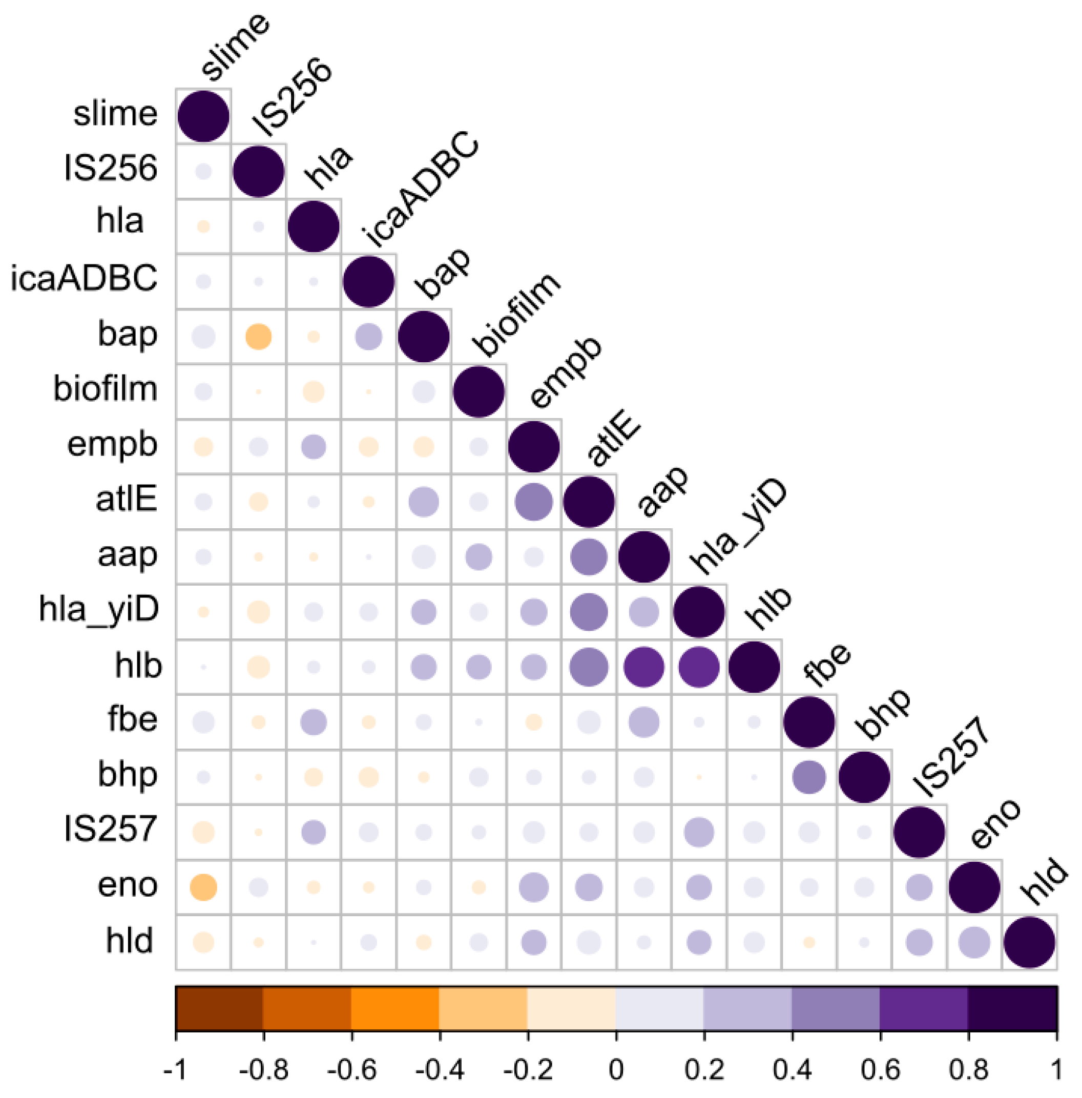
3.4. Phenotype vs. the Genotype Correlations

The question of the phenotype–genotype and the gene–gene correlations is a complex issue, given the relatively large number of resistance and virulence genes that could be compared. To systematically detect all such associations in an unbiased way, we analyzed all potential pairs of variables in the contingency tables, and statistically assessed their dependence by Fisher’s exact test. In addition, using a pseudo-numeric binary matrix (in which the presence of the genes and biofilm/slime production ability is marked as 1 and the absence as 0) we estimated the correlation coefficients among all potential pairs (Figure 1). These analyses highlighted that the ability to form a biofilm by the CoNS is correlated to the genetic determinants, as aap, atlE, hla_yiD, hlb, bhp and bap, while it is not correlated to icaADBC. However, the slime production capacity is correlated to icaADBC. Interestingly, we also observed that most strains carrying icaADBC, simultaneously, carry IS257 in their genomes.

4. Discussion

The ability to form a biofilm is one of the most important virulence factors in staphylococci [40]. In the present study, the majority (62.4%) of the CoNS strains isolated from RTE food were capable of forming a biofilm, and 41.7% were classified as strong or moderate biofilm producers. Furthermore, different biofilm-forming capacities were observed among the strains, even belonging to the same species. The highest percentage of strains with a strong and moderate biofilm-forming capability was observed in the species: S. xylosus (100%), S. lentus (100%), S. piscifermentas (100%), S. simulans (77.8%) and S. warneri (57.1%).
It is relevant to highlight that among the CoNS, strains belonging to S. xylosus, S. carnosus and S. warneri, they are commonly exploited, as starter cultures, in ripened food production, e.g., cheese and fermented meat [41,42]. For this reason, due to its positive link with food fermentation processes, the biofilm production can be perceived as a desired feature, as both the adhesion and the biofilm formation can increase the starter assertiveness towards autochthonous microbiota, inducing, concomitantly, a resistance to the colonization in a specific ecological niche. Moreover, the biofilms provide physical protection to bacteria against stressors, including anti-microbial substances. However, given the increasing role of the CoNS in inducing human infections, most recently they tend to be perceived as potential pathogens. Therefore, it is extremely important to provide data characterizing the strains and demonstrate a link among the various virulence factors. Such a link among the virulence factors, as biofilm formation and hemolytic activity, as demonstrated in the present study, plays a crucial role in identifying the pathogenic strains.
Staphylococci, which are ica-positive, i.e., can synthesize the PIA, often exhibit the ability to produce slime on agar with Congo red [43]. The current study did not show any statistically significant correlation between the ability to produce slime and the presence of the ica operon genes. This can stem from numerous factors, one of them being the absence of a regulatory gene necessary for the expression of a phenotypic trait or from a mutation. The findings of this and our earlier research emphasize the importance to deeply explore the biofilm formation mechanism, independently from the ica presence in staphylococci [35].
Therefore, the role of MSCRAMMs in biofilm formation within the CoNS was also evaluated. Various MSCRAMMs were found in the CoNS isolated from RTE food, such as the biofilm-associated protein (bap), laminin-binding protein (eno), that exhibit a strain-specific variability, as previously reported by other authors [44,45,46]. Bacteria surface proteins, such as cell wall-anchored proteins (CWA), are regarded as virulence factors among gram-positive bacteria pathogens, and they play a key role in the microbial adherence to the host’s tissues, avoiding the host’s defense systems and biofilm formation [47]. The current study confirmed that a large majority of S. epidermidis isolated from RTE food contained the atlE, aap and embP genes, as previously described for S. epidermidis isolated from blood infections associated with catheter and prosthetic joint infections (PJIs), or for the commensal S. epidermidis, for which the proteins are important, both in infection and in colonization [48]. Moreover, these determinants were found, not only in S. epidermidis, regarded as a potential human pathogen, but also in strains of S. lentus, S. haemolyticus and S. lugdenensis. The latest discoveries and updates concerning the CoNS species and subspecies confirmed this group as non-uniform, from non-pathogenic to potentially pathogenic, with different virulence levels [49]. Recent reports have highlighted that some species, such as S. lugdunensis, due to their higher virulent potential, are regarded as highly virulent pathogenic bacteria [50]. S. lugdunensis can cause very acute and destructive endocarditis (IE), with a higher mortality rate than other CoNS species, which generally cause less severe infections [51].
Compared to S. aureus, the CoNS have been studied less extensively. However, these species deserve special attention because of their growing importance, both from the clinical perspective and as a food-affecting agent, due to the multiple virulence factors. The surface colonization and the biofilm formation in the CoNS has been regarded as the main virulence factors because the heterogeneity of these bacteria in the biofilms is known to contribute to their survival, with emphasis on the viable but nonculturable (VBNC) cells and small colony variants (SCVs). Furthermore, the antibiotic resistance and enterotoxin production, which we have described in our earlier papers, also contribute to their virulence [2,3,8,52]. Given the toxin-formation potential and the observed increasing antibiotic and disinfectant resistance among the CoNS, the ability to form a strong biofilm observed in this study is not a cause for optimism. The cells in a biofilm exhibit a much greater tolerance to anti-microbial agents [53] and can easily survive adverse conditions during food processing and storage. Due to the high hemolytic activity observed in the CoNS isolated from RTE food, we can conclude that the CoNS should not be omitted in food microbiological analyses.
The current study also analyzed the link between the biofilm-forming capability and the hemolytic activity, finding a correlation between the biofilm-formation capability and the presence of the hla_yiD and hlb_epi genes. The hemolysin-encoding genes were also correlated to many genetic determinants responsible for the adhesion, such as the fibronectin-binding protein (embP), laminin-binding protein (eno), biofilm external matrix protein (aap) and adhesion to abiotic surfaces (atlE). Moreover, the presence of the hla_yiD and hlb genes was correlated to IS257, as hla to IS256. A significant correlation between the presence of IS256 and the hemolysin production has been recently observed by Nasaj et al. (2020) [38], within clinical isolates of the CoNS. Furthermore, a correlation between the biofilm formation and the hemolysin production in clinical strains was also demonstrated by Koskela et al. (2009) [54]. In addition, similar findings were observed by Huseby et al., who showed how beta-toxin stimulates the biofilm formation [55] within S. aureus, consistently with the current observations on the CoNS strains. Hemolysins, which belong to a group of cytolysins, are among the factors affecting the pathogenic potential of staphylococci [56].
The CoNS isolated from RTE food exhibited enough virulence and strategy factors to act as opportunistic pathogens, determined by the host’s specific conditions and by specific species- and strain-dependent traits. The CoNS clinical isolates contribute to the overall morbidity, mortality and socio-economic costs, being a non-solved medical issue likely to become exacerbated in the future. Finally, the alleged role of the CoNS as a dormant reservoir of antibiotic resistance, enterotoxicity and virulence factors, is still an underestimated issue, requiring greater scientific attention. All of this should induce food technologists to regard the resistant and virulent CoNS isolates as food pathogens, which should remain under supervision and control. Many CoNS pathogenicity determinants raise concerns, especially since their genes can be transmitted by a horizontal transfer to the S.aureus strains, thereby increasing their pathogenicity.

5. Conclusions

This study is the first study to be conducted on the CoNS isolated from RTE food to show a correlation between the biofilm formation and hemolytic activity. It has been demonstrated that hemolysins are correlated to multiple genes responsible for biofilm formation, apart from the PIA, rather than with icaADBC. Furthermore, the insertion sequences, such as IS256 and IS257, are associated with the slime production and some hemolysins, whereas the presence of those elements is not correlated with the biofilm-forming capability. The ability of the CoNS isolated from RTE food to form a strong biofilm, as observed in this study, as well as a potentially high hemolytic activity, indicates that the CoNS acquires characteristic features increasingly typical of the pathogenic S. aureus. More in-depth focus on these microorganisms is needed, especially since the coagulase-negative staphylococci are still not contemplated into routine microbiological food analyses.

Supplementary Materials

The following supporting information can be downloaded at: https://www.mdpi.com/article/10.3390/ijerph20021375/s1. Table S1. List of the primer sequences used in the PCR reactions, Refs. [57,58,59,60,61,62] are cited in Supplementary Materials. Table S2. The results of the ica genes’ presence in the CoNS. Table S3. Hemolysin profiles of the CoNS strains isolated from ready-to-eat food. Table S4. Detailed characteristic of all the strains used in the study.

Author Contributions

Conceptualization, W.C.-W.; methodology, W.C.-W. and J.G.; software, W.C.-W., J.G. and A.Z.; validation, W.C.-W., J.G. and A.J.Z.; formal analysis, W.C.-W. and J.G.; investigation, W.C.-W. and J.G.; resources, W.C.-W.; data curation, W.C.-W. and J.G.; writing—original draft preparation, W.C.-W.; writing—review and editing, W.C.-W. and J.G.; visualization, W.C.-W. and J.G.; supervision, C.C. and A.Z.; project administration, W.C.-W.; funding acquisition, W.C.-W. All authors have read and agreed to the published version of the manuscript.

Funding

This research was financed by the National Science Centre, allocated on the basis of a decision number DEC-2016/23/D/NZ9/01404. The APC was funded by the University of Warmia and Mazury in Olsztyn, Poland.

Institutional Review Board Statement

Not applicable.

Informed Consent Statement

Not applicable.

Data Availability Statement

Not applicable.

Acknowledgments

The publication was written as a result of the author’s (W.C.-W.) internship in the Department of Agriculture, Food and Environment (Di3A), University of Catania, Via Santa Sofia 100, 95123 Catania, Italy, co-financed by the European Union under the European Social Fund (Operational Program Knowledge Education Development), carried out in the project Development Program at the University of Warmia and Mazury in Olsztyn, Poland (POWR.03.05. 00-00-Z310/17).

Conflicts of Interest

The authors declare no conflict of interest.

References

  1. Seo, K.S.; Bohach, G.A. Staphylococcus aureus. In Food Microbiology: Fundamentals and Frontiers, 4th ed.; Doyle, M.P., Buchanan, R.L., Eds.; ASM Press: Washington, DC, USA, 2013; pp. 547–573. [Google Scholar]
  2. Chajecka-Wierzchowska, W.; Zadernowska, A.; Nalepa, B.; Sierpińska, M.; Laniewska-Trokenheim, L. Coagulase-negative staphylococci (CoNS) isolated from ready-to-eat food of animal origin-Phenotypic and genotypic antibiotic resistance. Food Microbiol. 2015, 46, 222–226. [Google Scholar] [CrossRef] [PubMed]
  3. Chajȩcka-Wierzchowska, W.; Zadernowska, A.; Nalepa, B.; Sierpińska, M.; Łaniewska-Trokenheim, Ł. Retail ready-to-eat food as a potential vehicle for Staphylococcus spp. Harboring antibiotic resistance genes. J. Food Prot. 2014, 77, 993–998. [Google Scholar] [CrossRef] [PubMed]
  4. Becker, K.; Both, A.; Weißelberg, S.; Heilmann, C.; Rohde, H. Emergence of coagulase-negative staphylococci. Expert Rev. Anti-Infect. Ther. 2020, 18, 349–366. [Google Scholar] [CrossRef] [PubMed]
  5. Fontana, C.; Favaro, M. Coagulase-Positive and Coagulase-Negative Staphylococci in Human Disease. In Pet-to-Man Travelling Staphylococci: A World in Progress; Academic Press: Cambridge, MA, USA, 2018; pp. 25–42. [Google Scholar]
  6. Kosecka-Strojek, M.; Buda, A.; Miȩdzobrodzki, J. Staphylococcal Ecology and Epidemiology. In Pet-to-Man Travelling Staphylococci: A World in Progress; Academic Press: Cambridge, MA, USA, 2018; pp. 11–24. [Google Scholar]
  7. Becker, K.; Heilmann, C.; Peters, G. Coagulase-negative staphylococci. Clin. Microbiol. Rev. 2014, 27, 870–926. [Google Scholar] [CrossRef] [PubMed] [Green Version]
  8. Chajęcka-Wierzchowska, W.; Gajewska, J.; Wiśniewski, P.; Zadernowska, A. Enterotoxigenic potential of coagulase-negative staphylococci from ready-to-eat food. Pathogens 2020, 9, 734. [Google Scholar] [CrossRef]
  9. Otto, M. Staphylococcal Biofilms. Microbiol. Spectr. 2018, 6, 1–26. [Google Scholar] [CrossRef]
  10. Azara, E.; Longheu, C.; Sanna, G.; Tola, S. Biofilm formation and virulence factor analysis of Staphylococcus aureus isolates collected from ovine mastitis. J. Appl. Microbiol. 2017, 123, 372–379. [Google Scholar] [CrossRef]
  11. Singh, R.; Ray, P.; Das, A.; Sharma, M. Penetration of antibiotics through Staphylococcus aureus and Staphylococcus epidermidis biofilms. J. Antimicrob. Chemother. 2010, 65, 1955–1958. [Google Scholar] [CrossRef] [Green Version]
  12. Vázquez-Sánchez, D.; Habimana, O.; Holck, A. Impact of food-related environmental factors on the adherence and biofilm formation of natural staphylococcus aureus isolates. Curr. Microbiol. 2013, 66, 110–121. [Google Scholar] [CrossRef] [Green Version]
  13. Chen, Q.; Xie, S.; Lou, X.; Cheng, S.; Liu, X.; Zheng, W.; Zheng, Z.; Wang, H. Biofilm formation and prevalence of adhesion genes among Staphylococcus aureus isolates from different food sources. Microbiologyopen 2020, 9, e00946. [Google Scholar] [CrossRef]
  14. Mack, D.; Becker, P.; Chatterjee, I.; Dobinsky, S.; Knobloch, J.K.M.; Peters, G.; Rohde, H.; Herrmann, M. Mechanisms of biofilm formation in Staphylococcus epidermidis and Staphylococcus aureus: Functional molecules, regulatory circuits, and adaptive responses. Int. J. Med. Microbiol. 2004, 294, 203–212. [Google Scholar] [CrossRef]
  15. Heilmann, C.; Hussain, M.; Peters, G.; Götz, F. Evidence for autolysin-mediated primary attachment of Staphylococcus epidermidis to a polystyrene surface. Mol. Microbiol. 1997, 24, 1013–1024. [Google Scholar] [CrossRef]
  16. Nilsson, M.; Frykberg, L.; Flock, J.I.; Pei, L.; Lindberg, M.; Guss, B. A fibrinogen-binding protein of Staphylococcus epidermidis. Infect. Immun. 1998, 66, 2666–2673. [Google Scholar] [CrossRef] [Green Version]
  17. Williams, R.J.; Henderson, B.; Sharp, L.J.; Nair, S.P. Identification of a fibronectin-binding protein from Staphylococcus epidermidis. Infect. Immun. 2002, 70, 6805–6810. [Google Scholar] [CrossRef] [Green Version]
  18. Heilmann, C.; Thumm, G.; Chhatwal, G.S.; Hartleib, J.; Uekötter, A.; Peters, G. Identification and characterization of a novel autolysin (Aae) with adhesive properties from Staphylococcus epidermidis. Microbiology 2003, 149, 2769–2778. [Google Scholar] [CrossRef] [Green Version]
  19. Löffler, B.; Tuchscherr, L.; Niemann, S.; Peters, G. Staphylococcus aureus persistence in non-professional phagocytes. Int. J. Med. Microbiol. 2014, 304, 170–176. [Google Scholar] [CrossRef]
  20. Moormeier, D.E.; Bayles, K.W. Staphylococcus aureus Biofilm: A Complex Developmental Organism Graphical Abstract HHS Public Access. Mol. Microbiol. 2017, 104, 365–376. [Google Scholar] [CrossRef] [Green Version]
  21. Mack, D.; Fischer, W.; Krokotsch, A.; Leopold, K.; Hartmann, R.; Egge, H.; Laufs, R. The intercellular adhesin involved in biofilm accumulation of Staphylococcus epidermidis is a linear β-1,6-linked glucosaminoglycan: Purification and structural analysis. J. Bacteriol. 1996, 178, 175–183. [Google Scholar] [CrossRef] [Green Version]
  22. Mack, D.; Riedewald, J.; Rohde, H.; Magnus, T.; Feucht, H.H.; Elsner, H.A.; Laufs, R.; Rupp, M.E. Essential functional role of the polysaccharide intercellular adhesin of Staphylococcus epidermidis in hemagglutination. Infect. Immun. 1999, 67, 1004–1008. [Google Scholar] [CrossRef] [Green Version]
  23. Arciola, C.R.; Campoccia, D.; Ravaioli, S.; Montanaro, L. Polysaccharide intercellular adhesin in biofilm: Structural and regulatory aspects. Front. Cell. Infect. Microbiol. 2015, 5, 7. [Google Scholar] [CrossRef]
  24. Agarwal, A.; Singh, K.P.; Jain, A. Medical significance and management of staphylococcal biofilm. FEMS Immunol. Med. Microbiol. 2010, 58, 147–160. [Google Scholar] [CrossRef] [PubMed]
  25. Magro, G.; Biffani, S.; Minozzi, G.; Ehricht, R.; Monecke, S.; Luini, M.; Piccinini, R. Virulence genes of S. aureus from dairy cow mastitis and contagiousness risk. Toxins 2017, 9, 195. [Google Scholar] [CrossRef] [PubMed] [Green Version]
  26. Puah, S.M.; Tan, J.A.M.A.; Chew, C.H.; Chua, K.H. Diverse Profiles of Biofilm and Adhesion Genes in Staphylococcus Aureus Food Strains Isolated from Sushi and Sashimi. J. Food Sci. 2018, 83, 2337–2342. [Google Scholar] [CrossRef] [PubMed]
  27. Moraveji, Z.; Tabatabaei, M.; Shirzad Aski, H.; Khoshbakht, R. Characterization of hemolysins of Staphylococcus strains isolated from human and bovine, southern Iran. Iran. J. Vet. Res. 2014, 15, 326–330. [Google Scholar] [PubMed]
  28. Ebrahimi, A.; Taheri, M.A. Characteristics of staphylococci isolated from clinical and subclinical mastitis cows in Shahrekord, Iran. Iran. J. Vet. Res. 2009, 10, 273–277. [Google Scholar]
  29. Zhang, Y.Q.; Ren, S.X.; Li, H.L.; Wang, Y.X.; Fu, G.; Yang, J.; Qin, Z.Q.; Miao, Y.G.; Wang, W.Y.; Chen, R.S.; et al. Genome-based analysis of virulence genes in a non-biofilm-forming Staphylococcus epidermidis strain (ATCC 12228). Mol. Microbiol. 2003, 49, 1577–1593. [Google Scholar] [CrossRef] [Green Version]
  30. Novick, R.P. Mobile genetic elements and bacterial toxinoses: The superantigen-encoding pathogenicity islands of Staphylococcus aureus. Plasmid 2003, 49, 93–105. [Google Scholar] [CrossRef]
  31. Murugesan, S.; Mani, S.; Kuppusamy, I.; Krishnan, P. Role of insertion sequence element is256 as a virulence marker and its association with biofilm formation among methicillin-resistant Staphylococcus epidermidis from hospital and community settings in Chennai, South India. Indian J. Med. Microbiol. 2018, 36, 124–126. [Google Scholar] [CrossRef]
  32. Zakrzewski, A.J.; Zarzecka, U.; Chajęcka-Wierzchowska, W.; Zadernowska, A. A Comparison of Methods for Identifying Enterobacterales Isolates from Fish and Prawns. Pathogens 2022, 11, 410. [Google Scholar] [CrossRef]
  33. Li, X.; Xing, J.; Li, B.; Wang, P.; Liu, J. Use of tuf as a target for sequence-based identification of Gram-positive cocci of the genus Enterococcus, Streptococcus, coagulase-negative Staphylococcus, and Lactococcus. Ann. Clin. Microbiol. Antimicrob. 2012, 11, 31. [Google Scholar] [CrossRef] [Green Version]
  34. Mathur, T.; Singhal, S.; Khan, S.; Upadhyay, D.J.; Fatma, T.; Rattan, A. Detection of biofilm formation among the clinical isolates of staphylococci: An evaluation of three different screening methods. Indian J. Med. Microbiol. 2006, 24, 25–29. [Google Scholar] [CrossRef]
  35. Gajewska, J.; Chajęcka-Wierzchowska, W. Biofilm formation ability and presence of adhesion genes among coagulase-negative and coagulase-positive staphylococci isolates from raw cow’s milk. Pathogens 2020, 9, 654. [Google Scholar] [CrossRef]
  36. Stepanović, S.; Vuković, D.; Hola, V.; Di Bonaventura, G.; Djukić, S.; Ćircović, I.; Ruzicka, F. Quantification of biofilm in microtiter plates. Apmis 2007, 115, 891–899. [Google Scholar] [CrossRef]
  37. Chajecka-Wierzchowska, W.; Zadernowska, A.; Łaniewska-Trokenheim, Ł. Virulence factors, antimicrobial resistance and biofilm formation in Enterococcus spp. isolated from retail shrimps. LWT Food Sci. Technol. 2016, 69, 117–122. [Google Scholar] [CrossRef]
  38. Nasaj, M.; Saeidi, Z.; Asghari, B.; Roshanaei, G.; Arabestani, M.R. Identification of hemolysin encoding genes and their association with antimicrobial resistance pattern among clinical isolates of coagulase-negative Staphylococci. BMC Res. Notes 2020, 13, 4–9. [Google Scholar] [CrossRef] [Green Version]
  39. Chessa, D.; Ganau, G.; Spiga, L.; Bulla, A.; Mazzarello, V.; Campus, G.V.; Rubino, S. Staphylococcus aureus and Staphylococcus epidermidis virulence strains as causative agents of persistent Infections in breast implants. PLoS ONE 2016, 11, e0146668. [Google Scholar] [CrossRef]
  40. Gonçalves, T.G.; Timm, C.D. Biofilm production by coagulase-negative Staphylococcus: A review. Arq. Inst. Biol. 2020, 87, 1–9. [Google Scholar] [CrossRef]
  41. Irlinger, F. Safety assessment of dairy microorganisms: Coagulase-negative staphylococci. Int. J. Food Microbiol. 2008, 126, 302–310. [Google Scholar] [CrossRef]
  42. Landeta, G.; Curiel, J.A.; Carrascosa, A.V.; Muñoz, R.; de las Rivas, B. Characterization of coagulase-negative staphylococci isolated from Spanish dry cured meat products. Meat Sci. 2013, 93, 387–396. [Google Scholar] [CrossRef] [Green Version]
  43. Petrelli, D.; Zampaloni, C.; D’ercole, S.; Prenna, M.; Ballarini, P.; Ripa, S.; Vitali, L.A. Analysis of different genetic traits and their association with biofilm formation in Staphylococcus epidermidis isolates from central venous catheter infections. Eur. J. Clin. Microbiol. Infect. Dis. 2006, 25, 773–781. [Google Scholar] [CrossRef]
  44. Atshan, S.S.; Nor Shamsudin, M.; Sekawi, Z.; Lung, L.T.T.; Hamat, R.A.; Karunanidhi, A.; Mateg Ali, A.; Ghaznavi-Rad, E.; Ghasemzadeh-Moghaddam, H.; Chong Seng, J.S.; et al. Prevalence of adhesion and regulation of biofilm-related genes in different clones of Staphylococcus aureus. J. Biomed. Biotechnol. 2012, 2012, e976972. [Google Scholar] [CrossRef] [PubMed]
  45. Serray, B.; Oufrid, S.; Hannaoui, I.; Bourjilate, F.; Soraa, N.; Mliji, M.; Sobh, M.; Hammoumi, A.; Timinouni, M.; Azhari, M. El Genes encoding adhesion factors and biofilm formation in methicillinresistant Staphylococcus aureus in Morocco. J. Infect. Dev. Ctries 2016, 10, 863–869. [Google Scholar] [CrossRef] [PubMed] [Green Version]
  46. Tang, J.; Chen, J.; Li, H.; Zeng, P.; Li, J. Characterization of adhesin genes, staphylococcal nuclease, hemolysis, and biofilm formation among staphylococcus aureus strains isolated from different sources. Foodborne Pathog. Dis. 2013, 10, 757–763. [Google Scholar] [CrossRef] [PubMed]
  47. Foster, T.J.; Geoghegan, J.A.; Ganesh, V.K.; Hook, M. Adhesion, invasion and evasion: The many functions of the surface proteins of Staphylococcus aureus. Nat. Rev. Microbiol. 2014, 12, 46–62. [Google Scholar] [CrossRef] [PubMed] [Green Version]
  48. Barbieri, R.; Pesce, M.; Franchelli, S.; Baldelli, I.; De Maria, A.; Marchese, A. Phenotypic and genotypic characterization of Staphylococci causing breast peri-implant infections in oncologic patients. BMC Microbiol. 2015, 15, 26. [Google Scholar] [CrossRef] [Green Version]
  49. França, A.; Gaio, V.; Lopes, N.; Melo, L.D.R. Virulence Factors in Coagulase-Negative Staphylococci. Pathogens 2021, 10, 170. [Google Scholar] [CrossRef]
  50. Heilbronner, S.; Foster, T.J. Staphylococcus lugdunensis: A skin commensal with invasive pathogenic potential. Clin. Microbiol. Rev. 2021, 34, e00205-20. [Google Scholar] [CrossRef]
  51. Argemi, X.; Matelska, D.; Ginalski, K.; Riegel, P.; Hansmann, Y.; Bloom, J.; Pestel-Caron, M.; Dahyot, S.; Lebeurre, J.; Prévost, G. Comparative genomic analysis of Staphylococcus lugdunensis shows a closed pan-genome and multiple barriers to horizontal gene transfer. BMC Genom. 2018, 19, 621. [Google Scholar] [CrossRef]
  52. Chajęcka-Wierzchowska, W.; Zadernowska, A.; Gajewska, J.S. epidermidis strains from artisanal cheese made from unpasteurized milk in Poland—Genetic characterization of antimicrobial resistance and virulence determinants. Int. J. Food Microbiol. 2019, 294, 55–59. [Google Scholar] [CrossRef]
  53. Mah, T. Biofilm-specific antibiotic resistance. Future Microbiol. 2012, 7, 1061–1072. [Google Scholar] [CrossRef] [Green Version]
  54. Koskela, A.; Nilsdotter-Augustinsson, Å.; Persson, L.; Söderquist, B. Prevalence of the ica operon and insertion sequence IS256 among Staphylococcus epidermidis prosthetic joint infection isolates. Eur. J. Clin. Microbiol. Infect. Dis. 2009, 28, 655–660. [Google Scholar] [CrossRef]
  55. Huseby, M.J.; Kruse, A.C.; Digre, J.; Kohler, P.L.; Vocke, J.A.; Mann, E.E.; Bayles, K.W.; Bohach, G.A.; Schlievert, P.M.; Ohlendorf, D.H.; et al. Beta toxin catalyzes formation of nucleoprotein matrix in staphylococcal biofilms. Proc. Natl. Acad. Sci. USA 2010, 107, 14407–14412. [Google Scholar] [CrossRef] [Green Version]
  56. Kmieciak, W.; Szewczyk, E.M. Cytolizyny-Czynniki zjadliwości Staphylococcus intermedius i Staphylococcus pseudintermedius. Postep. Mikrobiol. 2015, 54, 354–363. [Google Scholar]
  57. Arciola, C.R.; Baldassarri, L.; Montanaro, L. Presence of icaA and icaD genes and slime production in a collection of Staphylococcal strains from catheter-associated infections. J. Clin. Microbiol. 2001, 39, 2151–2156. [Google Scholar] [CrossRef] [Green Version]
  58. Solati, S.M.; Tajbakhsh, E.; Khamesipour, F.; Gugnani, H.C. Prevalence of virulence genes of biofilm producing strains of Staphylococcus epidermidis isolated from clinical samples in Iran. AMB Express 2015, 5, 47. [Google Scholar] [CrossRef] [Green Version]
  59. Cucarella, C.; Solano, C.; Valle, J.; Amorena, B.; Lasa, I.; Penadés, J.R. Bap, a Staphylococcus aureus Surface Protein Involved in Biofilm Formation Staphylococcus aureus Surface Protein Involved in Biofilm Formation. J. Bacteriol. 2001, 183, 2888–2896. [Google Scholar] [CrossRef] [Green Version]
  60. Tristan, A.; Ying, L.; Bes, M.; Etienne, J.; Vandenesch, F.; Lina, G. Use of multiplex PCR to identify Staphylococcus aureus adhesins involved in human hematogenous infections. J. Clin. Microbiol. 2003, 41, 4465–4467. [Google Scholar] [CrossRef] [Green Version]
  61. Rohde, H.; Burdelski, C.; Bartscht, K.; Hussain, M.; Buck, F.; Horstkotte, M.A.; Knobloch, J.K.M.; Heilmann, C.; Herrmann, M.; Mack, D. Induction of Staphylococcus epidermidis biofilm formation via proteolytic processing of the accumulation-associated protein by staphylococcal and host proteases. Mol. Microbiol. 2005, 55, 1883–1895. [Google Scholar] [CrossRef]
  62. Rohde, H.; Burandt, E.C.; Siemssen, N.; Frommelt, L.; Burdelski, C.; Wurster, S.; Scherpe, S.; Davies, A.P.; Harris, L.G.; Horstkotte, M.A.; et al. Polysaccharide intercellular adhesin or protein factors in biofilm accumulation of Staphylococcus epidermidis and Staphylococcus aureus isolated from prosthetic hip and knee joint infections. Biomaterials 2007, 28, 1711–1720. [Google Scholar] [CrossRef]
Figure 1. Correlation matrix indicating all correlations between the pairs of genetic or phenotypic determinants (biofilm and slime phenotypes). Shades of purple represent the positive correlations, while shades of orange represent the negative correlations.
Ijerph 20 01375 g001
Table 1. Biofilm formation of the tested CoNS by the quantitative (MTP) and qualitative (CRA) methods.
SpeciesBiofilm Formation (MTP)Slime Production (CRA)
StrongModerateWeakNo BiofilmPositiveNegative
S. epidermidis (n = 21)10 (47.6%)1 (4.8%)010 (47.6%)6 (28.6%)15 (71.4%)
S. warneri (n = 14)8 (57.1%)1 (7.1%)2 (14.3%)3 (21.4%)2 (14.3%)12 (85.7%)
S. carnosus (n = 9)2 (22.2%)007 (77.8%)3 (33.3%)6 (66.7%)
S. simulans (n = 9)7 (77.8%)002 (22.2%)7 (77.8%)2 (22.2%)
S. xylosus (n = 8)8 (100%)0006 (75%)2 (25.0%)
S. saprophyticus (n = 6)1 (16.7%)1 (16.7%)04 (66.7%)1 (16.7%)5 (83.3%)
S. pasteuri (n = 5)2 (40.0%)003 (60.0%)05 (100%)
S. heamolyticus (n = 4)01 (25.0%)1 (25.0%)2 (50.0%)1 (25%)3 (75.0%)
S. petrasii subsp. petrasii (n = 4)2 (50.0%)101 (25.0%)2 (50%)2 (50.0%)
S. lentus (n = 2)2 (100%)0001 (50%)1 (50.0%)
S. piscifermentas (n = 2)2 (100%)00002 (100%)
S. lugdenensis (n = 1)001 (100%)01 (100%)0
Total (n = 85)44 (51.8%)5 (5.9%)4 (4.7%)32 (37.6%)30 (35.3%)55 (64.7%)
Table 2. Comparison of the ica operon detection and the biofilm and slime production within the CoNS strains.
Biofilm Formation (MTP)ica+ (n = 39)ica (n = 46)Total (n = 85)
n(%)n(%)n(%)
strong 20(51.3)24(52.1)44(51.8)
moderate 1(2.6)4(8.7)5(5.9)
weak 1(2.6)3(6.5)4(4.7)
no biofilm 13(33.3)19(4.1)32(37.6)
Slime production CRAn(%)n(%)n(%)
positive 12(25.6)18(41.3)30(35.3)
negative 23(56.4)32(69.6)55(64.7)
n-number of strains.
Table 3. Detection of the biofilm-associated genes among the CoNS staphylococci.
SpeciesNo. of StrainsicaADBCbapenoaapfbebhpembPatlEBiofilm Positive Slime Production
S. epidermidis218 (38.1%)020 (95.2%)15 (71.4%)2 (9.5%)012 (57.1%)12 (57.1%)11 (52.4%)6 (28.6%)
S. warneri146 (42.9%)03 (21.4%)6 (42.9%)1 (7.1%)03 (21.4%)1 (7.1%)11 (78.6%)2 (14.3%)
S. carnosus93 (33.3%)06 (66.7%)2 (22.2%)00002 (22.2%)3 (33.3%)
S. simulans97 (77.8%)4 (44.4%)4 (44.4%)6 (66.7%)2 (22.2%)006 (66.7%)7 (77.8%)7 (77.8%)
S. xylosus84 (50%)01 (12.5%)6 (75%)1 (12.5%)0008 (100%)6 (75%)
S. saprophyticus64 (66.7%)06 (100%)3 (50%)2 (33.3%)1 (16.7%)002 (66.7%)1 (16.7%)
S. pasteuri52 (40%)01 (20%)2 (40%)002 (40%)2 (40%)2 (40%)0
S. heamolyticus43 (75%)03 (75%)3 (75%)1 (25%)04 (100%)3 (75%)2 (50%)1 (25%)
S. petrasii subsp. petrasii41 (25%)01 (25%)0001 (25%)03 (75%)2 (50%)
S. lentus2002 (100%)2 (100%)1 (50%)02 (100%)2 (100%)2 (100%)1 (50%)
S. piscifermentas21 (50%)02 (100%)2 (100%)00002 (100%)0
S. lugdenensis10001 (100%)0001 (100%)1 (100%)1 (100%)
Total8539 (45.9%)4 (4.7%)49 (57.6%)48 (56.5%)10 (11.8%)1 (1.2%)24 (28.2%)27 (31.8%)53 (62.4%)30 (35.3%)
Table 4. Prevalence of the hemolysin encoding genes and the insertion sequences IS256 and IS257 among the CoNS.
SpeciesNo. of Strains hla_haemhla_yiDhlbhldIS256IS257
S. epidermidis215 (23.8%)14 (66.7%)14 (66.7%)16 (76.2%)14 (66.7%)19 (90.5%)
S. warneri148 (57.1%)5 (35.7%)6 (42.9%)8 (57.1%)6 (42.9%)13 (92.9%)
S. carnosus905 (55.6%)3 (33.3%)1 (11.1%)6 (66.7%)8 (88.9%)
S. simulans93 (33.3%)7 (77.8%)6 (66.7%)3 (33.3%)1 (11.1%)9 (100%)
S. xylosus80001 (12.5%)7 (87.5%)5 (62.5%)
S. saprophyticus62 (33.3%)003 (50.0%)2 (33.3%)5 (83.5%)
S. pasteuri53 (60.0%)4 (80.0%)2 (40.0%)1 (20.0%)2 (40.0%)5 (100%)
S. heamolyticus44 (100%)3 (75.0%)3 (75.0%)1 (25.0%)3 (75.0%)4 (100%)
S. petrasii subsp. petrasii43 (75.0%)1 (25.0%)2 (50.0%)1 (25.0%)1 (25.0%)2 (50.0%)
S. lentus202 (100%)2 (100%)02 (100%)2 (100%)
S. piscifermentas21 (50.0%)1 (50.0%)2 (100%)002 (100%)
S. lugdenensis101 (100%)1 (100%)01 (100%)0
Total8529 (34.1%)43 (50.6%)41 (48.2%)35 (41.2%)45 (52.9%)74 (87.1%)
Table 5. Comparison of the prevalence rates of the genes involved in the hemolysin production, icaADBC operon and insertion elements.
Hemolysin Genesica+icaTotal (n = 85)
No(%)No(%)No(%)
hla+14(48.3)15(51.7)29(34.1)
hla_yiD+22(51.1)21(48.8)43(50.6)
hlb+19(46.3)22(53.7)41(48.2)
hld+19(54.3)16(45.7)35(41.2)
Insertion sequencesNo(%)No(%)No(%)
IS256+21(46.7)24(53.3)45(52.9)
IS257+35(47.3)39(52.7)74(87.1)
Disclaimer/Publisher’s Note: The statements, opinions and data contained in all publications are solely those of the individual author(s) and contributor(s) and not of MDPI and/or the editor(s). MDPI and/or the editor(s) disclaim responsibility for any injury to people or property resulting from any ideas, methods, instructions or products referred to in the content.

