Communication

Influence of Agaricus bisporus Mushroom on Pb Toxicokinetic in Pregnant Rats

by
Érika Leão Ajala Caetano
1,
Tatiana Pedron
2,3,
Bruna Moreira Freire
2,
Camila Neves Lange
2,
Bruno Lemos Batista
2 and
Denise Grotto
2,*
1
Toxicological Research Laboratory–Lapetox, University of Sorocaba, Sorocaba 18023-000, SP, Brazil
2
Center for Natural and Human Sciences, Federal University of ABC, Santo André 09210-580, SP, Brazil
3
CIQ-UP Department of Chemistry and Biochemistry, Faculty of Sciences, University of Porto, 4099-002 Porto, Portugal
*
Author to whom correspondence should be addressed.
Int. J. Environ. Res. Public Health 2023, 20(4), 3114; https://doi.org/10.3390/ijerph20043114
Submission received: 18 January 2023 / Revised: 3 February 2023 / Accepted: 8 February 2023 / Published: 10 February 2023

Abstract

:
(Pb) is a toxic metal, responsible for several damages to human health. Agaricus bisporus (Ab) is a mushroom with promising antioxidant properties to be used as an alternative chelator in Pb intoxication. The aim was to understand the Pb toxicokinetic and the potential of Ab as a protective agent. A total of 20 female Wistar rats were distributed into 4 groups (n = 5/group): Control (receiving water); Group Ab 100 mg/kg (gavage); Group Pb 100 mg/L in water; and Group Ab + Pb—100 mg/kg + 100 mg/L (gavage and water). Pb administration occurred daily until the 19th day of pregnancy. On day 19 of gestation, the rats were euthanized, and the blood and tissues were collected for Pb measurement, using an inductively coupled plasma mass spectrometer. The results showed that the levels of Pb in the blood, placenta, and liver of the mothers, and in the brain of the fetuses increased significantly in the Pb group. On the other hand, the combined exposure to Pb + Ab showed a significant decrease in the metal concentration in relation to the Pb group, returning to normal levels. Kidney and bone lead levels also increased significantly in the Pb group. However, in the combined exposure group, levels did not return to the control amounts; there was protection, but the Pb concentration was still significantly higher than in the control. In the brain, no significant differences were observed. In conclusion, we suggest A. bisporus is a natural chelator, because the co-administration of the mushroom was able to interact with Pb ions, minimizing the Pb absorption and distribution. These effects are suggested since A. bisporus have antioxidants and beta glucan that interact with Pb, chelating it and, thus, reducing its toxic effects.

1. Introduction

Lead (Pb) is a non-essential element with a proven neurotoxic effect, and with a significant risk for humans exposed to it by environmental, occupational, or dietary factors [1]. Sensitivity to Pb is greater in children; therefore, pregnant women should be aware of the risk of exposure to Pb, because the metal can pass to the fetus during pregnancy through the placenta, and breastfeeding can also be another source of exposure for babies [2,3].
In the general population, gastrointestinal absorption is the most common route, and adults can absorb a maximum of 10% of ingested Pb, in contrast to children (between 2 months and 6 years of age), who can absorb up to 50% of the amount ingested. Considering the toxic effects, in adults, the most common damages are cardiovascular, hypertension, loss of appetite, hallucinations, headache, insomnia, and joint pain [4], while children are susceptible to neurological damage, delay in psychomotor development, and learning difficulties, among others [5,6]. Another well-known toxic effect is the inhibition of the body’s ability to produce hemoglobin, affecting the synthesis of the heme group, by inhibiting the activity in the enzymes, δ-aminolevulinic acid dehydratase (ALA-D), coproporphyrinogen oxidase, and ferrochelatase. Such inhibition causes mild to moderate anemia in adults and severe anemia in children [7]. From the blood, Pb is distributed to all tissues (especially soft tissues such as the kidneys and liver), including the fetus during pregnancy. Subsequently, about 90% of the Pb is deposited in bones [8,9].
The World Health Organization’s 2021 update on the impact of chemicals on public health determined that, of the 2 million people who died in 2019 from exposure to known chemicals, nearly half were due to Pb exposure. Pb exposure is estimated to be responsible for 21.7 million years lost to disability and death [10]. Regarding blood Pb reference values, the Centers for Disease Control and Prevention (CDC) uses a reference value of 3.5 μg/dL to identify children with high Pb levels. The CDC estimates that approximately 800 million children worldwide have Pb in their blood [3]. In addition to affecting adults and children, the release of Pb into the environment can cause mortality and also limit population size among non-human animals, such as populations of wild animals and waterfowl species [11].
The administration of a chelating agent is the current therapy for metal intoxication [12]. EDTACaNa2 (disodium calcium ethylenediaminetetracetic acid), BAL (Dimercaprol), and DMSA (dimercaptosuccinic acid) are some of the synthetic chelating agents used to remove Pb from the body. However, some adverse reactions have already been reported during treatment, such as gastrointestinal discomfort, skin reactions, and elevation of liver enzymes, in addition to the chelation of essential metals [13,14]. In this way, alternative therapies using natural substances such as garlic, vitamin C, and vitamin E have been studied. Some findings have shown the use of different nutrients; for example, vitamins, flavonoids, and mineral elements as defenders against Pb poisoning [15,16,17].
Garlic, a natural substance used in food, contains several essential nutrients and antioxidants, as well as being rich in flavonoids and selenium [18]. It is recommended as an active antioxidant to combat toxicity caused by heavy metals, and the natural chelating ability of the allicin and sulfhydryl groups present in garlic makes it a strong antioxidant in the treatment of lead-induced toxicity, especially long-term toxicity [17,19]. Vitamin E acts as an antioxidant, inhibiting lipid peroxidation. Vitamin E neutralizes the oxidative stress generated by Pb, inhibiting the additional production of free radicals by chelation [17,20].
In this way, culinary and medicinal mushrooms can also be suggested as a complementary therapy. The mushroom Agaricus bisporus is highly nutritious [21]. This mushroom contains many bioactive compounds, including alkaloids, flavonoids, steroids, terpenes, and phenolic compounds responsible for reducing oxidative damage [22,23]. In addition, A. bisporus also has glucans, fibers, and these polysaccharides, especially the triple helix 1,3-1,6-β-d-glucans, which showed chelating characteristics [24], along with chitin and chitosan, which are known for their antioxidant activity, have the ability to eliminate different species of oxygen radicals, such as alkyl, superoxide, and hydroxyl. The mechanism is still unclear but must be related to the chelation of free metal ions by hydroxyl and amino polysaccharide groups, which leads to the formation of a stable system [25]. In waste treatment, chitosan is investigated as a heavy metal chelator [25,26].
In this way, we evaluated the presence of Pb in the blood and tissues of pregnant rats exposed to the metal, and in the brains of their fetuses, aiming to understand the Pb toxicokinetic in the presence of A. bisporus as a protective agent.

2. Materials and Methods

Fresh A. bisporus mushrooms were obtained commercially from a local producer. The mushrooms were sliced and dried in a ventilated oven at 38 ± 2 °C, until constant mass. The dried mushroom was ground in a mill to obtain a kind of thin flour. The mushroom flour was given to the animals every day as described below.

2.1. Experimental Design

Healthy Wistar rats, male and female, were obtained from the UNICAMP vivarium, Campinas, São Paulo state. The experiment was approved by the Committee for the Care and Use of Experimental Animals of the University of Sorocaba, under the protocol number 175/2020. The research was developed in the experimental animal vivarium at the Laboratory of Toxicological Research-LAPETOX, University of Sorocaba. The procedures followed the ethical precepts and the animal welfare standards [27]. Rats were kept in boxes with constant cycles of light and dark cycles—12 h each—at a temperature of 21–23 °C, with free access to a standard chow diet and filtered water. The animals were kept for a week in adaptation before starting the procedures.
For the induction of pregnancy, 01 male and 03 females were mated at night in an independent box, in the proestrus and estrus phases of the estrous cycle. Day 1 of gestation occurred when sperm were found in the vaginal wash, performed in the morning after mating [28].
All pregnant rats were randomly divided into 4 groups (n = 5 per group): Group I—Control (received water); Group II—Ab 100 mg/kg (received A. bisporus mushroom); Group III—Pb 100 mg/L (received Pb); Group IV—Ab + Pb (100 mg/kg +100 mg/L; combined exposure). Exposure to Pb was made in drinking water, while Ab was solubilized in water and administered orally (by gavage). The administration of Pb and Ab was carried out until the 19th day.
The dose of 100 mg/kg/day of A. bisporus was defined based on previous studies from our team [29,30]. The Pb dose of 100 mg/L was defined in a pilot study, which found out that the dose represented values above 90 μg/dL in the blood of pregnant rats. These levels can cross the placental barrier, causing damage to the cognitive development of the fetus [31,32]. At the end of the experiment, the animals were euthanized with ketamine, xylazine, and acepromazine. Blood and tissues were collected for Pb measurement.

2.2. Determination of Pb Levels

Pb was quantified using an inductively coupled plasma mass spectrometer, and the operating conditions are reported in Table 1. Reference materials (blood and tissue) were used as a control of the quality of the analysis. Recovery of blood reference material (Seronorm Trace Elements Blood L-2, ALS Scandinavia AB, Lulea, Sweden) was (103%). Concerning tissue (CRL-ISS 12th Proficiency Test Bovine meat), the recovery was (105.48%).
For blood and tissue, the methodological limits of detection were 1.9535 µg/L and 0.0435 µg/L, respectively. The methodology of Batista and colleagues [33] with some modifications was used for the determination of Pb in blood. In metal-free tubes, 100 µL of blood was added with 4.9 mL of a solution containing 0.5% v v−1 of HNO3 and 0.01% v v−1 of Triton X-100. The samples of blood were shaken and analyzed by ICP-MS.
Tissues were lyophilized for 48 h (lyophilizer Liotop l101, Liobras, São Carlos, SP, Brazil). After that, 35–250 mg of the dried sample was added in metal-free tubes and pre-digested for 48 h with 2 mL of sub-distilled HNO3. The samples were then heated at 90 °C, for 4 h. After cooling, the volume was made up to 50 mL with ultrapure water for analysis by ICP-MS [34], with modifications.

2.3. Statistical Analysis

Data were reported as mean ± standard deviation (SD). The comparison of the mean values among the experimental groups were determined by one-way nonparametric ANOVA (Kruskal–Wallis test), followed by Tukey–Kramer’s interval tests. Differences were considered statistically significant when p < 0.05. The results were analyzed using the Statistica® 8.0 and Graph Pad Prism® 5 programs.

3. Results

The Pb concentrations in the pregnant rats in the blood, bone, brain, placenta, liver, and kidney samples are shown in Figure 1 and in the Supplementary Material.
A significant increase in the Pb concentration was observed in the blood, placenta, and liver in the Pb group. On the other hand, the group that received both Pb + Ab showed a significant drop in the metal concentration in relation to the Pb group. The blood, brain, placenta, and liver did not present statistical differences in lead levels when comparing the control and Pb + Ab. Numerically, it seems to be a difference; however, the large variation within each group (see Supplementary Material) may have been responsible for the lack of significance. In the placenta, a tendency was found when comparing the Pb + Ab group to the control (p = 0.62). The lead concentration in the kidneys and bones also increased significantly in the Pb group. However, in the Pb + Ab group, despite the levels did not return to the similarity of the control, the lead levels diminished compared to the Pb group.
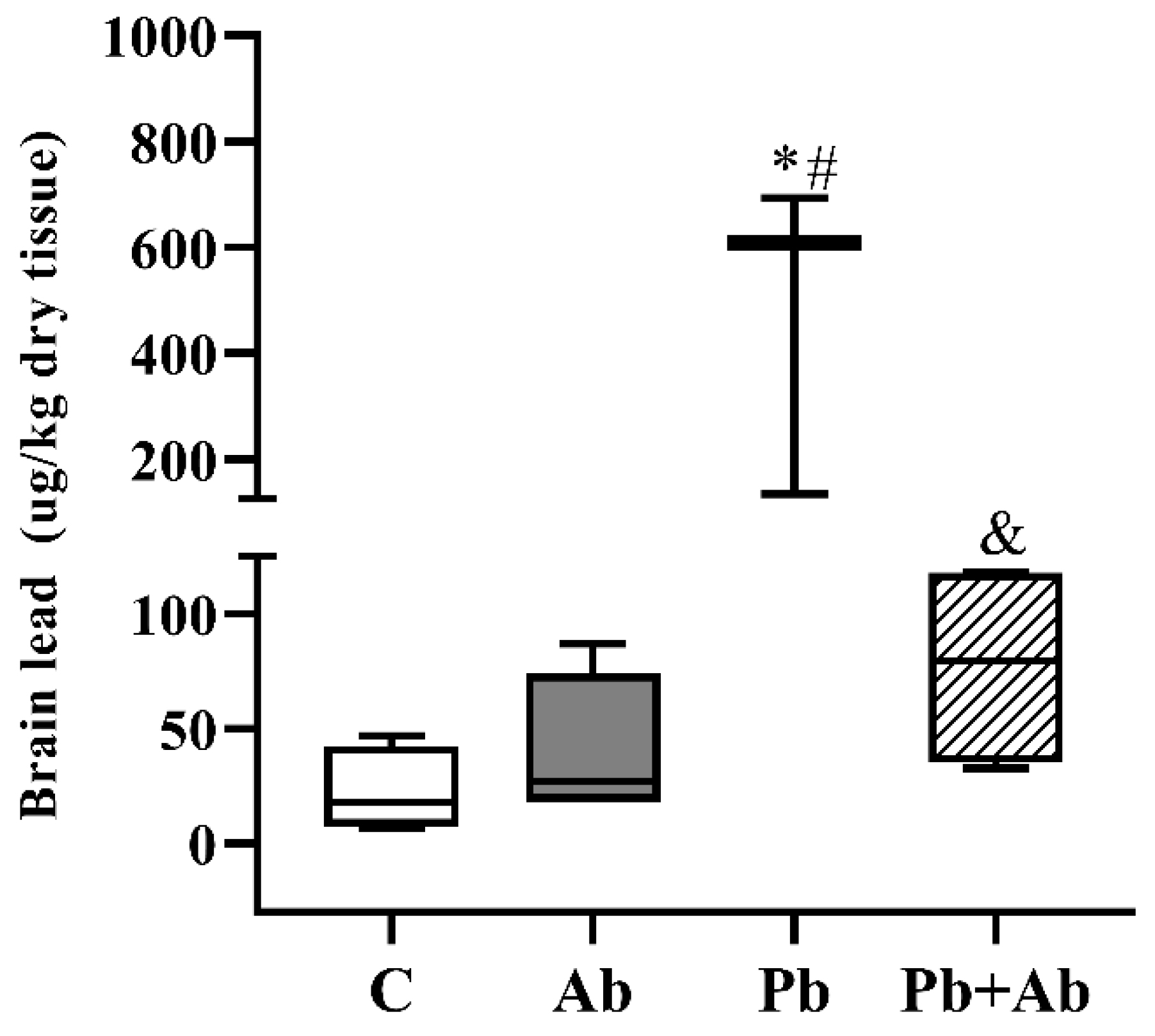
Concerning the brain of the fetuses, the Pb levels are shown in Figure 2. A significant increase in the Pb concentration was observed in the Pb group, compared to the control. On the other hand, the Pb + Ab group showed a significant drop in the metal levels in relation to the Pb group, showing the mushroom has a chelator potential.

4. Discussion

Nowadays, Pb toxicity is still a key environmental health problem causing harmful effects for children and pregnant women, but not restricted to them. Several and distinct species of wild animals are affected by Pb poisoning in the environment [11]. Pb toxic effects on pregnancy are complex, and Pb can affect the mother and fetus with harmful effects and prolonged consequences [35].
The concentrations of Pb in the blood reflect the amount that enters the body through all exposure routes through the respiratory and gastrointestinal tracts, and skin [36]. Thus, we quantified the Pb concentrations in the blood of pregnant rats to assess their level of exposure. The mean blood Pb level for the pregnant rats in the Pb group was around 140 μg/L. This value is two times greater than the maximum allowed biological index—60 µg/L—for Pb in the blood, in Brazil [37]. This finding was similar to those reported by Saleh et al. (2018) [38], with pregnant rats exposed to 160 and 320 mg Pb/kg for 20 days. Those authors found blood lead concentrations of 100 and 125 μg/dL, respectively, and these concentrations are clinically toxic.
We positively observed a decrease in the level of Pb in maternal blood with the A. bisporus co-exposures (around 50 μg/dL, almost three times lower than the Pb group) being below the maximum permitted biological index. Comparatively, similar data were reported by Sadeghi et al. (2021) [39], in which pregnant rats were exposed to Pb and treated with vitamin C and garlic extract. These findings corroborated with several studies and indicated that antioxidants can reduce toxic metals in the blood [40,41].
In the current study, a significant rise in the Pb level was observed in the kidney and hepatic tissues of the Pb-exposed group. This increase was in accordance with results reported by Takano et al. (2015) [42], for animals exposed to Pb for 4 weeks. The high concentrations of Pb in the maternal liver were probably caused by an altered Pb pharmacokinetics in pregnancy. Since the kidney was identified as a soft tissue target for Pb accumulation, research has been published on the Pb concentration in animal kidneys [43,44]. One possible explanation is that the Pb distribution is due to its reabsorption. In the kidney, Pb is filtered in the glomerulus; however, most of the Pb filtered is reabsorbed by the distal tubule and collecting duct [45].
During pregnancy, the internal store of Pb can escape from the bone, due to high bone turnover, and cross the placenta by diffusion in the developing embryo [46]; in our study, we found high concentrations of bone lead in the group exposed to Pb (3621.20 ± 1249.23). However, in the Pb + Ab group (1932.40 ± 657.63), the concentrations did not return to the similarity of the control group. This finding is in agreement with that reported by Mumtaz et al. (2020) [6], who reported that bones are one of the main storage sites for Pb, with the bone matrix being the main target due to its ability to replace cations (Fe2+, Ca2+, and Mg2+ Na+) in the body, altering the mineral metabolism.
The accumulation of Pb in the body of women of reproductive age can be transferred to the fetus during pregnancy through the placenta, the structure responsible for fetal nutrition, allowing the passage of Pb to the fetus, a fact that increases the risk of pregnancy complications and appearance of diseases after birth [47]. Thus, concentrations of Pb in the placenta can be good biomarkers of fetal exposure to Pb [48]. In our study, we found high Pb concentrations in the placenta of the group exposed to Pb (around 652 µg/L). On the other hand, A. bisporus co-exposure decreased the Pb concentration by half (about 340 µg/L), suggesting that the co-administration of the mushroom A. bisporus was beneficial.
In fetuses, the blood–brain barrier, because it is not fully developed, becomes the gateway to toxic products. For this reason, studies suggest that Pb can enter the brain more easily, becoming more harmful to newborns than adults [49,50]. In our study, an increase in the Pb concentrations in the brain of the fetuses in the group exposed to Pb (478.33 ± 301.21) was found, unlike in the Pb + Ab group where we observed a decrease in the metal (77.61 ± 46.42), returning to normal concentrations. In a study carried out by Antonio-Garcia and Masso-Gonzalez (2008) [51], rats were exposed to 300 mg Pb/L, a higher dose than in our study, and to a natural antioxidant, during pregnancy and lactation. The results were similar to ours, with an increase in Pb levels in the brain of the fetuses, showing an alteration in the antioxidant defense systems, as well as a reduction in the Pb concentrations in the groups exposed to antioxidants such as vitamin C. Thus, we can hypothesize that the exogenous supplementation of antioxidant agents, such as the mushroom A. bisporus, may be an alternative therapy for Pb toxicity due to its ability to cross the blood–brain barrier and exert its inhibitory effects on the brain. In studies carried out by Pachauri et al. (2008) [52] and Flora et al. (2003) [53], both highlighted the importance of antioxidant supplementation in metal poisoning, as supplementation can be beneficial to increase metal mobilization. In addition to the mushroom having antioxidants, it has important amounts of beta glucan, a soluble fiber that is directly related to the Pb metabolism. According to Visweswar et al. (2008) [54], three cyclic beta glucans interact with Pb, chelating it and, thus, reducing its toxic effects.

5. Conclusions

In this study, we observed that Pb has a rapid distribution for both the mother and fetus. Therefore, we conclude that there is a link between the Pb concentrations in the blood and tissues of the mothers and in the pups’ brain, demonstrating Pb transfer across the placenta. In addition, co-administration of the mushroom A. bisporus was able to interact with Pb ions. These results highlight the potential use of A. bisporus as neuroprotective agents from Pb-induced harmful effects.

Supplementary Materials

The following supporting information can be downloaded at: https://www.mdpi.com/article/10.3390/ijerph20043114/s1, Table S1: Lead levels in different tissues.

Author Contributions

Conceptualization, É.L.A.C. and D.G.; methodology, É.L.A.C., T.P., B.M.F., C.N.L. and B.L.B.; software, É.L.A.C. and D.G.; formal analysis, D.G.; investigation, T.P., B.M.F., C.N.L. and B.L.B.; writing—original draft preparation, É.L.A.C.; writing—review and editing, É.L.A.C., T.P., B.M.F., C.N.L. and B.L.B.; supervision, D.G.; project administration, D.G. All authors have read and agreed to the published version of the manuscript.

Funding

This research was funded by the University of Sorocaba (Uniso) and by the Coordination for Higher Level Graduate Improvements (CAPES/Brazil, finance code 001).

Institutional Review Board Statement

The animal study protocol was approved by the Ethics Committee of the University of Sorocaba (CEUA-Uniso), Protocol no 175/2020.

Informed Consent Statement

Not applicable.

Data Availability Statement

Data and publication materials are available from the corresponding author upon a reasonable request.

Conflicts of Interest

The authors declare no conflict of interest.

References

  1. Needleman, H. Lead poisoning. Annu. Rev. Med. 2004, 55, 209–222. [Google Scholar] [CrossRef] [PubMed]
  2. Neuwirth, L.S.; Masood, S.; Anderson, D.W.; Schneider, J.S. The a ttention set-shifting test is sensitive for revealing sex-based impairments in executive functions following developmental lead exposure in rats. Behav. Brain Res. 2019, 366, 126–134. [Google Scholar] [CrossRef]
  3. US CDC Advisory Committee on Childhood Lead Poisoning Prevention. CDC Updates Blood Lead Reference Value to 3.5µg/dL. Atlanta: US Centres for Disease Control and Prevention. 2021. Available online: https://www.cdc.gov/nceh/lead/news/cdc-updates-blood-lead-reference-value.html (accessed on 9 January 2023).
  4. Al-Masri, S.A. Effect of pumpkin oil and vitamin E on lead induced testicular toxicity in male rats. J. Anim. Plant Sci. 2015, 25, 72–77. [Google Scholar]
  5. CDC (Centers for Disease Control and Prevention). Blood Lead Levels in Children. 2020. Available online: https://www.cdc.gov/nceh/lead/prevention/blood-lead-levels.htm (accessed on 21 December 2022).
  6. de Freitas, C.U.; De Capitani, E.M.; Gouveia, N.; Simonetti, M.H.; Silva, M.R.D.P.E.; Kira, C.S.; Sakuma, A.M.; Carvalho, M.D.F.H.; Duran, M.C.; Tiglea, P.; et al. Lead exposure in an urban community: Investigation of risk factors and assessment of the impact of lead abatement measures. Environ. Res. 2007, 103, 338–344. [Google Scholar] [CrossRef]
  7. El-Boshy, M.E.; Refaat, B.; Qasem, A.H.; Khan, A.; Ghaith, M.; Almasmoum, H.; Mahbub, A.; Almaimani, R.A. The remedial effect of Thymus vulgaris extract against lead toxicity-induced oxidative stress, hepatorenal damage, immunosuppression, and hematological disorders in rats. Environ. Sci. Pollut. Res. Int. 2019, 26, 22736–22746. [Google Scholar] [CrossRef] [PubMed]
  8. Khalil, N.; Faulkner, K.A.; Greenspan, S.L.; Cauley, J.A. Osteoporotic Fractures in Men Research Group. Associations between bone mineral density, grip strength, and lead body burden in older men. J. Am. Geriatr. Soc. 2014, 62, 141–146. [Google Scholar] [CrossRef]
  9. Dongre, N.N.; Suryakar, A.N.; Patil, A.J.; Hundekari, I.A.; Devarnavadagi, B.B. Biochemical effects of lead exposure on battery manufacture workers with reference to blood pressure, calcium metabolism and bone mineral density. Indian J. Clin. Biochem. 2013, 28, 65–70. [Google Scholar] [CrossRef]
  10. World Health Organization. ‘Lead Poisoning and Health’ Fact Sheet. 2019. Available online: https://www.who.int/news-room/fact-sheets/detail/lead-poisoning-and-health (accessed on 9 January 2023).
  11. Wood, K.A.; Brown, M.J.; Cromie, R.L.; Hilton, G.M.; MacKenzie, C.; Newth, J.L.; Pain, D.J.; Perrins, C.M.; Rees, E.C. Regulation of lead fishing weights results in mute swan population recovery. Biol. Conserv. 2019, 230, 67–74. [Google Scholar] [CrossRef]
  12. Patrick, L. Lead toxicity, a review of the literature. Part 1: Exposure, evaluation, and treatment. Altern. Med. Rev. 2006, 11, 2–22. [Google Scholar] [PubMed]
  13. Mazumdar, I.; Goswami, K. Congenital lead poisoning: An unusual presentation. Indian J. Clin. Biochem. 2014, 29, 257–259. [Google Scholar] [CrossRef]
  14. Andersen, O. Chemical and biological considerations in the treatment of metal intoxications by chelating agents. Mini-Rev. Med. Chem. 2004, 4, 11–21. [Google Scholar] [CrossRef] [PubMed]
  15. Patra, R.C.; Rautray, A.K.; Swarup, D. Oxidative stress in lead and cadmium toxicity and its amelioration. Vet. Med. Int. 2011, 2011, 457327. [Google Scholar] [CrossRef]
  16. Chong, P.S.; Khairuddin, S.; Tse, A.C.K.; Hiew, L.F.; Lau, C.L.; Tipoe, G.L.; Fung, M.-L.; Wong, K.H.; Lim, L.W. Hericium erinaceus potentially rescues be-havioural motor deficits through ERK-CREB-PSD95 neuroprotective mechanisms in rat model of 3-acetylpyridine-induced cerebellar ataxia. Sci. Rep. 2020, 10, 14945. [Google Scholar] [CrossRef] [PubMed]
  17. Mumtaz, S.; Ali, S.; Khan, R.; Shakir, H.A.; Tahir, H.M.; Mumtaz, S.; Andleeb, S. Therapeutic role of garlic and vitamins C and E against toxicity induced by lead on various organs. Environ. Sci. Pollut. Res. 2020, 27, 8953–8964. [Google Scholar] [CrossRef]
  18. Chen, S.; Shen, X.; Cheng, S.; Li, P.; Du, J.; Chang, Y.; Meng, H. Evaluation of garlic cultivars for polyphenolic content and antioxidant properties. PLoS ONE 2013, 8, e79730. [Google Scholar] [CrossRef] [PubMed]
  19. Ried, K.; Frank, O.R.; Stocks, N.P. Aged garlic extract reduces blood pressure in hypertensives: A dose–response trial. Eur. J. Clin. Nutr. 2013, 67, 64–72. [Google Scholar] [CrossRef]
  20. Hassan, A.A.; Jassim, H.M. Effect of treating lactating rats with lead acetate and its interaction with vitamin E or C on neurobehavior, development and some biochemical parameters in their pups. Iraqi J. Vet. Sci. 2010, 24, 45–52. [Google Scholar] [CrossRef]
  21. Atila, F.; Owaid, M.N.; Shariati, M.A. The nutritional and medical benefits of Agaricus bisporus: A review. J. Microbiol. Biotechnol. Food Sci. 2021, 7, 281–286. [Google Scholar] [CrossRef]
  22. Morosanova, M.A.; Fedorova, T.V.; Polyakova, A.; Morosanova, E.I. Agaricus bisporus Crude Extract: Characterization and Analytical Application. Molecules 2020, 25, 5996. [Google Scholar] [CrossRef]
  23. Elhusseiny, S.; El-Mahdy, T.; Awad, M.; Elleboudy, N.; Farag, M.; Aboshanab, K.; Yassien, M. Antiviral, Cytotoxic, and Antioxidant Activities of Three Edible Agaricomycetes Mushrooms: Pleurotus columbinus, Pleurotus sajor-caju, and Agaricus bisporus. J. Fungi 2021, 7, 645. [Google Scholar] [CrossRef]
  24. Mirończuk-Chodakowska, I.; Witkowska, A.M. Evaluation of Polish wild Mushrooms as Beta-Glucan Sources. Int. J. Environ. Res. Public Health 2020, 17, 7299. [Google Scholar] [CrossRef] [PubMed]
  25. Casadidio, C.; Peregrina, D.V.; Gigliobianco, M.R.; Deng, S.; Censi, R.; Di Martino, P. Chitin and Chitosans: Characteristics, Eco-Friendly Processes, and Applications in Cosmetic Science. Mar. Drugs 2019, 17, 369. [Google Scholar] [CrossRef]
  26. Li, L.; Luo, C.; Li, X.; Duan, H.; Wang, X. Preparation of magnetic ionic liquid/chitosan/graphene oxide composite and application for water treatment. Int. J. Biol. Macromol. 2014, 66, 172–178. [Google Scholar] [CrossRef] [PubMed]
  27. du Sert, N.P.; Hurst, V.; Ahluwalia, A.; Alam, S.; Avey, M.T.; Baker, M.; Browne, W.J.; Clark, A.; Cuthill, I.C.; Dirnagl, U.; et al. The ARRIVE guidelines 2.0: Updated guidelines for reporting animal research. PLoS Biol. 2020, 118, e3000410. [Google Scholar] [CrossRef]
  28. Gerenutti, M.; Del Fiol, F.S.; Groppo, F.C. Reproductive performance of pregnant rats and embryotoxic effects of ciprofloxacin. Pharmazie 2006, 61, 79–80. [Google Scholar] [PubMed]
  29. Gerenutti, M.; Tribuiani, N.; Oliveira, B.R.; Rosa-Castro, R.M.; Frizo, I.; Oshima-Franco, Y.; Grotto, D. Safety assessment of the royal sun mushroom, Agaricus brasiliensis (higher Basidiomycetes) intake during rat pregnancy. Int. J. Med. Mushrooms 2014, 16, 519–528. [Google Scholar] [CrossRef] [PubMed]
  30. Laurino, L.F.; Viroel, F.J.M.; Caetano, E.; Spim, S.; Pickler, T.B.; Rosa-Castro, R.M.; Vasconcelos, E.A.; Jozala, A.F.; Hataka, A.; Grotto, D.; et al. Lentinus edodes Exposure before and after Fetus Implantation: Materno-Fetal Development in Rats with Gestational Diabetes Mellitus. Nutrients 2019, 11, 2720. [Google Scholar] [CrossRef]
  31. Weizsaecker, K. Lead toxicity during pregnancy. Prim. Care Update OB/GYNS 2003, 10, 304–309. [Google Scholar] [CrossRef]
  32. Vieira, J.S. Efeitos da Exposição Pré-Natal ao Etanol e ao Chumbo, Isoladamente e em Associação, Sobre a Pressão Arterial e a Reatividade da Aorta de Ratos Recém-Desmamados. Diploma Thesis, Universidade Estadual Paulista, Instituto de Biociência de Botucatu, Botucatu, Brazil, 2014. [Google Scholar]
  33. Batista, B.L.; Jairo, L.R.; Luciano, T.; Adilson, J.C.; Fernando, B.J. Reference concentrations for trace elements in urine for the Brazilian population based on q-ICP-MS with a simple dilute-and-shoot procedure. J. Braz. Chem. Soc. 2009, 10, 1406–1413. [Google Scholar] [CrossRef]
  34. Paniz, F.P.; Tatiana, P.; Bruna, M.F.; Daiane, P.T. Effective procedures for the determination of As, Cd, Cu, Fe, Hg, Mg, Mn, Ni, Pb, Se, Th, Zn, U and rare earth elements in plants and foodstuffs. Anal. Methods 2018, 10, 4094–4103. [Google Scholar] [CrossRef]
  35. Ortega, D.R.; Esquivel, D.G.; Ayala, T.B.; Pineda, B.; Manzo, S.G.; Quino, J.M.; Mora, P.C.; de la Cruz, V.P. Cognitive Impairment Induced by Lead Exposure during Lifespan: Mechanisms of Lead Neurotoxicity. Toxics 2021, 9, 23. [Google Scholar] [CrossRef] [PubMed]
  36. CDC (Centers for Disease Control and Prevention) of U.S. Fourth National Report on Human Exposure to Environmental Chemicals. 2019. Available online: http://www.cdc.gov/exposurereport/ (accessed on 11 January 2023).
  37. Brasil Ministério da Saúde Secretaria de Atenção à Saúde Departamento de Ações Programáticas Estratégicas. Atenção à saúde dos trabalhadores expostos ao chumbo metálico; Ministério da Saúde, Secretaria de Atenção à Saúde, Departamento de Ações Programáticas Estratégicas, Ministério da Saúde: Brasília, Brazil, 2006.
  38. Saleh, H.A.; El-Aziz, G.A.; Mustafa, H.N.; Saleh, A.H.A.; Mal, A.O.; Deifalla, A.H.S.; Aburas, M. Protective effect of garlic extract against maternal and foetal cerebellar damage induced by lead administration during pregnancy in rats. Folia Morphol. 2018, 77, 1–15. [Google Scholar] [CrossRef] [PubMed]
  39. Sadeghi, A.; Khordad, E.; Ebrahimi, V.; Raoofi, A.; Alipour, F.; Ebrahimzadeh-Bideskan, A. Neuroprotective effects of vitamin C and garlic on glycoconjugates changes of cerebellar cortex in lead-exposed rat offspring. J. Chem. Neuroanat. 2021, 114, 101948. [Google Scholar] [CrossRef] [PubMed]
  40. Nam, S.M.; Choi, S.-H.; Cho, H.-J.; Seo, J.S.; Choi, M.; Nahm, S.-S.; Chang, B.-J.; Nah, S.-Y. Ginseng Gintonin Attenuates Lead-Induced Rat Cer-ebellar Impairments during Gestation and Lactation. Biomolecules 2020, 10, 385. [Google Scholar] [CrossRef] [PubMed]
  41. Grotto, D.; Barcelos, G.R.M.; Valentini, J.; Antunes, L.M.G.; Angeli, J.P.F.; Garcia, S.C.; Jr, F.B. Low levels of methylmercury induce DNA damage in rats: Protective effects of selenium. Arch. Toxicol. 2009, 83, 249–254. [Google Scholar] [CrossRef]
  42. Takano, T.; Okutomi, Y.; Mochizuki, M.; Ochiai, Y.; Yamada, F.; Mori, M.; Ueda, F. Biological index of environmental lead pol-lution: Accumulation of lead in liver and kidney in mice. Environ. Monit. Assess. 2015, 187, 744. [Google Scholar] [CrossRef]
  43. Bortey-Sam, N.; Nakayama, S.M.M.; Ikenaka, Y.; Akoto, O.; Baidoo, E.; Yohannes, Y.B.; Mizukawa, H.; Ishizuka, M. Human health risks from metals and metalloid via consumption of food animals near gold mines in Tarkwa, Ghana: Estimation of the daily intakes and target hazard quotients (THQs). Ecotoxicol. Environ. Saf. 2015, 111, 160–167. [Google Scholar] [CrossRef]
  44. Nakayama, S.M.M.; Ikenaka, Y.; Hamada, K.; Muzandu, K.; Choongo, K.; Yabe, J.; Umemura, T.; Ishizuka, M. Accumulation and biological effects of metals in wild rats in mining areas of Zambia. Environ. Monit. Assess. 2013, 185, 4907–4918. [Google Scholar] [CrossRef]
  45. Togao, M.; Nakayama, S.M.; Ikenaka, Y.; Mizukawa, H.; Makino, Y.; Kubota, A.; Matsukawa, T.; Yokoyama, K.; Hirata, T.; Ishizuka, M. Bioimaging of Pb and STIM1 in mice liver, kidney and brain using Laser Ablation Inductively Coupled Plasma Mass Spectrometry (LA-ICP-MS) and immuno-histochemistry. Chemosphere 2020, 238, 124581. [Google Scholar] [CrossRef]
  46. Riess, M.L.; Halm, J.K. Lead poisoning in an adult: Lead mobilization by pregnancy? J. Gen. Intern. Med. 2007, 22, 1212–1215. [Google Scholar] [CrossRef] [PubMed]
  47. CDC (Centers for Disease Control and Prevention). Morbidity and Mortality Weekly Report MMWR; CDC: Atlanta, GE, USA, 2007.
  48. Villeda-Hernández, J.; Méndez Armenta, M.; Barroso-Moguel, R.; Trejo-Solis, M.C.; Guevara, J.; Rios, C. Morphometric analysis of brain lesions in rat fetuses prenatally exposed to low-level lead acetate: Correlation with lipid peroxidation. Histol. Histopathol. 2006, 21, 609–617. [Google Scholar] [CrossRef] [PubMed]
  49. Müller, Y.M.; Rivero, L.B.; Carvalho, M.C.; Kobus, K.; Farina, M.; Nazari, E.M. Behavioral impairments related to lead-induced developmental neurotoxicity in chicks. Arch. Toxicol. 2008, 82, 445–451. [Google Scholar] [CrossRef] [PubMed]
  50. Sharifi, A.M.; Mousavi, S.H.; Jorjani, M. Effect of chronic lead exposure on pro-apoptotic Bax and anti-apoptotic Bcl-2 protein expression in rat hippocampus in vivo. Cell. Mol. Neurobiol. 2010, 30, 769–774. [Google Scholar] [CrossRef] [PubMed]
  51. Antonio-García, M.T.; Massó-Gonzalez, E.L. Toxic effects of perinatal lead exposure on the brain of rats: Involvement of oxidative stress and the beneficial role of antioxidants. Food Chem. Toxicol. 2008, 46, 2089–2095. [Google Scholar] [CrossRef]
  52. Pachauri, V.; Saxena, G.; Mehta, A.; Mishra, D.; Flora, S.J. Combinational chelation therapy abrogates lead-induced neuro-degeneration in rats. Toxicol. Appl. Pharmacol. 2009, 1240, 255–264. [Google Scholar] [CrossRef]
  53. Flora, S.J.; Pande, M.; Mehta, A. Beneficial effect of combined administration of some naturally occurring antioxidants (vitamins) and thiol chelators in the treatment of chronic lead intoxication. Chem. Biol. Interact. 2003, 145, 267–280. [Google Scholar] [CrossRef] [PubMed]
  54. Visweswar, K.N.S.; Sunil, A.; Harsha, A.S.; Janardhana, C. Interaction studies of lead(II) ion with cyclic β-(1→3),(1→6) glucans extracted from Bradyrhizobium japonicum based on ‘chelation enhanced fluorescence’ (CHEF) effect. Luminescence 2018, 33, 1202–1208. [Google Scholar] [CrossRef] [PubMed]
Figure 1. Lead concentrations (mean ± standard deviation) in blood and tissue samples from pregnant rats exposed to A. bisporus and lead, from gestation day 1 to 19. C (Control), Ab (A. bisporus 100 mg/kg/day), Pb (Lead 100 mg/L/day), Pb + Ab (100 mg/L/day of Pb + 100 mg/kg/day of A. bisporus), n = 5/group. * p < 0.05 in comparison to Control group, # in comparison to A. bisporus, and & in comparison to lead. Kruskal–Wallis test, followed by Tukey–Kramer’s.
Ijerph 20 03114 g001
Figure 2. Lead concentrations (mean ± standard deviation) in brain samples of fetuses from female rats exposed to A. bisporus and lead, from gestation day 1 to 19. C (Control), Ab (A. bisporus 100 mg/kg/day), Pb (Lead 100 mg/L/day), Pb + Ab (100 mg/L/day of Pb + 100 mg/kg/day of A. bisporus), n = 5/group. * p < 0.05 in comparison to Control group, # in comparison to A. bisporus, and & in comparison to lead. One-way ANOVA, followed by the Tukey–Kramer’s test.
Ijerph 20 03114 g002
Table 1. Operating conditions for ICP-MS.
ICP-MS: Operating Conditions
Monitored isotope208Pb
Radio frequency power1550 W
Argon flow15 L min−1
Nebulization chamber0.9 L min−1
Collision cellHelium (purity > 99.999%)
Nebulization chamberScott (double pass)
InterfaceNickel cones
Sample 0.90 mm
Skimmer0.45 mm
Disclaimer/Publisher’s Note: The statements, opinions and data contained in all publications are solely those of the individual author(s) and contributor(s) and not of MDPI and/or the editor(s). MDPI and/or the editor(s) disclaim responsibility for any injury to people or property resulting from any ideas, methods, instructions or products referred to in the content.