Article

European Black Elderberry Fruit Extract Inhibits Replication of SARS-CoV-2 In Vitro

1
Institute of Virology, Friedrich-Alexander University Erlangen-Nürnberg (FAU), 91054 Erlangen, Germany
2
Department of Immune Modulation, Universitätsklinikum Erlangen, 91054 Erlangen, Germany
3
IPRONA AG/SPA, Industriestraße 1/6, 39011 Lana, Italy
*
Authors to whom correspondence should be addressed.
Nutraceuticals 2023, 3(1), 91-106; https://doi.org/10.3390/nutraceuticals3010007
Submission received: 9 December 2022 / Revised: 3 January 2023 / Accepted: 6 January 2023 / Published: 13 January 2023
(This article belongs to the Special Issue Functional Foods as a New Therapeutic Strategy)

Abstract

:
Coronavirus disease-19 (COVID-19) is still affecting the lives of people round the globe and remains a major public health threat. The emergence of new variants more efficiently transmitted, more virulent and more capable of escaping naturally acquired and vaccine-induced immunity creates a long-term negative outlook for the management of the pandemic. The development of effective and viable prevention and treatment options to reduce viral transmission is of the utmost importance. The fruits of the European black elderberry and extracts thereof have been traditionally used to treat viral infections such as coughs, cold and flu. Specifically, its efficacy against the Influenza A virus has been shown in vitro as well as in human clinical trials. In the current project, we investigated the antiviral activity of a black elderberry extract, mainly containing anthocyanins and phenolic compounds, against SARS-CoV-2 and its variants of concern and explored the possible mode of action by performing time of addition experiments. The results revealed that the extract displayed a strong anti-SARS-CoV-2 activity against the Wuhan type as well as the variants of concern Alpha, Beta, Gamma, Delta and Omicron with a comparable antiviral activity. Based on cytotoxicity data, a 2-log theoretical therapeutic window was established. The data accumulated so far suggest that the viral replication cycle is inhibited at later stages, inasmuch as the replication process was affected after virus entry. Therefore, it would be legitimate to assume that black elderberry extract might have the potential to be an effective treatment option for SARS-CoV-2 infections.

1. Introduction

The Coronavirus disease 2019 (COVID-19), caused by the Severe Acute Respiratory Syndrome Coronavirus 2 (SARS-CoV-2), is responsible for short- and long-term complications, e.g., the need for respiratory support or persistent cardiovascular complications. To date, the pandemic has resulted in around 647 million global cases and over 6.6 million deaths [1]. An ongoing major problem is the continuous emergence and spread of SARS-CoV-2 variants determined as “Variants of Concern” (VoCs) by the WHO. These variants are able to evade vaccine- or infection-induced antiviral immune response [2,3].
Mutations in SARS-CoV-2 VoCs are mainly located within the spike glycoprotein and generally change the interaction with host receptors and thus affect the infectivity, transmissibility or pathogenicity of the virus [4,5,6,7]. The VoCs described to date include SARS-CoV-2 Alpha [8], SARS-CoV-2 Beta [9], SARS-CoV-2 Gamma [10], SARS-CoV-2 Delta [11] as well as SARS-CoV-2 Omicron [12]. The latter is subdivided in the sub-lineages BA.1, BA.2, BA.3, BA.4 and BA.5 [13], with BA.5 being predominant worldwide at the moment. With BQ.1.1 a new sub-lineage increases, especially in Europe and USA. This Omicron variant is resistant against all clinically available monoclonal antibodies [14]. In comparison to the other VoCs, Omicron variants contain a high number of deletions, insertions and mutations, especially in the spike protein [13,15]. Generally, VoCs were reported to show higher transmissibility and infectivity [4,16,17,18,19,20]. In light of the continuing COVID-19 pandemic and the ongoing occurrence of new SARS-CoV-2 variants, the development of broadly effective prophylactic and therapeutic countermeasures remains of the utmost importance.
At the beginning of 2022, the first antiviral small molecule drugs were approved for high risk patients and only for use at early stages following infection with SARS-CoV-2 [21]. Nirmatrelvir, an inhibitor of the 3-Chymotrypsin-like protease of SARS-CoV-2 combined with Ritonavir, an HIV-1-protease inhibitor, were distributed under the label Paxlovid® [22,23]. However, a recent study revealed that the use of Paxlovid® exhibits only beneficiary effects for patients >65 years following infection with the VoC Omicron [24]. In addition, Molnupiravir targeting the RNA-dependent RNA-Polymerase of SARS-CoV-2 received approval for high-risk COVID-19 patients [25]. Despite some severe side effects of these drugs, they also target mutation-prone viral components leading to a high risk of the development of drug-resistance.
Although several vaccines have been authorized to this point [26,27], herd-immunity might be difficult to achieve, as the vaccines do not confer complete immunity [28]. All these points underline that there is still an unmet need to develop prophylactically active as well as safe therapeutic agents, which should ideally be rapidly available and broadly acting against different viral strains of SARS-CoV-2. Regarding the time- and cost-consuming path for the development of new therapeutics, the investigation of natural substances for their antiviral activity against various SARS-CoV-2 VoCs represent a fast and promising alternative. Natural products have been shown repeatedly to have antiviral effects against a variety of viruses. Since the beginning of the SARS-CoV-2 pandemic, several natural substances have been tested for their potential effects against SARS-CoV-2 with promising results [29,30,31,32].
European black elderberries and extracts thereof have been used for centuries in traditional medicine to treat upper respiratory infections [33]. The antiviral effects of black elderberries have been evaluated in several in vivo and in vitro studies. Thereby, it was shown that liquid black elderberry extract has an inhibitory effect on the propagation of the human pathogenic Influenza A virus (IAV) lines KAN-1 and H5N1 [34]. In addition, elderberry extracts exhibit strong antiviral activity against Feline Immunodeficiency Virus (FIV) [35] as well as IAV by inhibiting hemagglutinin [36]. An ethanolic Sambucus Formosana Nakai (also known as Sambucus javanica) extract, a species of elderberry rich in phenolic acid components, exhibited antiviral activity against human coronavirus HCoV-NL63 in vitro [37]. Moreover, human clinical trials have shown that black elderberry extracts reduce symptom severity as well as the duration of viral infections, especially IAV and the Influenza B virus (IBV) [36,38,39,40]. A juice concentrate of black elderberry suppressed viral replication in the bronchoalveolar lavage fluids of mice infected with human IAV [41].
Although black elderberry has been tested against a variety of different viruses, there are no data available regarding an inhibitory activity on the replication of SARS-CoV-2 and its VoCs. Here, it is described for the first time that black elderberry fruit extracts exhibit antiviral activity not only against the SARS-CoV-2 Wuhan type but also the VoCs Alpha, Beta, Gamma, Delta and Omicron with comparable IC50 values in different human cell lines. Moreover, time of addition experiments revealed that the viral replication cycle is inhibited at later stages, as virus entry was not affected by the addition of the extracts. To characterize the main compounds present in the elderberry extracts, HPLC analysis was performed identifying the anthocyanins cyanidin-3-glucoside, cyanidin-3-sambubioside, cyanidin-3-sambubioside-5-glucoside as well as the phenolic compounds chlorogenic acid, rutin and isoquercitrin as the main components of the extract.

2. Materials and Methods

2.1. Inhibitors

Liquid and dry European black elderberry (Sambucus nigra L.) extract (brand name ElderCraft®) was provided by IPRONA AG/SPA, Italy, and was designated as EC 3.2 and EC 14, respectively. EC 3.2 is a water extract in liquid format, standardized to a minimum of 3.2% anthocyanin while EC 14 is a spray-dried water extract standardized to minimum of 14% anthocyanin. To compare the activity of the EC 3.2 and EC 14, the EC 14 powder was diluted in water and set to the same anthocyanin content as EC 3.2.

2.2. Viruses

The “Wuhan type” virus SARS-CoV-2PR-1, isolated from a 61-year-old patient, was amplified in Vero B4 cells as described in [29]. The virus strains SARS-CoV-2 Alpha, Beta, Gamma and Delta were obtained from Michael Schindler (University Hospital, Tübingen, Germany). The SARS-CoV-2 Alpha variant (210416_UKv) was generated as described in [31]. SARS-CoV-2 Beta was generated as described in [42]. The Gamma variant (210504_BRv) and the Delta variant (210601_INv) were isolated from throat swabs collected in May 2021 at the Institute for Medical Virology and Epidemiology of Viral Diseases, University Hospital, Tübingen, from PCR-positive patients and generated as described in [32]. The clinical SARS-CoV-2 Omicron variant was generated as described in [43]. SARS-CoV-2 Viral titers of each variant were determined by an endpoint titration assay. For the generation of new virus stock, virus-containing cell culture supernatant was harvested 72 h post-infection (hpi) and passed through a 0.45 μm pore-size filter. All virus stocks were stored at −80 °C until further usage.

2.3. Infection Experiments

For infection experiments, cells were inoculated with SARS-CoV-2PR-1 (Wuhan type) or the VoCs Alpha, Beta, Gamma, Delta and SARS-CoV-2 Omicron (multiplicity of infection (MOI): 2 × 10⁻²) for 1 h, washed and further treated with interventions. At 72 hpi, virus-containing cell culture supernatants were incubated for 10 min at 95 °C and finally used for qRT-PCR analysis. For titer determination of SARS-CoV-2 virus stocks, A549-ACE2/TMPRSS2 and Calu-3 cells were infected with serial dilutions of the virus stock over 72 h. Afterwards, cells were fixed (4% PFA), permeabilized (0.5% Triton/PBS), blocked (1% BSA/PBS-T) and finally stained with a SARS-CoV-2 NP antibody (Biozol, Eching, Germany). Endpoint of virus infection was analyzed via fluorescence microscopy and viral titer was calculated by the method of Reed and Muench [44].
For preincubation experiments, cells were preincubated either with or without inhibitors for 2 h at 37 °C. After 1 h of infection, the inoculum was removed and cells were incubated without treatment for another 3 days. At 72 hpi, supernatants were harvested and analyzed as described above. For the co-treatment experiments, inhibitors were present during the 1 h of infection, following removement of the inoculum and incubation of the cells for another 3 days without treatment. At 72 hpi, supernatants were harvested and analyzed as described above.

2.4. Cell Culture

Calu-3 cells were maintained in Minimal Essential Medium (MEM) containing 20% (v/v) inactivated fetal calf serum (FCS), 1 mM l-glutamine, 100 U/mL penicillin and 100 μg/mL streptomycin and 1 mM sodium pyruvate. Caco-2 (human colorectal adenocarcinoma) cells were cultured at 37 °C with 5% CO2 in DMEM containing 10% FCS, with 2 mM l-glutamine, 100 µg/mL penicillin-streptomycin and 1% non-essential amino acids. A549-cells expressing ACE2 and TMPRSS2 were generated by retroviral transduction as described in [29] and cultivated in RPMI 1640 medium containing 10% (v/v) inactivated FCS, 2 mM l-glutamine, 100 U/mL penicillin, 100 μg/mL streptomycin and 100 µg/mL blastomycin.

2.5. Assessment of Cell Viability

Viability of infected and treated cells was assessed by the water-soluble tetrazolium salt (WST)-1 assay (Cat.: 5015944001, Roche, Penzberg, Germany) according to the manufacturer’s instructions.

2.6. Determination of the Amount of Viral RNA Copies from Released Viruses by qRT-PCR

The amount of viral RNA copies in the virus–containing samples was quantified by real-time PCR Luna Universal Probe One-Step RT-PCR Kit from New England Biolabs (Cat: E3006L, Ipswich, MA, USA). This kit allows reverse transcription, cDNA synthesis and PCR amplification in a single step. Samples were analyzed by 7500 software v2.3 (Applied Biosystems, Waltham, MA, USA). As described previously in [45], PCR primers were designed and used. Thereby, the polynucleotide sequence contains parts of the SARS-CoV-2 Envelope (E) and RNA-dependent RNA-polymerase (RdRp) genes and was used as standard for the determination of viral RNA copies in the experiments. The sequences of the used primers were: RdRp_forward (fwd): 5′-GTG-ARA-TGG-TCA-TGT-GTG-GCG-G-3′ and RdRp_reverse (rev) 5′-CAR-ATG-TTA-AAS-ACA-CTA-TTA-GCA-TA-C-3′. Probe was 5′-CAG-GTG-GAA-/ZEN/CCT-CAT-CAG-GAG-ATG-C-3′ (Label: FAM/IBFQ Iowa Black FQ). A dsDNA-polynucleotide sequence (Integrated DNA Technologies, Coralville, IA, USA) was used as a positive control: 5′-TAA-TAC-GAC-TCA-CTA-TAG-GGT-ATT-GAG-TGA-AAT-GGT-CAT-GTG-TGG-CGG-TTC-ACT-ATA-TGT-TAA-ACC-AGG-TGG-AAC-CTC-ATC-AGG-AGA-TGC-CAC-AAC-TGC-TTA-TGC-TAA-TAG-TGT-TTT-TAA-CAT-TTG-GAA-GAG-ACA-GGT-ACG-TTA-ATA-GTT-AAT-AGC-GTA-CTT-CTT-TTT-CTT-GCT-TTC-GTG-GTA-TTC-TTG-CTA-GTT-ACA-CTA-GCC-ATC-CTT-ACT-GCG-CTT-CGA-TTG-TGT-GCG-TAC-TGC-TGC-AAT-ATT-GTT-3’. Generating a series of dilutions (104, 105, 106 and 107 copies/mL) of this standard, the experiments were quantified using a standard curve to obtain absolute values of RNA copies in the sample.

2.7. Software and Statistics

Microsoft Word and Excel were used. GraphPad Prism 9.0 was used for statistical analyses and to generate graphs. Figures were generated with CorelDrawX7. The 7500 software v2.3 was used to evaluate the results obtained by qRT-PCR. The HPLC analysis data were captured and evaluated using Chromeleon 7 (Thermo Fisher Scientific Inc., Waltham, MA, USA).

2.8. High Performance Liquid Chromatography (HPLC) Analysis of Elderberry Extracts

The HPLC analysis was performed as published by IFU No. 71 as established by International Fruit and Vegetable Juice Association with modifications [46]. The analysis was performed on an UltiMate 3000 HPLC device coupled with a diode array detector DAD-3000 (Thermo Fisher Scientific Inc., Waltham, MA, USA). For phenolic compounds, a wavelength of 350 nm and for anthocyanins a wavelength of 520 nm was used. Total anthocyanin content was calculated using cyanidin-chloride as standard with conversion factor of 1.393 to convert the result from cyanidin-chloride into cyanidin-3-glucoside.

3. Results

3.1. European Black Elderberry Extract Compositional Analysis

Initially, the main components of European black elderberry extract (EC 3.2) and European black elderberry extract dried power (EC 14; resolved in water), both used in this study, were determined. Therefore, the anthocyanin and polyphenol content of the extracts were analyzed using High Performance Liquid Chromatography (HPLC). The main anthocyanins were identified in both extracts as cyanidin-3-sambubioside-5-glucoside, cyanidin-3-sambubioside and cyanidin-3-glucoside (chromatogram of liquid extract EC 3.2 shown in Figure 1).
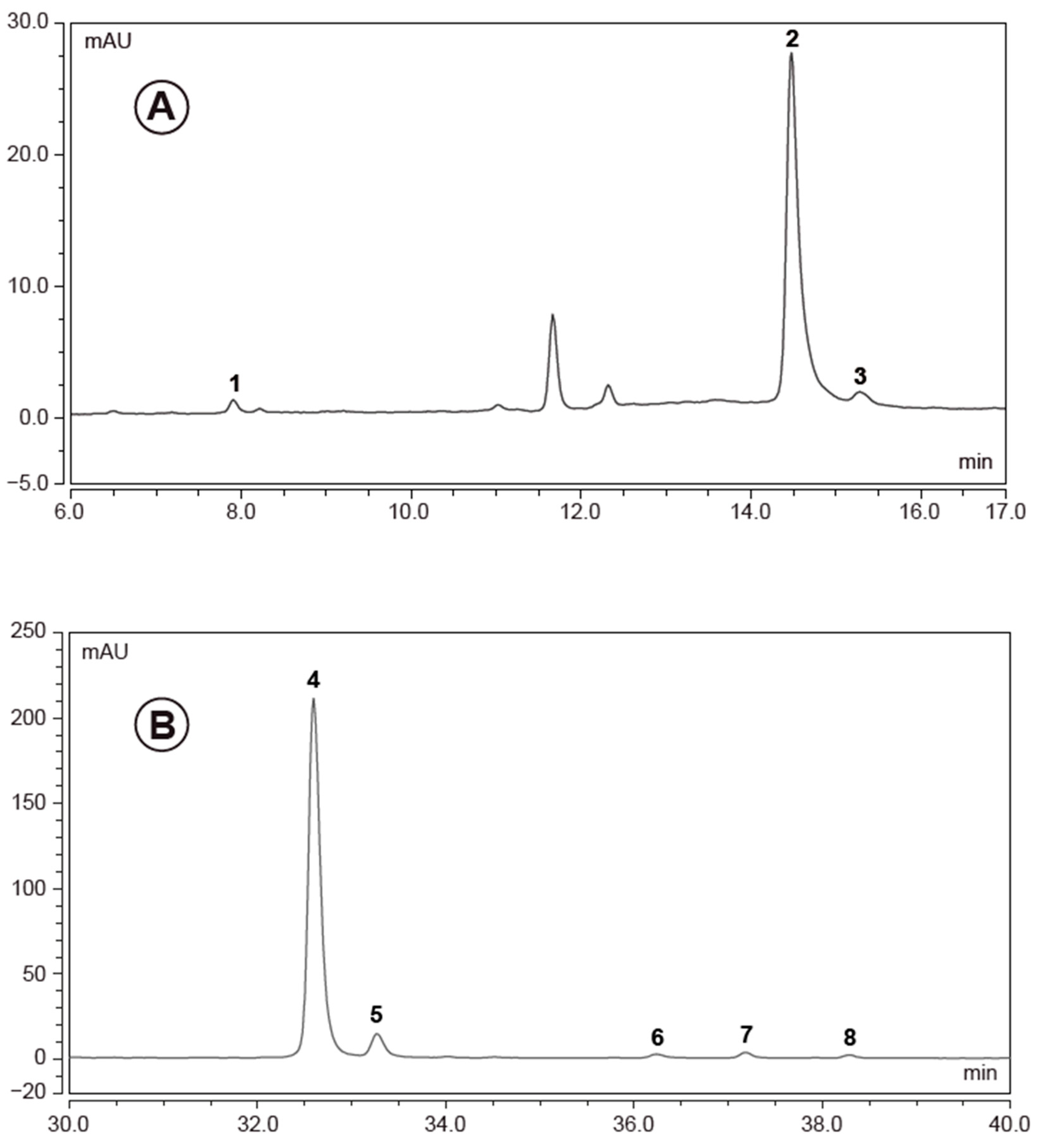
The analysis of the content of phenolic compounds revealed that, among others, mainly rutin, chlorogenic acid and isoquercitirin were present in both black elderberry extracts (chromatogram of liquid extract EC 3.2 shown in Figure 2).
Table 1 summarizes the total amounts of the identified compounds of EC 3.2 and EC 14.

3.2. European Black Elderberry Extract Exhibits Efficient Antiviral Activity against SARS-CoV-2 in Different Cell Lines

In order to determine whether liquid European black elderberry extract (EC 3.2) exhibits antiviral activity against SARS-CoV-2, human Caco-2 colon carcinoma-derived epithelial cells [47] and Calu-3 human lung cells, the most extensively studied surrogate lung cell infection model that expresses ACE2 and TMPRSS2 endogenously [48], were infected with SARS-CoV-2 Wuhan type (Figure 3).
One hour post-infection, different dilutions of EC 3.2 were added to the cell cultures. Three days post-infection (dpi), cell culture supernatants were harvested and virus production was analyzed by quantitative RT-PCR (qRT-PCR) (Figure 3).
Treatment with EC 3.2 led to a dose dependent reduction of virus replication in all infected cell lines. At a dilution of 1:100, EC 3.2 almost completely blocked the production of progeny virions, which was comparable to 1 µM Remdesivir. Thereby, the IC50 values varied between ~1:800 in Calu-3 cells and ~1:200 in Caco-2 cells (Figure 3).
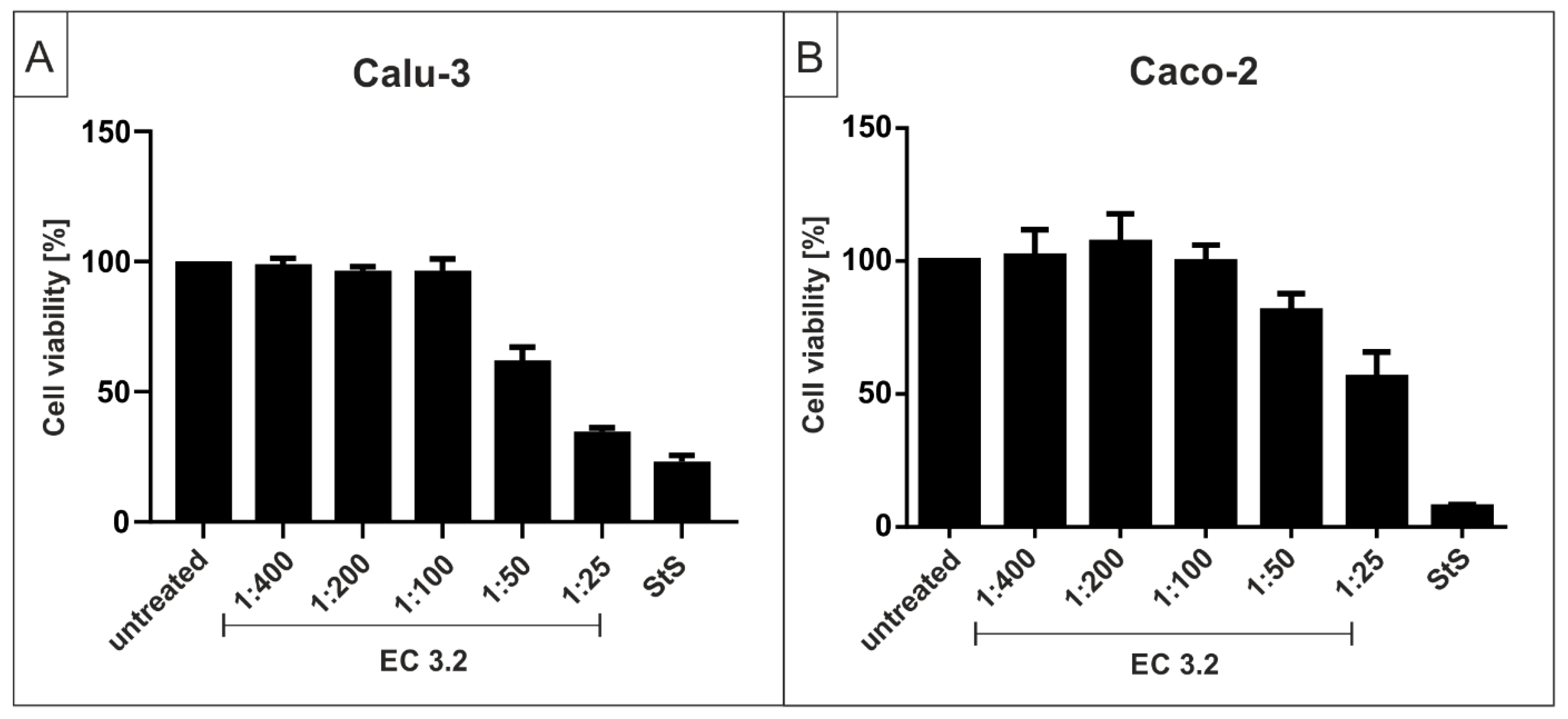
To control for the potential unspecific effects of EC 3.2 treatment on cell viability, water-soluble tetrazolium salt (WST)-1 assays were performed in uninfected Caco-2 or Calu-3 cells under otherwise identical conditions as for the virus infection experiments. The results, summarized in Figure 4, demonstrate that treatment with EC 3.2 at dilutions up to 1:100, and thus in a concentration range where the replication of SARS-CoV-2 was completely blocked, had no impact on cell viability in both investigated cell types (Figure 4). The TD50 values for EC 3.2 were ~1:25 in Caco-2 and 1:50 in Calu-3 cells. Staurosporine (StS) was used as a positive control at a concentration of 1 µM.
Next, it was determined if European black elderberry dried power (EC 14) resolved in water also showed a similar antiviral activity to EC 3.2. As this dried powder is commonly used in food supplements, it is of interest whether the drying process has any negative influence on its antiviral activity.
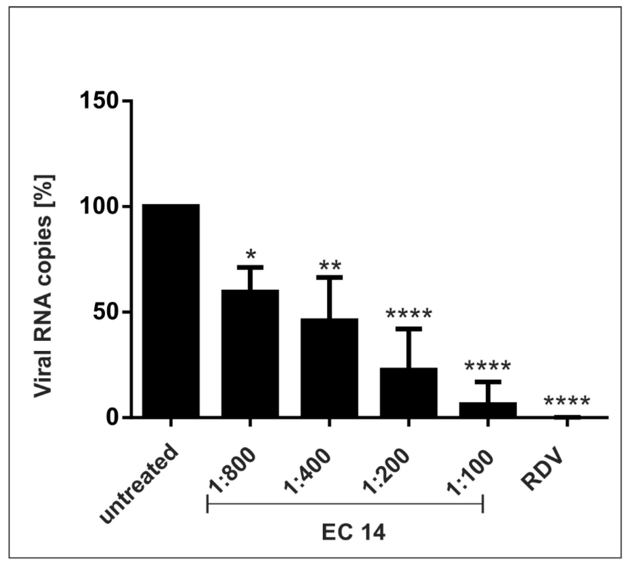
Therefore, Calu-3 cells were infected with the SARS-CoV-2 Wuhan type as described before and qRT-PCR analysis was performed (Figure 5). Similarly to EC 3.2, EC 14 also inhibits the replication of SARS-CoV-2 in a dose-dependent manner with a comparable IC50 value of ~1:800 (Figure 5).

3.3. EC 3.2 Exhibits Comparable Antiviral Activity against All SARS-CoV-2 Variants of Concern

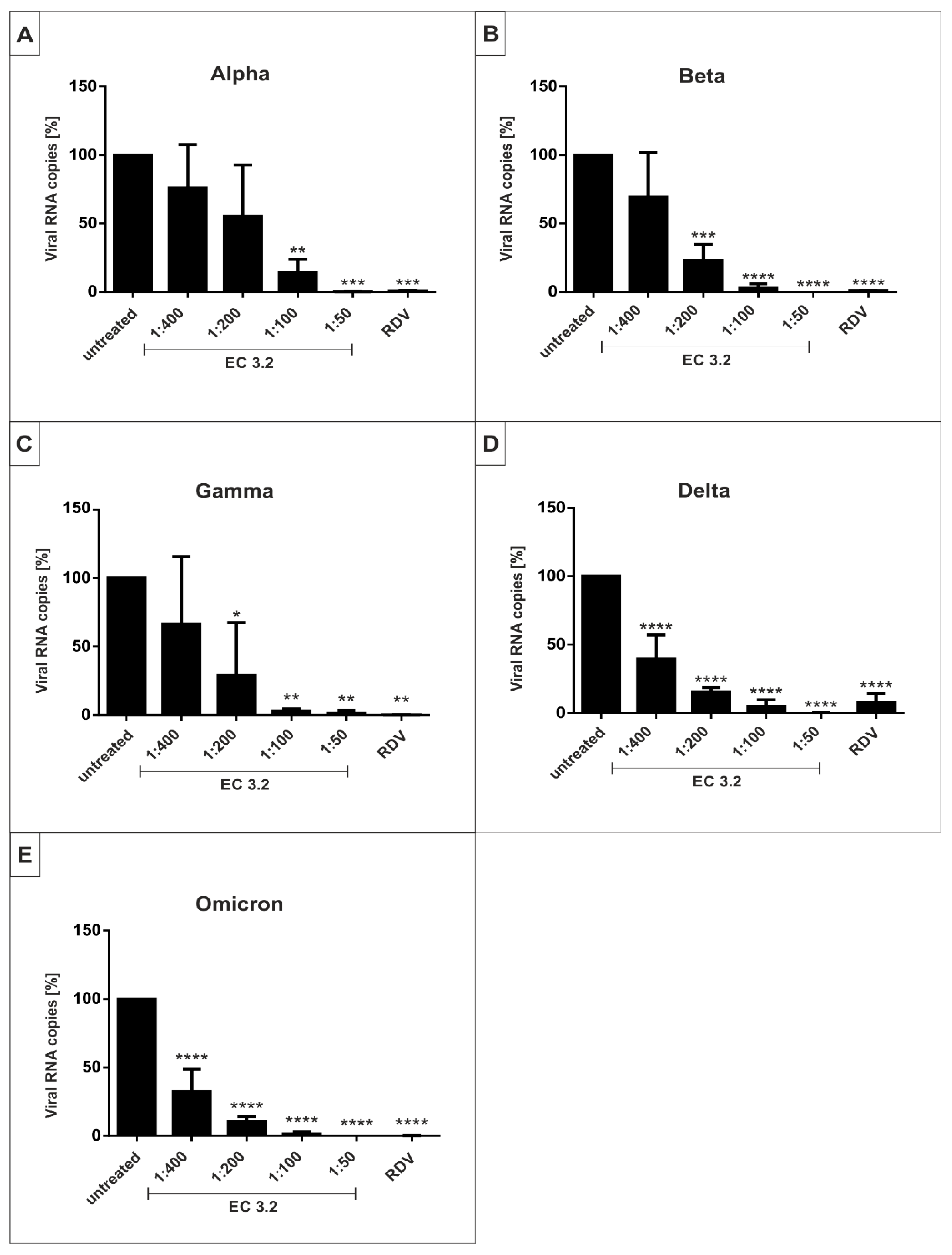
In order to determine if EC 3.2 exhibits a comparable, broad antiviral activity against all described VoCs of SARS-CoV-2, Calu-3 human lung cells were infected with the VoCs Alpha, Beta, Gamma, Delta and Omicron (Figure 6).
One hour post-infection, different dilutions of EC 3.2 were added to the cells. Three dpi cell culture supernatants were harvested and virus production was analyzed by qRT-PCR (Figure 6).
Treatment with EC 3.2 led to a dose-dependent reduction of virus replication that occurred with comparable efficacy for all VoCs (Figure 6). The IC50 values varied between ~1:800 for the Wuhan type (Figure 3) and ~1:200 for Alpha (Figure 6). However, the IC90 was ~1:100 for all VoCs and thus in a similar range (Figure 6).
The IC50 and IC90 values for EC 3.2 following infection with the Wuhan type and respective VoCs are summarized in Table 2.
Next, we calculated the IC50 values for the main compounds of EC 3.2, based on the results of the HPLC analysis (Figure 1 and Figure 2 and Table 1). The results are summarized in Table 3.

3.4. Treatment with EC 3.2 Does Not Affect Early Steps of the Replication of SARS-CoV-2

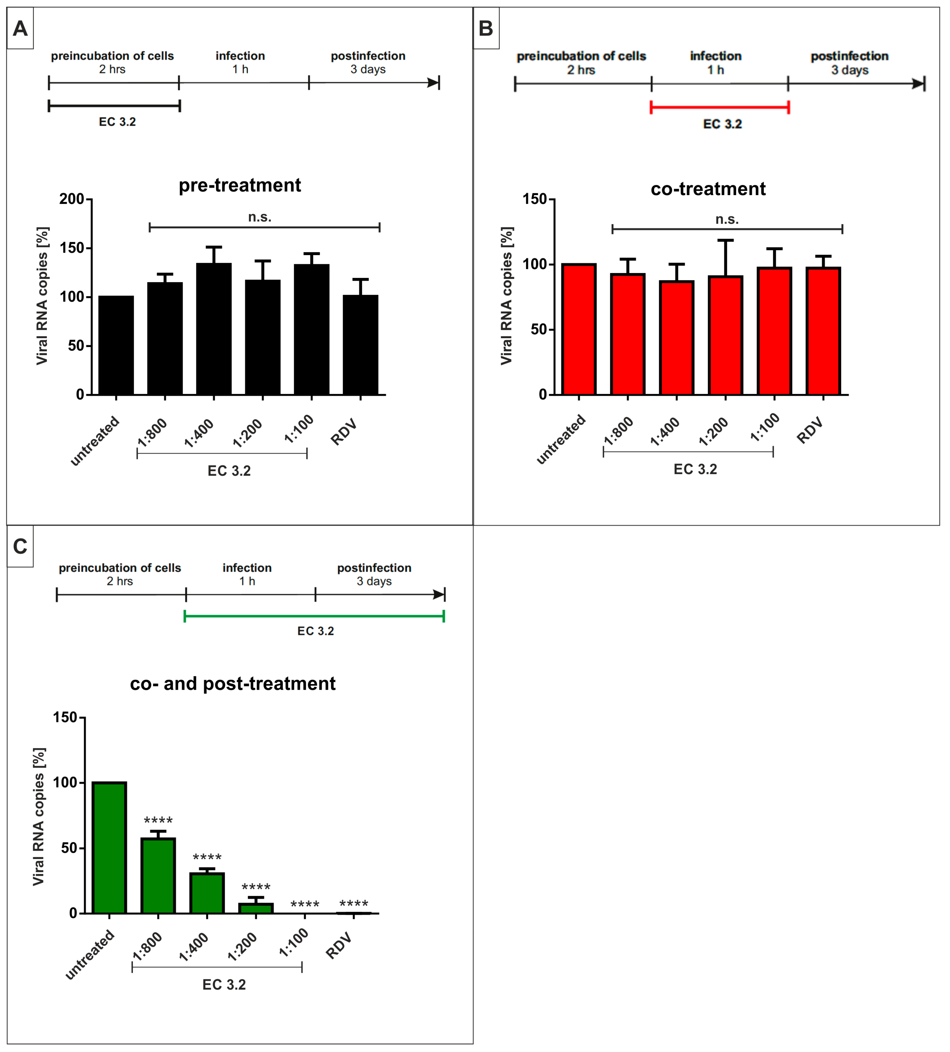
Next, it was analyzed if EC 3.2 blocks the replication of SARS-CoV-2 by interfering with the early steps of the viral replication cycle. Therefore, time of addition (TOA) experiments were performed. First, Calu-3 cells were preincubated with different concentrations of EC 3.2 for 2 h at 37 °C (see treatment scheme Figure 7A). Following two washing steps with PBS to remove EC 3.2, cells were infected with SARS-CoV-2 Wuhan type and cell culture supernatants were harvested after 3 days and analyzed by qRT-PCR.
The data revealed that the preincubation of the cells without further treatment during the infection and post-infection period does not interfere with SARS-CoV-2 replication (Figure 7A). Remdesivir, an inhibitor of RNA metabolism [49], was used as a control and also exhibit no antiviral activity in this experimental setup (Figure 7A).
Next, EC 3.2 was added to Calu-3 cells only during the time of infection with SARS-CoV-2 Wuhan type for 1 h and without applying further treatment to the cells afterwards (treatment scheme Figure 7B). After 1 h, the infectious supernatants were removed and the cells were incubated without further treatment for three days, followed by q RT-PCR analysis of the supernatants. As described for the pre-treatment, the co-treatment with EC 3.2 also has no influence on the replication capacity of SARS-CoV-2 (Figure 7B).
Within a final setup, Calu-3 cells were treated during the 1 h infection period with SARS-CoV-2 Wuhan type and, additionally, during three days post-infection with EC 3.2 (treatment scheme Figure 7C). Thereby, a similar dose-dependent reduction of viral replication (Figure 7C) was detected as with the post-treatment alone (Figure 3A). The IC50 was ~1:800; thus, the additional co-treatment with EC 3.2 has no further influence on the replication capacity of SARS-CoV-2 (Figure 7C). In summary, the TOA experiments revealed that EC 3.2 does not influence early steps in the replication cycle of SARS-CoV-2 but rather exerts its antiviral activity effect during the later steps of viral replication.

4. Discussion

COVID-19, caused by SARS-CoV-2, is still responsible for the ongoing worldwide pandemic that led to a global health and socioeconomic crisis. Similar to the occurrence of SARS-CoV, Middle East respiratory syndrome-related coronavirus (MERS-CoV) and SARS-CoV-2, it can be expected that in the future new coronaviruses will emerge by zoonotic transmission from animals to humans potentially leading to new threats. This demonstrates the need for pandemic preparedness.
Regarding viral infections, vaccines are valuable but have limitations due to the high mutation rate of viruses, especially for RNA viruses such as SARS-CoV-2. This causes the continuous appearance of escape mutants, which are able to evade vaccine-induced immunity. Thus, there is an urgent need for new therapeutics that are available within a short period of time, broadly active, safe, cost-effective and easily distributable for patients all over the world when compared to standard antivirals.
In this study, an antiviral effect of European black elderberry fruit extract against SARS-CoV-2 and its VoCs was shown for the first time. In order to analyze whether black elderberry extract exhibits antiviral activity against various VoCs, Calu-3 cells were infected with SARS-CoV-2 Alpha, Beta, Gamma, Delta and Omicron. The results showed that the replication of these variants could be blocked in a comparable concentration range (Figure 6). This suggests an effective inhibition of viral replication independent of the current and possibly future variants of SARS-CoV-2.
Fruit extracts and juices have previously been shown to be a potential source of antiviral agents (for review, see [50]). Thereby, fruit extracts from, e.g., blackberry, blackcurrant, mulberry and pomegranate exhibit antiviral properties against various viruses such as the Dengue virus, IAV, Zika, Hepatitis C virus (HCV), Human immunodeficiency virus 1 (HIV-1) and the polio virus [50]. For black elderberry extracts, several in vitro studies show an antiviral activity against IAV and IBV, as well as FIV [34,35,36,51,52]. Moreover, an ethanolic Sambucus Formosana Nakai extract exhibits antiviral activity against the endemic human coronavirus HCoV-NL63 [37]. Most importantly, human clinical trials have shown that a Sambucus nigra extract significantly reduces the total duration and severity of upper respiratory symptoms following common cold or IAV infections [38]. In addition, black elderberry extracts exhibit an immunomodulatory effect, which seems to be attributed to the polysaccharide fraction. In this context, it was shown that Sambucus nigra fruits contain peptic polysaccharides influencing the immune system via the activation of macrophages and dendritic cells [53,54,55], which might also contribute to the therapeutic effects seen in human clinical trials.
The black elderberry extract used in this study is a standardized European black elderberry fruit extract (Sambucus nigra, variety ‘Haschberg’) with a total polyphenol content of 4.6% and a total anthocyanin content of 3.5%. The used extract in this study consists of a mixture of organic compounds including but not limited to polyphenols, anthocyanin, mono- and disaccharides, proteins, lipids and carbohydrates. Therefore, the observed antiviral effect of the whole elderberry extract cannot be attributed to single compounds. Further studies including the identified main compounds of the used black elderberry fruit extract are necessary to designate the anti-SARS-CoV-2 effect to specific compounds of the whole extract.
The antiviral activities of polyphenols as well as anthocyanins against various viruses were extensively described previously (for review, see [56,57]). The phenolic compounds of various plants are discussed to be effective against SARS-CoV-2 [58]. For the extracts of some plants, not including black elderberry, an antiviral activity of phenolic compounds against SARS-CoV-2 was shown in vitro [58]. In this study, we identified as the main anthocyanins cyanidin-3-sambubioside-5-glucoside, cyanidin-3-sambubioside and cyanidin-3-glucoside (Figure 1). The main polyphenols were identified as chlorogenic acid, rutin and isoquercetin (Figure 2). This is consistent with previous reports on the phenolic content of elderberry fruits [59,60,61]. For some of these identified compounds, an antiviral activity was reported previously. For instance, isoquercitrin displayed potent antiviral activity against the Varicella Zoster virus (VZV) and human Cytomegalovirus (hCMV) [62]. In addition, a very recent study shows that isoquercitrin inhibits the replication of IAV [63]. Moreover, it was proposed that the combinational treatment of quercetin and vitamin C might be an effective therapy for the prevention and treatment of COVID-19 [64]. For chlorogenic acid, an antiviral activity was described against different viral strains of IAV as well as for Enterovirus 71 [65,66]. However, to our knowledge, the specific anti-SARS-CoV-2 activities of these compounds have not been reported yet.
Analyzing the potential antiviral mode of action of phenolic compounds, it was shown that the flavonoids present in elderberry fruits can directly bind to H1N1 Influenza virus particles, thereby inhibiting the entry of the virus into the host cells [52]. Another study indicates that an ethanolic extract of Sambucus nigra fruits can inhibit the infectious bronchitis virus (IBV), a pathogenic chicken coronavirus, at early points during replication [67]. By performing time of addition experiments, Cho et al. showed that isoquercetin inhibits viral attachment and the entry of IAV [63]. Such a mechanism inhibiting the early stages of the replication of SARS-CoV-2 is unlikely to be the case for European black elderberry extract inasmuch as the time of addition experiments performed in this study displayed antiviral activity only for the later stages of virus replication. Treatment before or during the infection with SARS-CoV-2 had no influence on the replication capacity (Figure 7), strongly indicating an inhibitory effect after virus entry.
However, there are also several reports showing an antiviral effect of anthocyanins or phenolic compounds at later stages in the viral replication cycle. For instance, the main anthocyanin present in European black elderberry fruits, i.e., cyanidin-3-sambubiocide, can bind and inhibit the active pocket of the IAV neuraminidase [51]. In another study, it was demonstrated that natural phenolic compounds are able to inhibit the papain-like protease (PLpro) of SARS-CoV-2 with an IC50 of 4–10 µM [68]. The papain-like protease is a viral protease with multiple functions and is crucial for the virus replication of SARS-CoV-2. Such a mechanism might also be responsible for the observed antiviral effect of black elderberry fruit extract (Figure 3, Figure 5 and Figure 6 and Table 1 and Table 2) and will be the subject of further investigations. For rutin, which is also a main phenolic compound in the black elderberry extract used in this study (Figure 2 and Table 1), an inhibition of the second SARS-CoV-2 protease 3CLpro was shown in a low micromolar range [69,70,71], which might also, at least partially, be responsible for the antiviral effect of European black elderberry fruit extract.
Due to the history of using European black elderberry, it can be considered as safe for ingestion. The unripe berries of black elderberry can contain cyanogenic compounds such a sambunigrin, which is readily degraded during a short heat treatment. Thus, both extracts used in this study, EC 3.2 and EC 14, undergo a short heat treatment during production. This is in concert with the data in this study, which showed no toxic effect on Caco-2 and Calu-3 cells when treated with different concentrations of black elderberry extract up to a dilution of 1:50, which points towards a broad therapeutic window (Figure 4).
The results of this study suggest that European black elderberry fruit extracts could provide beneficial effects in therapeutic settings following a SARS-CoV-2 infection. Their low cytotoxicity and wide availability in nature would make them a readily distributable treatment option for current and future pandemics.

5. Patents

IPRONA AG/SPA has filed a PCT and EP patent entitled “Elderberry extract for use in a method of preventing or treating a SARS-CoV-2 infection” claiming the priority date of 06.12.2021.

Author Contributions

Conceptualization, C.S., U.S., A.S. and S.P.; methodology, C.S., M.F., P.R., M.G. and J.A.; validation, C.S., M.F., S.P. and U.S.; investigation, C.S., M.F., M.G., J.A. and P.R.; data curation, U.S.; writing—original draft preparation, C.S., S.P., A.S. and U.S.; writing—review and editing, C.S., S.P., A.S. and U.S.; visualization, C.S., M.F. and J.A.; supervision, U.S. and S.P.; project administration, C.S., S.P. and U.S.; funding acquisition, S.P. and U.S. All authors have read and agreed to the published version of the manuscript.

Funding

This research was funded by the Deutsche Forschungsgemeinschaft (DFG, German Research Foundation)—401821119/GRK2504 to U.S and supported by the Interdisciplinary Center for Clinical Research (IZKF) at the University Hospital of the University of Erlangen-Nuremberg to J.A. and M.F. (MD-Thesis Scholarship Programme). Additional funding was received by the Autonomous Province of Bozen/Bolzano (Italy)—File 106/2021, decree 14449.

Institutional Review Board Statement

Not applicable.

Informed Consent Statement

Not applicable.

Data Availability Statement

Data are included in the article.

Acknowledgments

The present work was performed in partial fulfillment of the requirements for obtaining Dr. med. degree at the Friedrich-Alexander University Erlangen-Nuremberg for M.F. and J.A.

Conflicts of Interest

The authors have read the journal’s policy and declare that the author Stephan Plattner is employed by IPRONA AG/SPA. Stephan Plattner, Ulrich Schubert, Christian Setz and Alexander Steinkasserer are inventors of a patent submission related to the content of the manuscript; the number of this patent application is EP21212602. The funders had no role in the design of the study; in the collection, analyses, or interpretation of data; in the writing of the manuscript, or in the decision to publish the results. All other authors declare no conflict of interest.

References

  1. Medicine, J.H.U. COVID-19 Dashboard by the Center for Systems Science and Engineering (CSSE) at Johns Hopkins University. Available online: https://coronavirus.jhu.edu/map.html (accessed on 8 December 2022).
  2. Collier, D.A.; De Marco, A.; Ferreira, I.; Meng, B.; Datir, R.P.; Walls, A.C.; Kemp, S.A.; Bassi, J.; Pinto, D.; Silacci-Fregni, C.; et al. Sensitivity of SARS-CoV-2 B.1.1.7 to mRNA vaccine-elicited antibodies. Nature 2021, 593, 136–141. [Google Scholar] [CrossRef] [PubMed]
  3. Wibmer, C.K.; Ayres, F.; Hermanus, T.; Madzivhandila, M.; Kgagudi, P.; Oosthuysen, B.; Lambson, B.E.; de Oliveira, T.; Vermeulen, M.; van der Berg, K.; et al. SARS-CoV-2 501Y.V2 escapes neutralization by South African COVID-19 donor plasma. Nat. Med. 2021, 27, 622–625. [Google Scholar] [CrossRef] [PubMed]
  4. Korber, B.; Fischer, W.M.; Gnanakaran, S.; Yoon, H.; Theiler, J.; Abfalterer, W.; Hengartner, N.; Giorgi, E.E.; Bhattacharya, T.; Foley, B.; et al. Tracking Changes in SARS-CoV-2 Spike: Evidence that D614G Increases Infectivity of the COVID-19 Virus. Cell 2020, 182, 812–827.e19. [Google Scholar] [CrossRef] [PubMed]
  5. Galloway, S.E.; Paul, P.; MacCannell, D.R.; Johansson, M.A.; Brooks, J.T.; MacNeil, A.; Slayton, R.B.; Tong, S.; Silk, B.J.; Armstrong, G.L.; et al. Emergence of SARS-CoV-2 B.1.1.7 Lineage—United States, December 29, 2020–January 12, 2021. MMWR Morb. Mortal. Wkly. Rep. 2021, 70, 95–99. [Google Scholar] [CrossRef] [PubMed]
  6. Meng, B.; Kemp, S.A.; Papa, G.; Datir, R.; Ferreira, I.; Marelli, S.; Harvey, W.T.; Lytras, S.; Mohamed, A.; Gallo, G.; et al. Recurrent emergence of SARS-CoV-2 spike deletion H69/V70 and its role in the Alpha variant B.1.1.7. Cell Rep. 2021, 35, 109292. [Google Scholar] [CrossRef]
  7. Tegally, H.; Wilkinson, E.; Giovanetti, M.; Iranzadeh, A.; Fonseca, V.; Giandhari, J.; Doolabh, D.; Pillay, S.; San, E.J.; Msomi, N.; et al. Detection of a SARS-CoV-2 variant of concern in South Africa. Nature 2021, 592, 438–443. [Google Scholar] [CrossRef]
  8. Public Health England Investigation of SARS-CoV-2 Variants of Concern: Technical Briefings. Available online: https://www.gov.uk/government/publications/investigation-of-novel-sars-cov-2-variant-variant-of-concern-20201201 (accessed on 13 April 2022).
  9. Mwenda, M.; Saasa, N.; Sinyange, N.; Busby, G.; Chipimo, P.J.; Hendry, J.; Kapona, O.; Yingst, S.; Hines, J.Z.; Minchella, P.; et al. Detection of B.1.351 SARS-CoV-2 Variant Strain—Zambia, December 2020. MMWR Morb. Mortal. Wkly. Rep. 2021, 70, 280–282. [Google Scholar] [CrossRef]
  10. National Institute of Infectious Diseases (NIID) of Japan Brief Report: New Variant Strain of SARS-CoV-2 Identified in Travelers from Brazil. Available online: https://www.niid.go.jp/niid/en/2019-ncov-e/10108-covid19-33-en.html (accessed on 29 July 2022).
  11. Cherian, S.; Potdar, V.; Jadhav, S.; Yadav, P.; Gupta, N.; Das, M.; Rakshit, P.; Singh, S.; Abraham, P.; Panda, S.; et al. SARS-CoV-2 Spike Mutations, L452R, T478K, E484Q and P681R, in the Second Wave of COVID-19 in Maharashtra, India. Microorganisms 2021, 9, 1542. [Google Scholar] [CrossRef]
  12. WHO. Classification of Omicron (B. 1.1. 529): SARS-CoV-2 Variant of Concern; World Health Organization: Geneva, Switzerland, 2021; Available online: https://www.who.int/news/item/26-11-2021-classification-of-omicron-(b.1.1.529)-sars-cov-2-variant-of-concern (accessed on 28 November 2021).
  13. Ke, H.; Chang, M.R.; Marasco, W.A. Immune Evasion of SARS-CoV-2 Omicron Subvariants. Vaccines 2022, 10, 1545. [Google Scholar] [CrossRef]
  14. Arora, P.; Kempf, A.; Nehlmeier, I.; Schulz, S.R.; Jäck, H.M.; Pöhlmann, S.; Hoffmann, M. Omicron sublineage BQ.1.1 resistance to monoclonal antibodies. Lancet Infect. Dis. 2022, 23, 22–23. [Google Scholar] [CrossRef]
  15. Karim, S.S.A.; Karim, Q.A. Omicron SARS-CoV-2 variant: A new chapter in the COVID-19 pandemic. Lancet 2021, 398, 2126–2128. [Google Scholar] [CrossRef] [PubMed]
  16. Volz, E.; Mishra, S.; Chand, M.; Barrett, J.C.; Johnson, R.; Geidelberg, L.; Hinsley, W.R.; Laydon, D.J.; Dabrera, G.; O'Toole, Á.; et al. Assessing transmissibility of SARS-CoV-2 lineage B.1.1.7 in England. Nature 2021, 593, 266–269. [Google Scholar] [CrossRef] [PubMed]
  17. Davies, N.G.; Abbott, S.; Barnard, R.C.; Jarvis, C.I.; Kucharski, A.J.; Munday, J.D.; Pearson, C.A.B.; Russell, T.W.; Tully, D.C.; Washburne, A.D.; et al. Estimated transmissibility and impact of SARS-CoV-2 lineage B.1.1.7 in England. Science 2021, 372, eabg3055. [Google Scholar] [CrossRef]
  18. Kim, Y.J.; Jang, U.S.; Soh, S.M.; Lee, J.Y.; Lee, H.R. The Impact on Infectivity and Neutralization Efficiency of SARS-CoV-2 Lineage B.1.351 Pseudovirus. Viruses 2021, 13, 633. [Google Scholar] [CrossRef] [PubMed]
  19. Planas, D.; Veyer, D.; Baidaliuk, A.; Staropoli, I.; Guivel-Benhassine, F.; Rajah, M.M.; Planchais, C.; Porrot, F.; Robillard, N.; Puech, J.; et al. Reduced sensitivity of SARS-CoV-2 variant Delta to antibody neutralization. Nature 2021, 596, 276–280. [Google Scholar] [CrossRef] [PubMed]
  20. Wang, P.; Casner, R.G.; Nair, M.S.; Wang, M.; Yu, J.; Cerutti, G.; Liu, L.; Kwong, P.D.; Huang, Y.; Shapiro, L.; et al. Increased Resistance of SARS-CoV-2 Variant P.1 to Antibody Neutralization. bioRxiv 2021. [Google Scholar] [CrossRef] [PubMed]
  21. WHO Therapeutics and COVID-19: Living Guideline. Available online: https://www.who.int/publications/i/item/WHO-2019-nCoV-therapeutics-2022.1 (accessed on 8 February 2022).
  22. EMA EMA Issues Advice on Use of Paxlovid (PF-07321332 and Ritonavir) for the Treatment of COVID-19: Rolling Review Starts in Parallel. Available online: https://www.ema.europa.eu/en/news/ema-issues-advice-use-paxlovid-pf-07321332-ritonavir-treatment-covid-19-rolling-review-starts (accessed on 8 February 2022).
  23. National Insitutes of Health COVID-19 Treatment Guidelines—Therapeutic Management of Nonhospitalized Adults with COVID-19. Available online: https://www.covid19treatmentguidelines.nih.gov/management/clinical-management/nonhospitalized-adults--therapeutic-management/ (accessed on 28 July 2022).
  24. Arbel, R.; Wolff Sagy, Y.; Hoshen, M.; Battat, E.; Lavie, G.; Sergienko, R.; Friger, M.; Waxman, J.G.; Dagan, N.; Balicer, R.; et al. Nirmatrelvir Use and Severe COVID-19 Outcomes during the Omicron Surge. N. Engl. J. Med. 2022, 387, 790–798. [Google Scholar] [CrossRef]
  25. Singh, A.K.; Singh, A.; Singh, R.; Misra, A. Molnupiravir in COVID-19: A systematic review of literature. Diabetes Metab. Syndr. 2021, 15, 102329. [Google Scholar] [CrossRef]
  26. Creech, C.B.; Walker, S.C.; Samuels, R.J. SARS-CoV-2 Vaccines. JAMA 2021, 325, 1318–1320. [Google Scholar] [CrossRef]
  27. European Medicines Agency. COVID-19 Vaccines: Authorised. Available online: https://www.ema.europa.eu/en/human-regulatory/overview/public-health-threats/coronavirus-disease-covid-19/treatments-vaccines/vaccines-covid-19/covid-19-vaccines-authorised#authorised-covid-19-vaccines-section (accessed on 22 March 2022).
  28. European Centre for Disease Prevention and Control Risk of SARS-CoV-2 Transmission from Newly-Infected Individuals with Documented Previous Infection or Vaccination. Available online: https://www.ecdc.europa.eu/en/publications-data/sars-cov-2-transmission-newly-infected-individuals-previous-infection#copy-to-clipboard (accessed on 18 August 2022).
  29. Große, M.; Ruetalo, N.; Layer, M.; Hu, D.; Businger, R.; Rheber, S.; Setz, C.; Rauch, P.; Auth, J.; Fröba, M.; et al. Quinine Inhibits Infection of Human Cell Lines with SARS-CoV-2. Viruses 2021, 13, 647. [Google Scholar] [CrossRef]
  30. Mani, J.S.; Johnson, J.B.; Steel, J.C.; Broszczak, D.A.; Neilsen, P.M.; Walsh, K.B.; Naiker, M. Natural product-derived phytochemicals as potential agents against coronaviruses: A review. Virus Res. 2020, 284, 197989. [Google Scholar] [CrossRef] [PubMed]
  31. Auth, J.; Fröba, M.; Große, M.; Rauch, P.; Ruetalo, N.; Schindler, M.; Morokutti-Kurz, M.; Graf, P.; Dolischka, A.; Prieschl-Grassauer, E.; et al. Lectin from Triticum vulgaris (WGA) Inhibits Infection with SARS-CoV-2 and Its Variants of Concern Alpha and Beta. Int. J. Mol. Sci. 2021, 22, 10205. [Google Scholar] [CrossRef] [PubMed]
  32. Fröba, M.; Große, M.; Setz, C.; Rauch, P.; Auth, J.; Spanaus, L.; Münch, J.; Ruetalo, N.; Schindler, M.; Morokutti-Kurz, M.; et al. Iota-Carrageenan Inhibits Replication of SARS-CoV-2 and the Respective Variants of Concern Alpha, Beta, Gamma and Delta. Int. J. Mol. Sci. 2021, 22, 13202. [Google Scholar] [CrossRef]
  33. Roxas, M.; Jurenka, J. Colds and influenza: A review of diagnosis and conventional, botanical, and nutritional considerations. Altern. Med. Rev. J. Clin. Ther. 2007, 12, 25–48. [Google Scholar]
  34. Krawitz, C.; Mraheil, M.A.; Stein, M.; Imirzalioglu, C.; Domann, E.; Pleschka, S.; Hain, T. Inhibitory activity of a standardized elderberry liquid extract against clinically-relevant human respiratory bacterial pathogens and influenza A and B viruses. BMC Complement. Altern. Med. 2011, 11, 16. [Google Scholar] [CrossRef] [PubMed] [Green Version]
  35. Uncini Manganelli, R.E.; Zaccaro, L.; Tomei, P.E. Antiviral activity in vitro of Urtica dioica L. Parietaria diffusa M. et K. and Sambucus nigra L. J. Ethnopharmacol. 2005, 98, 323–327. [Google Scholar] [CrossRef] [PubMed]
  36. Zakay-Rones, Z.; Varsano, N.; Zlotnik, M.; Manor, O.; Regev, L.; Schlesinger, M.; Mumcuoglu, M. Inhibition of several strains of influenza virus in vitro and reduction of symptoms by an elderberry extract (Sambucus nigra L.) during an outbreak of influenza B Panama. J. Altern. Complement. Med. 1995, 1, 361–369. [Google Scholar] [CrossRef]
  37. Weng, J.R.; Lin, C.S.; Lai, H.C.; Lin, Y.P.; Wang, C.Y.; Tsai, Y.C.; Wu, K.C.; Huang, S.H.; Lin, C.W. Antiviral activity of Sambucus FormosanaNakai ethanol extract and related phenolic acid constituents against human coronavirus NL63. Virus Res. 2019, 273, 197767. [Google Scholar] [CrossRef]
  38. Hawkins, J.; Baker, C.; Cherry, L.; Dunne, E. Black elderberry (Sambucus nigra) supplementation effectively treats upper respiratory symptoms: A meta-analysis of randomized, controlled clinical trials. Complement. Ther. Med. 2019, 42, 361–365. [Google Scholar] [CrossRef]
  39. Tiralongo, E.; Wee, S.S.; Lea, R.A. Elderberry Supplementation Reduces Cold Duration and Symptoms in Air-Travellers: A Randomized, Double-Blind Placebo-Controlled Clinical Trial. Nutrients 2016, 8, 182. [Google Scholar] [CrossRef] [Green Version]
  40. Zakay-Rones, Z.; Thom, E.; Wollan, T.; Wadstein, J. Randomized study of the efficacy and safety of oral elderberry extract in the treatment of influenza A and B virus infections. J. Int. Med. Res. 2004, 32, 132–140. [Google Scholar] [CrossRef] [PubMed]
  41. Kinoshita, E.; Hayashi, K.; Katayama, H.; Hayashi, T.; Obata, A. Anti-influenza virus effects of elderberry juice and its fractions. Biosci. Biotechnol. Biochem. 2012, 76, 1633–1638. [Google Scholar] [CrossRef] [PubMed]
  42. Becker, M.; Dulovic, A.; Junker, D.; Ruetalo, N.; Kaiser, P.D.; Pinilla, Y.T.; Heinzel, C.; Haering, J.; Traenkle, B.; Wagner, T.R.; et al. Immune response to SARS-CoV-2 variants of concern in vaccinated individuals. Nat. Commun. 2021, 12, 3109. [Google Scholar] [CrossRef] [PubMed]
  43. Setz, C.; Große, M.; Auth, J.; Fröba, M.; Rauch, P.; Bausch, A.; Wright, M.; Schubert, U. Synergistic Antiviral Activity of Pamapimod and Pioglitazone against SARS-CoV-2 and Its Variants of Concern. Int. J. Mol. Sci. 2022, 23, 6830. [Google Scholar] [CrossRef]
  44. Reed, L.J.M. A Simple Method of Estimating Fifty Per Cent Endpoints. Am. J. Epidemiol. 1936, 27, 493–497. [Google Scholar] [CrossRef]
  45. Corman, V.M.; Landt, O.; Kaiser, M.; Molenkamp, R.; Meijer, A.; Chu, D.K.; Bleicker, T.; Brünink, S.; Schneider, J.; Schmidt, M.L.; et al. Detection of 2019 novel coronavirus (2019-nCoV) by real-time RT-PCR. Euro Surveill. 2020, 25, 2000045. [Google Scholar] [CrossRef] [Green Version]
  46. Anon, M. Determination of Anthocyanins by HPLC No. 71. In Handbook of Analytical Methods; The international Fruit Juice Union: Paris, France, 1999. [Google Scholar]
  47. Bertram, S.; Glowacka, I.; Blazejewska, P.; Soilleux, E.; Allen, P.; Danisch, S.; Steffen, I.; Choi, S.Y.; Park, Y.; Schneider, H.; et al. TMPRSS2 and TMPRSS4 facilitate trypsin-independent spread of influenza virus in Caco-2 cells. J. Virol. 2010, 84, 10016–10025. [Google Scholar] [CrossRef] [Green Version]
  48. Aguiar, J.A.; Tremblay, B.J.; Mansfield, M.J.; Woody, O.; Lobb, B.; Banerjee, A.; Chandiramohan, A.; Tiessen, N.; Cao, Q.; Dvorkin-Gheva, A.; et al. Gene expression and in situ protein profiling of candidate SARS-CoV-2 receptors in human airway epithelial cells and lung tissue. Eur. Respir. J. 2020, 56, 2001123. [Google Scholar] [CrossRef]
  49. Kokic, G.; Hillen, H.S.; Tegunov, D.; Dienemann, C.; Seitz, F.; Schmitzova, J.; Farnung, L.; Siewert, A.; Höbartner, C.; Cramer, P. Mechanism of SARS-CoV-2 polymerase stalling by remdesivir. Nat. Commun. 2021, 12, 279. [Google Scholar] [CrossRef]
  50. Santhi, V.P.; Sriramavaratharajan, V.; Murugan, R.; Masilamani, P.; Gurav, S.S.; Sarasu, V.P.; Parthiban, S.; Ayyanar, M. Edible fruit extracts and fruit juices as potential source of antiviral agents: A review. J. Food Meas. Charact. 2021, 15, 5181–5190. [Google Scholar] [CrossRef]
  51. Swaminathan, K.; Dyason, J.C.; Maggioni, A.; von Itzstein, M.; Downard, K.M. Binding of a natural anthocyanin inhibitor to influenza neuraminidase by mass spectrometry. Anal. Bioanal. Chem. 2013, 405, 6563–6572. [Google Scholar] [CrossRef] [PubMed]
  52. Roschek, B., Jr.; Fink, R.C.; McMichael, M.D.; Li, D.; Alberte, R.S. Elderberry flavonoids bind to and prevent H1N1 infection in vitro. Phytochemistry 2009, 70, 1255–1261. [Google Scholar] [CrossRef] [PubMed]
  53. Ho, G.T.; Ahmed, A.; Zou, Y.F.; Aslaksen, T.; Wangensteen, H.; Barsett, H. Structure-activity relationship of immunomodulating pectins from elderberries. Carbohydr. Polym. 2015, 125, 314–322. [Google Scholar] [CrossRef] [PubMed] [Green Version]
  54. Ho, G.T.; Wangensteen, H.; Barsett, H. Elderberry and Elderflower Extracts, Phenolic Compounds, and Metabolites and Their Effect on Complement, RAW 264.7 Macrophages and Dendritic Cells. Int. J. Mol. Sci. 2017, 18, 584. [Google Scholar] [CrossRef] [Green Version]
  55. Stich, L.; Plattner, S.; McDougall, G.; Austin, C.; Steinkasserer, A. Polysaccharides from European Black Elderberry Extract Enhance Dendritic Cell Mediated T Cell Immune Responses. Int. J. Mol. Sci. 2022, 23, 3949. [Google Scholar] [CrossRef]
  56. Badshah, S.L.; Faisal, S.; Muhammad, A.; Poulson, B.G.; Emwas, A.H.; Jaremko, M. Antiviral activities of flavonoids. Biomed. Pharmacother. 2021, 140, 111596. [Google Scholar] [CrossRef]
  57. Mohammadi Pour, P.; Fakhri, S.; Asgary, S.; Farzaei, M.H.; Echeverría, J. The Signaling Pathways, and Therapeutic Targets of Antiviral Agents: Focusing on the Antiviral Approaches and Clinical Perspectives of Anthocyanins in the Management of Viral Diseases. Front. Pharmacol. 2019, 10, 1207. [Google Scholar] [CrossRef] [Green Version]
  58. Tirado-Kulieva, V.A.; Hernández-Martínez, E.; Choque-Rivera, T.J. Phenolic compounds versus SARS-CoV-2: An update on the main findings against COVID-19. Heliyon 2022, 8, e10702. [Google Scholar] [CrossRef]
  59. Młynarczyk, K.; Walkowiak-Tomczak, D.; Łysiak, G.P. Bioactive properties of Sambucus nigra L. as a functional ingredient for food and pharmaceutical industry. J. Funct. Foods 2018, 40, 377–390. [Google Scholar] [CrossRef]
  60. Mocanu, M.L.; Amariei, S. Elderberries-A Source of Bioactive Compounds with Antiviral Action. Plants 2022, 11, 740. [Google Scholar] [CrossRef]
  61. Lee, J.; Finn, C.E. Anthocyanins and other polyphenolics in American elderberry (Sambucus canadensis) and European elderberry (S. nigra) cultivars. J. Sci. Food Agric. 2007, 87, 2665–2675. [Google Scholar] [CrossRef] [PubMed]
  62. Kim, C.H.; Kim, J.E.; Song, Y.J. Antiviral Activities of Quercetin and Isoquercitrin against Human Herpesviruses. Molecules 2020, 25, 2379. [Google Scholar] [CrossRef] [PubMed]
  63. Cho, W.K.; Lee, M.M.; Ma, J.Y. Antiviral Effect of Isoquercitrin against Influenza a Viral Infection via Modulating Hemagglutinin and Neuraminidase. Int. J. Mol. Sci. 2022, 23, 13112. [Google Scholar] [CrossRef] [PubMed]
  64. Colunga Biancatelli, R.M.L.; Berrill, M.; Catravas, J.D.; Marik, P.E. Quercetin and Vitamin C: An Experimental, Synergistic Therapy for the Prevention and Treatment of SARS-CoV-2 Related Disease (COVID-19). Front. Immunol. 2020, 11, 1451. [Google Scholar] [CrossRef] [PubMed]
  65. Li, X.; Liu, Y.; Hou, X.; Peng, H.; Zhang, L.; Jiang, Q.; Shi, M.; Ji, Y.; Wang, Y.; Shi, W. Chlorogenic acid inhibits the replication and viability of enterovirus 71 in vitro. PLoS ONE 2013, 8, e76007. [Google Scholar] [CrossRef] [Green Version]
  66. Ding, Y.; Cao, Z.; Cao, L.; Ding, G.; Wang, Z.; Xiao, W. Antiviral activity of chlorogenic acid against influenza A (H1N1/H3N2) virus and its inhibition of neuraminidase. Sci. Rep. 2017, 7, 45723. [Google Scholar] [CrossRef] [Green Version]
  67. Chen, C.; Zuckerman, D.M.; Brantley, S.; Sharpe, M.; Childress, K.; Hoiczyk, E.; Pendleton, A.R. Sambucus nigra extracts inhibit infectious bronchitis virus at an early point during replication. BMC Vet. Res. 2014, 10, 24. [Google Scholar] [CrossRef] [Green Version]
  68. Srinivasan, V.; Brognaro, H.; Prabhu, P.R.; de Souza, E.E.; Günther, S.; Reinke, P.Y.A.; Lane, T.J.; Ginn, H.; Han, H.; Ewert, W.; et al. Antiviral activity of natural phenolic compounds in complex at an allosteric site of SARS-CoV-2 papain-like protease. Commun. Biol. 2022, 5, 805. [Google Scholar] [CrossRef]
  69. Agrawal, P.K.; Agrawal, C.; Blunden, G. Rutin: A Potential Antiviral for Repurposing as a SARS-CoV-2 Main Protease (Mpro) Inhibitor. Nat. Prod. Commun. 2021, 16, 1934578X21991723. [Google Scholar] [CrossRef]
  70. Rahman, F.; Tabrez, S.; Ali, R.; Alqahtani, A.S.; Ahmed, M.Z.; Rub, A. Molecular docking analysis of rutin reveals possible inhibition of SARS-CoV-2 vital proteins. J. Tradit. Complement. Med. 2021, 11, 173–179. [Google Scholar] [CrossRef]
  71. Rizzuti, B.; Grande, F.; Conforti, F.; Jimenez-Alesanco, A.; Ceballos-Laita, L.; Ortega-Alarcon, D.; Vega, S.; Reyburn, H.T.; Abian, O.; Velazquez-Campoy, A. Rutin Is a Low Micromolar Inhibitor of SARS-CoV-2 Main Protease 3CLpro: Implications for Drug Design of Quercetin Analogs. Biomedicines 2021, 9, 375. [Google Scholar] [CrossRef] [PubMed]
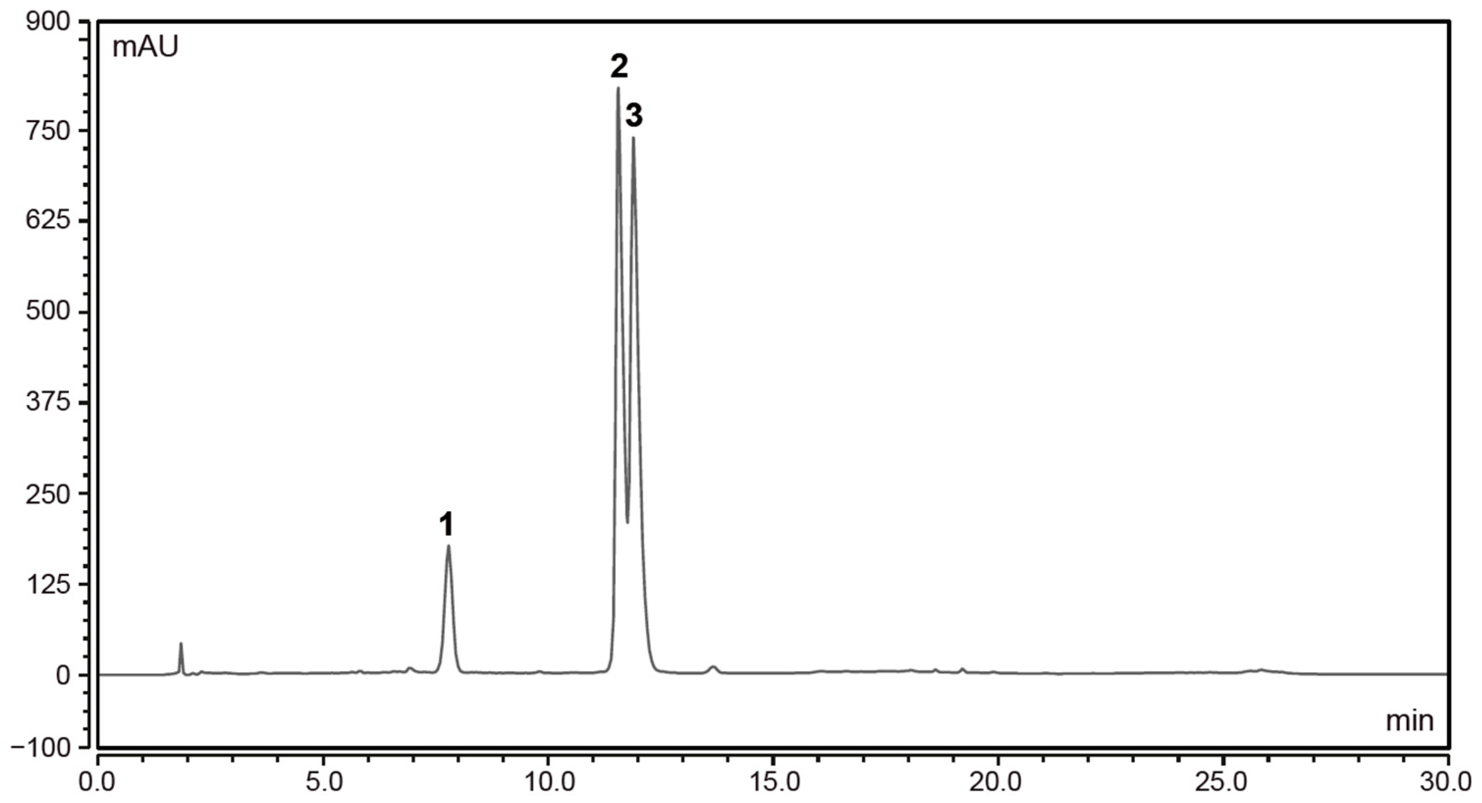
Figure 1. The HPLC chromatogram of anthocyanins in EC 3.2. Peak 1: cyanidin-3-sambubioside-5-glucoside; peak 2: cyanidin-3-sambubioside; peak 3: cyanidin-3-glucoside. Absorbance (mAU) measured at 520 nm.
Nutraceuticals 03 00007 g001
Figure 2. The HPLC chromatogram of phenolic compounds in EC 3.2. (A): peak 1: neochlorogenic acid; peak 2: chlorogenic acid; peak 3: cryptochlorogenic acid. (B): peak 4: rutin; peak 5: isoquercitrin; peak 6: kaempferol-3-rutinoside; peak 7: isorhamnetin-3-rutinoside; peak 8: isorhamnetin-3-glucoside.
Nutraceuticals 03 00007 g002
Figure 3. Liquid European black elderberry (EC 3.2) extract inhibits replication of SARS-CoV-2 Wuhan type in Calu-3 and Caco-2 cells. Calu-3 (A) and Caco-2 cells (B) were infected with the clinical isolate SARS-CoV-2PR-1 at a MOI of 2 × 10⁻². One hour after infection and removal of input virus, cells were treated with indicated dilution steps of EC 3.2. A total of 1 µM Remdesivir (RDV) was included as a positive control. Cell culture supernatants were harvested at 3 dpi. The virions were purified and analyzed by qRT-PCR. Bars show mean values of three independent experiments ± standard deviation. Statistical analysis was performed using a multiple comparison Kruskal–Wallis test (Anova) followed by Dunn’s post hoc test (*** p < 0.001; **** p < 0.0001 versus the untreated control).
Nutraceuticals 03 00007 g003
Figure 4. Influence of EC 3.2 on the cell viability of Calu-3 (A) and Caco-2 cells (B). Following treatment with different dilutions of EC 3.2 (indicated at the x-axis) for three days, the influence on cell viability was measured by water-soluble tetrazolium salt (WST)-1 assay. Bars represent means of 3 independent experiments ± SD. Staurosprine (StS, 1 µM) was used as a positive control.
Nutraceuticals 03 00007 g004
Figure 5. European black elderberry dried powder (EC 14) resolved in water inhibits replication of SARS-CoV-2 Wuhan type. Calu-3 cells were infected with the clinical isolate SARS-CoV-2PR-1 at a MOI of 2 × 10⁻². One hour after infection and removal of input virus, cells were treated with indicated dilution steps of EC 14. A total of 1 µM RDV was included as a positive control. Cell culture supernatants were harvested at 3 dpi. The virions were purified and analyzed by qRT-PCR. Bars show mean values of three independent experiments ± standard deviation. Statistical analysis was performed using a multiple comparison Kruskal–Wallis test (Anova) followed by Dunn’s post hoc test (* p < 0.05; ** p < 0.01; **** p < 0.0001 versus the untreated control).
Nutraceuticals 03 00007 g005
Figure 6. EC 3.2 inhibits replication of the SARS-CoV-2 Variants of Concern (VoCs) Alpha, Beta, Gamma, Delta and Omicron with comparable antiviral efficacy. Calu-3 cells were infected with clinical isolates of the SARS-CoV-2 VoCs Alpha (A), Beta (B), Gamma (C), Delta (D) or Omicron (E) at a MOI of 2 × 10⁻². One hour after infection and removal of input virus, cells were treated with the indicated dilutions of EC 3.2. A total of 1 µM RDV was included as a positive control. Cell culture supernatants were harvested at 3 dpi. The virions were purified and analyzed by qRT-PCR. Bars show mean values of three independent experiments ± standard deviation. Statistical analysis was performed using a multiple comparison Kruskal–Wallis test (Anova) followed by Dunn’s post hoc test (* p < 0.05; ** p < 0.01; *** p < 0.001; **** p < 0.0001 versus the untreated control).
Nutraceuticals 03 00007 g006
Figure 7. Treatment with EC 3.2 before or during the infection with SARS-CoV-2 has no influence on viral replication. (A) Time of addition (TOA) of EC 3.2 with the indicated dilutions. Calu-3 cells were preincubated with EC 3.2 for 2 h following two washing steps with PBS. (B) The TOA of EC 3.2 was only during infection of Calu-3 cells for 1 h. (C) The TOA of EC 3.2 was during infection of cells for 1 h and following removal of input virus, for 3 days post-infection. (AC) Cells were infected with the clinical isolate SARS-CoV-2PR-1 at a MOI of 2 × 10⁻² for 1 h. A total of 1 µM RDV was included as a positive control. Cell culture supernatants were harvested at 3 dpi. The virions were purified and analyzed by qRT-PCR. Bars show mean values of three independent experiments ± standard deviation. Statistical analysis was performed using a multiple comparison Kruskal–Wallis test (Anova) followed by Dunn’s post hoc test (n.s. = not significant; **** p < 0.0001 versus the untreated control).
Nutraceuticals 03 00007 g007
Table 1. Quantitative analysis of compounds in black elderberry extracts EC 3.2 and EC 14.
Compound EC 3.2 (mg/kg)EC 14 (mg/kg)
Cyanidin-3-glucoside14,88958,336
Cyanidin-3-sambubioside16,58477,507
Cyanidin-3-sambubioside-5-glucoside355813,682
Neochlorogenic acid42151
Chlorogenic acid18905953
Cryptochlorogenic acid72359
Rutin841931,999
Isoquercitrin5071784
Kaempferol-3-rutinoside69195
Isorhamnetin-3-rutinoside67221
Isorhamnetin-3-glucoside48215
Table 2. IC50 and IC90 values of EC 3.2 against SARS-CoV-2 Wuhan type and VoCs in Calu-3 cells.
EC 3.2
IC50IC90
Wuhan Type~1:800~1:100
Alpha~1:200~1:100
Beta~1:300~1:100
Gamma~1:300~1:100
Delta~1:400~1:100
Omicron~1:400~1:100
Table 3. The IC50 values [µM] of the main compounds of EC 3.2 following infection of Calu-3 cells with SARS-CoV-2 Wuhan Type and VoCs.
IC50 [µM]
Wuhan TypeAlphaBetaGammaDeltaOmicron
Cyanidin-3-sambubioside-5-glucoside62418181212
Cyanidin-3-sambubioside351421071077171
Cyanidin-3-glucoside411651231238282
Chlorogenic acid6.62720201313
Rutin176851513434
Isoquercitrin1.35.4442.72.7
Disclaimer/Publisher’s Note: The statements, opinions and data contained in all publications are solely those of the individual author(s) and contributor(s) and not of MDPI and/or the editor(s). MDPI and/or the editor(s) disclaim responsibility for any injury to people or property resulting from any ideas, methods, instructions or products referred to in the content.