Article

A Chemical Investigation of the Antioxidant Capacity of Extracts from Red Macroalga Gracilaria domingensis

by
Priscila Torres
*,
Fungyi Chow
* and
Deborah Yara Alves Cursino dos Santos
*
Department of Botany, Institute of Biosciences, University of São Paulo, São Paulo 05508-090, Brazil
*
Authors to whom correspondence should be addressed.
Phycology 2022, 2(3), 332-343; https://doi.org/10.3390/phycology2030018
Submission received: 27 July 2022 / Revised: 17 August 2022 / Accepted: 22 August 2022 / Published: 25 August 2022

Abstract

:
Extracts that were obtained with solvents of increasing polarity (hexane, dichloromethane, methanol, 80% methanol, and water) from the red macroalga Gracilaria domingensis were evaluated by reducing power with ferric reduction antioxidant power (FRAP) and Folin–Ciocalteu (FC) assays, lipid peroxidation inhibition by β-carotene-linoleic acid assay, and metal chelating ability based on the iron-ferrozine system. The highest antioxidant capacity was reported for the hexane (Hx) extract by the FRAP, metal chelating, and lipid peroxidation inhibition assays. An activity-guided fractionation of the Hx extract was carried out for the identification of its active constituents. The primary components were the most active antioxidant compounds. Despite the high antioxidant activities, the Hx extract was not active in the FC assay. In this assay, the activities were found in the methanol (M) and 80% methanol (80M) extracts. The FC assay is commonly used to measure the total phenolic compounds. However, no phenolic compounds were detected by GC-MS and HPLC analyses in the M and 80M extracts. Thus, non-phenolic components influenced the FC assay. The M and 80M extracts showed high content of mycosporine-like amino acids (MAAs). A fraction contained two MAAs (porphyra-334 and shinorine) (156 mg GAE·g−1) showed a similar performance to the values that were found for well-known antioxidants (BHT = 156 mg GAE·g−1 and Trolox = 166 mg GAE·g−1) and 30 times higher than those of the original extracts (~5 mg GAE·g−1) in the FC assay. Thus, MAAs contribute to the antioxidant activities that were observed in the FC assay within the studied samples. Together, these results advance our understanding of the antioxidant properties of algal extracts.

1. Introduction

Reactive oxygen species (ROS) are produced in the organism naturally as a byproduct of metabolism. An imbalance between ROS production and antioxidant protection creates oxidative stress, which can lead to biomolecule damage. Antioxidants play a crucial role in biological systems since they prevent or delay the oxidation of these biomolecules. Evaluating the antioxidant capacity provides relevant information that may be used in different research fields, such as studies of the abundance and distribution of species in ecology [1] and understanding of the physiological state of species under stress [2]. Besides, in vitro antioxidant assays are low-cost and require simple laboratory procedures.
There are several in vitro assays to measure the antioxidant capacities. These in vitro assays can evaluate different mechanisms of antioxidant action [3]. Assays that are based on hydrogen atom transfer (HAT) measure the free-radical scavenging capacity of an antioxidant by hydrogen donation [4]. The inhibition of β-carotene bleaching that is caused by peroxidation of linoleic acid is a well-known HAT-based assay. In the assays that are based on electron-transfer (ET) reaction, the free radical is stabilized by donating an electron from an antioxidant [5]. The ability to reduce ferric iron (Fe3+) to ferrous iron (Fe2+) measured on the FRAP (ferric reduction antioxidant power) and the ability to reduce the Folin–Ciocalteu (FC) reagent that was measured in the FC assay are based on ET reactions. In the literature, the FC assay is commonly used for the quantification of the total phenolic content (TPC). Despite the popularity for this use, the electron transfer reactions to the FC reagent are not performed exclusively by the phenolic compounds [6,7], so the scientific community has discussed the use of the FC assay for measuring concentrations of TPC [8]. Yet, a third kind of assay can measure the ability of the antioxidant to chelate transition metals. Metal chelating assays measure the ability to sequester transition metals that cause free radical generation. A chelator agent such as ferrozine is used in these assays.
Due to the diversity of mechanisms of antioxidant action and radical types, an antioxidant may react in an assay, but it may be not sensitive in others. Consequently, more than one assay should be used to evaluate the antioxidant capacity of a sample. This capacity is related to the interaction effects (synergistic, additive, and antagonistic) between compounds. Detterweiler et al. [9] proposed an index to evaluate how much the interaction between the extract components is responsible for biological activities. Interactions between antioxidant components are often in complex mixtures such as extracts [10]. Thus, the activity-guided fractionation can provide us with a better understanding of the antioxidant properties of crude extracts.
Antioxidant capacity has been studied in numerous genera of macroalgae. Part of these studies searches for natural antioxidants and extracts for health, food, and cosmetic applications [11,12] and other topics such as the evaluation of the effect of abiotic stresses, or variation among seasons. For example, the antioxidant capacity from Gracilaria cornea (Rhodophyta) increased under UV exposure [13], and the antioxidant capacity differed between dry and rainy seasons for Sargassum vulgare (Ochrophyta) and Palisada flagellifera (Rhodophyta), with the highest antioxidant activities in the rainy season for the first and in the dry season for the last [14].
Gracilaria (Gracilariaceae, Rhodophyta) is a genus of red macroalga comprising of about 197 species that are distributed nearly worldwide [15]. This genus includes species of economic value due to the agar, a phycocolloid that is widely used in the food and biotechnology industries [16]. The antioxidant potentials from Gracilaria species are reported mainly for crude extracts [17], with promising results for some species. Methanolic extracts from Gracilaria corticata and Gracilaria edulis have significant activities in the FRAP assay when compared to BHT (butylated hydroxytoluene) [18], and some crude extracts from Gracilaria gracilis have higher or similar reducing activities than BHT and gallic acid. [19].
Compounds with antioxidant properties that are already identified in the genus include bromophenols from Gracilaria edulis [20], carotenoids from Gracilaria birdiae and Gracilaria tenuistipitata [21,22], floridoside from G. tenuistipitata [23], citric acid from Gracilaria vermiculophylla [24], and mycosporine-like amino acids (MAAs) from several species [13,25,26,27]. Phenolic compounds are among the most potent antioxidants in nature, and they are found in abundance in land plants and brown algae. Benzoic acid, gallic acid, gentisic acid, p-hydroxybenzoic acid, protocatechuic acid, salicylic acid, syringic acid, and vanillic acid are phenolic compounds that have been identified in Gracilaria [17].
Gracilaria domingensis (Kützing) Sonder ex Dickie is distributed in the Western Atlantic Ocean from Mexico to northeast Brazil. The antioxidant potential of G. domingensis was evaluated in two studies. One study evaluated only crude extracts [21], and another study focused on the algal nutritional potential using bleached samples [25]. Unlike previous studies, we investigate the components that are responsible for the antioxidant capacities of the crude extracts using activity-guided fractionation. This procedure allows locating the compounds that are active. To this end, we selected assays that evaluate the main mechanisms of antioxidant action: electron-transfer reaction (FRAP and Folin–Ciocalteu assays), hydrogen atom transfer reaction (β-carotene-linoleic acid assay), and the ability to chelate transition metals based on the measurement of the iron-ferrozine complex.

2. Material and Methods

2.1. Algal Samples

Gracilaria domingensis was collected in the intertidal zone at “Morro de Pernambuco” Beach in the city of Ilhéus in Bahia state (14°48′21.6″ S, 39°01′25.6″ W). The fresh sample (10 kg) was rinsed in tap water and triaged for removal of epibionts. DNA barcoding (COI-5P—mitochondrial cytochrome c oxidase subunit I gene) and morphological characters were used for taxonomical identification of the species [28,29]. The samples were initially air-dried at room temperature in a shaded environment (~one day), and then in an air circulation oven at 40 °C (~two days).

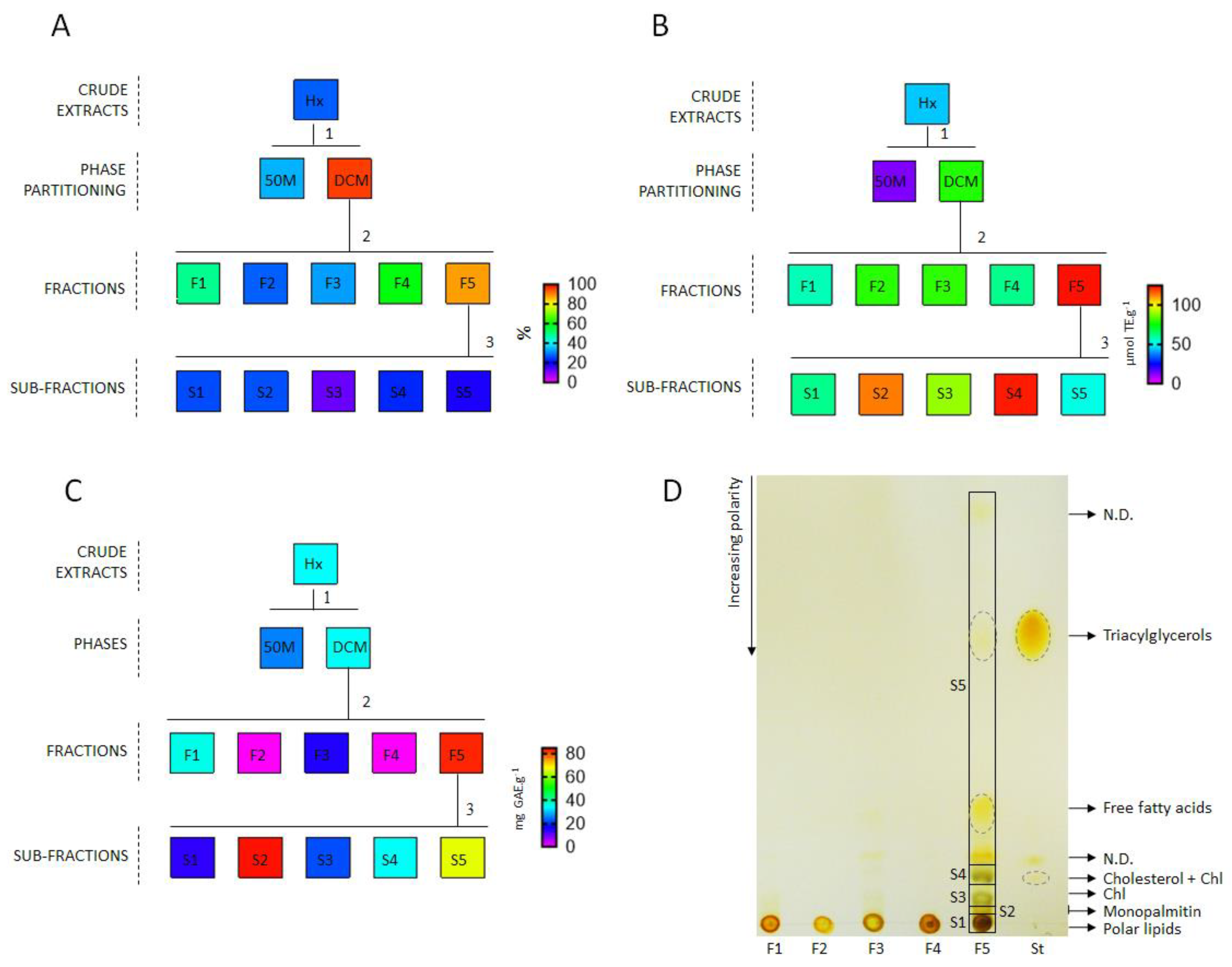
2.2. Preparation of the Crude Extracts and Fractionation of the Active Extracts

Figure 1 depicts the general scheme of the extraction and fractionation procedure. The dried samples were ground in a knife mill (30-mesh; Fortinox® STAR FT 80, Piracicaba, Brazil). The algal powder (1 kg) was macerated at 50 ± 5 °C for 8 h in organic solvents (1:10, w/v) in increasing order of polarity starting with hexane, followed by dichloromethane, methanol, and 80% methanol. The final residue was macerated in ultrapure water (1:20, w/v) at 50 ± 5 °C for 8 h. The dried methanolic and hydromethanolic extracts were desalted using methanol precipitation by mixing with one volume of methanol. After the homogenization, the sample was allowed to sit at room temperature for 15–30 min, and the supernatant was collected. The organic extracts were concentrated in a rotary evaporator (<45 °C), and the aqueous extract was freeze-dried. There were five crude extracts that were obtained: hexane (Hx = 4.2 g), dichloromethane (DCM = 2.9 g), methanol (M = 65 g), 80% methanol (80M = 29 g), and aqueous (Aq = 340 g).
The Hx and M extracts were partitioned and fractionated. The Hx extract was dissolved in DCM and partitioned with 50% methanol (yields of 3.5 g and 0.5 g, respectively). Part of the DCM phase (2.5 g) was fractionated in silica column chromatography (silanized silica gel 60, Merck Millipore, Darmstadt, Germany) using as mobile phase a solvent gradient in decreasing order of polarity (water—250 mL, 25% methanol—250 mL, 50% methanol—250 mL, 80% methanol—250 mL, methanol—250 mL, methanol:dichloromethane (7:3)—250 mL, methanol:dichloromethane (3:7)—2 × 250 mL, dichloromethane—6 × 250 mL, and hexane—250 mL). All the fractions were monitored by analytical thin-layer chromatography (TLC; Merck Silica Gel 60 F254, Darmstadt, Germany), visualized using iodine vapor, and pooled together according to elution similarity, resulting in five groups (F1—20 mg, F2—16 mg, F3—165 mg, F4—632 mg, and F5—1.6 g). Part of the group F5 (41 mg) was fractionated in preparative thin-layer chromatography (TLC; 60G silica gel, MerckMillipore, impregnated with 0.02% sodium fluorescein). The mobile phase was hexane:diethyl ether:acetic acid (80:20:1, v/v/v). The plates were visualized under UV light, and five bands were scraped off the silica gel, resulting in the subfractions S1 (3.4 mg), S2 (11 mg), S3 (3 mg), S4 (4.3 mg), and S5 (19 mg). The M extract was dissolved in 50% methanol and partitioned with DCM (yields of 59 g and 5 g, respectively). Part of the 50% methanol phase (531 mg) was fractionated by semi-preparative high-performance liquid chromatography (HPLC; Agilent 1200 series, USA) equipment using a semi-preparative column (Zorbax RX-SIL HILIC, 9.4 mm × 250 mm, 5 μm, USA), with a mobile phase of acetonitrile (solvent A) and 0.2% acetic acid (solvent B) in isocratic elution with 68% A for 20 min, and flow rate of 4 mL min−1. This fractionation procedure resulted in a fraction that was rich in two MAAs (31 mg; porphyra-334 and shinorine, 70% and 30% respectively).
The chemical characterization of the active extracts and fractions was performed using analytical TLC plates, GC-MS (Gas Chromatography-Mass Spectrometry), and an analytical HPLC system. The group F5 was analyzed by analytical TLC (Merck Silica Gel 60 F254, Darmstadt, Germany) [30]. The mobile phase was hexane:diethyl ether:acetic acid (80:20:1, v/v/v), and the plates were visualized using iodine vapor. The M and 80M extracts and subfractions S1 to S5 were analyzed by GC-MS and HPLC with Diode Array Detection (HPLC-DAD). For the GC-MS analysis, the samples (100 µg) were dissolved in 50 μL of pyridine and derivatized by reaction with 50 μL of BSTFA for 30 min at 80 °C. The derivatized samples were evaluated by GC-MS (Agilent 6890N/5975, Malaysia) that was equipped with an HP-5MS column (30 m × 0.25 mm, 0.25 µm, USA) [31]. For the HPLC–DAD analysis, the samples were injected into a 1260 Infinity LC System (Agilent, Santa Clara, CA, USA). A ZORBAX Eclipse plus C18 column (150 mm × 4.6 mm, 3.5 μm, USA) was used for the analysis of the metabolites, and the mobile phase was a gradient of 0.1% acetic acid and acetonitrile [32]. The M and 80M extracts were also analyzed for MAAs through HPLC-DAD using an HILIC column (Zorbax RX-SIL, 250 mm × 4.6 mm, 5 µm, USA), and a gradient of acetonitrile:5 mM ammonium acetate (9:1, v/v) and acetonitrile:5 mM ammonium acetate (1:1, v/v) as mobile phase [33]. The MAAs were identified by comparison with isolated standards and quantified using a calibration curve of porphyra-334 (R2 > 0.99).

2.3. Determination of the Antioxidant Capacity

All the assays were performed in 96-well clear polystyrene microplates using a microplate reader (Synergy™ H1 BioTek®, Winooski, VT, USA). All the samples (crude extracts, phase partitioning, fractions, and subfractions) were dissolved in DMSO and diluted in ultrapure water to obtain sample concentrations between 0.6 and 6 mg mL−1 (final concentrations in the wells between 40 and 400 µg mL−1) and a DMSO concentration of 10% (v/v). In all the assays, a negative control (sample volume was replaced by 10% DMSO), positive control (sample volume was replaced by standard), and sample blank (sample + reagents of the assay, but without one reagent to avoid the reaction) were included. Sample blanks are used to avoid errors from color or turbidity that may exist in the sample. Only corrected absorbances by subtracting the sample blank absorbance from the absorbance after reaction were used for calculations. There were two natural antioxidants (rutin and p-coumaric acid) and two synthetic antioxidants (Trolox and BHT—butylated hydroxytoluene) that were used as positive controls. The results were expressed in gallic acid equivalents (mg GAE·g−1 dry extract) for the Folin–Ciocalteu and metal chelating assays, Trolox equivalent (µmol TE·g−1 dry extract) for the FRAP assay, and protection percentages from the negative control for the β-carotene-linoleic acid assay.

2.4. Ferric Reduction Antioxidant Power (FRAP) Assay

The reducing power by the FRAP assay was conducted following the method that was described previously [34]. The FRAP solution was prepared mixing 0.3 M acetate buffer (pH 3.6), 10 mM TPTZ in 40 mM hydrochloric acid, and 20 mM ferric chloride in a ratio of 10:1:1. Briefly, a volume of 20 µL of sample (final concentration in the well: 400 µg mL−1 for crude extracts and 150 µg mL−1 for other samples), negative control, or standard was added to each well along with 15 µL of ultrapure water and 265 µL of FRAP solution. After incubation at 37 °C for 20 min, the absorbances were measured at 595 nm. For the sample blanks, a FRAP solution was prepared without TPTZ. A calibration curve was constructed using Trolox (final concentrations in well between 0.5 and 7 µg mL−1, R2 > 0.99).

2.5. Metal Chelating Ability Based on the Measurement of Iron-Ferrozine Complex

The metal chelating ability assay was performed as the method in the microplate format that was described previously [35]. Briefly, a volume of 20 µL of sample (final concentration in the well: 400 µg mL−1 for crude extracts and 150 µg mL−1 for other samples), negative control, or standard was added to each well, followed by the addition of 260 µL of 10% (w/v) ammonium acetate and 10 µL of 1 mM ferrous ammonium sulfate. After incubation for 5 min, a volume of 10 µL of 6.1 mM ferrozine was added to each well. The microplate was incubated for 10 min, and the absorbances were measured at 562 nm. For the sample blanks, the volume of the ferrozine solution in the well was replaced by ultrapure water. A calibration curve was constructed using gallic acid (final concentrations in the wells between 1 and 14 µg mL−1, R2 > 0.99).

2.6. Lipid Peroxidation Inhibition Using the β-Carotene-Linoleic Acid Assay

The assay of inhibition of β-carotene bleaching was performed with modifications from the methods that were described previously [14]. The reactive solution was prepared by mixing 16 µL of linoleic acid, 160 µL of tween 40, and 200 µL of β-carotene (2 mg mL−1 in dichloromethane). The solution was evaporated until the complete removal of the dichloromethane (<45 °C), and then 50 mL of ultrapure water that was bubbled with air for 30 min was added. The absorbance was adjusted to 0.90 ± 0.10 at 450 nm. For the assay, 20 µL of sample (final concentration in the well: 100 µg mL−1), negative control, or standard was added to each well with 280 µL of reactive solution. The microplate was incubated at 45 °C under stirring, and the absorbance was measured at 450 nm in the time 0 min (sample blank) and 120 min.

2.7. Folin–Ciocalteu (FC) Assay

This assay was performed based on a previously described method [36]. Into each well containing 200 µL of ultrapure water, 20 µL of sample (final concentration in the well: 400 µg mL−1 for crude extracts and 40 µg mL−1 for fraction), negative control, or standard was added along with 20 µL of Folin–Ciocalteu reagent, and 60 µL of saturated sodium carbonate (25%; w/v). After incubation for 30 min, the absorbances were measured at 760 nm. For the sample blanks, the volume of the FC reagent in the well was replaced by ultrapure water. A calibration curve was constructed using gallic acid (final concentrations in the well between 1 and 10 µg mL−1, R2 > 0.99).

2.8. Interaction Indexes

Interaction indexes (Ii) were calculated based on Dettweiler et al. [9], with adaptations to use for values that are expressed in mass equivalent of the reference antioxidant or inhibition percentage. Values ≤ 0.5 indicate synergy, values between 0.5–4 indicate non-interaction, and values > 4 indicate antagonism. The index was calculated using the following formula:   ( A s   ×   Y s ) / A f , where As = antioxidant value of the fraction/subfraction, Ys = yield of the fraction/subfraction, and Af = antioxidant value of the original sample. The formula was applied to each fraction/subfraction, and the sum of all the values is the interaction index.

2.9. Statistical Analysis
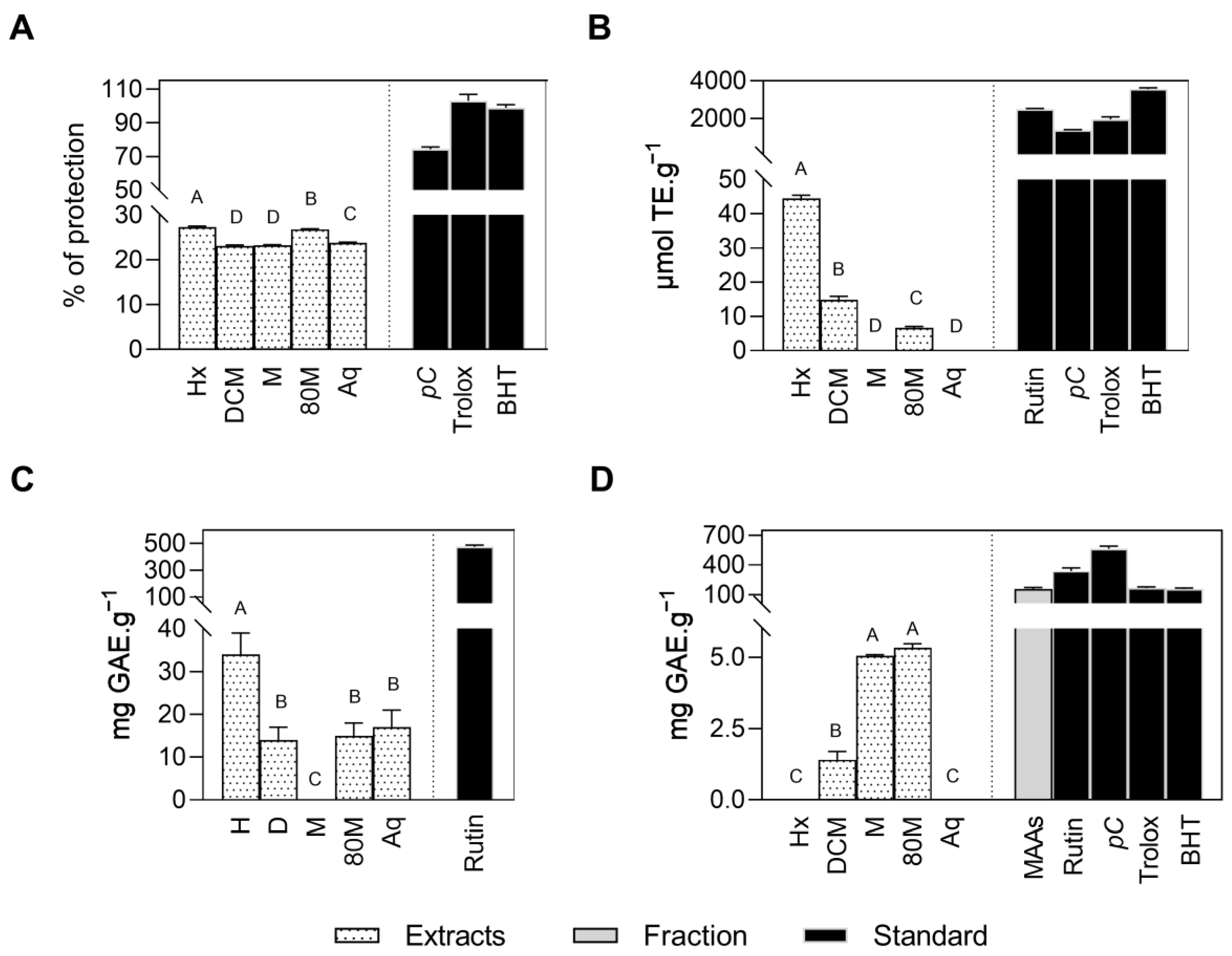
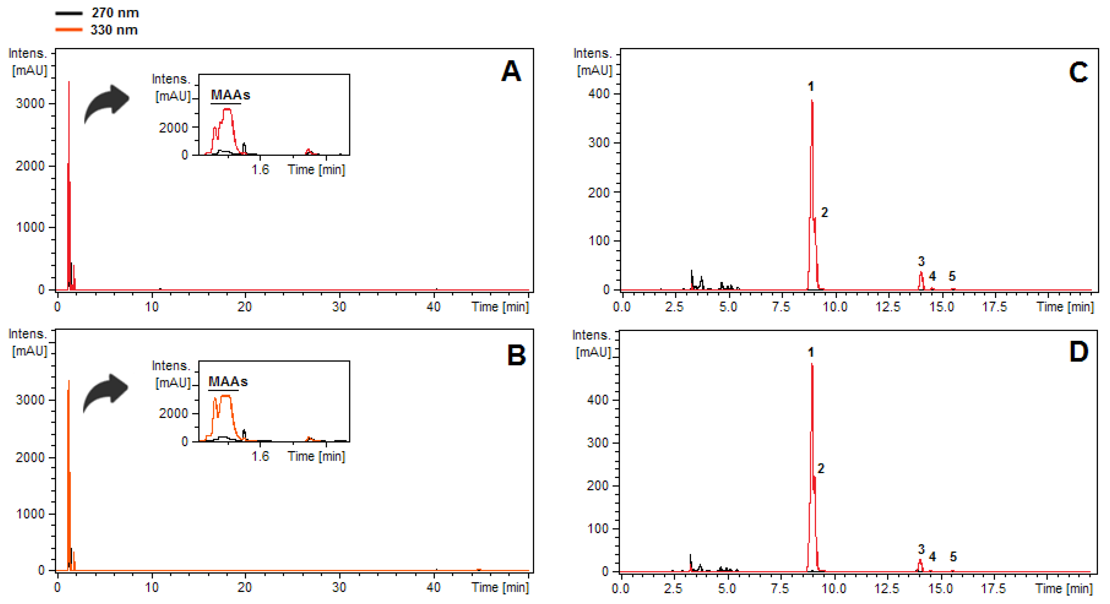
The data analysis and graphing software was GraphPad 8®. The results were expressed as the mean ± standard deviation (SD; n = 3, technical triplicate). The comparisons between the means were performed by one-way ANOVA. The Tukey test was used in post hoc analysis (p < 0.05).

3. Results

The Aq extract showed the highest yield (34% on dry algal mass), while the Hx and DCM extracts gave the lowest yields (values < 1%). The M and 80M extracts yielded 6.5% and 3%, respectively. The Hx extract was the most effective in three-antioxidant assays: lipid peroxidation inhibition (Figure 2A), FRAP (Figure 2B), and metal chelating assay (Figure 2C). However, this extract had no activity in the FC assay (Figure 2D). The DCM extract showed weak to moderate activities in the four assays (Figure 2). The M and 80M extracts were the most effective in the FC assay (Figure 2D), while the Aq extract showed lipid peroxidation inhibition (Figure 2A) and metal chelating ability (Figure 2C).
An activity-guided fractionation of the Hx extract was carried out for the identification of its active constituents (Figure 3). The fractionation procedure resulted in the fraction F5 with higher antioxidant activities than those of the crude extract (Figure 3). The level of lipid peroxidation inhibition for the fraction F5 (85% of protection) was higher than the value for p-coumaric acid—74% of protection (100 µg mL−1). However, further purifications from this fraction resulted in a loss of lipid peroxidation inhibition, with values ranging from 10% to 20% of protection for all subfractionations (Figure 3A). The interaction index (Ii) among them was 0.23, suggesting that the subfractions from F5 have a strong synergistic effect. On the other hand, a synergistic effect was not observed among subfractions on FRAP and metal chelating assays (interaction index between 0.5–4). Similar activities to fraction F5 were found for subfractions S2 and S4 in the FRAP assay (Figure 3B) and for S2 in the metal chelating assay (Figure 3C).
The chemical composition of the fraction F5 includes free fatty acids, triacylglycerols, monopalmitin, chlorophyll a and its derivatives, cholesterol, and polar lipids (Figure 3D). The latter is also found in fractions F1 to F4 (Figure 3D). Concerning active subfractions, GC-MS analysis confirmed monopalmitin (40%) as the main peak in the subfraction S2 and cholesterol (97%) in the subfraction S4. Chlorophyll a and its derivatives were confirmed in the subfractions S3 and S4 using HPLC analysis.
Once the M and 80M extracts were the most active in the FC assay (Figure 2D), the search for phenolic compounds was carried out in these extracts. However, even after analysis in GC-MS (Figure S1) and HPLC (Figure 4A,B), no phenolic compounds were detected in these extracts. Carboxylic acids (e.g., citric acid), free amino acids (e.g., glutamine), heterosides (floridoside and isofloridoside), monosaccharides, and isethionic acid were characteristic components of these extracts that were detected by GC-MS (Figure S1 and Table S1), while MAAs were practically the unique class of compounds that were found in the HPLC-DAD chromatograms (Figure 4A,B). The methodology for analysis for specific MAAs allowed identification of porphyra-334 as the major MAAs of the polar extracts, followed by shinorine, palythine, asterina-330, and palythinol (Figure 4C,D).
The polar extracts contained a high amount of MAAs, with contents of 7.8% and 9.6% (dry mass of the extract), respectively, for the M and 80M extracts. Due to the remarkably high MAAs contents in the M and 80M extracts, a fraction of MAA (porphyra-334 + shinorine, 70% and 30%, respectively) was isolated and evaluated in the FC assay. This fraction (156 mg GAE·g−1) showed a similar performance to the values that were found for well-known synthetic antioxidants (BHT = 156 mg GAE·g−1 and Trolox = 166 mg GAE·g−1) and 30 times greater than those of the original extracts (~5 mg GAE·g−1) (Figure 2D).

4. Discussion

Hexane (Hx) and methanolic (M and 80M) extracts of Gracilaria domingensis presented the higher antioxidant activities, enabling them for further fractionation and compound identification. The synergetic effects between compounds might be responsible for the antioxidant activities that were observed for a fraction of Hx extract against lipid peroxidation inhibition. MAAs were the main components of the polar extracts. An enriched fraction with porphyra-334 and shinorine presented a Folin–Ciocalteu assay value that was 30 times greater than those of the original extracts.
Despite the low yield, the Hx extract was the most active extract, with significant activities in the three assays (FRAP, metal chelating, and lipid peroxidation inhibition). Similar results for non-polar extracts of Gracilaria gracilis were obtained previously with the FRAP assay [19]. The fractionation procedure from the Hx extract allowed to observe a loss in the lipid peroxidation inhibition (Figure 3A), suggesting synergistic interaction effects between the compounds. Synergistic interactions between antioxidant components are common in complex mixtures such as crude extracts [10]. Algal extracts have shown synergistic antioxidant activities when they are supplemented with isolated compounds [37] and other crude extracts [38].
In contrast to lipid peroxidation inhibition, the fractionation procedure led to the subfractions with activities that were similar to the original fraction in the FRAP and ferrous chelating assays. The main compounds from fraction F5 and its subfractions are primary metabolites with several reports of antioxidant activities. For example, chlorophyll a and its derivatives have iron-chelating ability [39], and monoacylglycerols are described with reducing activities in the FRAP assay [40].
In contrast to the Hx extract, the Aq extract showed the highest yield, but only weak antioxidant activities in ferrous chelating (Figure 2C) and inhibition of lipid peroxidation (Figure 2A) assays. Our previous study showed sulfated polysaccharides as the main component of this Aq extract [31]. The antioxidant properties, including lipid peroxidation inhibition and ferrous chelating activity, have already been reported for sulfated polysaccharides from Gracilaria [41,42].
The M and 80M extracts were the most active extracts in the FC assay. The activities for these extracts (~5 mg GAE·g−1) were higher or similar to those that were seen in another study with Gracilaria, such as G. birdiae (~1 mg GAE·g−1) [43], G. cornea (~1 mg GAE·g−1) [43], Gracilaria corticata (~2 mg GAE·g−1) [44], G. edulis (~4 mg GAE·g−1) [45], G. gracilis (3 to 6 mg GAE·g−1) [46], and Gracilaria manilaensis (0 to 6 mg GAE·g−1) [47]. However, surprisingly, no phenolic compounds were detected in the extracts, suggesting that the concentration of these compounds is lower than the minimum detectable amount, or they are absent in G. domingensis. Despite its popularity, the FC method is nonspecific since reducing non-phenolic compounds can also react with the FC reagent [6,7]. Thus, our results lead us to think that the activities using the FC assay can have a strong influence of other non-phenolic components.
Among the non-phenolic components, the M and 80M extracts have a high content of MAAs. The reducing power of MAAs was suggested by Coba et al. [48]. These authors demonstrated a powerful reducing capability of MAAs in alkaline conditions with ABTS•+ assay. More recently, a comparative study of the antioxidant capacity of isolated MAAs using several assays has demonstrated that these compounds have strong antioxidant activity in the FC assay in comparison to a reference antioxidant (Trolox) [33]. This is not surprising since the FC assay is performed in a high pH of ~10, an ideal condition for the higher reducing potential of MAAs. Our results show that the reducing activities of a fraction of MAA were notably higher than those of the crude extracts (Figure 2D). Thus, within the studied samples, MAAs contributed to the antioxidant activities that were observed in the FC assay.

5. Conclusions

Chemical investigation of the antioxidant potential of Gracilaria species is rarely described in the literature. The studies that carried out chemical investigation tried to establish a relationship between antioxidant capacity and phenolic compounds through searches of these components in the extracts [11,43] or the use the Folin–Ciocalteu assay for quantification of the total phenolic content [44,45,46,47]. In the present study, we use the antioxidant activity-guided fractionation that provided us with a better understanding of the antioxidant properties of the crude extracts. The primary components were found as the most active antioxidants from non-polar extracts by the FRAP, metal chelating, and lipid peroxidation inhibition assays. On the other hand, MAAs seem to be responsible for the antioxidant potential by the FC assay. The interference of non-phenolic compounds on the FC assay should not be ignored in algal samples that are rich in MAAs, especially red algae, or other reducing components (e.g., ascorbic acid). A critical appraisal of the use of the FC assay for the total phenolic compounds determination is suggested for algae.

Supplementary Materials

The following supporting information can be downloaded at: https://www.mdpi.com/article/10.3390/phycology2030018/s1, Figure S1: GC-MS chromatograms for the methanol (A) and 80% methanol (B) extracts from Gracilaria domingensis, showing the abundant peaks. For peak annotation, see Table S1; Table S1: Relative abundance (%) of the compounds identified in the methanol (M) and 80% methanol (80M) extracts from Gracilaria domingensis. Values comparable only within a same chemical class. RT—retention time, (-)—absence, and tr—trace.

Author Contributions

Conceptualization, D.Y.A.C.d.S.; methodology, P.T. and D.Y.A.C.d.S.; formal analysis, P.T.; investigation, P.T.; resources, F.C. and D.Y.A.C.d.S.; writing—original draft preparation, P.T.; writing—review and editing, F.C. and D.Y.A.C.d.S.; visualization, P.T.; supervision, D.Y.A.C.d.S.; project administration, D.Y.A.C.d.S.; funding acquisition, F.C. and D.Y.A.C.d.S. All authors have read and agreed to the published version of the manuscript.

Funding

This work was financially supported by CNPq (Conselho Nacional de Desenvolvimento Científico e Tecnológico; 140073/2013-2) and FAPESP (Fundação de Amparo à Pesquisa do Estado de São Paulo; 2013/07543-0 and Biota/FAPESP 2013/50731-1).

Institutional Review Board Statement

Not applicable.

Informed Consent Statement

Not applicable.

Data Availability Statement

The data that support the findings of this study are available on request from the corresponding author.

Conflicts of Interest

The authors declare no conflict of interest.

References

  1. Kaur, P.; Chaudhary, A.; Singh, R.D.; Gopichand; Prasad, R.; Singh, B. Spatial and temporal variation of secondary metabolite profiles in Ginkgo biloba leaves. Chem. Biodivers. 2012, 9, 409–417. [Google Scholar] [CrossRef]
  2. Urrea-Victoria, V.; Furlan, C.M.; dos Santos, D.Y.A.C.; Chow, F. Antioxidant potential of two Brazilian seaweeds in response to temperature: Pyropia spiralis (red alga) and Sargassum stenophyllum (brown alga). J. Exp. Mar. Biol. Ecol. 2022, 549, 151706. [Google Scholar] [CrossRef]
  3. Huang, D.; Ou, B.; Prior, R.L. The chemistry behind antioxidant capacity assays. J. Agric. Food Chem. 2005, 53, 1841–1856. [Google Scholar] [CrossRef]
  4. Apak, R.; Özyürek, M.; Güçlu, K.; çapanoğlu, E. Antioxidant Activity/Capacity Measurement. 2. Hydrogen Atom Transfer (HAT)-Based, Mixed-Mode (Electron Transfer (ET)/HAT), and Lipid Peroxidation Assays. J. Agric. Food Chem. 2016, 64, 1028–1045. [Google Scholar] [CrossRef]
  5. Apak, R.; Özyürek, M.; Güçlu, K.; çapanoğlu, E. Antioxidant Activity/Capacity Measurement. 1. Classification, Physicochemical Principles, Mechanisms, and Electron Transfer (ET)-Based Assays. J. Agric. Food Chem. 2016, 64, 997–1027. [Google Scholar] [CrossRef]
  6. MacDonald-Wicks, L.K.; Wood, L.G.; Garg, M.L. Methodology for the determination of biological antioxidant capacity in vitro: A review. J. Sci. Food Agric. 2006, 86, 2046–2056. [Google Scholar] [CrossRef]
  7. Singleton, V.L.; Orthofer, R.; Lamuela-Raventós, R.M. Analysis of total phenols and other oxidation substrates and antioxidants by means of Folin-Ciocalteu reagent. Methods Enzymol. 1999, 3, 152–178. [Google Scholar] [CrossRef]
  8. Granato, D.; Shahidi, F.; Wrolstad, R.; Kilmartin, P.; Melton, L.D.; Hidalgo, F.J.; Miyashita, K.; van Camp, J.; Alasalvar, C.; Ismail, A.B.; et al. Antioxidant activity, total phenolics and flavonoids contents: Should we ban in vitro screening methods? Food Chem. 2018, 264, 471–475. [Google Scholar] [CrossRef]
  9. Dettweiler, M.; Marquez, L.; Bao, M.; Quave, C.L. Quantifying synergy in the bioassay-guided fractionation of natural product extracts. PLoS ONE 2020, 15, 0235723. [Google Scholar] [CrossRef]
  10. Olszowy-Tomczyk, M. Synergistic, antagonistic and additive antioxidant effects in the binary mixtures. Phytochem. Rev. 2020, 19, 63–103. [Google Scholar] [CrossRef]
  11. Farvin, K.H.S.; Jacobsen, C. Phenolic compounds and antioxidant activities of selected species of seaweeds from Danish coast. Food Chem. 2013, 138, 1670–1681. [Google Scholar] [CrossRef]
  12. Zubia, M.; Robledo, D.; Freile-Pelegrin, Y. Antioxidant activities in tropical marine macroalgae from the Yucatan peninsula, Mexico. J. Appl. Phycol. 2007, 19, 449–458. [Google Scholar] [CrossRef]
  13. Álvarez-Gómez, F.; Korbee, N.; Figueroa, F.L. Effects of UV radiation on photosynthesis, antioxidant capacity and the accumulation of bioactive compounds in Gracilariopsis longissima, Hydropuntia Cornea and Halopithys Incurva (Rhodophyta). J. Phycol. 2019, 55, 1258–1273. [Google Scholar] [CrossRef]
  14. Santos, J.P.; Torres, P.B.; dos Santos, D.Y.A.C.; Motta, L.B.; Chow, F. Seasonal effects on antioxidant and anti-HIV activities of Brazilian seaweeds. J. Appl. Phycol. 2019, 31, 1333–1341. [Google Scholar] [CrossRef]
  15. Guiry, M.D.; AlgaeBase. World-Wide Electronic Publication. Available online: https://www.algaebase.org (accessed on 14 April 2022).
  16. Armisen, R. Agar. In Thickening and Gelling Agents for Food; Imeson, A.P., Ed.; Springer: Boston, MA, USA, 1997; pp. 1–21. [Google Scholar]
  17. Torres, P.; Pires, J.; Chow, F.; Santos, D.Y.A.C. A comprehensive review of traditional uses, bioactivity potential, and chemical diversity of the genus Gracilaria (Gracilariales, Rhodophyta). Algal Res. 2019, 37, 288–306. [Google Scholar] [CrossRef]
  18. Arulkumar, A.; Rosemary, T.; Paramasivam, S.; Rajendran, R.B. Phytochemical composition, in vitro antioxidant, antibacterial potential and GC-MS analysis of red seaweeds (Gracilaria Corticata and Gracilaria Edulis) from Palk Bay, India. Biocatal. Agric. Biotechnol. 2018, 15, 63–71. [Google Scholar] [CrossRef]
  19. Francavilla, M.; Franchi, M.; Monteleone, M.; Caroppo, C. The red seaweed Gracilaria gracilis as a multi products source. Mar. Drugs 2013, 11, 3754–3776. [Google Scholar] [CrossRef]
  20. Whitfield, F.B.; Helidoniotis, F.; Shaw, K.J.; Svoronos, D. Distribution of bromophenols in species of marine algae from Eastern Australia. J. Agric. Food Chem. 1999, 47, 2367–2373. [Google Scholar] [CrossRef]
  21. Guaratini, T.; Lopes, N.P.; Marinho-Soriano, E.; Colepicolo, P.; Pinto, E. Antioxidant activity and chemical composition of the non polar fraction of Gracilaria Domingensis (Kützing) Sonder Ex Dickie and Gracilaria Birdiae (Plastino & Oliveira). Braz. J. Pharmacogn. 2012, 22, 724–729. [Google Scholar] [CrossRef]
  22. Torres, P.; Chow, F.; Furlan, C.M.; Mandelli, F.; Mercadante, A.; dos Santos, D.Y.A.C. Standardization of a protocol to extract and analyze chlorophyll a and carotenoids in Gracilaria tenuistipitata Var. Liui. Zhang and Xia (Rhodophyta). Braz. J. Oceanogr. 2014, 62, 57–63. [Google Scholar] [CrossRef] [Green Version]
  23. Collén, P.N.; Camitz, A.; Hancock, R.D.; Viola, R.; Pedersén, M. Effect of nutrient deprivation and resupply on metabolites and enzymes related to carbon allocation in Gracilaria tenuistipitata (Rhodophyta). J. Phycol. 2004, 40, 305–314. [Google Scholar] [CrossRef]
  24. Nylund, G.M.; Weinberger, F.; Rempt, M.; Pohnert, G. Metabolomic assessment of induced and activated chemical defence in the invasive red alga Gracilaria vermiculophylla. PLoS ONE 2011, 6, 0029359. [Google Scholar] [CrossRef]
  25. Torres, P.; Nagai, A.; Teixeira, D.I.A.; Marinho-Soriano, E.; Chow, F.; dos Santos, D.Y.A.C. Brazilian native species of Gracilaria (Gracilariales, Rhodophyta) as a source of valuable compounds and as nutritional supplements. J. Appl. Phycol. 2019, 31, 3163–3173. [Google Scholar] [CrossRef]
  26. Torres, P.B.; Chow, F.; Ferreira, M.J.P.; dos Santos, D.Y.A.C. Mycosporine-like Amino acids from Gracilariopsis tenuifrons (Gracilariales, Rhodophyta) and its variation under high light. J. Appl. Phycol. 2016, 28, 2035–2040. [Google Scholar] [CrossRef]
  27. Álvarez-Gómez, F.; Korbee, N.; Figueroa, F.L. Analysis of antioxidant capacity and bioactive compounds in marine macroalgal and lichenic extracts using different solvents and evaluation methods. Cienc. Mar. 2016, 42, 271–288. [Google Scholar] [CrossRef]
  28. Saunders, G.W. Applying DNA barcoding to red macroalgae: A preliminary appraisal holds promise for future applications. Philos. Trans. R. Soc. B Biol. Sci. 2005, 360, 1879–1888. [Google Scholar] [CrossRef]
  29. Marinho-Soriano, E.; Carneiro, M.A.A.; Soriano, J.P. Manual de Identificação das Macroalgas Marinhas do Litoral do Rio Grande do Norte; UFRN: Natal, RN, Brazil, 2009. [Google Scholar]
  30. Bartz, R.; Li, W.-H.; Venables, B.; Zehmer, J.K.; Roth, M.R.; Welti, R.; Anderson, R.G.W.; Liu, P.; Chapman, K.D. Lipidomics reveals that adiposomes store ether lipids and mediate phospholipid traffic. J. Lipid Res. 2007, 48, 837–847. [Google Scholar] [CrossRef]
  31. Torres, P.; Novaes, P.; Ferreira, L.G.; Santos, J.P.; Mazepa, E.; Duarte, M.E.R.; Noseda, M.D.; Chow, F.; dos Santos, D.Y.A.C. Effects of extracts and isolated molecules of two species of Gracilaria (Gracilariales, Rhodophyta) on early growth of lettuce. Algal Res. 2018, 32, 142–149. [Google Scholar] [CrossRef]
  32. Santos, K.P.; Sedano-Partida, M.D.; Sala-Carvalho, W.R.; Loureiro, B.O.S.J.; da Silva-Luz, C.L.; Furlan, C.M. Biological activity of Hyptis Jacq. (Lamiaceae) is determined by the environment. Ind. Crops Prod. 2018, 112, 705–715. [Google Scholar] [CrossRef]
  33. Torres, P.; Santos, J.P.; Chow, F.; Pena Ferreira, M.J.; dos Santos, D.Y.A.C. Comparative analysis of in vitro antioxidant capacities of Mycosporine-like Amino Acids (MAAs). Algal Res. 2018, 34, 57–67. [Google Scholar] [CrossRef]
  34. Urrea-Victoria, V.; Pires, J.; Torres, P.B.; dos Santos, D.Y.A.C.; Chow, F. Ensaio Antioxidante em Microplaca do Poder de Redução do Ferro (FRAP) para Extratos de Algas, 1st ed.; Instituto de Biociências, Universidade de São Paulo: São Paulo, Brazil, 2016. [Google Scholar]
  35. Harb, T.B.; Torres, P.B.; Pires, J.S.; Santos, D.Y.A.C.; Chow, F. Ensaio em Microplaca do Potencial Antioxidante Através do Sistema Quelante de Metais para Extratos de Algas, 1st ed.; Instituto de Biociências, Universidade de São Paulo: São Paulo, Brazil, 2016. [Google Scholar]
  36. Pires, J.S.; Torres, P.B.; Santos, D.Y.A.C.; Chow, F. Ensaio em Microplaca de Substâncias Redutoras pelo Método do Folin-Ciocalteu para Extratos de Algas, 1st ed.; Instituto de Biociências, Universidade de São Paulo: São Paulo, Brazil, 2017; ISBN 978-85-85658-70-0. [Google Scholar]
  37. Le Tutour, B. Antioxidative activities of algal extracts, synergistic effect with vitamin E. Phytochemistry 1990, 29, 3759–3765. [Google Scholar] [CrossRef]
  38. Dell’Anno, M.; Sotira, S.; Rebucci, R.; Reggi, S.; Castiglioni, B.; Rossi, L. In vitro evaluation of antimicrobial and antioxidant activities of algal extracts. Ital. J. Anim. Sci. 2020, 19, 103–113. [Google Scholar] [CrossRef]
  39. Hsu, C.-Y.; Chao, P.-Y.; Hu, S.-P.; Yang, C.-M. The antioxidant and free radical scavenging activities of chlorophylls and pheophytins. Food Nutr. Sci. 2013, 4, 1–8. [Google Scholar] [CrossRef]
  40. Cho, K.-H.; Hong, J.-H.; Lee, K.-T. Monoacylglycerol (MAG)-oleic acid has stronger antioxidant, anti-atherosclerotic, and protein glycation inhibitory activities than MAG-palmitic acid. J. Med. Food 2010, 13, 99–107. [Google Scholar] [CrossRef]
  41. Di, T.; Chen, G.; Sun, Y.; Ou, S.; Zeng, X.; Ye, H. Antioxidant and immunostimulating activities in vitro of sulfated polysaccharides isolated from Gracilaria rubra. J. Funct. Foods 2017, 28, 64–75. [Google Scholar] [CrossRef]
  42. Yeh, C.-C.; Huang, H.-W.; Wu, Y.-C.; Chung, C.-C.; Yuan, S.-S.F.; Chang, F.-R.; Chang, H.-W. Antioxidant potential of solvent partitioned extraction from aqueous extract of Gracilaria tenuistipitata. Curr. Org. Chem. 2014, 19, 39–44. [Google Scholar] [CrossRef]
  43. Souza, B.W.S.; Cerqueira, M.A.; Martins, J.T.; Quintas, M.A.C.; Ferreira, A.C.S.; Teixeira, J.A.; Vicente, A.A. Antioxidant Potential of two red seaweeds from the Brazilian coasts. J. Agric. Food Chem. 2011, 59, 5589–5594. [Google Scholar] [CrossRef]
  44. Narasimhan, M.K.; Pavithra, S.K.; Krishnan, V.; Chandrasekaran, M. In vitro analysis of antioxidant, antimicrobial and antiproliferative activity of Enteromorpha antenna, Enteromorpha linza and Gracilaria corticata extracts. Jundishapur J. Nat. Pharm. Prod. 2013, 8, 151–159. [Google Scholar] [CrossRef]
  45. Ganesan, P.; Kumar, C.S.; Bhaskar, N. Antioxidant properties of methanol extract and its solvent fractions obtained from selected Indian red seaweeds. Bioresour. Technol. 2008, 99, 2717–2723. [Google Scholar] [CrossRef]
  46. Heffernan, N.; Smyth, T.J.; Fitzgerald, R.J.; Soler-Vila, A.; Brunton, N. Antioxidant Activity and phenolic content of pressurised liquid and solid-liquid extracts from four Irish origin macroalgae. Int. J. Food Sci. Technol. 2014, 49, 1765–1772. [Google Scholar] [CrossRef]
  47. Pang, J.R.; Goh, V.M.J.; Tan, C.Y.; Phang, S.M.; Wong, K.H.; Yow, Y.Y. Neuritogenic and in vitro antioxidant activities of malaysian Gracilaria manilaensis Yamamoto & Trono. J. Appl. Phycol. 2018, 30, 3253–3260. [Google Scholar] [CrossRef]
  48. De La Coba, F.; Aguilera, J.; Figueroa, F.L.; De Gálvez, M.V.; Herrera, E. Antioxidant activity of Mycosporine-like Amino Acids isolated from three red macroalgae and one marine lichen. J. Appl. Phycol. 2009, 21, 161–169. [Google Scholar] [CrossRef]
Figure 1. Scheme of the extraction and fractionation procedure depicting the crude extracts (Hx—hexane, DCM—dichloromethane, M—methanol, 80M—80% methanol, Aq—aqueous), the phase partitioning (50M—50% methanol and DCM—dichloromethane), the fractions F1 to F5, a fraction rich in two MAAs (mycosporine-like amino acids; porphyra-334 and shinorine), and the subfractions S1 to S5. 1. Extraction. 2. Desalination. 3. Partition, 4. Silica column chromatography, 5. Preparative thin layer chromatography (TLC), and 6. Semi-preparative high-performance liquid chromatography (HPLC).
Phycology 02 00018 g001
Figure 2. Antioxidant activities of crudes extracts that were obtained with increasing polarity solvents and MAAs (porphyra-334 + shinorine, 70% and 30%, respectively) from Gracilaria domingensis and standard references for lipid peroxidation inhibition (A), FRAP (B), ferrous chelating (C), and Folin–Ciocalteu (D) assays. The values (means ± SD; n = 3) were expressed in mg GAE (Gallic Acid Equivalent)·g−1 of dry extract or µmol TE (Trolox Equivalent)·g−1 of dry extract or % of protection. Hx—hexane, DCM—dichloromethane, M—methanol, 80M—80% methanol, A—aqueous, MAAs—mycosporine-like amino acids, BHT—butylated hydroxytoluene, and pC—p-coumaric acid.
Phycology 02 00018 g002
Figure 3. Variation of the antioxidant activities through color scale using the scheme of the fractionation of hexane crude extract that was guided by lipid peroxidation inhibition (A), FRAP (B), and ferrous chelating capacity (C). Thin-layer chromatography (TLC) (D) of the main lipid classes of the fractions F1 to F5, and indications of the subfractions S1 to S5 that were obtained from the fraction F5. Hx = Hexane, DCM = Dichloromethane, M = Methanol, 80M = 80% Methanol, and 50M = 50% Methanol. 1. Partition, 2. Silica column chromatography, and 3. Preparative thin layer chromatography. St = standard containing cholesterol and triacylglycerol. Chl = chlorophyll a and its derivatives. N.D. = not determined.
Phycology 02 00018 g003
Figure 4. Global analysis of metabolites (A,B), respectively, methanol and 80% methanol extracts and specific analysis of MAAs (mycosporine-like amino acids) (C,D), respectively, methanol and 80% methanol extracts from polar extracts of Gracilaria domingensis. 1. Porphyra-334, 2. Shinorine, 3. Palythine, 4. Palythinol, and 5. Asterina-330.
Phycology 02 00018 g004
Publisher’s Note: MDPI stays neutral with regard to jurisdictional claims in published maps and institutional affiliations.

