Article

Transcriptional Response of Circadian Clock Genes to an ‘Artificial Light at Night’ Pulse in the Cricket Gryllus bimaculatus

1
School of Zoology, Tel Aviv University, Tel-Aviv 6997801, Israel
2
Department of Evolutionary and Environmental Biology, Institute of Evolution, University of Haifa, Haifa 3498838, Israel
3
Department of Natural and Life Sciences, The Open University of Israel, Raanana 4353701, Israel
4
Sagol School of Neuroscience, Tel Aviv University, Tel-Aviv 6997801, Israel
*
Authors to whom correspondence should be addressed.
Int. J. Mol. Sci. 2022, 23(19), 11358; https://doi.org/10.3390/ijms231911358
Submission received: 31 August 2022 / Revised: 22 September 2022 / Accepted: 22 September 2022 / Published: 26 September 2022

Abstract

:
Light is the major signal entraining the circadian clock that regulates physiological and behavioral rhythms in most organisms, including insects. Artificial light at night (ALAN) disrupts the natural light–dark cycle and negatively impacts animals at various levels. We simulated ALAN using dim light stimuli and tested their impact on gene expression in the cricket Gryllus bimaculatus, a model of insect physiology and chronobiology. At night, adult light–dark-regime-raised crickets were exposed for 30 min to a light pulse of 2–40 lx. The relative expression of five circadian-clock-associated genes was compared using qPCR. A dim ALAN pulse elicited tissue-dependent differential expression in some of these genes. The strongest effect was observed in the brain and in the optic lobe, the cricket’s circadian pacemaker. The expression of opsin-Long Wave (opLW) was upregulated, as well as cryptochrome1-2 (cry) and period (per). Our findings demonstrate that even a dim ALAN exposure may affect insects at the molecular level, underscoring the impact of ALAN on the circadian clock system.

1. Introduction

For most animal species, light detection is vital for their temporal adaptation to the earth’s diel cycles. Monitoring the annual change in the daily light duration (photoperiod) is also used by many organisms in adapting to the seasonal cycle and conditions [1,2,3]. Many behaviors depend on such cycles: daily periods of activity and rest, sleep, foraging, courtship and mating, ecdysis in insects, migration, as well as diapause, to name just a few [1,4,5,6,7]. Consequently, many living organisms have evolved sensitive mechanism for light detection, and utilize light for other functions in addition to vision, such as entrainment of the circadian clock, timing of gene expression, sexual maturation, and hormonal regulation [4,5,8,9,10].
Artificial light at night (ALAN) is a fast-growing worldwide phenomenon [11,12]. ALAN refers to both an increase in light intensity and changes in the naturally occurring light spectrum. ALAN causes changes in the behavior and temporal activity of many animals, such as birds [13,14], rodents [15], anurans [16], and insects [17,18]. ALAN impairs sleeping behavior [19], camouflage, and population synchronization [17,20,21]. Moreover, it affects predation [22,23], orientation [24], and pollination [25,26], causes high insect mortality [27,28,29], changes in community structures, and changes in biodiversity [30,31,32]. Although the specific impacts of ALAN on insects have recently received considerable attention [33], the underlying molecular mechanisms are still poorly understood. Here, we specifically focus on the effect of dim ALAN on the circadian system, which has been well studied in vertebrates [34,35,36], but to a lesser extent in insects.
The circadian clock and its light input pathways in insects have been studied extensively in the fruit fly, Drosophila melanogaster [37,38], as well as in cockroaches, crickets, and bees (reviewed in [39]). The cricket Gryllus bimaculatus has been widely used as a model for behavior and circadian activity [9,40]. In contrast to the fruit fly, whose clock neurons are located in the brain, the circadian pacemaker of the cricket is located in the optic lobes [39,41,42,43,44,45,46,47,48]. Light entrainment of the circadian system in the cricket is mediated by green-sensitive opsins in the insect’s compound eyes [43]. The pacemaker consists of two major feedback loops, one of which is based on the period (per) and timeless (tim) genes, repressing their own transcription by inhibiting the transcription of Clock (Clk) and cycle (cyc). The other feedback loop is based on the two cry genes, cry1 (a Drosophila-type cry), which is a light sensitive photopigment, and cry2, a light insensitive mammalian-type cry [39,49]. This second feedback loop involves the upregulation of PAR domain protein 1 (Pdp1) and c-fosB [49,50,51].
Recently, we have demonstrated that the lifelong exposure of male crickets to dim ALAN as low as 2 lx leads to a loss of rhythmicity and desynchronization of stridulation and locomotion behavior [17]. Stridulation serves to attract females and is therefore crucial for the species’ fitness. We have reasoned that these behavioral changes are mediated by changes in gene expression. Indeed, recent studies on birds [52,53], amphibians [54], and glow-worms [55] have revealed altered gene expression following exposure to dim ALAN, an effect that was present in both the visual system and various other tissues. Here, we sought to investigate the effect of exposure to dim ALAN on gene expression in four different tissues of the cricket G. bimaculatus. Due the effect of ALAN on male behavior (locomotion, stridulation), we have focused on transcriptional response in males. Understanding the effect of dim ALAN on internal processes and pathways is crucial for assessing the threats of ALAN to pollinators and ground-dwelling insects, as well as for predicting the long-term effects on ecosystems.

2. Results

The relative levels of gene expression in the different cricket tissues and hemolymph samples following a 30 min light pulse of 2, 5, or 40 lx, and a no-pulse control, are shown in Figure 1. In the brain, the expression of most genes was affected by the different light intensities (Figure 1A). The expression levels of both per and cry2 significantly increased with increasing light pulse intensity (F3,21 = 3.70, p = 0.028, and F3,24 = 3.04, p = 0.048, respectively, Figure 1A). A dependence on light pulse intensity was also observed in cry1, although the effect was only marginally significant (F3,21 = 2.96, p = 0.056). The expression of opLW differed significantly only when light treatment experiments were pooled and compared with the control (F1,26 = 4.90, p = 0.036). The c-fosB transcription in the brain did not differ among the different treatments (F3,24 = 1.15, p = 0.35).
In the optic lobe, the expression of opLW was 100-fold higher compared to that seen in the brain but did not change significantly following exposure to light (Figure 1B). In contrast to the brain (Figure 1A), in which most genes exhibited light-induced upregulation, in the optic lobe the light stimuli elicited a decrease in the expression of most genes. Downregulation was observed in cry2 although only marginally significant (F3,16 = 2.86, p = 0.07) and a similar trend was observed in cry1 (Figure 1B). No change in per and c-fosB expression was observed (F3,17 = 1.45, p = 0.26, F3,18 = 0.98, p = 0.42, respectively).
In the Malpighian tubules most gene expression did not change with increased light pulse intensity (Figure 1C). A marginally significant effect was observed for opLW (F3,42 = 2.58, p = 0.066) and no differences were found for cry2 (F3,42 = 0.80, p = 0.501), per (F3,40 = 1.14, p = 0.34), or c-fosB (F3,42 = 0.18, p = 0.91). However, opLW expression was higher in the control, compared to the light treatments pooled together (F1,44 = 6.84, p = 0.012), and the expression of cry1 significantly diminished following exposure to any of the light pulses (F3,41 = 7.21, p = 0.0005, Figure 1C).
Interestingly, we found traces of expression of all genes in the hemolymph (Figure 1D). Gene expression did not differ significantly among samples. However, when light treatments were pooled together, upregulation in per was observed (F1,18 = 5.83, p = 0.027, Figure 1D).
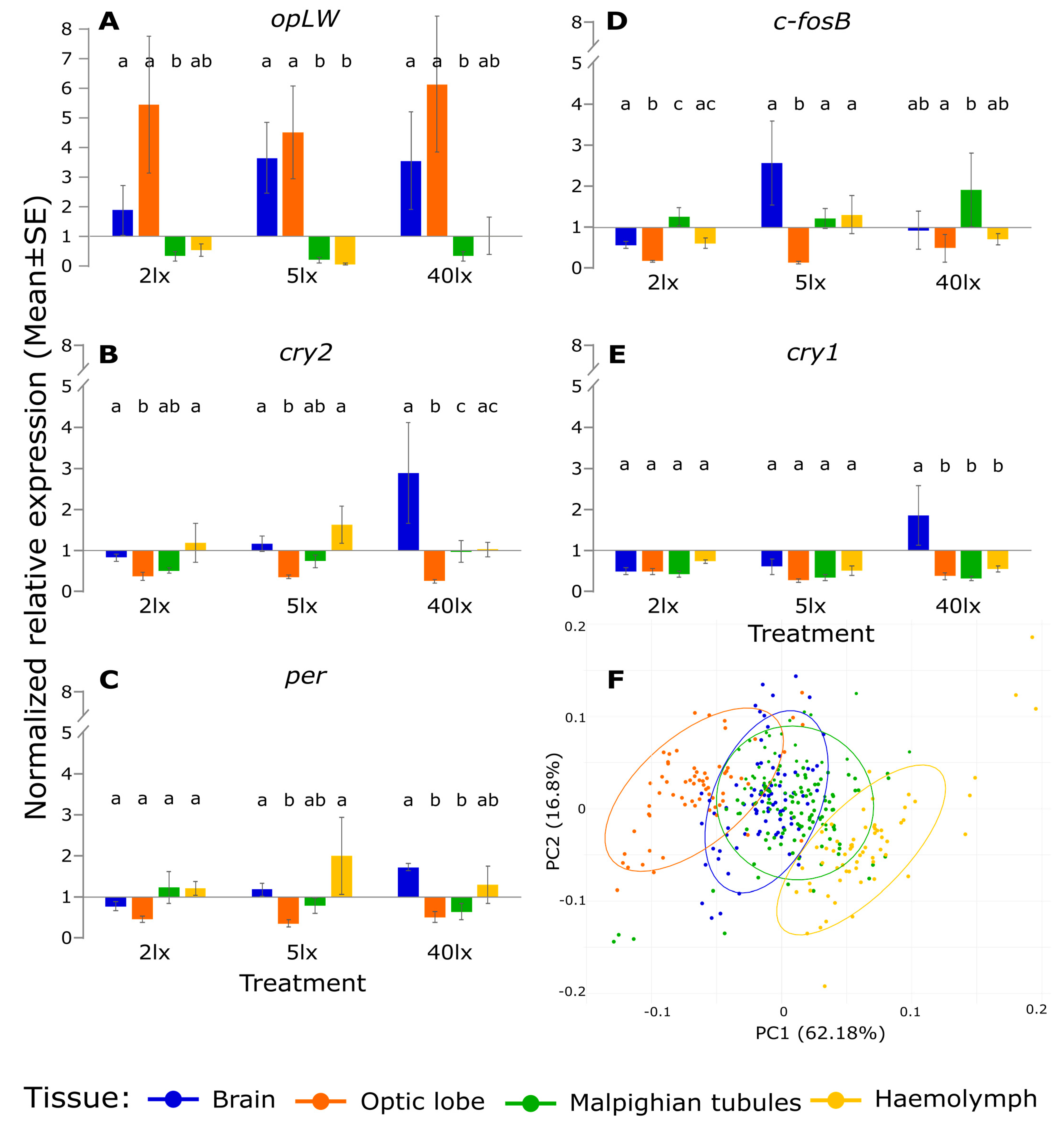
In order to enable a better comparison of gene expression among tissues, we repeated the analysis using within-tissue normalization of the expression values (relative to the control mean expression values in each tissue, Figure 2). Gene expression patterns varied substantially among tissues. The expression of opLW was strongly upregulated in the brain and in the optic lobe throughout all three light treatments, while being downregulated in the Malpighian tubules (Figure 2A). The light stimuli elicited upregulation of cry2 in the brain and downregulation in the optic lobe (Figure 2B). The transcriptional response of per showed a similar pattern, being upregulated in the brain and downregulated in the Malpighian tubules (Figure 2C). The expression of c-fosB showed no significant changes in the brain, while being downregulated in the optic lobe. c-fosB was the only gene that was upregulated in the Malpighian tubules (Figure 2D). Expression of cry1 was downregulated in all tissues except the brain, where it was upregulated under 40 lx only (F3,23 = 7.77, p < 0.001. Figure 2E). The PCA of the relative gene expression clearly separated the optic lobe and the hemolymph from the brain and the Malpighian tubules, while the latter two mostly overlapped (Figure 2F).
To simultaneously analyze the transcriptional differences between the treatments, we used Linear Discriminant Analysis (LDA). The first two LD functions explained 67.12% of the variation in the brain, 73.29% in the optic lobes, 66.87% in the Malpighian tubules and, 73.76% in the hemolymph (Figure 3). In the brain, the control group was clearly separated from the 5 and 40 lx treatments, but not from the 2 lx one (Figure 3A). In contrast, the clustering of the treatments in the optic lobe revealed a substantial difference between the control and all ALAN treatments (Figure 3B). In the Malpighian tubules and in the hemolymph, the discriminant map did not reveal any significant difference between the control and the ALAN treatments (Figure 3C,D).

3. Discussion

Insects have served for almost a century as successful models in laboratory studies on the role of light in regulating circadian behavior [7], and in raising questions about the underlying molecular mechanisms. Here, we investigated the effect of a short, dim ALAN pulse on the gene expression of five genes associated with the circadian system of the cricket Gryllus bimaculatus.
This work substantiates the findings of previous studies [56] that uncovered the heterogeneity of circadian pacemakers across various tissues in G. bimaculatus (see PCA analysis, Figure 2F), and demonstrates the tissue-specific effects of ALAN on transcription. In the optic lobe, where the cricket’s central pacemaker is located [46,48,57,58], the response to ALAN exposures was clearly distinct from that of the control, presumably due to the strong effect of dim ALAN on opLW expression. In the brain, transcriptional responses of the control and the 2 lx treatment were largely overlapping, and to a lesser extent also with those of the 5 lx and 40 lx treatments. The Malpighian tubules and hemolymph seemed less affected by the light exposures. Indeed, a previous study [56] suggested that G. bimaculatus tubules do not harbor a circadian pacemaker, as per transcript levels lacked any diurnal rhythm (in contrast to in Drosophila [59]).
The expression of circadian clock genes in G. bimaculatus in tissues other than the optic lobes has been previously reported [56]. Particularly interesting is the role of these genes in the brain. Expression of per oscillates under constant darkness (DD), and this oscillation persists after removal of the optic lobes. However, in the absence of the optic lobes, the phase of the brain per mRNA rhythm is aberrant. These results allude to a brain circadian pacemaker that is subordinate to the central clock in the optic lobes [56].
In the hemolymph, we found minute levels of circadian clock transcripts, which to the best of our knowledge is the first report of extracellular RNA (exRNA) in circadian clock transcripts. exRNA is emerging as a newly discovered form of intracellular signaling [60]. It is secreted to the biofluids, encapsulated by protecting extracellular vesicles. The exRNA is largely composed of small non-coding RNA, and to a lesser extent messenger RNA [60]. exRNA have been shown to be an effective biomarker for diagnostic purposes, but their functional role awaits further study.
The rather rapid transcriptional response to light of clock genes such as per or cry, is intriguing, and the underlying molecular mechanism warrants further investigation. In Drosophila, a similar rapid change of per mRNA following a light pulse at zeitgeber time (ZT) 15 has been previously reported [61,62], although, in contrast to the cricket, light evoked downregulation of per. It was suggested that the response to light is mediated by an histone acetylation mechanism. In accord with studies in other insects (e.g., ants [63] and lepidoptera [64]), we observed very high levels of opLW in the crickets’ optic lobe. All ALAN treatments induced some upregulation in opLW expression in the optic lobe, although not significant (we sampled 4.5 h post lights-off, while 6 h after onset of darkness a substantial downregulation has been reported [43], Figure 1 therein). Given that the circadian photosensitivity of G. bimaculatus varies during the day, as was shown by the phase response curve of this species [65,66], one can predict that the effect of transient ALAN would be time dependent.
The disruption caused by ALAN to the circadian transcriptional cycle (Figure 1) is likely to disrupt the diurnal pattern of behavior (locomotor, stridulation), but the specific behavioral output awaits further study.
The circadian disruption caused by dim (2–5 lx) ALAN may have ecological implications, which should be taken into consideration when designing regulations concerning suitable light intensity for park and city lights [67]. Not only flying insects, which are attracted to light [28], are threatened by ALAN, but also ground-dwelling insects [21,24,68]. The deep-level effect of ALAN may therefore threaten many more species of insects than currently assumed, and add to the ongoing insect decline described in the last century [69,70]. Our own work in crickets [17] demonstrated that ALAN can lead to arrhythmicity in locomotory behavior and male stridulation and therefore may affect the survival and reproduction of exposed individuals.
It should also be noted that outdoors, especially in proximity to cities and streetlights, insects are not exposed to a single light pulse, but rather to night-long, and even lifelong ALAN [17]. Populations of the small ermine moth for example, were found to reduce their flight-to-light behavior following long-term exposure to ALAN, which may result in lower predation risk and mortality in this population [71]. It remains unknown whether crickets, and indeed other insects, have developed similar adaptations to chronic ALAN exposure.

4. Materials and Methods

4.1. Insect Rearing Conditions

Gryllus bimaculatus crickets were reared under a constant temperature of 26 ± 1 °C and white fluorescent light (5W white CFL bulb, NeptOn, 6500 K, 380–780 nm, peak: 547 and 612 nm, Figure S1), under a 12 h light:12 h dark cycle. Actual light intensity measured at the top of the containers ranged from 250 to 350 lx, while the intensity under the provided shelter (egg cartons) ranged from 20 to 60 lx. Crickets were fed three times a week with dog-food pellets and vegetables. The rearing boxes contained water flasks with absorbent cotton wool.

4.2. Light-Pulse Experiments and Sample Preparation

Individual males, 3–5 days post adult emergence, were removed from the breeding colony and maintained individually, under similar 12 h light:12 h dark conditions as above, for three days. On the fourth night, three hours post lights-off (i.e., at zeitgeber time ZT 15), the experimental animals were exposed to a 30 min long light pulse of 2, 5, or 40 lx (by partially covering the same 5 W white CFL bulb as above). An additional group (no pulse) served as control.
One hour after pulse termination (ZT 16.5), the insects were decapitated under red light (led, 600–670 nm, peak: 642 nm), and their brain, optic lobes, and Malpighian tubules were removed over ice. Hemolymph (25 µL) was also extracted from each animal. Samples were placed separately into individually marked PCR tubes containing 100 µL RNAlater (Thermo Fisher Scientific), and immediately frozen at −20 °C.
Total RNA was extracted from each sample using the Purelink RNA mini kit (Invitrogen), according to the manufacturer’s instructions. Samples with genomic DNA residuals were treated with the PureLink™ DNase Set (Invitrogen, Waltham, MA, USA). RNA concentration was determined by NanoDrop One (Thermo Fisher Scientific, Waltham, MA, USA). First strand cDNA synthesis was carried out using the RT Random Primers of the High-Capacity cDNA Reverse Transcription kit (Applied Biosystems™, Waltham, MA, USA) and treated with RNase Inhibitor (Applied Biosystems™) according to the manufacturer’s instructions. cDNA was synthesized out of 20 ng/µL RNA and was then 10× diluted.
The number of successfully collected samples per experimental group varied as follows: 5–11 brain samples, 4–6 optic lobe samples, 9–15 Malpighian tubules, and 5–7 hemolymph samples. A detailed list of the final sample sizes is given in Table S1.

4.3. Primers and qPCR

The expression of two housekeeping genes (Actin and Rpl18a) and five target genes (opLW, per, cry1, cry2, c-fosB) was determined in all samples. Primer sequences are listed in Table S2. A standard curve was generated for each of the genes by using a serial dilution (1:2) of the pooled cDNA [72]. qPCR (Quantstudio 3, Thermo Fisher Scientific) was run using 59 °C as annealing temperature. Fast SYBR® Green Master Mix (Applied Biosystems™) was used. The cycle point was calculated using QuantStudio™ Design and Analysis Desktop Software version 1.5.1 (Applied Biosystems).

4.4. Data-Processing and Statistical Analysis

Gene expression was quantified using the relative standard curve method and following primer efficiencies: opLW: 94.38%, cry2: 105.95%, per: 100.16%, c-fosB: 105.02%, and cry1: 93.57%. The target gene expression was normalized by dividing its values by the corresponding geometric mean of both housekeeping genes. The expression values were log transformed and a nested ANOVA was applied using R version 4.1.3 [73] and the nlme package [74]. A mixed model was used, with light treatment and cDNA replicate as fixed and random effects, respectively. A principal component analysis (PCA) was applied to all expression values and the first two principal components (PC1 and PC2) of the PCA were used as explanatory variables for a linear discriminant analysis (LDA), with the treatments as response variables.

Supplementary Materials

The supporting information can be downloaded at https://www.mdpi.com/article/10.3390/ijms231911358/s1.

Author Contributions

Conceptualization, A.B., A.A., E.T. and K.L.; methodology, A.A., E.T. and B.F.; validation, K.L., B.F. and E.T.; formal analysis, K.L. and E.T.; investigation, K.L.; resources, A.B. and E.T.; writing—original draft preparation, K.L.; writing—review and editing, E.T., A.A. and A.B.; funding acquisition, A.B. All authors have read and agreed to the published version of the manuscript.

Funding

This research was funded by The Open University of Israel Research Fund and by the Constantiner Institute for Molecular Genetics of Tel Aviv University.

Institutional Review Board Statement

Not applicable.

Informed Consent Statement

Not applicable.

Data Availability Statement

The data presented in this study are openly available in FigShare at doi: 10.6084/m9.figshare.20721265 [75].

Acknowledgments

We thank Odelia Pisanti, Yael Diesendruck, Daniel Knebel, David Burg, Alex Slavenko, Lilach Raijman, Omer Lavy, Michaela Kolker-Ghatan, Izhak David, and Talma and Rami Zelingher for their assistance. This work is dedicated to our mentor Meir Paul Pener (1930–2021).

Conflicts of Interest

The authors declare no conflict of interest.

References

  1. Borges, R.M. Impacts of Artificial Light at Night on Biological Timings. Indian J. Entomol. 2022, 84, 483–492. [Google Scholar] [CrossRef]
  2. Saunders, D.S. Insect Photoperiodism: Seeing the Light. Physiol. Entomol. 2012, 37, 207–218. [Google Scholar] [CrossRef]
  3. Pittendrigh, C.S. Circadian Rhythms and the Circadian Organization of Living Systems. Cold Spring Harb. Symp. Quant. Biol. 1960, 25, 159–184. [Google Scholar] [CrossRef]
  4. Kronfeld-Schor, N.; Visser, M.E.; Salis, L.; van Gils, J.A. Chronobiology of Interspecific Interactions in a Changing World. Philos. Trans. R. Soc. B Biol. Sci. 2017, 372, 20160248. [Google Scholar] [CrossRef]
  5. Ampleford, E.J.; Steel, C.G.H. Circadian Control of Ecdysis in Rhodnius prloixus (Hempitera). J. Comp. Physiol. A 1982, 147, 281–286. [Google Scholar] [CrossRef]
  6. Helm, B.; Visser, M.E.; Schwartz, W.; Kronfeld-Schor, N.; Gerkema, M.; Piersma, T.; Bloch, G. Two Sides of a Coin: Ecological and Chronobiological Perspectives of Timing in the Wild. Philos. Trans. R. Soc. B Biol. Sci. 2017, 372, 20160246. [Google Scholar] [CrossRef]
  7. Saunders, D.S.; Steel, C.G.H.; Vafopoulou, X.; Lewis, R.D. Circadian Rhythms of Activity in Individual Insects. In Insect Clocks; Elsevier: Amsterdam, The Netherlands, 2002; pp. 7–42. [Google Scholar]
  8. Merrow, M.; Spoelstra, K.; Roenneberg, T. The Circadian Cycle: Daily Rhythms from Behaviour to Genes. EMBO Rep. 2005, 6, 930–935. [Google Scholar] [CrossRef]
  9. Horch, H.W.; Mito, T.; Popadi, A.; Ohuchi, H.; Noji, S. The Cricket as a Model Organism; Springer: Tokyo, Japan, 2017; ISBN 978-4-431-56476-8. [Google Scholar]
  10. Moaraf, S.; Vistoropsky, Y.; Pozner, T.; Heiblum, R.; Okuliarová, M.; Zeman, M.; Barnea, A. Artificial Light at Night Affects Brain Plasticity and Melatonin in Birds. Neurosci. Lett. 2020, 716, 134639. [Google Scholar] [CrossRef]
  11. Falchi, F.; Furgoni, R.; Gallaway, T.A.; Rybnikova, N.A.; Portnov, B.A.; Baugh, K.; Cinzano, P.; Elvidge, C.D. Light Pollution in USA and Europe: The Good, the Bad and the Ugly. J. Environ. Manag. 2019, 248, 109227. [Google Scholar] [CrossRef]
  12. Hölker, F.; Wolter, C.; Perkin, E.K.; Tockner, K. Light Pollution as a Biodiversity Threat. Trends Ecol. Evol. 2010, 25, 681–682. [Google Scholar] [CrossRef]
  13. Dominoni, D.; Quetting, M.; Partecke, J. Artificial Light at Night Advances Avian Reproductive Physiology. Proc. R. Soc. B Biol. Sci. 2013, 280, 20123017. [Google Scholar] [CrossRef]
  14. Amichai, E.; Kronfeld-Schor, N. Artificial Light at Night Promotes Activity Throughout the Night in Nesting Common Swifts (Apus apus). Sci. Rep. 2019, 9, 11052. [Google Scholar] [CrossRef]
  15. Kumar, D.; Soni, S.K.; Kronfeld-Schor, N.; Singaravel, M. Wheel-Running Activity Rhythms and Masking Responses in the Diurnal Palm Squirrel, Funambulus pennantii. Chronobiol. Int. 2020, 37, 1693–1708. [Google Scholar] [CrossRef]
  16. Buchanan, B.W. Observed and Potential Effects of Artificial Night Lighting on Anuran Amphibians. Ecol. Conseq. Artif. Night Light. 2006, 09, 192–220. [Google Scholar]
  17. Levy, K.; Wegrzyn, Y.; Efronny, R.; Barnea, A.; Ayali, A. Lifelong Exposure to Artificial Light at Night Impacts Stridulation and Locomotion Activity Patterns in the Cricket Gryllus bimaculatus. Proc. R. Soc. B Biol. Sci. 2021, 288, 20211626. [Google Scholar] [CrossRef]
  18. Durrant, J.; Michaelides, E.B.; Rupasinghe, T.; Tull, D.; Green, M.P.; Jones, T.M. Constant Illumination Reduces Circulating Melatonin and Impairs Immune Function in the Cricket Teleogryllus commodus. PeerJ 2015, 3, e1075. [Google Scholar] [CrossRef]
  19. Raap, T.; Pinxten, R.; Eens, M. Light Pollution Disrupts Sleep in Free-Living Animals. Sci. Rep. 2015, 5, 13557. [Google Scholar] [CrossRef]
  20. Gaston, K.J.; Davies, T.W.; Nedelec, S.L.; Holt, L.A. Impacts of Artificial Light at Night on Biological Timings. Annu. Rev. Ecol. Evol. Syst. 2017, 48, 49–68. [Google Scholar] [CrossRef]
  21. Owens, A.C.S.; Lewis, S.M. The Impact of Artificial Light at Night on Nocturnal Insects: A Review and Synthesis. Ecol. Evol. 2018, 8, 11337–11358. [Google Scholar] [CrossRef]
  22. Sanders, D.; Kehoe, R.; Cruse, D.; van Veen, F.J.F.; Gaston, K.J. Low Levels of Artificial Light at Night Strengthen Top-Down Control in Insect Food Web. Curr. Biol. 2018, 28, 2474–2478.e3. [Google Scholar] [CrossRef]
  23. Bolliger, J.; Hennet, T.; Wermelinger, B.; Bösch, R.; Pazur, R.; Blum, S.; Haller, J.; Obrist, M.K. Effects of Traffic-Regulated Street Lighting on Nocturnal Insect Abundance and Bat Activity. Basic Appl. Ecol. 2020, 47, 44–56. [Google Scholar] [CrossRef]
  24. Foster, J.J.; Tocco, C.; Smolka, J.; Khaldy, L.; Baird, E.; Byrne, M.J.; Nilsson, D.E.; Dacke, M. Light Pollution Forces a Change in Dung Beetle Orientation Behavior. Curr. Biol. 2021, 31, 3935–3942.e3. [Google Scholar] [CrossRef]
  25. Giavi, S.; Blösch, S.; Schuster, G.; Knop, E. Artificial Light at Night Can Modify Ecosystem Functioning beyond the Lit Area. Sci. Rep. 2020, 10, 11870. [Google Scholar] [CrossRef]
  26. Knop, E.; Zoller, L.; Ryser, R.; Gerpe, C.; Hörler, M.; Fontaine, C. Artificial Light at Night as a New Threat to Pollination. Nature 2017, 548, 206–209. [Google Scholar] [CrossRef]
  27. Rich, C.; Longcore, T. Ecological Consequences of Artificial Night Lighting; Island Press: Washington, DC, USA, 2006; ISBN 9781559631297. [Google Scholar]
  28. Eisenbeis, G.; Hänel, A. Light Pollution and the Impact of Artificial Night Lighting on Insects. In Ecology of Cities and Towns: A Comparative Approach; McDonnell, M.J., Hahs, A.K., Breuste, J.H., Eds.; Cambridge University Press: Cambridge, UK, 2009; pp. 243–263. ISBN 9780511609763. [Google Scholar]
  29. Perkin, E.K.; Hölker, F.; Tockner, K. The Effects of Artificial Lighting on Adult Aquatic and Terrestrial Insects. Freshw. Biol. 2014, 59, 368–377. [Google Scholar] [CrossRef]
  30. Davies, T.W.; Bennie, J.; Gaston, K.J. Street Lighting Changes the Composition of Invertebrate Communities. Biol. Lett. 2012, 8, 764–767. [Google Scholar] [CrossRef]
  31. Sanders, D.; Frago, E.; Kehoe, R.; Patterson, C.; Gaston, K.J. A Meta-Analysis of Biological Impacts of Artificial Light at Night. Nat. Ecol. Evol. 2021, 5, 74–81. [Google Scholar] [CrossRef]
  32. Sanders, D.; Gaston, K.J. How Ecological Communities Respond to Artificial Light at Night. J. Exp. Zool. Part A Ecol. Integr. Physiol. 2018, 329, 394–400. [Google Scholar] [CrossRef]
  33. Owens, A.C.S.; Cochard, P.; Durrant, J.; Farnworth, B.; Perkin, E.K.; Seymoure, B. Light Pollution Is a Driver of Insect Declines. Biol. Conserv. 2020, 241, 108259. [Google Scholar] [CrossRef]
  34. Bachleitner, W.; Kempinger, L.; Wülbeck, C.; Rieger, D.; Helfrich-Förster, C. Moonlight Shifts the Endogenous Clock of Drosophila melanogaster. Proc. Natl. Acad. Sci. USA 2007, 104, 3538–3543. [Google Scholar] [CrossRef]
  35. Kempinger, L.; Dittmann, R.; Rieger, D.; Helfrich-Förster, C. The Nocturnal Activity of Fruit Flies Exposed to Artificial Moonlight Is Partly Caused by Direct Light Effects on the Activity Level That Bypass the Endogenous Clock. Chronobiol. Int. 2009, 26, 151–166. [Google Scholar] [CrossRef]
  36. Kronfeld-Schor, N.; Dominoni, D.; la Iglesia, H.D.; Levy, O.; Herzog, E.D.; Dayan, T.; Helfrich-Forster, C. Chronobiology by Moonlight. Proc. R. Soc. B Biol. Sci. 2013, 1765, 20123088. [Google Scholar] [CrossRef]
  37. Helfrich-Förster, C. The Circadian Clock in the Brain: A Structural and Functional Comparison between Mammals and Insects. J. Comp. Physiol. A Neuroethol. Sens. Neural Behav. Physiol. 2004, 190, 601–613. [Google Scholar] [CrossRef]
  38. Tomioka, K.; Matsumoto, A. The Circadian System in Insects: Cellular, Molecular, and Functional Organization. In Advances in Insect Physiology; Academic Press Inc.: Cambridge, MA, USA, 2019; Volume 56, pp. 73–115. ISBN 9780081028421. [Google Scholar]
  39. Helfrich-Förster, C. Light Input Pathways to the Circadian Clock of Insects with an Emphasis on the Fruit Fly Drosophila melanogaster. J. Comp. Physiol. A Neuroethol. Sens. Neural Behav. Physiol. 2020, 206, 259–272. [Google Scholar] [CrossRef]
  40. Tomioka, K.; Chiba, Y. Post-Embryonic Development of Circadian Rhythm in the Cricket, Gryllus bimaculatus: A Rhythm Reversal. J. Comp. Physiol. A 1982, 147, 299–304. [Google Scholar] [CrossRef]
  41. Tomioka, K.; Chiba, Y. Circadian Rhythm in the Neurally Isolated Lamina-Medulla-Complex of the Cricket, Gryllus bimaculatus. J. Insect Physiol. 1986, 32, 747–755. [Google Scholar] [CrossRef]
  42. Abe, Y.; Ushirogawa, H.; Tomioka, K. Circadian Locomotor Rhythms in the Cricket, Gryllodes sigillatus I. Localization of the Pacemaker and the Photoreceptor. Zool. Sci. 1997, 14, 719–727. [Google Scholar] [CrossRef]
  43. Komada, S.; Kamae, Y.; Koyanagi, M.; Tatewaki, K.; Hassaneen, E.; Saifullah, A.; Yoshii, T.; Terakita, A.; Tomioka, K. Green-Sensitive Opsin Is the Photoreceptor for Photic Entrainment of an Insect Circadian Clock. Zool. Lett. 2015, 1, 11. [Google Scholar] [CrossRef]
  44. Tomioka, K. Chronobiology of Crickets: A Review. Zool. Sci. 2014, 31, 624–632. [Google Scholar] [CrossRef]
  45. Loher, W. Temporal Organization of Reproductive Behavior. In Cricket Behavior and Neurobiology; Huber, F., Moore, T.E., Loher, W., Eds.; Cornell University Press: Ithaca, NY, USA; London, UK, 1989; pp. 83–113. [Google Scholar]
  46. Moriyama, Y.; Kamae, Y.; Uryu, O.; Tomioka, K. Gb’clock Is Expressed in the Optic Lobe and Is Required for the Circadian Clock in the Cricket Gryllus bimaculatus. J. Biol. Rhythm. 2012, 27, 467–477. [Google Scholar] [CrossRef]
  47. Masayuki, I.; Kenji, T. Temperature Dependency of the Circadian Locomotor Rhythm in the Cricket Gryllus bimaculatus. Zool. Sci. 1993, 10, 597–604. [Google Scholar] [CrossRef]
  48. Tomioka, K. Analysis of Coupling between Optic Lobe Circadian Pacemakers in the Cricket Gryllus bimaculatus. J. Comp. Physiol. A 1993, 172, 401–408. [Google Scholar] [CrossRef]
  49. Tokuoka, A.; Itoh, T.Q.; Hori, S.; Uryu, O.; Danbara, Y.; Nose, M.; Bando, T.; Tanimura, T.; Tomioka, K. Cryptochrome Genes Form an Oscillatory Loop Independent of the per/Tim Loop in the Circadian Clockwork of the Cricket Gryllus bimaculatus. Zool. Lett. 2017, 3, 1–14. [Google Scholar] [CrossRef]
  50. Kutaragi, Y.; Tokuoka, A.; Tomiyama, Y.; Nose, M.; Watanabe, T.; Bando, T.; Moriyama, Y.; Tomioka, K. A Novel Photic Entrainment Mechanism for the Circadian Clock in an Insect: Involvement of c-Fos and Cryptochromes. Zool. Lett. 2018, 4, 26. [Google Scholar] [CrossRef]
  51. Moriyama, Y.; Takeuchi, K.; Shinohara, T.; Miyagawa, K.; Matsuka, M.; Yoshii, T.; Tomioka, K. Timeless Plays an Important Role in Compound Eye-Dependent Photic Entrainment of the Circadian Rhythm in the Cricket Gryllus bimaculatus. Zool. Sci. 2022, 39, 397–405. [Google Scholar] [CrossRef]
  52. Alaasam, V.J.; Liu, X.; Niu, Y.; Habibian, J.S.; Pieraut, S.; Ferguson, B.S.; Zhang, Y.; Ouyang, J.Q. Effects of Dim Artificial Light at Night on Locomotor Activity, Cardiovascular Physiology, and Circadian Clock Genes in a Diurnal Songbird. Environ. Pollut. 2021, 282, 117036. [Google Scholar] [CrossRef]
  53. Dominoni, D.M.; de Jong, M.; van Oers, K.; O’Shaughnessy, P.; Blackburn, G.J.; Atema, E.; Mateman, A.C.; D’Amelio, P.B.; Trost, L.; Bellingham, M.; et al. Integrated Molecular and Behavioural Data Reveal Deep Circadian Disruption in Response to Artificial Light at Night in Male Great Tits (Parus major). Sci. Rep. 2022, 12, 1553. [Google Scholar] [CrossRef]
  54. Touzot, M.; Lefebure, T.; Lengagne, T.; Secondi, J.; Dumet, A.; Konecny-Dupre, L.; Veber, P.; Navratil, V.; Duchamp, C.; Mondy, N. Transcriptome-Wide Deregulation of Gene Expression by Artificial Light at Night in Tadpoles of Common Toads. Sci. Total Environ. 2022, 818, 151734. [Google Scholar] [CrossRef] [PubMed]
  55. Chen, Y.; Wei, W.; Tzeng, D.T.W.; Owens, A.C.S.; Tang, H.; Wu, C.; Lin, S.; Zhong, S.; Yang, E. Effects of Artificial Light at Night (ALAN) on Gene Expression of Aquatica ficta Fire Fly Larvae. Environ. Pollut. 2021, 281, 116944. [Google Scholar] [CrossRef]
  56. Uryu, O.; Tomioka, K. Circadian Oscillations Outside the Optic Lobe in the Cricket Gryllus bimaculatus. J. Insect Physiol. 2010, 56, 1284–1290. [Google Scholar] [CrossRef]
  57. Tomioka, K.; Abdelsalam, S. Circadian Organization in Hemimetabolous Insects. Zool. Sci. 2004, 21, 1153–1162. [Google Scholar] [CrossRef] [Green Version]
  58. Tomioka, K.; Chiba, Y. Photoperiodic Entrainment of Locomotor Activity in Crickets (Gryllus bimaculatus) Lacking the Optic Lobe Pacemaker. J. Insect Physiol. 1989, 35, 827–835. [Google Scholar] [CrossRef]
  59. Hege, D.M.; Stanewsky, R.; Hall, J.C.; Giebultowicz, J.M. Rhythmic Expression of a PER-Reporter in the Malpighian Tubules of Decapitated Drosophila: Evidence for a Brain-Independent Circadian Clock. J. Biol. Rhythm. 1997, 12, 300–308. [Google Scholar] [CrossRef]
  60. Sadik, N.; Cruz, L.; Gurtner, A.; Rodosthenous, R.S.; Dusoswa, S.A.; Ziegler, O.; Van Solinge, T.S.; Wei, Z.; Salvador-Garicano, A.M.; Gyorgy, B.; et al. Extracellular RNAs: A New Awareness of Old Perspectives. In Extracellular RNA. Methods in Molecular Biology; Patel, T., Ed.; Springer: New York, NY, USA, 2018; Volume 1740, pp. 1–15. ISBN 978-1-4939-7652-2. [Google Scholar]
  61. Adewoye, A.B.; Kyriacou, C.P.; Tauber, E. Identification and Functional Analysis of Early Gene Expression Induced by Circadian Light-Resetting in Drosophila. BMC Genom. 2015, 16, 570. [Google Scholar] [CrossRef] [PubMed]
  62. Lee, C.; Parikh, V.; Itsukaichi, T.; Bae, K.; Edery, I. Resetting the Drosophila Clock by Photic Regulation of PER and a PER-TIM Complex. Science 1996, 271, 1740–1744. [Google Scholar] [CrossRef] [PubMed]
  63. Yilmaz, A.; Lindenberg, A.; Albert, S.; Grübel, K.; Spaethe, J.; Rössler, W.; Groh, C. Age-Related and Light-Induced Plasticity in Opsin Gene Expression and in Primary and Secondary Visual Centers of the Nectar-Feeding Ant Camponotus rufipes. Dev. Neurobiol. 2016, 76, 1041–1057. [Google Scholar] [CrossRef]
  64. Yan, S.; Zhu, J.; Zhu, W.; Zhang, X.; Li, Z.; Liu, X.; Zhang, Q. The Expression of Three Opsin Genes from the Compound Eye of Helicoverpa armigera (Lepidoptera: Noctuidae) Is Regulated by a Circadian Clock, Light Conditions and Nutritional Status. PLoS ONE 2014, 9, e111683. [Google Scholar] [CrossRef]
  65. Tomioka, K. Light and Serotonin Phase-Shift the Circadian Clock in the Cricket Optic Lobe in Vitro. J. Comp. Physiol.—A Sens. Neural Behav. Physiol. 1999, 185, 437–444. [Google Scholar] [CrossRef]
  66. Okada, Y.; Tomioka, K.; Chiba, Y. Circadian Phase-Response Curves for Light in Nymphal and Adult Crickets, Gryllus bimaculatus. J. Insect Physiol. 1991, 37, 583–590. [Google Scholar] [CrossRef]
  67. Hölker, F.; Bolliger, J.; Davies, T.W.; Giavi, S.; Jechow, A.; Kalinkat, G.; Longcore, T.; Spoelstra, K.; Tidau, S.; Visser, M.E.; et al. 11 Pressing Research Questions on How Light Pollution Affects Biodiversity. Front. Ecol. Evol. 2021, 9, 767177. [Google Scholar] [CrossRef]
  68. Grubisic, M.; van Grunsven, R.H. Artificial Light at Night Disrupts Species Interactions and Changes Insect Communities. Curr. Opin. Insect Sci. 2021, 47, 136–141. [Google Scholar] [CrossRef]
  69. Shortall, R.C.; Moore, A.; Smith, E.; Hall, J.M.; Woiwod, P.I.; Harrington, R. Long-Term Changes in the Abundance of Flying Insects. Insect Conserv. Divers. 2009, 2, 251–260. [Google Scholar] [CrossRef]
  70. Hallmann, C.A.; Sorg, M.; Jongejans, E.; Siepel, H.; Hofland, N.; Schwan, H.; Stenmans, W.; Müller, A.; Sumser, H.; Hörren, T.; et al. More than 75 Percent Decline over 27 Years in Total Flying Insect Biomass in Protected Areas. PLoS ONE 2017, 12, e0185809. [Google Scholar] [CrossRef]
  71. Altermatt, F.; Ebert, D. Reduced Flight-to-Light Behaviour of Moth Populations Exposed to Long-Term Urban Light Pollution. Biol. Lett. 2016, 12, 2016–2019. [Google Scholar] [CrossRef]
  72. Taylor, S.C.; Nadeau, K.; Abbasi, M.; Lachance, C.; Nguyen, M.; Fenrich, J. The Ultimate QPCR Experiment: Producing Publication Quality, Reproducible Data the First Time. Trends Biotechnol. 2019, 37, 761–774. [Google Scholar] [CrossRef]
  73. R Core Team. R: A Language and Environment for Statistical Computing; R Foundation for Statistical Computing: Vienna, Austria, 2022. [Google Scholar]
  74. Pinheiro, J.B. Nlme: Linear and Nonlinear Mixed Effects Models; R Package Version 3.1-157; R Core Team: Vienna, Austria, 2022. [Google Scholar]
  75. Levy, K.; Fishman, B.; Barnea, A.; Ayali, A.; Tauber, E. Supplementary material and data from: Transcriptional Response of Circadian Clock Genes to an ‘Artificial Light at Night’ Pulse in the Cricket Gryllus bimaculatus. Figshare 2022. [CrossRef]
Figure 1. Transcriptional response of G. bimaculatus to dim ALAN. The relative gene expression (mean ± s.e.) following 30 min light pulse of 2 lx (dark grey), 5 lx (light grey) or 40 lx (yellow), and no-pulse control (black). Results are shown for the brain (A), optic lobe (B), Malpighian tubules (C), and hemolymph (D). * p < 0.05.
Ijms 23 11358 g001
Figure 2. Normalized mean (±s.e.) of the relative gene expression of (A) opLW, (B) cry2, (C) per, (D) c-fosB, (E) cry1, and (F) Principal component analysis (PCA) of relative gene expression in the brain (blue), optic lobe (orange), Malpighian tubules (green), and hemolymph (yellow). Within-tissue normalization was performed relative to the mean of control. Different letters indicate significant differences. In the PCA, each point represents one individual sample and ellipses contain 95% of the group. PCA plot is illustrated on the two principal axes, explaining 78.98% of the variability.
Ijms 23 11358 g002
Figure 3. Linear discriminant analysis of transcriptional response to ALAN. The analysis was carried out under four different light treatments: 0 lx (black), 2 lx (dark grey), 5 lx (light grey), 40 lx (yellow); sampled in the brain (A), optic lobe (B), Malpighian tubules (C), and hemolymph (D). Each point represents one individual sample while ellipses represent a 95% confidence level. Each plot is illustrated on the two principal axes of the LDA, explaining >65% of the variability.
Ijms 23 11358 g003
Publisher’s Note: MDPI stays neutral with regard to jurisdictional claims in published maps and institutional affiliations.