
Complex Permittivity of Ex-Vivo Human, Bovine and Porcine Brain Tissues in the Microwave Frequency Range

Abstract
:1. Introduction
2. Materials and Methods
2.1. Complex Permittivity
2.2. Measurement Method
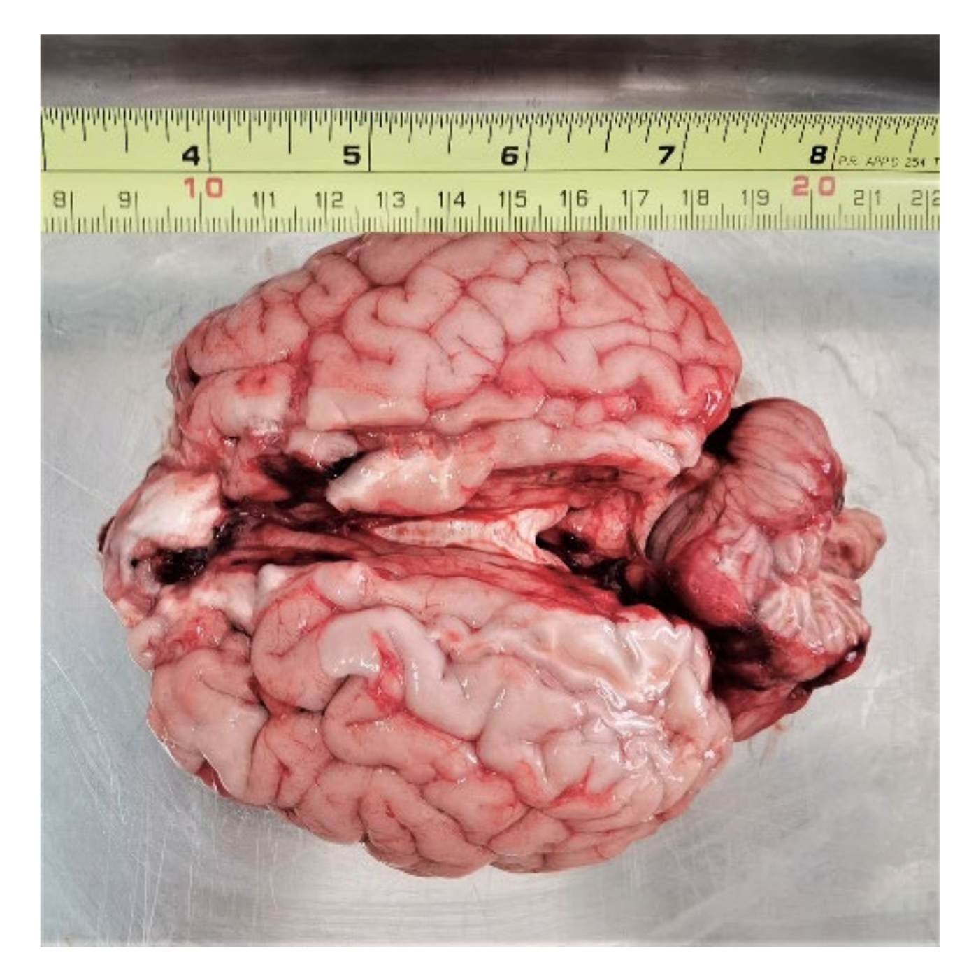
2.3. Materials under Test
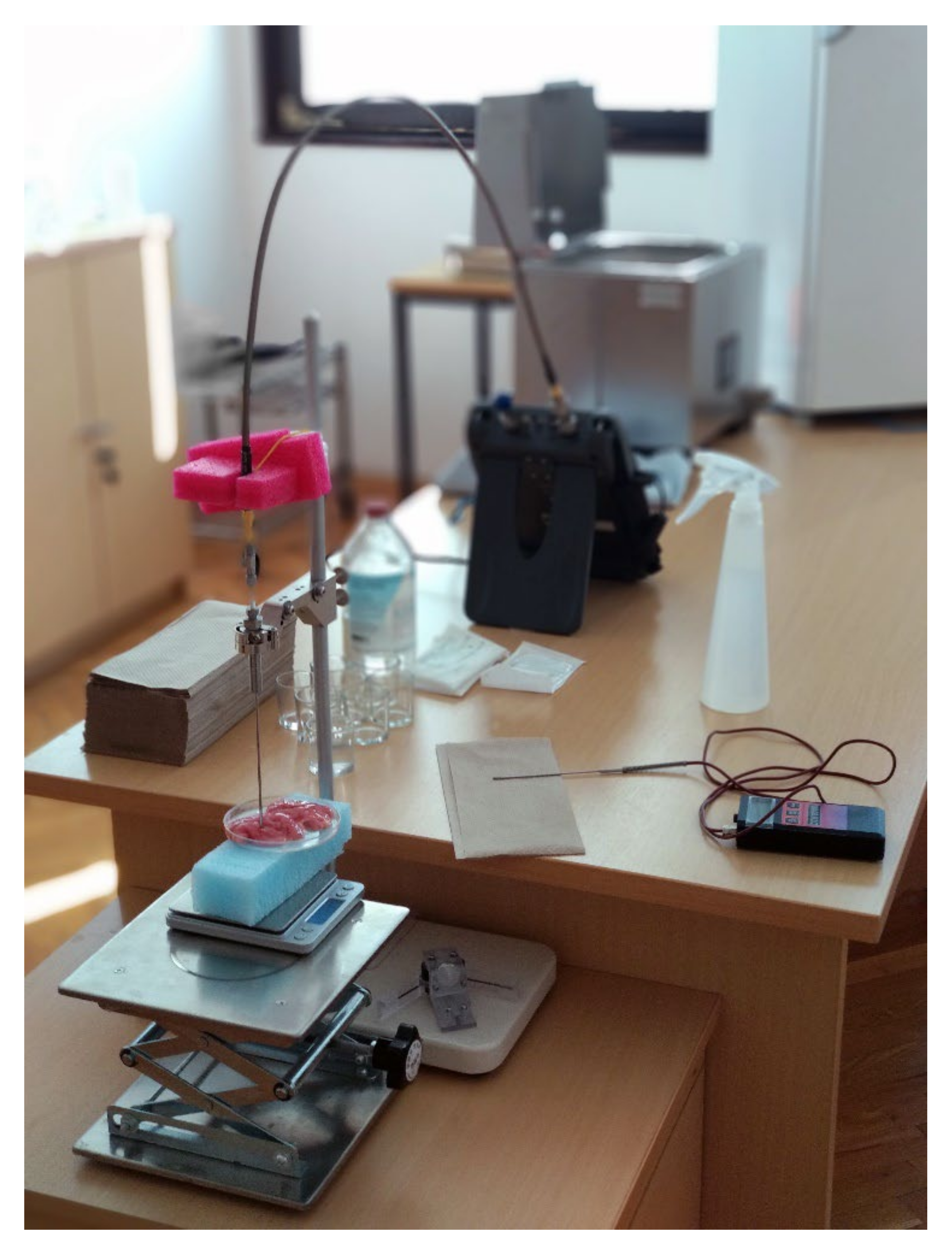
2.4. Measurement Setup
3. Results
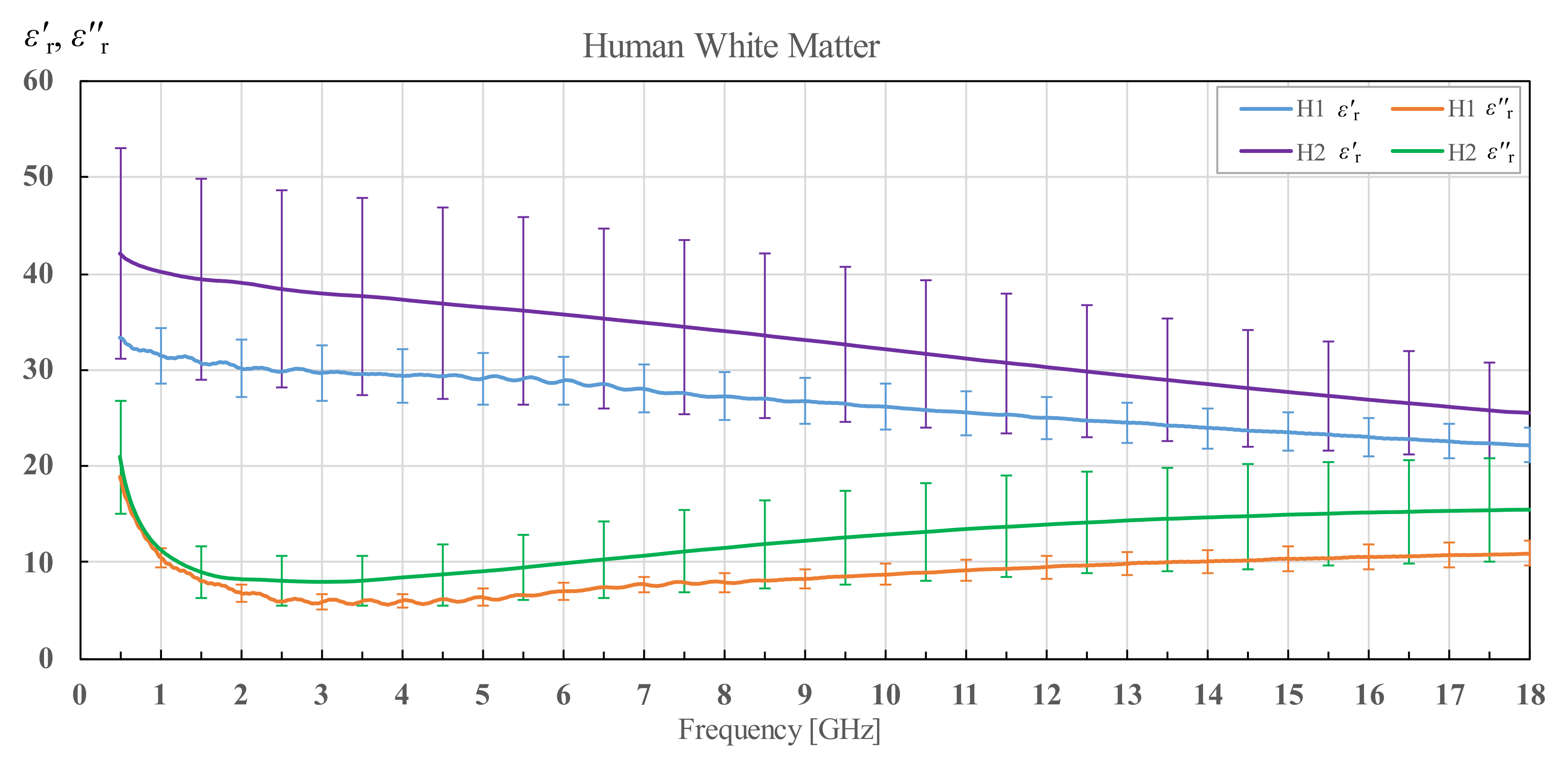
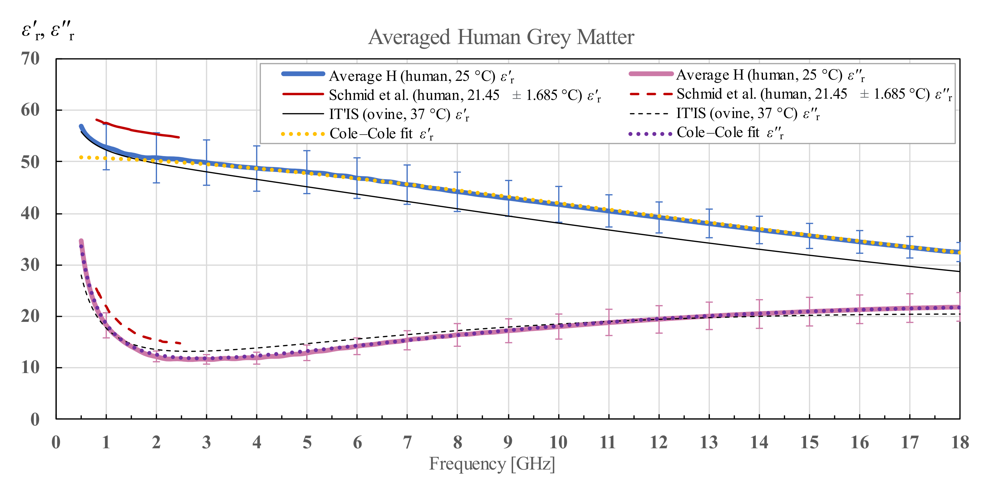
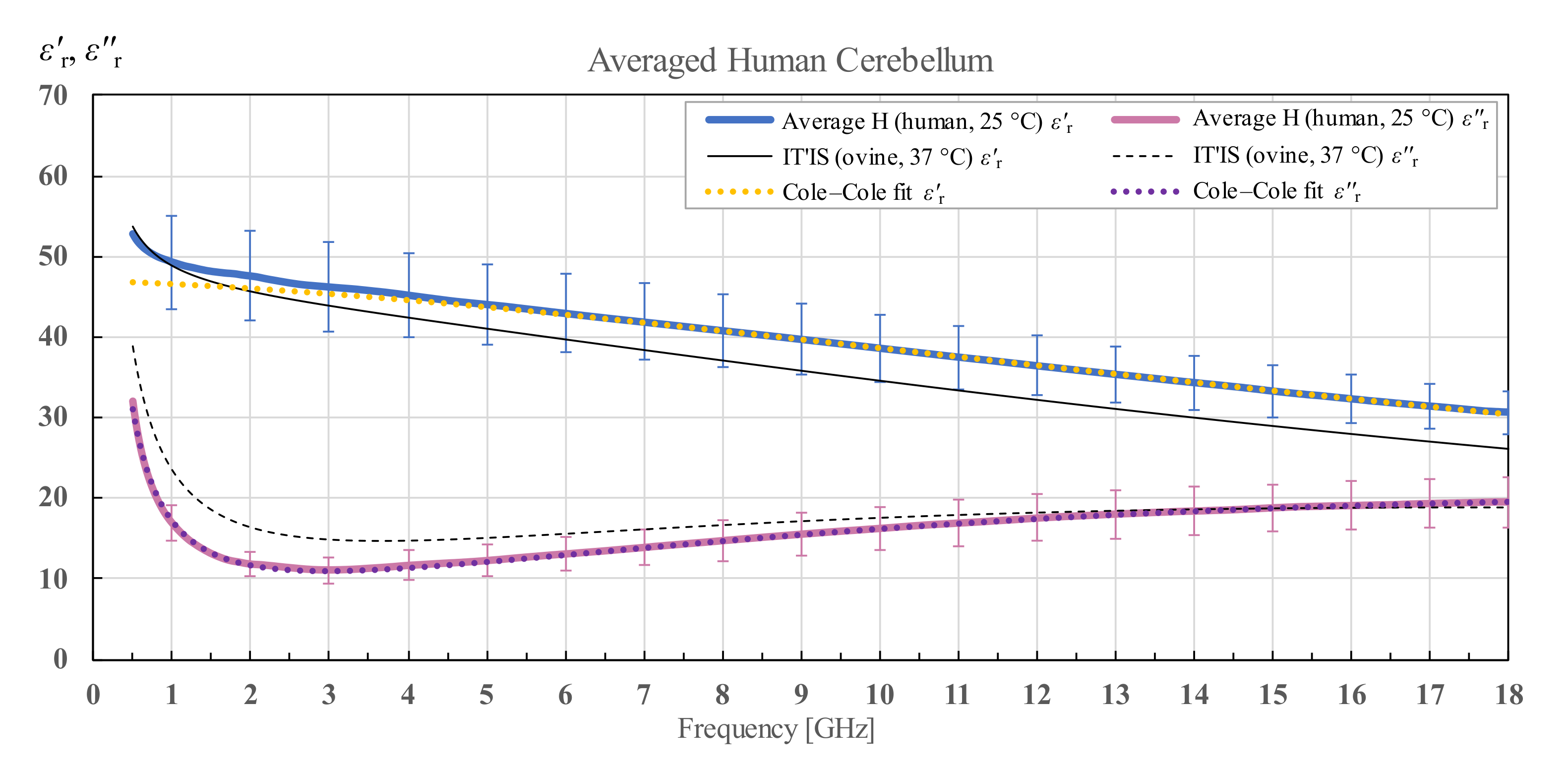
3.1. Human Brain Permittivity
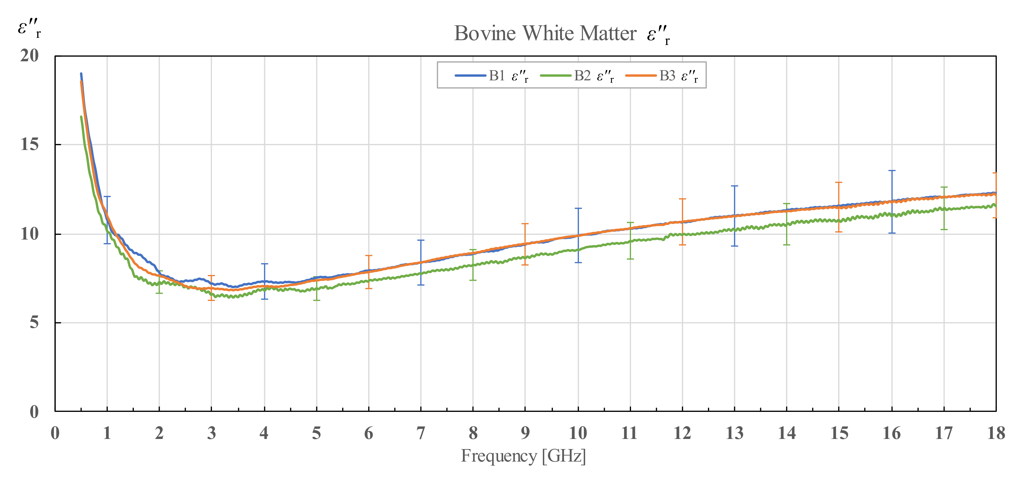
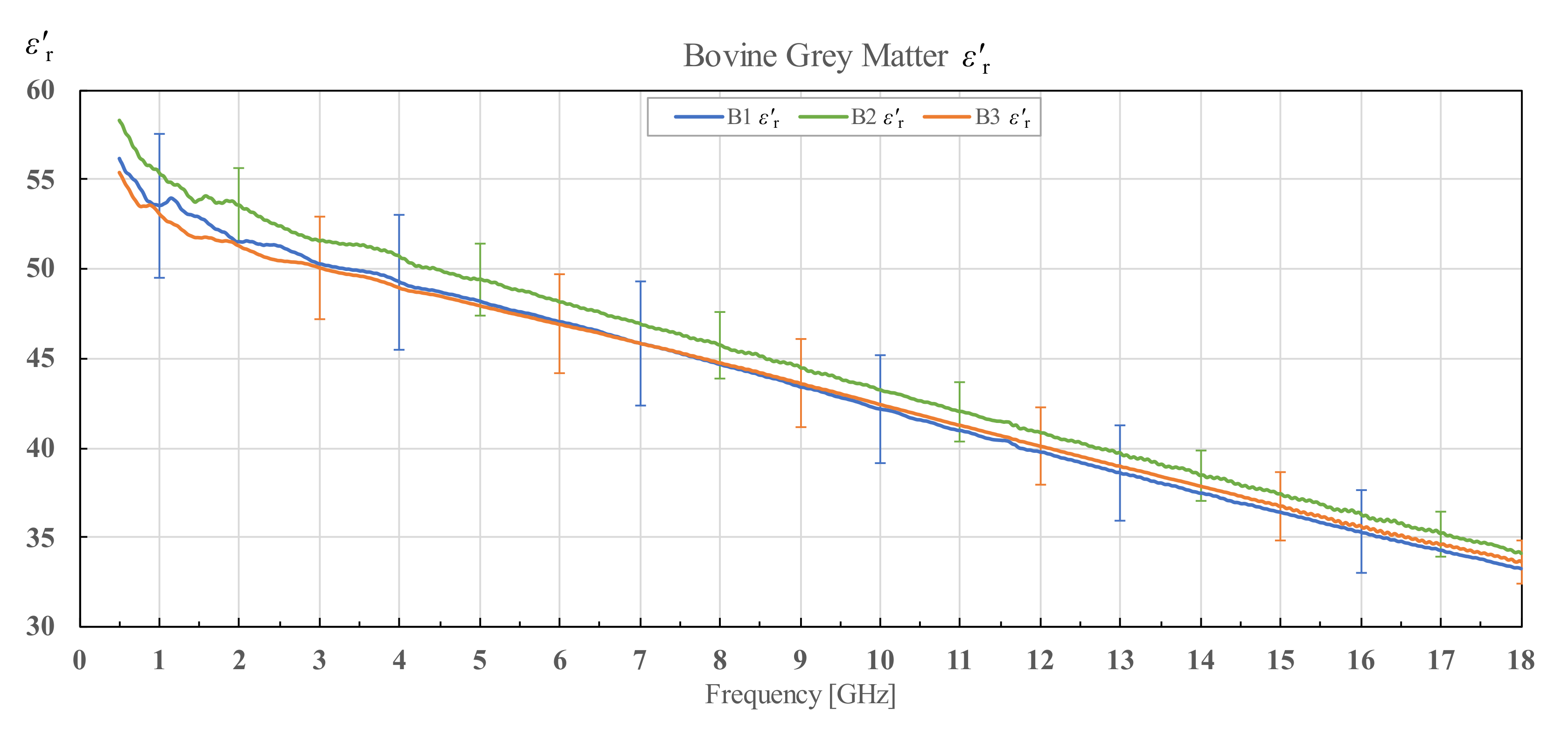
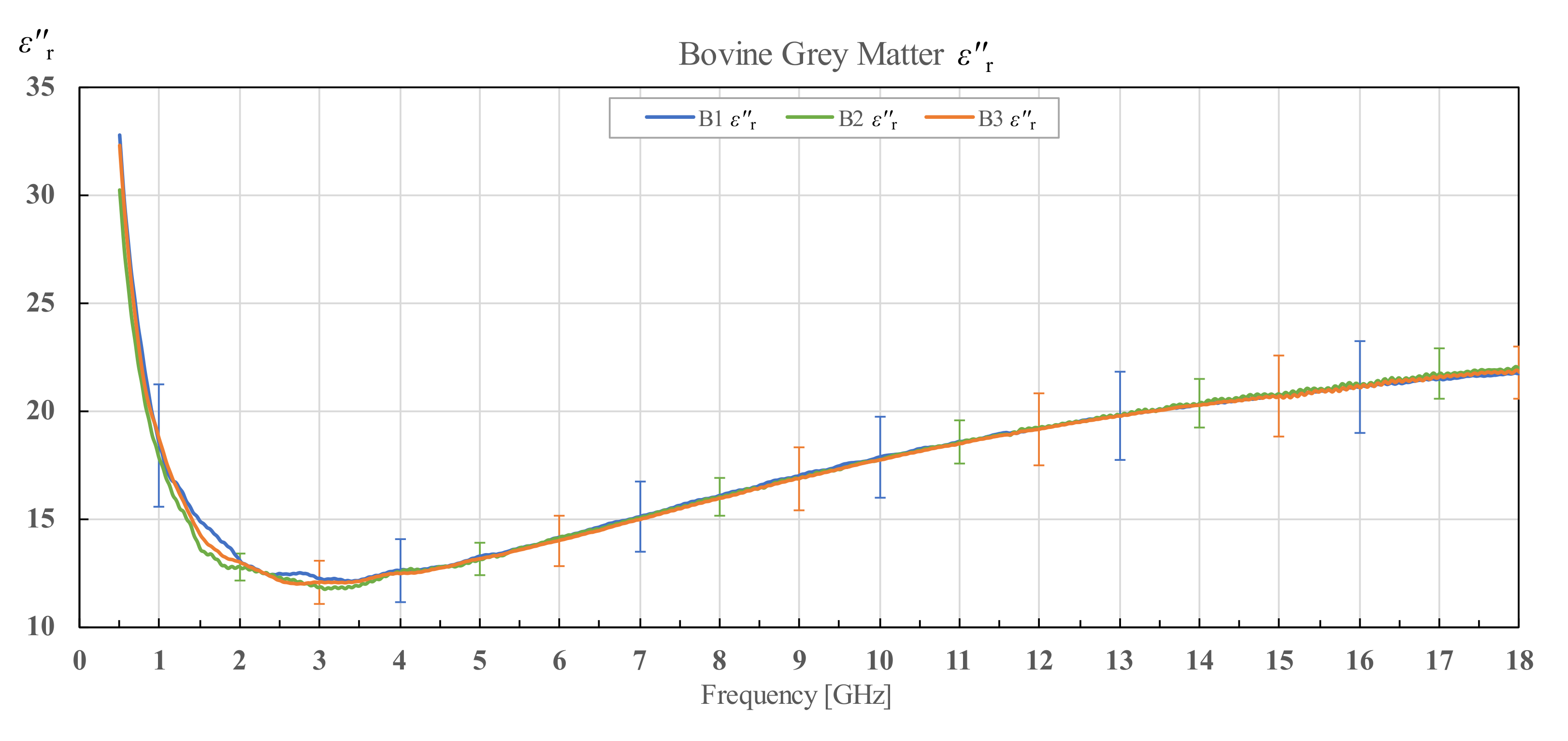
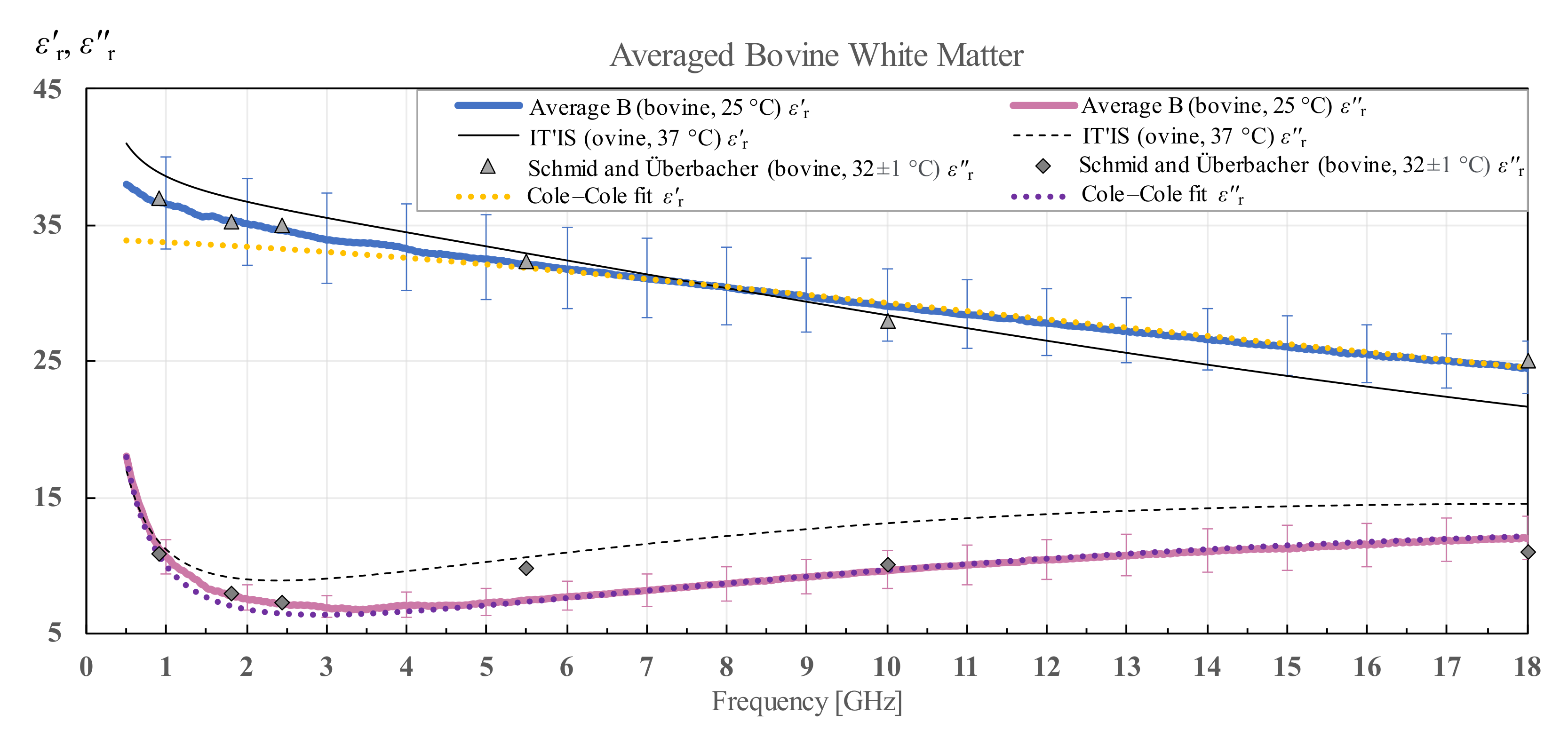
3.2. Bovine Brain Permittivity
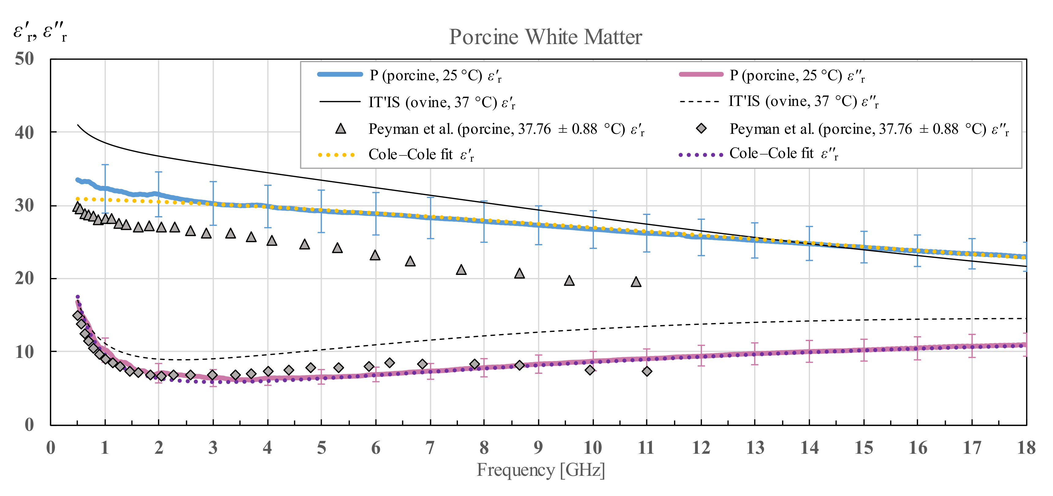
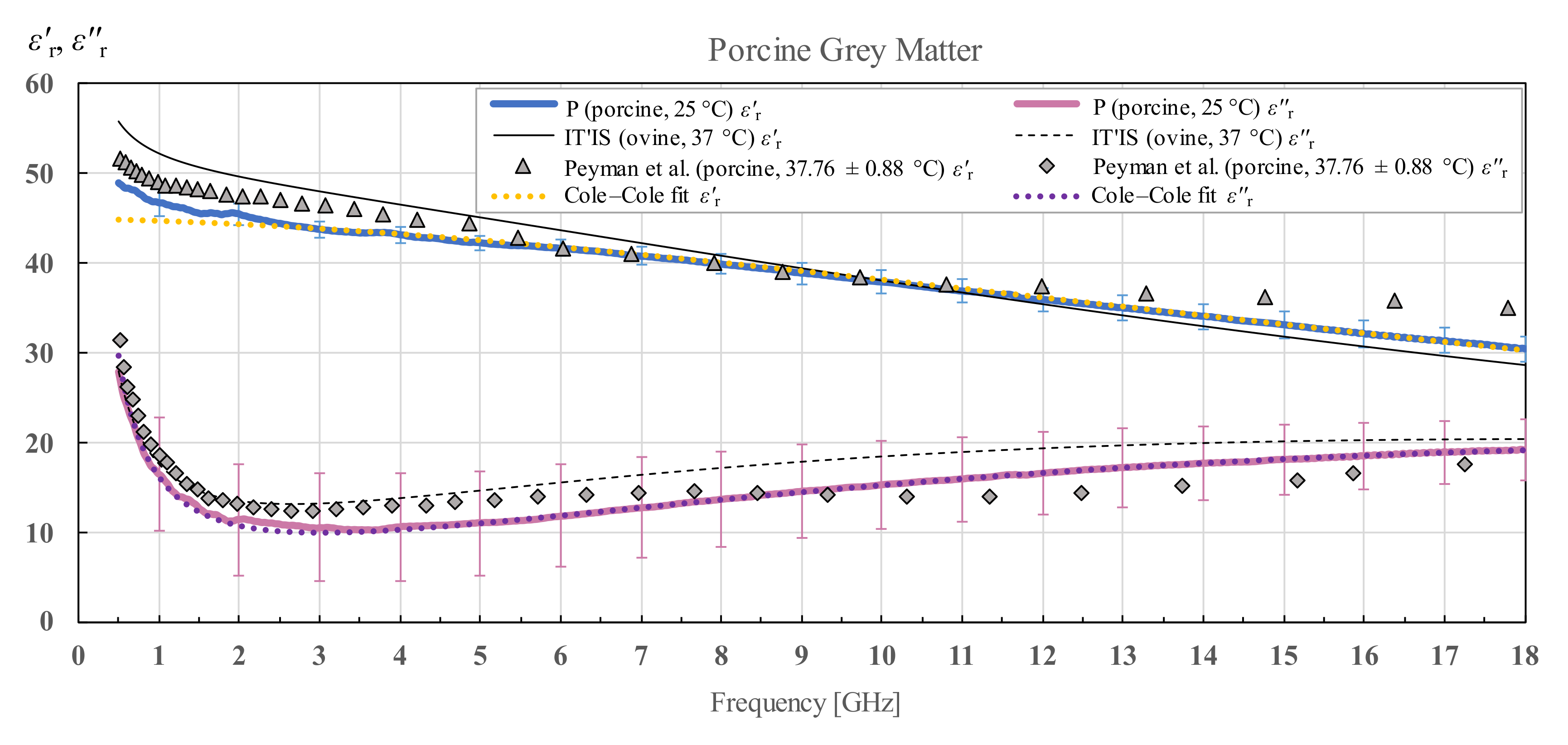
3.3. Porcine Brain Permittivity
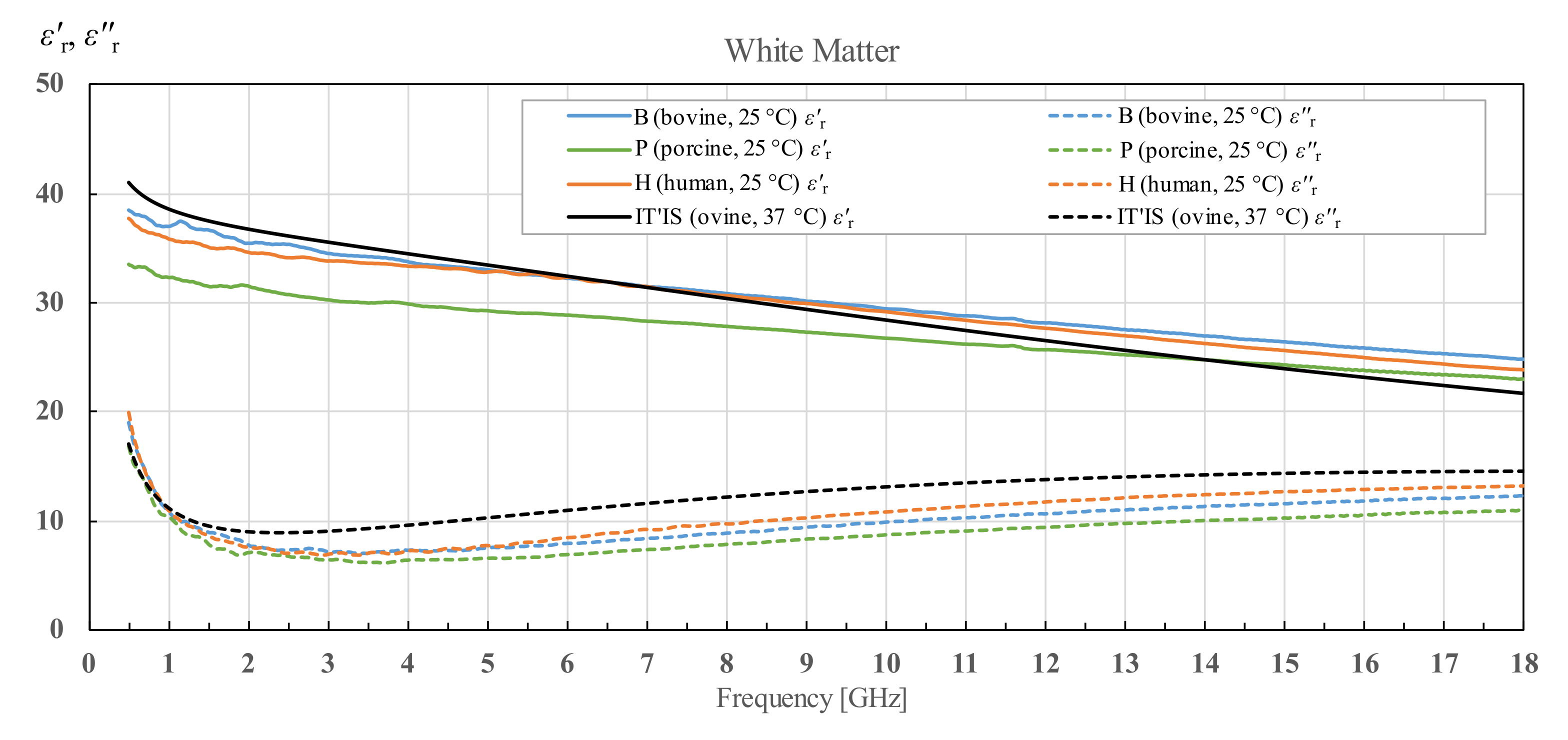
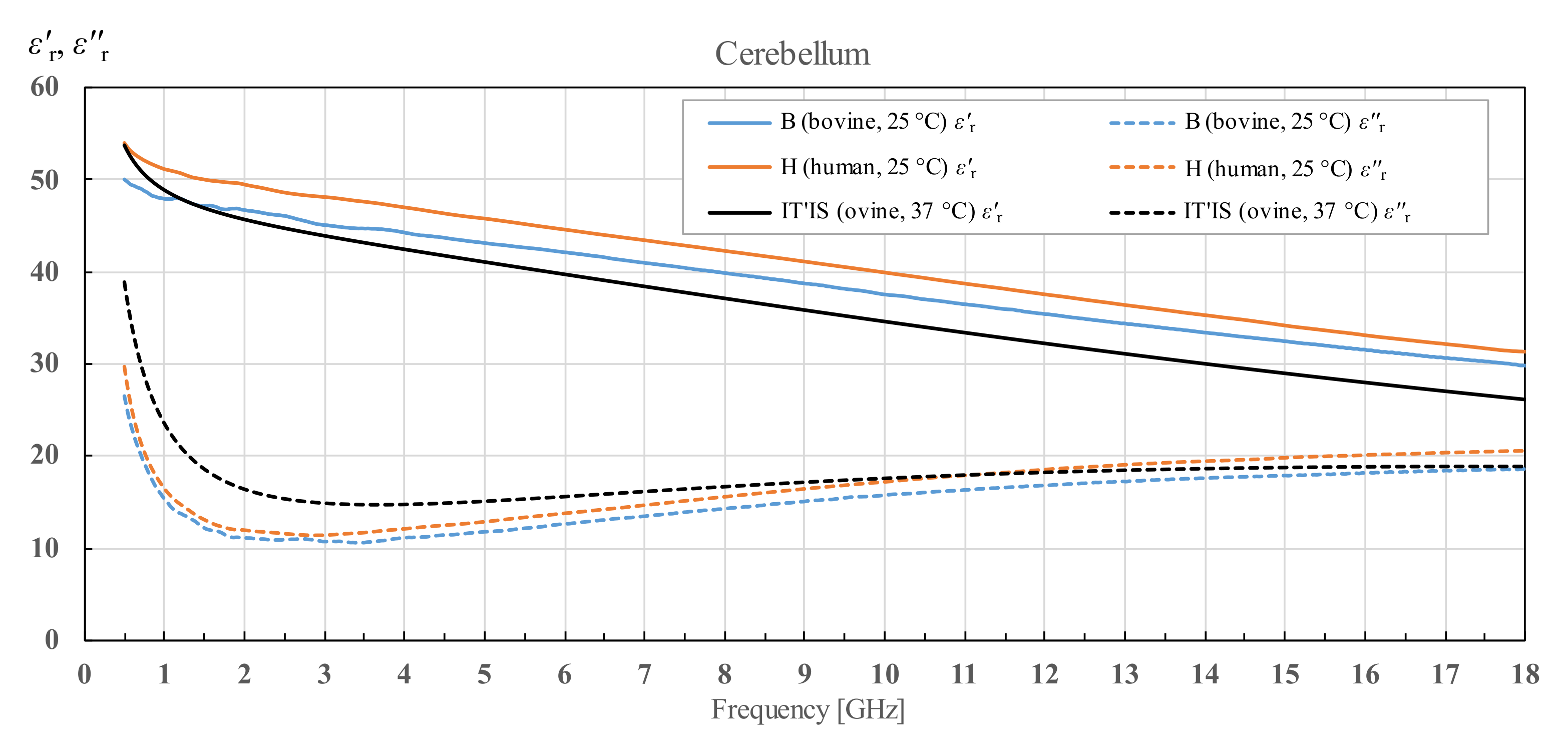
3.4. Interspecies Variability in Permittivity of White and Grey Matter and Cerebellum
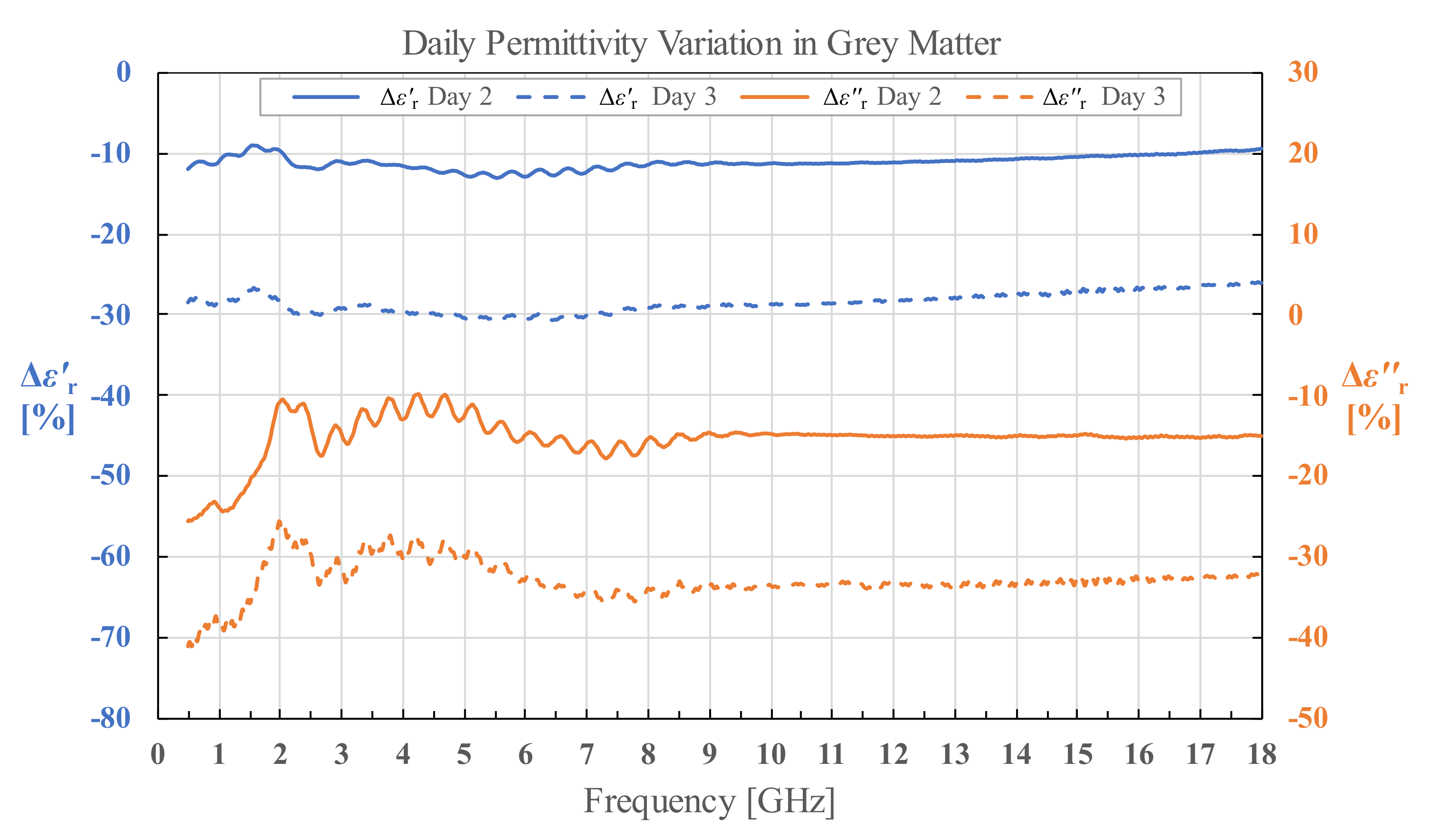
3.5. Human White and Grey Matter Permittivity on Day 2 and Day 3
3.6. Cole–Cole Models of the Data
3.7. Supplementary Materials
- human white matter, grey matter and cerebellum permittivity for brains H1 and H2, with the average permittivity of human white matter, grey matter and cerebellum;
- bovine white matter and grey matter permittivity for brains B1, B2 and B3, and cerebellum permittivity for brain B1, with the average permittivity of bovine white matter and grey matter;
- porcine white matter and grey matter permittivity;
- human white matter permittivity for brain H1 measured along the course of three days;
- Cole–Cole models for the average permittivity of each tissue.
4. Discussion
4.1. Results Comparison with the Published Literature
4.1.1. General Remarks
- rabbit specimen at 37 °C [31] (white and grey matter),
- mouse specimen at 37 °C [32] (grey matter),
- rat specimen in vivo at 32 °C [33] (grey matter),
- feline specimen in vivo at 36 °C [33] (grey matter),
- canine specimen in situ at 36 °C [34] (white and grey matter),
- canine specimen at 20 °C [35] (white and grey matter),
- feline specimen in vivo at 33 °C [36] (white and grey matter),
- canine specimen at 37 °C [37] (white and grey matter),
4.1.2. Human Tissues
4.1.3. Bovine Tissues
4.1.4. Porcine Tissues
4.2. Interspecies Variability
4.3. Tissue Degradation over Time
4.4. Intraspecies Variability
4.4.1. Age-Related Factors
4.4.2. Medical Condition of the Patients
4.4.3. Tissue Variability
5. Conclusions
Supplementary Materials
Author Contributions
Funding
Institutional Review Board Statement
Informed Consent Statement
Data Availability Statement
Acknowledgments
Conflicts of Interest
References
- Crocco, L.; Karanasiou, I.; James, M.L.; Conceição, R.C. (Eds.) Emerging Electromagnetic Technologies for Brain Diseases Diagnostics, Monitoring and Therapy; Springer International Publishing: Cham, Switzerland, 2018; ISBN 978-3-319-75006-4. [Google Scholar]
- Saied, I.; Arslan, T.; Chandran, S.; Smith, C.; Spires-Jones, T.; Pal, S. Non-Invasive RF Technique for Detecting Different Stages of Alzheimer’s Disease and Imaging Beta-Amyloid Plaques and Tau Tangles in the Brain. IEEE Trans. Med. Imaging 2020, 39, 4060–4070. [Google Scholar] [CrossRef] [PubMed]
- Persson, M.; Fhager, A.; Trefna, H.D.; Yu, Y.; McKelvey, T.; Pegenius, G.; Karlsson, J.-E.; Elam, M. Microwave-Based Stroke Diagnosis Making Global Prehospital Thrombolytic Treatment Possible. IEEE Trans. Biomed. Eng. 2014, 61, 2806–2817. [Google Scholar] [CrossRef] [PubMed] [Green Version]
- Ljungqvist, J.; Candefjord, S.; Persson, M.; Jönsson, L.; Skoglund, T.; Elam, M. Clinical Evaluation of a Microwave-Based Device for Detection of Traumatic Intracranial Hemorrhage. J. Neurotrauma 2017, 34, 2176–2182. [Google Scholar] [CrossRef] [PubMed]
- El rube’, I.; Heatley, D.; Abdel-Maguid, M. Detecting a Stroke-Affected Region in the Brain by Scanning with Low-Intensity Electromagnetic Waves in the Radio Frequency/Microwave Band. Healthcare 2021, 9, 1170. [Google Scholar] [CrossRef] [PubMed]
- Vedin, T.; Bergenfeldt, H.; Holmström, E.; Lundager-Forberg, J.; Edelhamre, M. Microwave scan and brain biomarkers to rule out intracranial hemorrhage: Study protocol of a planned prospective study (MBI01). Eur. J. Trauma Emerg. Surg. 2022, 48, 1335–1342. [Google Scholar] [CrossRef]
- Lazebnik, M.; Popovic, D.; McCartney, L.; Watkins, C.B.; Lindstrom, M.J.; Harter, J.; Sewall, S.; Ogilvie, T.; Magliocco, A.; Breslin, T.M.; et al. A large-scale study of the ultrawideband microwave dielectric properties of normal, benign and malignant breast tissues obtained from cancer surgeries. Phys. Med. Biol. 2007, 52, 6093–6115. [Google Scholar] [CrossRef]
- Thill, M. MarginProbe ®: Intraoperative margin assessment during breast conserving surgery by using radiofrequency spectroscopy. Expert Rev. Med. Devices 2013, 10, 301–315. [Google Scholar] [CrossRef]
- Burfeindt, M.J.; Zastrow, E.; Hagness, S.C.; Van Veen, B.D.; Medow, J.E. Microwave beamforming for non-invasive patient-specific hyperthermia treatment of pediatric brain cancer. Phys. Med. Biol. 2011, 56, 2743–2754. [Google Scholar] [CrossRef]
- Schooneveldt, G.; Dobšíček Trefná, H.; Persson, M.; de Reijke, T.M.; Blomgren, K.; Kok, H.P.; Crezee, H. Hyperthermia Treatment Planning Including Convective Flow in Cerebrospinal Fluid for Brain Tumour Hyperthermia Treatment Using a Novel Dedicated Paediatric Brain Applicator. Cancers 2019, 11, 1183. [Google Scholar] [CrossRef] [Green Version]
- Drizdal, T.; Paulides, M.M.; van Holthe, N.; van Rhoon, G.C. Hyperthermia treatment planning guided applicator selection for sub-superficial head and neck tumors heating. Int. J. Hyperthermia 2018, 34, 704–713. [Google Scholar] [CrossRef]
- Prasad, B.; Kim, J.K.; Kim, S. Role of Simulations in the Treatment Planning of Radiofrequency Hyperthermia Therapy in Clinics. J. Oncol. 2019, 2019, 9685476. [Google Scholar] [CrossRef] [PubMed] [Green Version]
- Datta, N.R.; Jain, B.M.; Mathi, Z.; Datta, S.; Johari, S.; Singh, A.R.; Kalbande, P.; Kale, P.; Shivkumar, V.; Bodis, S. Hyperthermia: A Potential Game-Changer in the Management of Cancers in Low-Middle-Income Group Countries. Cancers 2022, 14, 315. [Google Scholar] [CrossRef] [PubMed]
- Kroesen, M.; van Holthe, N.; Sumser, K.; Chitu, D.; Vernhout, R.; Verduijn, G.; Franckena, M.; Hardillo, J.; van Rhoon, G.; Paulides, M. Feasibility, SAR Distribution, and Clinical Outcome upon Reirradiation and Deep Hyperthermia Using the Hypercollar3D in Head and Neck Cancer Patients. Cancers 2021, 13, 6149. [Google Scholar] [CrossRef] [PubMed]
- Ramírez, A.; Rodríguez, P.; Gutiérrez, S.E.; Ávila, O. Preliminary experience in the management of brain and skull-base tumors with microwave ablation; feasibility guided by ultrasound, report from 23 cases. Surg. Neurol. Int. 2019, 10, 17. [Google Scholar] [CrossRef]
- Redr, J.; Pokorny, T.; Drizdal, T.; Fiser, O.; Brunat, M.; Vrba, J.; Vrba, D. Microwave Hyperthermia of Brain Tumors: A 2D Assessment Parametric Numerical Study. Sensors 2022, 22, 6115. [Google Scholar] [CrossRef]
- International Commission on Non-Ionizing Radiation Protection (ICNIRP) Guidelines for Limiting Exposure to Electromagnetic Fields (100 kHz to 300 GHz). Health Phys. 2020, 118, 483–524. [CrossRef]
- Gosselin, M.-C.; Neufeld, E.; Moser, H.; Huber, E.; Farcito, S.; Gerber, L.; Jedensjö, M.; Hilber, I.; Gennaro, F.D.; Lloyd, B.; et al. Development of a new generation of high-resolution anatomical models for medical device evaluation: The Virtual Population 3.0. Phys. Med. Biol. 2014, 59, 5287–5303. [Google Scholar] [CrossRef] [Green Version]
- ZMT Zurich MedTech, AG. Sim4Life. Available online: https://zmt.swiss/sim4life/ (accessed on 18 August 2022).
- Gabriel, C. Compilation of the Dielectric Properties of Body Tissues at RF and Microwave Frequencies; Occupational and Environmental Health Directorate, Radiofrequency Radiation Division, Brooks Air Force Base: San Antonio, TX, USA, 1996. [Google Scholar]
- Hasgall, P.A.; Di Gennaro, F.; Baumgartner, C.; Neufeld, E.; Lloyd, B.; Gosselin, M.C.; Payne, D.; Klingenböck, A.; Kuster, N. IT’IS Database for Thermal and Electromagnetic Parameters of Biological Tissues, Version 4.1. 2022. Available online: itis.swiss/database (accessed on 21 August 2022).
- Cole, K.S.; Cole, R.H. Dispersion and Absorption in Dielectrics I. Alternating Current Characteristics. J. Chem. Phys. 1941, 9, 341–351. [Google Scholar] [CrossRef] [Green Version]
- Šarolić, A.; Matković, A. Dielectric Permittivity Measurement Using Open-Ended Coaxial Probe—Modeling and Simulation Based on the Simple Capacitive-Load Model. Sensors 2022, 22, 6024. [Google Scholar] [CrossRef]
- Keysight Technologies. Keysight N1501A Dielectric Probe Kit 10 MHz to 50 GHz—Technical Overview; Keysight Technologies: Santa Rosa, CA, USA, 2018. [Google Scholar]
- Keysight Technologies. Field Fox Handheld Analyzers—Technical Overview 2021; Keysight Technologies: Santa Rosa, CA, USA, 2021. [Google Scholar]
- La Gioia, A.; Porter, E.; Merunka, I.; Shahzad, A.; Salahuddin, S.; Jones, M.; O’Halloran, M. Open-Ended Coaxial Probe Technique for Dielectric Measurement of Biological Tissues: Challenges and Common Practices. Diagnostics 2018, 8, 40. [Google Scholar] [CrossRef]
- Keysight Technologies. N1500A Materials Measurement Suite—Technical Overview 2021; Keysight Technologies: Santa Rosa, CA, USA, 2021. [Google Scholar]
- Schmid, G.; Neubauer, G.; Mazal, P.R. Dielectric properties of human brain tissue measured less than 10 h postmortem at frequencies from 800 to 2450 MHz. Bioelectromagnetics 2003, 24, 423–430. [Google Scholar] [CrossRef] [PubMed]
- Schmid, G.; Überbacher, R. Age dependence of dielectric properties of bovine brain and ocular tissues in the frequency range of 400 MHz to 18 GHz. Phys. Med. Biol. 2005, 50, 4711–4720. [Google Scholar] [CrossRef] [PubMed]
- Peyman, A.; Holden, S.J.; Watts, S.; Perrott, R.; Gabriel, C. Dielectric properties of porcine cerebrospinal tissues at microwave frequencies: In vivo, in vitro and systematic variation with age. Phys. Med. Biol. 2007, 52, 2229–2245. [Google Scholar] [CrossRef] [PubMed]
- Steel, M.C.; Sheppard, R.J. Dielectric properties of mammalian brain tissue between 1 and 18 GHz. Phys. Med. Biol. 1985, 30, 621–630. [Google Scholar] [CrossRef]
- Thurai, M.; Goodridge, V.D.; Sheppard, R.J.; Grant, E.H. Variation with age of the dielectric properties of mouse brain cerebrum. Phys. Med. Biol. 1984, 29, 1133–1136. [Google Scholar] [CrossRef]
- Kraszewski, A.; Stuchly, M.A.; Stuchly, S.S.; Smith, A.M. In vivo and in vitro dielectric properties of animal tissues at radio frequencies. Bioelectromagnetics 1982, 3, 421–432. [Google Scholar] [CrossRef]
- Burdette, E.C.; Friederich, P.G.; Seaman, R.L.; Larsen, L.E. In Situ Permittivity of Canine Brain: Regional Variations and Postmortem Changes. IEEE Trans. Microw. Theory Tech. 1986, 34, 38–50. [Google Scholar] [CrossRef]
- Xu, D.; Liu, L.; Jiang, Z. Measurement of the Dielectric Properties of Biological Substances Using an Improved Open-Ended Coaxial Line Resonator Method. IEEE Trans. Microw. Theory Tech. 1987, 35, 1424–1428. [Google Scholar] [CrossRef]
- Stuchly, M.A.; Athey, T.W.; Stuchly, S.S.; Samaras, G.M.; Taylor, G. Dielectric properties of animal tissues in vivo at frequencies 10 MHz–1 GHz. Bioelectromagnetics 1981, 2, 93–103. [Google Scholar] [CrossRef]
- Foster, K.R.; Schepps, J.L.; Stoy, R.D.; Schwan, H.P. Dielectric properties of brain tissue between 0.01 and 10 GHz. Phys. Med. Biol. 1979, 24, 1177–1187. [Google Scholar] [CrossRef]
- Rossmann, C.; Haemmerich, D. Review of Temperature Dependence of Thermal Properties, Dielectric Properties, and Perfusion of Biological Tissues at Hyperthermic and Ablation Temperatures. Crit. Rev. Biomed. Eng. 2014, 42, 467–492. [Google Scholar] [CrossRef] [PubMed] [Green Version]
- Michel, E.; Hernandez, D.; Lee, S.Y. Electrical conductivity and permittivity maps of brain tissues derived from water content based on T1-weighted acquisition. Magn. Reson. Med. 2017, 77, 1094–1103. [Google Scholar] [CrossRef] [PubMed]
- Mohammed, B.; Bialkowski, K.; Abbosh, A.; Mills, P.C.; Bradley, A.P. Closed-form equation to estimate the dielectric properties of biological tissues as a function of age: Dielectric Properties of Biological Tissues. Bioelectromagnetics 2017, 38, 474–481. [Google Scholar] [CrossRef] [PubMed]
- Gullett, J.M.; O’Shea, A.; Lamb, D.G.; Porges, E.C.; O’Shea, D.M.; Pasternak, O.; Cohen, R.A.; Woods, A.J. The association of white matter free water with cognition in older adults. NeuroImage 2020, 219, 117040. [Google Scholar] [CrossRef]
- Jones, S.B.; Sheng, W.; Or, D. Dielectric Measurement of Agricultural Grain Moisture—Theory and Applications. Sensors 2022, 22, 2083. [Google Scholar] [CrossRef]
- Siwach, P.; Levy, E.; Livshits, L.; Feldman, Y.; Kaganovich, D. Water is a biomarker of changes in the cellular environment in live animals. Sci. Rep. 2020, 10, 9095. [Google Scholar] [CrossRef]
- Lewis, S.R.; Pritchard, M.W.; Evans, D.J.; Butler, A.R.; Alderson, P.; Smith, A.F.; Roberts, I. Colloids versus crystalloids for fluid resuscitation in critically ill people. Cochrane Database Syst. Rev. 2018, 8, CD000567. [Google Scholar] [CrossRef]
- Schrier, R.W.; Wang, W.; Poole, B.; Mitra, A. Acute renal failure: Definitions, diagnosis, pathogenesis, and therapy. J. Clin. Investig. 2004, 114, 5–14. [Google Scholar] [CrossRef] [Green Version]
- Haller, M.; Schelling, G. Medizin aktuell: Aktuelle Strategien Klinischer Ernährung II. Anaesthesist 2000, 49, 349–352. [Google Scholar] [CrossRef]
- DeSai, C.; Hays Shapshak, A. Cerebral Ischemia. In StatPearls; StatPearls Publishing: Treasure Island, FL, USA, 2022. [Google Scholar]
- Zhang, Z.; Guo, Q.; Wang, E. Hyperventilation in neurological patients: From physiology to outcome evidence. Curr. Opin. Anaesthesiol. 2019, 32, 568–573. [Google Scholar] [CrossRef]
- Fornes-Leal, A.; Garcia-Pardo, C.; Frasson, M.; Pons Beltrán, V.; Cardona, N. Dielectric characterization of healthy and malignant colon tissues in the 0.5–18 GHz frequency band. Phys. Med. Biol. 2016, 61, 7334–7346. [Google Scholar] [CrossRef] [PubMed]
- Ištuk, N.; Porter, E.; O’Loughlin, D.; McDermott, B.; Santorelli, A.; Abedi, S.; Joachimowicz, N.; Roussel, H.; O’Halloran, M. Dielectric Properties of Ovine Heart at Microwave Frequencies. Diagnostics 2021, 11, 531. [Google Scholar] [CrossRef] [PubMed]


| Species | Brain | Age | White Matter | Grey Matter | Cerebellum |
|---|---|---|---|---|---|
| Human | H1 | 50 years | 14 | 15 | 15 |
| H2 | 84 years | 18 | 10 | 5 | |
| Bovine | B1 | Under 12 months | 72 | 70 | 13 |
| B2 | 38 | 32 | - | ||
| B3 | 38 | 32 | - | ||
| Porcine | P1 | Unknown | 38 | 40 | - |
| Day 2 | Day 3 | |||
|---|---|---|---|---|
| Grey Matter | −11.19% | −15.29% | −28.55% | −32.81% |
| White Matter | 8.97% | 8.24% | −20.63% | −24.34% |
| Porcine Brain | White Matter | 5 | 31 | 0.09 | 0.47 | 5.4 |
| Grey Matter | 3 | 45 | 0.05 | 0.8 | 6.2 | |
| Bovine Brain | White Matter | 5 | 34 | 0.09 | 0.48 | 5.6 |
| Grey Matter | 5 | 51 | 0.05 | 0.85 | 6.8 | |
| Cerebellum | 5 | 47 | 0.1 | 0.8 | 7.0 | |
| Human Brain | White Matter | 2 | 36 | 0.13 | 0.5 | 5.8 |
| Grey Matter | 5 | 51 | 0.05 | 0.9 | 7.1 | |
| Cerebellum | 3 | 47 | 0.08 | 0.83 | 6.5 |
| Brain Sample | White Matter | Grey Matter | Cerebellum | |||
|---|---|---|---|---|---|---|
| H1 | 2.4 | 1.1 | 2.9 | 1.6 | 3.9 | 2.3 |
| H2 | 8.1 | 4.5 | 3.1 | 1.9 | 2.1 | 1.5 |
| B1 | 3.0 | 1.4 | 3.1 | 1.9 | 6.0 | 3.6 |
| B2 | 2.1 | 0.9 | 1.7 | 0.9 | - | - |
| B3 | 2.1 | 1.1 | 2.3 | 1.5 | - | - |
| P | 2.6 | 1.3 | 1.2 | 4.9 | - | - |
Publisher’s Note: MDPI stays neutral with regard to jurisdictional claims in published maps and institutional affiliations. |
© 2022 by the authors. Licensee MDPI, Basel, Switzerland. This article is an open access article distributed under the terms and conditions of the Creative Commons Attribution (CC BY) license (https://creativecommons.org/licenses/by/4.0/).
Share and Cite
Matković, A.; Kordić, A.; Jakovčević, A.; Šarolić, A. Complex Permittivity of Ex-Vivo Human, Bovine and Porcine Brain Tissues in the Microwave Frequency Range. Diagnostics 2022, 12, 2580. https://doi.org/10.3390/diagnostics12112580
Matković A, Kordić A, Jakovčević A, Šarolić A. Complex Permittivity of Ex-Vivo Human, Bovine and Porcine Brain Tissues in the Microwave Frequency Range. Diagnostics. 2022; 12(11):2580. https://doi.org/10.3390/diagnostics12112580
Chicago/Turabian StyleMatković, Anđela, Anton Kordić, Antonia Jakovčević, and Antonio Šarolić. 2022. "Complex Permittivity of Ex-Vivo Human, Bovine and Porcine Brain Tissues in the Microwave Frequency Range" Diagnostics 12, no. 11: 2580. https://doi.org/10.3390/diagnostics12112580
APA StyleMatković, A., Kordić, A., Jakovčević, A., & Šarolić, A. (2022). Complex Permittivity of Ex-Vivo Human, Bovine and Porcine Brain Tissues in the Microwave Frequency Range. Diagnostics, 12(11), 2580. https://doi.org/10.3390/diagnostics12112580