


Abstract
Background: Flatfoot is an alteration of the normal structure of the foot, characterized by a partial or total reduction of the medial longitudinal plantar arch, valgus deformity of the heel, and abduction of the forefoot. While treatments often include strengthening of the intrinsic foot muscles, evidence of its efficacy in adults with flatfoot remains limited. Objectives: The main objective of this review was to evaluate the effects of strengthening the plantar intrinsic muscles in adults with flatfoot. Methods: Searches were conducted in PubMed, Embase, Cochrane, PEDro, and Web of Science databases up to October 2023. The review protocol was developed and followed according to the PRISMA Extension for Scoping Reviews (PRISMA-ScR) guidelines. Studies included were those published on intrinsic muscle strengthening in adult populations. A qualitative synthesis of all included articles was performed, along with a quantitative sub-analysis of randomized controlled trials and a critical methodological assessment. Results: Eleven studies involving a total of 374 participants were selected. Most studies identified the “short foot exercise” as the optimal exercise for isolating and training the plantar intrinsic foot muscles. The most commonly analyzed variables were the Foot Posture Index and the Navicular Drop Test. Conclusions: Strengthening the plantar intrinsic muscles enhances the height of the medial longitudinal arch, improves hindfoot posture and balance, and increases hallux abductor muscle activity. This strengthening, whether achieved through short foot exercises alone or in combination with other techniques, is effective in treating adult flatfoot. Current literature suggests that a duration of 4–6 weeks may be sufficient to achieve beneficial outcomes.
1. Introduction
Adult acquired flatfoot (AAFF) is a common deformity in the general population that develops after skeletal maturity. It is characterized by a partial or complete reduction in the medial longitudinal arch (MLA) and a heel valgus deformity caused by the downward, inward, and forward displacement of the talus relative to the calcaneus. Symptoms typically begin in the fourth or fifth decade of life, with peak incidence around age 55 [1]. This deformity can be a casual finding or a frequent reason for medical consultation, ranging from occasional discomfort to significant limitations in daily activities. It is about 25% more prevalent in females, in individuals with obesity, and in those with a history of childhood flat feet [1].
Identifying the type of flatfoot (flexible or rigid) is crucial for determining the appropriate treatment, whether conservative or surgical [2,3]. Treatment is challenging because this condition involves multiple static and dynamic deformities [4,5]. Commonly used conservative treatments include plantar orthoses, anti-pronation taping, and motion control shoes [6,7]. An active exercise intervention offers additional benefits over passive bracing by improving foot arches through the strengthening of intrinsic foot muscles. While intrinsic foot muscle (IPM) strength is recognized as vital for maintaining foot arch integrity and function, there is a significant gap in the literature regarding exercise interventions specifically targeting IPM in adult acquired flatfoot deformity (AAFF). Most research has concentrated on surgical and orthotic management rather than on conservative, exercise-based approaches. This is concerning because talus displacement and heel valgus in AAFF not only disrupt foot biomechanics but also affect the stability and function of the entire lower limb, including the ankle, knee, and hip. Existing studies often overlook the broader impact of IPM strengthening on overall foot biomechanics and lower extremity function, underscoring the need for evidence-based exercise protocols. Addressing this gap could enhance non-surgical interventions, improving balance, stability, and overall outcomes in AAFF management [6,7].
Given the high number of patients presenting with this deformity and the evidence supporting the role of IPM strengthening in other conditions, such as ankle instability, plantar fasciitis, and hallux valgus [1,4,5,8,9,10], the clinical application of training programs targeting flatfoot as part of conservative treatment is of significant interest. Therefore, the purpose of this review is to evaluate the effects of intrinsic plantar muscle strengthening in adults with flatfoot and to detail the exercise prescriptions used in IPM strengthening programs.
2. Methods
This scoping review was conducted in accordance with the Preferred Reporting Items for Systematic Reviews and Meta-Analyses (PRISMA) Extension for Scoping Reviews (PRISMA-ScR) guidelines [11] and the 6-stage framework described by Arksey and O’Malley, as reviewed by Levac et al. and O’Brien et al. [12,13,14] (Table S1). This methodology includes: (1) identifying the research question; (2) identifying relevant studies; (3) selecting studies; (4) charting data; (5) collating, summarizing, and reporting results; and (6) an optional final phase of consultation exercises. The review protocol was not registered, as registration is not mandatory for scoping reviews.
2.1. Phase 1: Identifying the Research Question
Our review was guided by the following research question: “Is IPM strengthening effective as a treatment for adult flatfoot?”
2.2. Phase 2: Identification of Relevant Studies
Two researchers (MMMF and PTV) conducted the literature search to identify, review, and select studies for inclusion. The search strategy was developed following the methodological procedures proposed by Arksey and O’Malley [12] and the Johanna Briggs Institute [15], using a 3-step approach:
- A pilot search of the PubMed database using the main search strategy ((“flatfoot” [Mesh]) OR “Flatfoot/rehabilitation” [Mesh]) OR (“Flatfoot/therapy” [Mesh]). The complete search strategy is available in Appendix A.
- The medical descriptors (Mesh) used included flatfoot, exercise therapy, and pronation. Additional synonyms such as muscle strength and pronated foot were also included.
- Execution of the final search strategy and subsequent review of reference lists from selected articles.
A comprehensive search was conducted across five recognized databases: PubMed, Embase, Cochrane, PEDro, and Web of Science, covering the period from February to October 2023 to ensure the inclusion of the most current articles on the topic. Doctoral theses were also reviewed through the international database ‘oatd.org,’ but no relevant results were obtained.
Article selection was carried out according to the PRISMA-ScR guidelines [11], which ensure transparency and quality in conducting systematic and scoping reviews. Articles were assessed by reviewers MMMF and PTV, who applied established inclusion and exclusion criteria detailed below. This rigorous methodology was employed to ensure that only relevant and high-quality articles were included in the final analysis, maintaining the integrity and objectivity of the review by relying on scientifically robust and up-to-date studies.
2.3. Phase 3: Selection of Studies
2.3.1. Population: Inclusion and Exclusion Criteria
Included studies were required to involve healthy adults (defined as individuals aged 18 years or older) with flexible flatfoot (e.g., obese individuals, those with patellofemoral pain, pronated feet, or instability), provided that measurements such as the Navicular Drop (ND) and Foot Posture Index (FPI) scores indicated the presence of flatfoot or pronation. Exclusion criteria included studies focused on pediatric populations, surgical interventions, or severe trauma. Although no specific age range was set, it is acknowledged that younger and older adults may respond differently to the same exercise program, which should be considered when interpreting the results.
2.3.2. Instrument: Inclusion and Exclusion Criteria
Studies that used the Navicular Drop (ND) and/or the Foot Posture Index (FPI) as clinical assessment tools for the dependent variable (measurement of the effectiveness of IPM strengthening) were included. The ND test is a cost-effective, easy, and quick method that assesses the difference in the navicular tuberosity height from the ground in millimeters using a measuring tape. Measurements are taken from an initial seated position with the subtalar joint in neutral and then from a standing position, corresponding to the drop of the navicular and, consequently, the lowering of the medial longitudinal arch (MLA). The difference between the original height of the navicular tuberosity in seated and weight-bearing positions represents the navicular drop, indicating the descent of the MLA [16].
The FPI quantifies the posture of each foot using six items, resulting in a total score ranging from −12 to +12 (reference values: 0 to +5 indicates a neutral position, +6 to +9 indicates a pronated position, +10 to +12 indicates a hyperpronated position, −1 to −5 indicates a supinated position, and −6 to −12 indicates a highly supinated position). The six items of the FPI include: (1) palpation of the talar head, (2) supra and infra malleolar curvature, (3) calcaneal position in the frontal plane, (4) prominence of the talonavicular region, (5) congruence of the medial longitudinal arch, and (6) abduction or adduction of the forefoot relative to the rearfoot. The FPI was assessed following the protocol by Redmond et al. [17], in which a score is obtained for each foot, with normal values set between 0 and +5.
2.3.3. Intervention: Inclusion and Exclusion Criteria
Studies describing a program of IPM strengthening among participants were included. Studies that combined these protocols with other types of conservative treatment were also considered. Given the interest in the effects of IPM strengthening on balance, studies evaluating the impact of strengthening on foot stability and function were included, regardless of the assessment tool used. Studies that did not explicitly address this variable were excluded.
In addition to these criteria, eligible studies met the following conditions: (1) published from 2014 to 2023, (2) published in English, German, or Spanish, and (3) contained relevant information on the effectiveness of IPM strengthening and the exercises required for it. During screening, we excluded: (1) literature reviews, (2) letters, editorials, government reports, guidelines, conference proceedings, and process development statements, (3) studies lacking methodological detail or scientific interest, and (4) studies without empirical data.
A two-stage screening process (titles/abstracts followed by full-text screening) was used to select eligible studies, with independent screenings conducted to identify relevant studies. Pilot screenings were performed using pre-defined inclusion and exclusion criteria to resolve any disagreements before the main screening process began. Discrepancies were resolved by consensus among all authors.
2.4. Phase 4: Data Charting
Two reviewers (MMMF and PTV) extracted data from the eligible studies to create an evidence table that recorded relevant information from the included articles. The extracted information from each study included: (1) author, study design, year, and countries where the studies were conducted; (2) type of interventions; (3) characteristics of the study population; (4) duration of the intervention; (5) frequency of exercise; and (6) muscle contraction duration.
2.5. Phase 5: Collate, Summarize, and Communicate the Results
A numerical analysis of the scope and nature of the studies is presented using a PRISMA flow chart, detailing the results of the search and screening process. Additionally, a qualitative synthesis of all included papers and an assessment of methodological quality were conducted. For this purpose, two reviewers (MMMF and PTV) independently evaluated the studies using the PEDro scale. Cohen’s Kappa statistic was employed to measure inter-rater agreement [18]. The PEDro scale comprises an 11-item checklist designed to assess methodological quality, with possible scores ranging from 0 to 10. Articles scoring above 6 were considered of good quality; scores between 4 and 6 were deemed fair quality; and scores below 4 were classified as poor quality [19].
3. Results
3.1. Selection of Studies
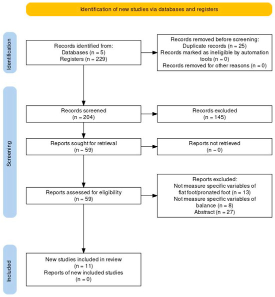
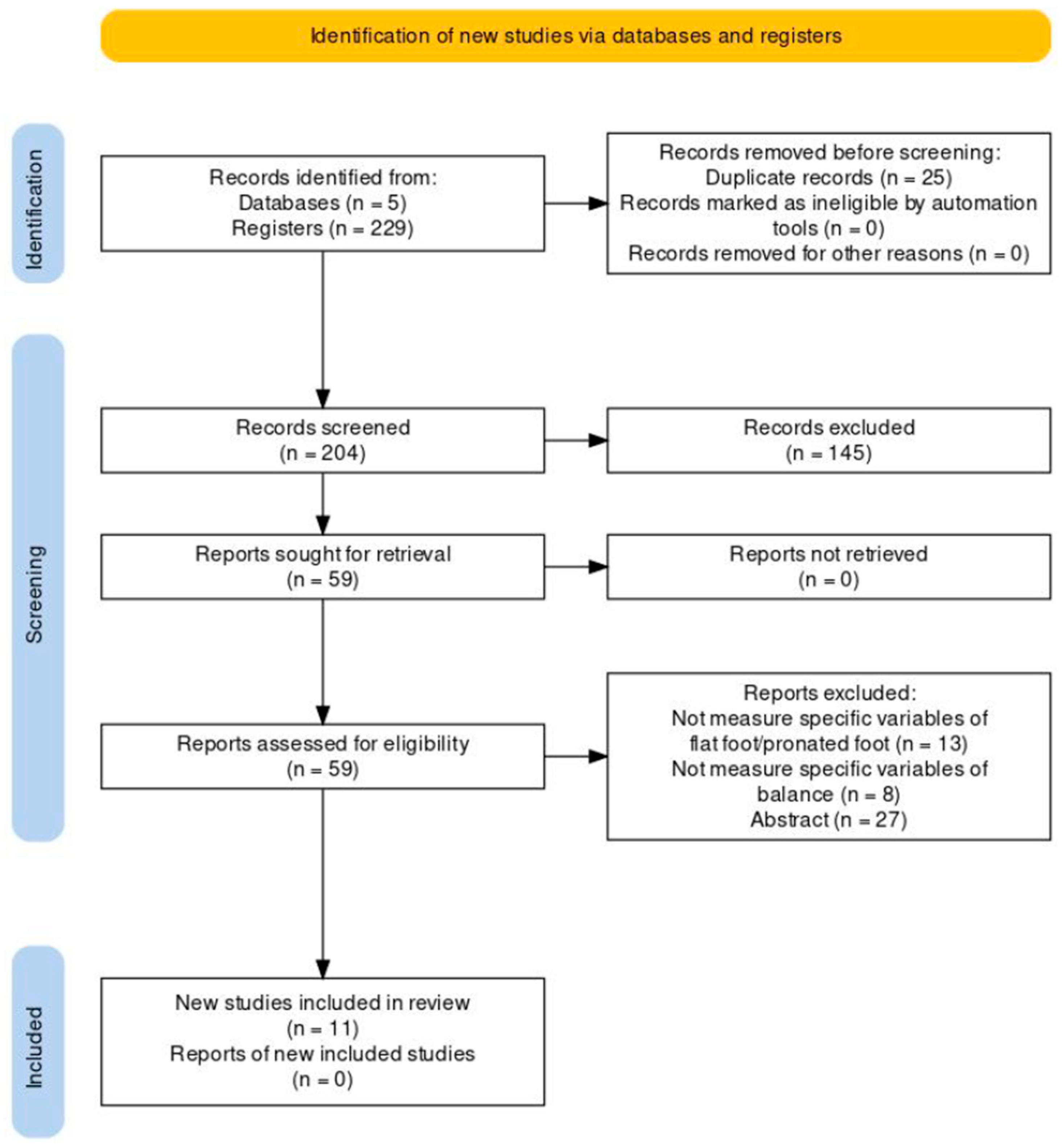
After completing the database searches using various keywords, a total of 229 documents were identified. After removing duplicates, 204 articles remained. Of these, 145 were excluded during the initial screening, resulting in 59 articles.
Out of the 59 articles, 48 were further excluded because they did not include variables specifically related to flatfoot and/or pronated foot, among others. Thus, 11 studies were included in the final review. The complete selection process across the various phases is detailed in Figure 1.
Figure 1.
Flow chart showing the selection and inclusion of studies according to the PRISMA guidelines.
Of the included studies, 9 provided relevant information on the measurement instruments used, along with their respective pre- and post-strengthening scores [20,21,22,23,24,25,26,27]. Additionally, only 4 recent articles explored the relationship between IPM strengthening and its effects on balance in individuals with flat feet [21,27,28,29], while a fifth study provided related information on this variable [30].
3.2. Descriptive Numerical Analysis
Of the eleven included studies, six used the ND [21,22,23,24,25,27] and five employed the FPI [22,23,26,27,31]. The selected articles included a total of 374 participants, with the proportion of females ranging from 43% to 57%. The mean height of the participants was 167.10 cm, and their mean BMI was 23.73 kg/m2. The study populations included flexible flatfoot [25], flexible flatfoot with ND > 10 mm [25,27,28], IPF > 6 [22,26], pronated feet with FPI > 6 [23,31], and right flat feet as measured by the arch height index [7]. Five studies were conducted in Europe [20,22,23,24,25,26], and six were conducted in Asia [21,22,25,27,28,30]. Nine studies took place in university settings [20,21,22,23,24,25,26,27,30], one in a clinical setting [31], and one in the general population [28]. All studies, except one [20], divided participants into experimental and control groups [21,22,23,24,25,26,27,28,30,31]. Details regarding intervention duration, training protocol, exercise frequency, and muscle contraction duration are summarized in Table 1.
Table 1.
Studies reporting intervention duration, training protocol, exercise frequency, and muscle contraction duration.
3.3. Methodological Quality
The methodological quality of the included clinical trials was assessed using the PEDro scale (Table 2). The quality scores of the included studies ranged from 1 to 10 out of a possible 10 (criterion 1 affects external validity but not internal validity, so it is not included in the total score). The mean PEDro score was 7, indicating that, overall, the studies were of good methodological quality. One study [20] demonstrated poor methodological quality, scoring 1/10. Two studies [26,28] were classified as fair quality, five studies [21,22,24,27,31] were considered good quality, and three studies [20,24,29] were rated as excellent quality. Two studies [23,25] blinded both participants and therapists. Nine studies [21,22,23,25,26,27,28,30,31] included between-group comparisons. The agreement between the two assessors was 91%. Cohen’s kappa coefficient for the measure of agreement between the reviewers on individual validity was 0.88, indicating almost excellent agreement.
Table 2.
Evaluation According to the PEDro scale. R1: reviewer 1; R2: reviewer.
4. Discussion
The main objective of this literature review was to evaluate the effectiveness and outcomes of IPM strengthening interventions in adults with flatfoot. Most articles classify the “short foot exercise” (SFE) as a form of sensorimotor training that activates the IPM and actively shapes the medial longitudinal arch (MLA) and transverse plantar arch [20,21,24]. Authors suggest that SFE is a valuable tool for treating conditions associated with excessive foot pronation, such as flexible flatfoot in adults, and contributes positively to improving the MLA [20,21,22,24,25,26,27].
The SFE involves moving the head of the first metatarsal closer to the heel while keeping the toes extended. The subtalar joint should remain in a neutral position, allowing the MLA to elevate without toe flexion [23]. In the studies selected for this section [20,21,22,23,24,25,26,27], the SFE was the predominant exercise performed by the experimental groups, except in the study by Sánchez-Rodríguez et al. [31], which used a toe exercise involving picking up objects.
Various clinical assessment tools were used to evaluate the effectiveness of IPM strengthening. The most recent study using the “Arch Height Index” measurement system was conducted by Ishiyama et al. [20] in 2022, which effectively identified individuals with scores below 0.310 for men and 0.290 for women as having flat feet. However, the Navicular Drop (ND) test was the most commonly used assessment method among the majority of authors. This test identifies flat feet in individuals who present a difference greater than 10 mm, as reported across all studies [21,22,23,25,27]. The second most common tool was the Foot Posture Index (FPI), used in five studies, classifying individuals with scores of 6 as having flat feet [22,23,26,27,31]. Park et al. [21] in 2021 and Kim et al. [27] in 2016 exclusively used the ND to assess efficacy. In Park et al.’s study involving 44 participants, one group performed the SFE, while the other underwent proprioceptive neuromuscular facilitation. Both groups showed significant improvement in the ND test after 4 weeks, with an increase in foot arch height during loading. However, proprioceptive neuromuscular facilitation reduced the height difference in the ND more than the SFE, likely because it improved the strength of the dorsiflexor and evertor muscles more effectively. Kim et al. [27] reported that the ND score decreased from 11.4 ± 1.6 mm to 7.7 ± 1.1 mm following the SFE intervention, concluding that the SFE was effective. Kim et al. [27] conducted the only study comparing the effects of the SFE with plantar supports for flat feet. They found that the SFE was more effective than plantar supports in improving the MLA and dynamic balance ability. The effectiveness of plantar supports depends heavily on usage frequency, and their impact in adults tends to be palliative rather than corrective, complicating data comparisons. Nonetheless, the authors indicated that combining both therapies enhances results.
Authors who used the ND along with other tests, including the FPI, were Pabón-Carrasco et al. [23], Sánchez-Rodríguez et al. [31], Kisacik et al. [24], Okamura et al. [22], and Namsawang et al. [25]. In 2020, the studies by Pabón-Carrasco et al. [23] and Kisacik et al. [24] did not include individuals with flat feet but are considered noteworthy because they demonstrated the efficacy of an intervention program incorporating the SFE. Both authors reported positive effects on the navicular position, showing a tendency toward a more neutral stance and an improvement in hindfoot pronation posture, as evidenced by enhancements in the FPI. Sánchez-Rodríguez et al. [31] did not use the SFE to strengthen the IPM but included a toe exercise for picking up objects as part of a joint muscle strengthening protocol (targeting both extrinsic and intrinsic musculature). Their results showed a 1.66-point reduction in the FPI score, indicating improved hyperpronation and a more neutral foot posture, aligning with findings from Pabón-Carrasco et al. [23] and Kisacik et al. [24].
Okamura et al. [22] were the only authors to evaluate the ND during gait. Their study found no change with the SFE intervention, likely due to the need for more precise measurement methods and the small sample size, which may have hindered detection of improvements. Nonetheless, they observed that the navicular slowed its descent during dynamic movement, suggesting improved foot stability.
In addition to the aforementioned tests, four studies evaluated muscle modifications to assess the effectiveness of the intervention [20,22,25,26]. Among these, Okamura et al. [22] and Namsawang et al. [25] were the only ones to combine the SFE with neuromuscular electrical stimulation (NMES). Okamura et al. [22] measured muscle thickness, while Namsawang et al. [25] focused on the cross-sectional area of the abductor hallucis muscle (AHM), with activity measured via wireless surface electromyography. Results were significantly better in the group that received SFE combined with NMES. Both studies confirmed the efficacy of the SFE alone but concluded that its effectiveness is enhanced when combined with NMES.
Regarding the effectiveness of the SFE, Namsawang et al. [25], Ishiyama et al. [20], and Unver et al. [26] observed an increase in AHM activity after the exercises. Ishiyama et al. [20] found that both AHM and MLA muscle activities increased during the SFE but emphasized that the effectiveness of the SFE depends on the type of flatfoot (flexible or rigid). They suggested that an assessment of flexibility is necessary before establishing a treatment plan. In contrast, Unver et al. [26] confirmed that the SFE was effective in reducing the ND and improving foot posture, as well as in reducing pain and disability while increasing maximal plantar strength in the midfoot. Because the SFE reduces the ND and FPI, it may prevent excessive pronation during gait, potentially increasing lateral plantar strength of the midfoot due to reduced pronation.
Regarding training protocols and intervention duration, the literature lacks a specific evidence-based training protocol for the SFE. Table 1 presents training protocols according to different authors based on intervention duration, training structure, exercise frequency, and muscle contraction duration, generally describing intervention programs ranging from 4 to 9 weeks.
IPM strengthening is effective in improving both dynamic and static balance, suggesting that the SFE may enhance functional movement balance in individuals with flat feet [21,23,27,28,31]. However, current evidence is insufficient to definitively determine the effect of foot core training on balance in adult flatfoot patients, as few studies have examined the direct effects of the SFE on postural stability. Therefore, future randomized controlled trials (RCTs) investigating proprioception in this condition are recommended.
Different measuring instruments were used to evaluate the effects of strengthening on balance. In the study by Park et al. [21], a Tekscan pressure mapping tool was used to measure changes in the center of pressure. The SFE significantly improved anterior–posterior and left-right balance, and it was determined that performing the SFE for 4 weeks effectively restored balance. Regarding static equilibrium, the study by Kim et al. [28] utilized visual feedback during exercise to prevent compensatory movements and achieve correct posture over 6 weeks. They reported a significant improvement in the flexible flatfoot group before and after the SFE intervention (p < 0.05). For dynamic balance, studies [27,29,30] employed the “Y-balance test” [27] and the “stability limit test” [29]. The results indicated that the SFE enhances dynamic stability. Kim et al. [27] attributed this improvement to the SFE’s ability to enhance the function and activity of the AHM and flexor hallucis brevis muscles, both of which play critical roles in foot stability during gait. Moon et al. [29], however, suggested that the SFE stimulates proprioceptors in the sole of the foot, increasing afferent input, thereby enhancing stability and voluntary muscle activities. This increased the contact pressure between the foot and the ground, intensifying cutaneous stimulation. They also proposed that the SFE can be a foundational step in sensorimotor training, improving proprioception and postural stability when combined with other exercises. Lee et al. [30] suggest that a stable core may reduce muscle fatigue and improve proprioception in the lower limbs, which could potentially decrease muscle fatigue in the lower limb and thereby enhance ankle proprioceptive function. However, our review did not find sufficient evidence indicating that muscle exhaustion is a significant issue or that longer than typical rest times are recommended.
4.1. Limitations
The limitations of this review include a small sample size, which restricts the generalizability of the findings to larger populations, as the results may not represent the general population. Most reviewed studies did not account for rest or recovery time between sets during training, a critical variable in strength training planning, as its omission could impact results and lead to muscle fatigue. There were discrepancies in training protocols, with variations in intervention duration, exercise frequency, and time commitment, complicating comparisons and the determination of an optimal strengthening protocol. Variability in measuring the effectiveness of exercises led to discrepancies in results, making it challenging to assess the effectiveness of different training approaches. Differences in the foot selected for evaluation further introduced variability and made comparisons between studies difficult. Most studies involved asymptomatic participants with similar characteristics (age and BMI), limiting the applicability of results to more diverse populations, including older adults, overweight or obese individuals, and those experiencing pain or discomfort related to flatfoot. Many studies also lacked information on the long-term effects of IPM strengthening in adult flatfoot; without adequate follow-up, it remains unclear whether the benefits persist beyond the exercise period. Poor technique execution could lead to inaccurate results, affecting the reliability and validity of findings. Selection bias due to evidence collection strategies, including specific selection criteria or methodological limitations, may have also influenced the representativeness of the available evidence.
4.2. Practical Implications
Based on the review findings, clinicians should consider including specific IPM strengthening exercises as part of flatfoot treatment. Recognizing the current lack of evidence, further research is needed to fill these gaps and establish stronger evidence for the efficacy of strengthening exercises in adult flatfoot (e.g., clinical trials). When designing IPM strengthening interventions, factors such as optimal exercise duration, frequency, progression, and correct technique execution should be considered. Additionally, the importance of rest between sets should be emphasized to prevent muscle exhaustion. Patients should be educated on the importance of strengthening these muscles to improve foot stability and function, and exercise recommendations should be tailored to individual needs for performance at home or in the gym.
5. Conclusions
There is evidence that IPM strengthening improves MLA height, hindfoot posture, balance, and AHM muscle activity. Strengthening the IPM through the SFE, whether alone or in combination with other techniques, has proven effective in treating adult flatfoot. Discrepancies were noted in the training protocols, with heterogeneity observed in intervention durations (ranging from 4 to 9 weeks), exercise frequency (2 to 3 times per week), and the number of sets and repetitions (1 to 3 sets of 4 to 30 repetitions per session). While the reviewed studies do not establish optimal duration and frequency, these ranges can provide a general guide. However, it is recommended that professionals customize exercise programs based on the individual needs and abilities of each patient, adjusting progression and intensity according to the observed response.
Supplementary Materials
The following supporting information can be downloaded at: https://www.mdpi.com/article/10.3390/japma116010008/s1, Table S1: The PRISMA-ScR checklist.
Author Contributions
Conceptualization, M.M.M.-F. and P.T.-V.; methodology, M.M.M.-F., P.T.-V. and P.V.M.-M.; software,. M.M.M.-F.; validation, M.M.M.-F., P.T.-V. and P.V.M.-M.; formal analysis, M.M.M.-F., P.T.-V. and P.V.M.-M.; investigation, M.M.M.-F.; resources, M.M.M.-F., P.T.-V. and P.V.M.-M.; data curation, M.M.M.-F., P.T.-V. and P.V.M.-M.; writing—original draft preparation, M.M.M.-F., P.T.-V., P.V.M.-M., L.R.-F. and J.M.C.-S.; writing—review and editing, M.M.M.-F., P.T.-V. and P.V.M.-M.; visualization, P.T.-V. and P.V.M.-M.; supervision, P.T.-V. and P.V.M.-M.; project administration; M.M.M.-F., P.T.-V. and P.V.M.-M. funding acquisition M.M.M.-F. All authors have read and agreed to the published version of the manuscript.
Funding
This research received no external funding.
Institutional Review Board Statement
Not applicable.
Informed Consent Statement
Not applicable.
Data Availability Statement
Not applicable.
Conflicts of Interest
The authors declare no conflicts of interest.
Appendix A
Search strategies applied in the different databases consulted:
- Mesh terms in Pubmed
First search:
("flatfoot"[Mesh]) AND "Flatfoot/rehabilitation"[Mesh] Filters: 2014-2023, abstract, randomised controlled trial, Adult: 19+ years
Second search:
("flatfoot"[Mesh]) AND "Flatfoot/therapy"[Mesh] Filters: 2014-2023, abstract, clinical trial, randomised controlled trial, Adult: 19+ years
Third search:
(("flatfoot"[Mesh]) OR "Flatfoot/rehabilitation"[Mesh]) OR ( "Flatfoot/therapy"[Mesh] ) Filters: 2014-2023, abstract, clinical trial, randomised controlled trial, Adult: 19+ years
Fourth search:
("pesplanus"[Mesh]) AND "Pesplanus/rehabilitation"[Mesh] Filters: 2014-2023, abstract, randomised controlled trial, Adult: 19+ years
Fifth search:
("pesplanus"[Mesh]) AND "Pesplanus/therapy"[Mesh] Filters: 2014-2023, abstract, clinical trial, randomised controlled trial, Adult: 19+ years
Sixth search:
(("pesplanus"[Mesh]) OR "Pesplanus/rehabilitation"[Mesh]) OR ( "Pesplanus/therapy"[Mesh] ) Filters: 2014-2023, abstract, clinical trial, randomised controlled trial, Adult: 19+ years
- Medline in PubMed
Fist search:
flat foot AND (“muscles exercise” OR “intrinsic”)
Second search:
((flat foot) OR (pronated foot)) AND (exercise therapy)
Third search:
((flat foot) OR (pronated foot)) AND (muscle strength)
Filters: 2014-2023, abstract, clinical trial, randomised controlled trial, Adult: 19+ years
- The Cochrane Library
(flatfoot OR pronated foot OR pes planus) AND (muscle strength OR exercise therapy)
Filters: 2014-2023, clinical article, randomised controlled trial
- PEDro
First search:
Flatfoot*
Second search:
Pronated foot*
Third search:
Pesplanus*
Not filters.
- Webof Science
((flatfoot OR pronated foot OR pes planus OR pronation)) AND ((muscle strength OR exercise OR exercise therapy))
Filters: 2014-2023, Web of Science core collection, clinical trial and article, MeSH qualifier: therapy
- Embase
(flatfoot OR pronated OR pes planus) AND foot AND (muscle AND strength OR exercise) AND therapy
Filters: 2014-2023, clinical article, randomised controlled trial
References
- Kodithuwakku Arachchige, S.N.K.; Chander, H.; Knight, A. Flatfeet: Biomechanical implications, assessment and management. Foot Edinb. Scotl. 2019, 38, 81–85. [Google Scholar] [CrossRef]
- Abousayed, M.M.; Tartaglione, J.P.; Rosenbaum, A.J.; Dipreta, J.A. Classifications in Brief: Johnson and Strom Classification of Adult-acquired Flatfoot Deformity. Clin. Orthop. 2016, 47, 588–593. [Google Scholar] [CrossRef]
- Raikin, S.M.; Winters, B.S.; Daniel, J.N. The RAM classification: A novel, systematic approach to the adult-acquired flatfoot. Foot Ankle Clin. 2012, 17, 169–181. [Google Scholar] [CrossRef] [PubMed]
- Gutteck, N.; Schilde, S.; Delank, K.S.; Arbab, D. Etiology, pathogenesis, clinical features, diagnostics and conservative treatment of adult flatfoot. Orthopade 2020, 49, 942–953. [Google Scholar] [CrossRef]
- Toullec, E. Adult flatfoot. Orthop. Traumatol. Surg. Res. 2015, 101, S11–S17. [Google Scholar] [CrossRef]
- Brijwasi, T.; Borkar, P. A comprehensive exercise program improves foot alignment in people with flexible flat foot: A randomised trial. J. Physiother. 2023, 69, 42–46. [Google Scholar] [CrossRef]
- Alam, F.; Raza, S.; Moiz, J.A.; Bhati, P.; Anwer, S.; Alghadir, A. Effects of selective strengthening of tibialis posterior and stretching of iliopsoas on navicular drop, dynamic balance, and lower limb muscle activity in pronated feet: A randomized clinical trial. Phys. Sportsmed. 2019, 47, 301–311. [Google Scholar] [CrossRef]
- Polichetti, C.; Borruto, M.I.; Lauriero, F.; Caravelli, S.; Mosca, M.; Maccauro, G.; Greco, T.; Perisano, C. Adult Acquired Flatfoot Deformity: A Narrative Review about Imaging Findings. Diagnosis 2023, 13, 225. [Google Scholar] [CrossRef] [PubMed]
- Flores, D.V.; Mejía Gómez, C.; Fernández Hernando, M.; Davis, M.A.; Pathria, M.N. Adult Acquired Flatfoot Deformity: Anatomy, Biomechanics, Staging, and Imaging Findings. Radiographics 2019, 39, 1437–1460. [Google Scholar] [CrossRef]
- Smyth, N.A.; Aiyer, A.A.; Kaplan, J.R.; Carmody, C.A.; Kadakia, A.R. Adult-acquired flatfoot deformity. Eur. J. Orthop. Surg. Traumatol. 2017, 2, 433–439. [Google Scholar] [CrossRef] [PubMed]
- Tricco, A.C.; Lillie, E.; Zarin, W.; O’Brien, K.K.; Colquhoun, H.; Levac, D.; Moher, D.; Peters, M.D.J.; Horsley, T.; Weeks, L.; et al. PRISMA Extension for Scoping Reviews (PRISMA-ScR): Checklist and Explanation. Ann. Intern. Med. 2018, 169, 467–473. [Google Scholar] [CrossRef]
- Arksey, H.; O’Malley, L. Scoping studies: Towards a methodological framework. Int. J. Soc. Res. Methodol. 2005, 8, 19–32. [Google Scholar] [CrossRef]
- Levac, D.; Colquhoun, H.; O’Brien, K.K. Scoping studies: Advancing the methodology. Implement. Sci. 2010, 5, 69. [Google Scholar] [CrossRef]
- O’Brien, K.K.; Colquhoun, H.; Levac, D.; Baxter, L.; Tricco, A.C.; Straus, S.; Wickerson, L.; Nayar, A.; Moher, D.; O’mAlley, L. Advancing scoping study methodology: A web-based survey and consultation of perceptions on terminology, definition and methodological steps. BMC Health Serv. Res. 2016, 16, 305. [Google Scholar] [CrossRef]
- Peters, M.D.J.; Godfrey, C.M.; Khalil, H.; McInerney, P.; Parker, D.; Soares, C.B. Guidance for conducting systematic scoping reviews. Int. J. Evid. Based Healthc. 2015, 13, 141–146. [Google Scholar] [CrossRef]
- Brody, D.M. Techniques in the evaluation and treatment of the injured runner. Orthop. Clin. N. Am. 1982, 13, 541–558. [Google Scholar] [CrossRef]
- Redmond, A.C.; Crosbie, J.; Ouvrier, R.A. Development and validation of a novel rating system for scoring standing foot posture: The Foot Posture Index. Clin. Biomech. 2006, 21, 89–98. [Google Scholar] [CrossRef] [PubMed]
- McHugh, M.L. Interrater reliability: The kappa statistic. Biochem. Medica 2012, 22, 276–282. [Google Scholar] [CrossRef]
- Tee, E.; Melbourne, J.; Sattler, L.; Hing, W. Evidence for Rehabilitation Interventions After Acute Lateral Ankle Sprains in Athletes: A Scoping Review. J. Sport Rehabil. 2022, 31, 457–464. [Google Scholar] [CrossRef]
- Ishiyama, H.; Takabayashi, T.; Hiratsuka, S.; Ito, W.; Kikumoto, T.; Kubo, M. Effect of Arch Height Flexibility in Individuals With Flatfoot on Abductor Hallucis Muscle Activity and Medial Longitudinal Arch Angle During Short Foot Exercises. J. Foot Ankle Surg. 2023, 62, 168–172. [Google Scholar] [CrossRef]
- Park, D.-J.; Lee, K.-S.; Park, S.-Y. Effects of Two Foot-Ankle Interventions on Foot Structure, Function, and Balance Ability in Obese People with Pes Planus. Healthcare 2021, 9, 667. [Google Scholar] [CrossRef]
- Okamura, K.; Fukuda, K.; Oki, S.; Ono, T.; Tanaka, S.; Kanai, S. Effects of plantar intrinsic foot muscle strengthening exercise on static and dynamic foot kinematics: A pilot randomized controlled single-blind trial in individuals with pes planus. Gait Posture 2020, 75, 40–45. [Google Scholar] [CrossRef]
- Pabón-Carrasco, M.; Castro-Méndez, A.; Vilar-Palomo, S.; Jiménez-Cebrián, A.M.; García-Paya, I.; Palomo-Toucedo, I.C. Randomized Clinical Trial: The Effect of Exercise of the Intrinsic Muscle on Foot Pronation. Int. J. Environ. Res. Public Health 2020, 17, 4882. [Google Scholar] [CrossRef]
- Kısacık, P.; Tunay, V.B.; Bek, N.; Atay, Ö.A.; Selfe, J.; Karaduman, A.A. Short foot exercises have additional effects on knee pain, foot biomechanics, and lower extremity muscle strength in patients with patellofemoral pain. J. Back Musculoskelet. Rehabil. 2021, 34, 1093–1104. [Google Scholar] [CrossRef]
- Namsawang, J.; Eungpinichpong, W.; Vichiansiri, R.; Rattanathongkom, S. Effects of the Short Foot Exercise With Neuromuscular Electrical Stimulation on Navicular Height in Flexible Flatfoot in Thailand: A Randomized Controlled Trial. J. Prev. Med. Public Health 2019, 52, 250–257. [Google Scholar] [CrossRef] [PubMed]
- Unver, B.; Erdem, E.U.; Akbas, E. Effects of Short-Foot Exercises on Foot Posture, Pain, Disability, and Plantar Pressure in Pes Planus. J. Sport Rehabil. 2020, 29, 436–440. [Google Scholar] [CrossRef]
- Kim, E.-K.; Kim, J.S. The effects of short foot exercises and arch support insoles on improvement in the medial longitudinal arch and dynamic balance of flexible flatfoot patients. J. Phys. Ther. Sci. 2016, 28, 3136–3139. [Google Scholar] [CrossRef] [PubMed]
- Kim, J.S.; Lee, M.Y. The effect of short foot exercise using visual feedback on the balance and accuracy of knee joint movement in subjects with flexible flatfoot. Medicine 2020, 99, e19260. [Google Scholar] [CrossRef] [PubMed]
- Moon, D.-C.; Kim, K.; Lee, S.-K. Immediate Effect of Short-foot Exercise on Dynamic Balance of Subjects with Excessively Pronated Feet. J. Phys. Ther. Sci. 2014, 26, 117–119. [Google Scholar] [CrossRef]
- Lee, E.; Cho, J.; Lee, S. Short-Foot Exercise Promotes Quantitative Somatosensory Function in Ankle Instability: A Randomized Controlled Trial. Med. Sci. Monit. 2019, 25, 618–626. [Google Scholar] [CrossRef]
- Sánchez-Rodríguez, R.; Valle-Estévez, S.; Fraile-García, P.A.; Martínez-Nova, A.; Gómez-Martín, B.; Escamilla-Martínez, E. Modification of Pronated Foot Posture after a Program of Therapeutic Exercises. Int. J. Environ. Res. Public Health 2020, 17, 8406. [Google Scholar] [CrossRef] [PubMed]

Disclaimer/Publisher’s Note: The statements, opinions and data contained in all publications are solely those of the individual author(s) and contributor(s) and not of MDPI and/or the editor(s). MDPI and/or the editor(s) disclaim responsibility for any injury to people or property resulting from any ideas, methods, instructions or products referred to in the content. |
© 2026 by the authors. Published by MDPI on behalf of the American Podiatric Medical Association (APMA). Licensee MDPI, Basel, Switzerland. This article is an open access article distributed under the terms and conditions of the Creative Commons Attribution (CC BY) license.