Article

Laser Ablation Synthesis of Silver Nanoparticles for Polymer Nanocomposites

1
Laboratory for Physics of Nanomaterials and Energy (LPNE), Research Institute for Materials Science and Engineering, University of Mons (UMONS), 20 Place du Parc, B-7000 Mons, Belgium
2
Physics of Materials and Optics Unit (LPMO), Research Institute for Materials Science and Engineering, University of Mons (UMONS), 20 Place du Parc, B-7000 Mons, Belgium
*
Author to whom correspondence should be addressed.
Energies 2023, 16(12), 4625; https://doi.org/10.3390/en16124625
Submission received: 13 April 2023 / Revised: 31 May 2023 / Accepted: 8 June 2023 / Published: 10 June 2023

Abstract

:
Silver nanoparticles were synthesized via laser ablation in two different organic solvents (tetrahydrofuran and toluene). The influence of solvent choice on the production and behavior of silver nanoparticles dispersed in a polystyrene matrix was investigated. UV–Vis spectroscopy, ellipsometry and scanning probe microscopy techniques were used for characterization. The silver nanoparticles’ optical properties were modified by the existence of a core-shell structure appearing in toluene-ablated particles. For both solvents and, in particular, for the toluene case, the particles showed good dispersion in the matrix. Additionally, the interphase behavior of the doped polymer films was influenced by the synthesis process, affecting the mechanical and optical (dielectric) properties. The observed results for the nanocomposite are attributed to the formation of a core-shell structure around the particles directly due to the ablation in organic solvents. These findings contribute to the understanding of silver/polystyrene nanocomposites and offer opportunities for developing tailored functional materials by using laser ablation in liquids.

1. Introduction

During the last decades, polymer nanocomposites took a growing share of interest in the area of material sciences [1,2,3,4]. It is a field of nanotechnology dedicated to the study of a class of materials made of a nanofiller dispersed in a polymer matrix. The incorporation of nanofillers, such as nanoparticles, nanotubes, or nanosheets, into polymers offers unique opportunities to tailor the properties of the resulting composite materials. Polymer nanocomposites find applications in various industrial and environmental applications [5]. They hold great potential for lightweight structural components, high-performance coatings, flexible electronics, energy storage devices, and drug delivery systems, among others [6].
In particular, studies on nanodielectrics have revealed unusual electrical properties that are not simply a weighted mean of the component properties. These changes have been observed in numerous nanocomposite properties, including electrical susceptibility and complex dielectric permittivity. For instance Yang et al. [7] show significant enhancement of the dielectric constant of polystyrene/BaTiO3. The measured changes have significant implications for the applications of high-performance nanodielectrics in energy-storage devices [8]. Overall, the changes observed in several nanocomposite properties show great promise for the development of new and improved materials in a variety of fields.
It has also been shown that the performance of polymer nanocomposite materials is strongly influenced by the presence of an interphase region at the interface between the filler and the matrix [9]. Because of the use of a nano-filler with a high surface-to-volume ratio, the interface effect plays a main contribution in the overall material properties. Consequently, interphase volume is significant compared to the undisturbed phases of the composite.
For these reasons, understanding the properties of this class of materials requires carefully examining the properties of the interphase. Several models were proposed relating interaction at the nanoscale and macroscopic behavior of the nanocomposite [10]. One of the most used interphase models is the Tanaka multi-core model [11]. It describes the interphase as three successive layers with distinct properties that are superimposed with an electric double layer. On top of affecting electrical behavior, it predicts an increase in the polymer free volume which could have an effect on the local mechanical properties. Other models have been published to cover the variety of observed behavior [12,13,14,15]. Yet, there is a need for experimental characterization of the interphase to establish a solid foundation of data.
For that purpose, nanoscale characterizations of various classes of nanocomposites such as epoxy/boehmite [16], polyimide/Si3N4 nanocomposite [17] or metal oxide/poly(vinylidene fluoride)-poly(ethylene glycol) blend [18] have been published.
Besides the theoretical aspect, the sample preparation has a strong influence on the performance [19]. In particular, optimizing the formation of the interphase during the sample preparation requires particular attention. It relies on the good dispersion of the filler within the matrix which remains a challenge. In this context, the surface modification of inorganic fillers using organic modifiers is often employed to improve the dispersion of the filler in the polymer matrix by enhancing the interfacial interaction and material compatibility [19]. Nevertheless, modification of the surface state of nanoparticles by organic compounds is not the only possible choice and, as we show in this study, the modification of nanoparticles by physical methods, in particular, laser ablation in a liquid medium, turns out to be an interesting alternative for preparing polymer nanocomposites.
Indeed, laser ablation in liquids (LAL) is now a common method for nanomaterials synthesis, avoiding the use of chemical reagents such as reducing agents (e.g., citrate, NaBH4...) or stabilizing ones (mainly surfactants). General aspects of the technique are presented in [20] and the work of Zhang and coworkers gives a very recent review of the methods [21]. Additional information concerning the reshaping of the particles interacting in the liquid medium with the laser beam is given in Mansour et al. [22].
The aim of this study is to investigate the impact of synthesis conditions and the core-shell structure of silver nanoparticles (AgNPs) on their dispersion in a polystyrene (PS) matrix. Indeed, surface modification of AgNPs is known to influence the particle dispersion in a polymer matrix [23]. In particular, the study aims to show that it is possible to improve the dispersion of AgNPs in a PS matrix by carefully controlling their synthesis conditions, specifically by changing the solvent used during LAL.
In this study, thin PS films (a few hundred nanometers thick) embedding AgNPs have been prepared and analyzed. In this model system, PS has been chosen as the matrix because its mechanical properties are studied for a long time and are well-known and documented in the literature. On the other hand, silver has the highest electrical and thermal conductivity among metals, as well as a very pronounced optical signature in the visible due to the plasmonic resonance, which is an undeniable benefit for the optical characterization of the materials.

2. Materials and Methods

2.1. Chemicals

Unless otherwise stated, chemicals were analytical grade and used as received, without further purification. Toluene and THF (VWR chemicals, Leuven, Belgium) are respectively HiPerSolv CHROMANORM and AnalaR NORMAPUR. PS (Aldrich Chemistry, Saint-Louis, USA) has an average molecular weight of 35 kDa. The silver ablation target is a 1-mm-thick foil (Alfa Aesar, Haverhill, USA; purity 99.9985%). Prior to each ablation experiment, the target is polished using a Tegrapol 25 polishing machine (Struers, Champigny-sur-Marne, France) (grain size ∼ 15 µm) before being thoroughly rinsed in acetone and methanol.

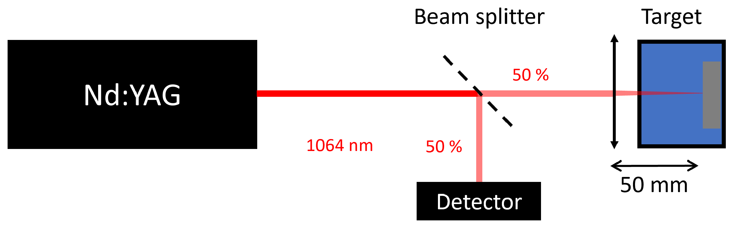
2.2. Laser Ablation in Liquids

Throughout this study, nanoparticles were produced by LAL (Nd-YAG in nanosecond regime) in two different organic solvents: toluene and tetrahydrofuran (THF). It is well established that photoablation allows the production of nanoparticles with good colloidal stability [24]. Moreover, the use of an organic solvent allows to vary the size, shape as well as structure of the nanoparticles without using additional chemicals as encapsulating agents. In particular, toluene and THF were chosen because of the PS solubility in both solvents. Besides, previous papers from Amendola et al. [25,26] have demonstrated that using these solvents for LAL could lead to a modification of AgNPs surface.
LAL was carried out using a Q-switched ns-YAG laser (Continuum Surelite II, Amplitude Système, Pessac, France) operated at 20 Hz and at the fundamental (1064 nm). The pulse duration was 5–7 ns. The optical scheme of the ablation setup is shown in Figure 1. The pulse energy is continuously monitored during the experiment using a 50–50 non-polarising beam splitter and a pyroelectric energy sensor (ES220C, Thorlabs) connected to a PM100USB energy meter (Thorlabs, Newton, NJ, USA). The beam (radius: 6 mm) is focused on the silver target using a +50 mm plano-convex lens (f-number: 8.3). Due to the focusing, the waist on the target w out is given by
w out = λ f π w in
where w in is the radius of the incoming beam, f the focal length of the lens and λ the laser wavelength. The target was vertically immersed in a 27-mL spectrophotometry cuvette filled with the solvent (toluene, THF, or water). To avoid multiple laser hits on the target, the cuvette containing the target is moved during the ablation process using a computer-controlled motorized XY stage. Typically, an average area of 3 mm × 3 mm was scanned by the laser beam. To avoid laser-induced damage to the cuvette, the energy per pulse was maintained below 30 mJ/pulse. Unless underwise stated, the following parameters were used λ = 1064 nm with an E pulse = 6.5 ± 0.1 mJ. Based on Equation (1), the estimated waist dimension in free space was 5.6 μ m, leading to a fluence on the target equal to ( 6.5 ± 0.1 ) 10 3 J/cm 2 . The ablation was carried out at room temperature for an estimated time of 5 min and the solution was magnetically stirred the ensure an appropriate dispersion of the NPs and minimize their interaction with the ablation beam [22]. In usual laser ablation experiments, the aim is to obtain nanoparticles with a size poly-dispersity as narrow as possible. In our experiments, we will advantage of the polydispersity to obtain a statistical distribution of particles and, as we expect, of interphases.

2.3. Silver Nanoparticles Characterisation by Ultraviolet–Visible Spectroscopy

After the laser ablation process, solutions of AgNPs were obtained. They are referred to as Ag-THF and Ag-Tol with THF and toluene as solvents respectively. Measurements of the solution optical properties were carried out using a Genesys 20 UV–Vis spectrometer (Thermo Fisher Scientific, Waltham, MA, USA). Quartz cuvettes with an optical path of 10.0 mm are used for each solution.

2.4. Substrate Cleaning and Nanocomposite Sample Preparation

Two types of substrates were considered in the experiments: silicon wafer fragments (Sb-doped, orientation <100>, ACM, Villers-Saint-Frédéric, France) and ITO substrates (sheet resistance 70–100 Ω /□, Sigma-Aldrich, Saint-Louis, MO, USA). They were respectively used for optical measurement and electrical characterization. Silicon substrates were cleaned as follows: they were rinsed with distilled water before being sonicated for 10 min in acetone and 10 min in methanol. The silicon fragments were dried under a Nitrogen flow just before spin coating. ITO substrates were cleaned with the following procedure: plunged for 5 min in a 3% RBS in water solution, rinsed with water, dried under a Nitrogen flow, and cleaned with an O2 plasma.
Ag-PS thin films were spin-coated with a Laurell WS-650-23B spin-coater. Film thickness was controlled by varying PS-solvent ratio (w:w) between 1 and 5% at a constant rotation speed (6000 rpm).

2.5. Thickness Control of the Composite Films

The thickness of the composite films was measured using Spectroscopic Ellipsometry (SE) with an EP3-SE imaging ellipsometer (Accurion GmbH, Göttingen, Germany) in the 360–1000 nm spectral range. The magnification of the ellipsometer objective was 10×. The angle of incidence was typically set at 60 degrees. SE is now considered as a reference experimental method to determine the refractive index and thickness of polymer films [27] and, in particular, of plasmonic nanocomposite ones (see e.g., [28,29] and the references therein).

2.6. Scanning Probe Microscopy Measurements

Scanning Probe Microscopy (SPM) is used to identify the produced particles and nanocomposite samples. Characterization of the sample mechanical properties is performed to emphasize the solvent effects.
In order to characterize the mechanical properties of our samples Peak Force Tapping Quantitative Nanomechanical mapping (PFT QNM) was used. It is a quasi-static Atomic Force Microscope (AFM) mode developed by Bruker Nano Inc. (Santa Barbara, CA, USA) [30]. While studying mechanical properties, the focus is on rigidity modulus and adhesion due to their significance to apprehend the composite mechanical behavior.
All the SPM measurements were performed with a Dimension Icon® AFM (Bruker Nano Inc., Santa Barbara, CA, USA). The analysis of the SPM data is performed with MountainsLab Premium V9 (Digital Surf, Besançon, France) and NanoScope Analysis 2.0 (Bruker corporation).
For the mechanical measurement, RTESP-300 probes were used: tip radius = 8 nm, spring constant = 40 N/m. Tips were calibrated on a sapphire test sample in a similar way than in Enrriques et al., study [31].

3. Results and Discussion

The results we present here are organized in two subsections: the results concerning the sole AgNPs and the ones concerning those dispersed in PS films.

3.1. AgNPs Characterization

With the setup and the parameters described in Section 2.2, we end up with solutions with different intensities and s for the naked eye. This is quantitatively assessed by the UV spectra. Absorption peaks observed in such spectra give insight into the particles: concentration (peak intensity), shape (peak central wavelength), and polydispersity (peak width at half maximum). The influence of the solvent is shown in Figure 2.
These peaks correspond to the energy absorption due to the localized surface plasmon resonance (LSPR): the impinging light interacts with the free electrons of the metal NPs which oscillate under appropriate resonance conditions. In the simplest case of a spherical particle of radius R in the quasi-static approximation, these are related to the polarizability of the particle α by the following relation [32]
α = 4 3 π R 3 ϵ m a t r i x ϵ m e t a l ϵ m a t r i x ϵ m e t a l + 2 ϵ m a t r i x
with ϵ m a t r i x and ϵ m e t a l the frequency dependent dielectric constants of the polymer matrix and of the metal nanoparticle, respectively. From Equation (2), one can see that the polarisability increases when ϵ m e t a l + 2 ϵ m a t r i x = 0 . This condition is named Frölich’s condition and is met for silver at wavelengths close to 400 nm [33].
In water, a symmetric absorption peak is observed at 398 nm with an absorption tail at wavelengths greater than 450 nm. In THF, two peaks are clearly visible: one at 410 nm and a broader one in the longer wavelengths. It can be interpreted as AgNPs polydispersity in the solution. Besides, for toluene no peak is observed within the 350–700-nm range. Aside from the polydispersity in the THF solution, these observations correspond to Amendola’s et al., results [25,26]. Nevertheless, the origin of this polydispersity can be explained by the lack of complete reshaping of the ablated AgNPs as a consequence of the interaction of the laser beam with the initially ablated particles [34]. This hypothesis is confirmed below using SPM analysis (Figure 3). Such a complete reshaping would have resulted in a sharp monomodal size distribution and, hence in a single peak in the absorption spectra.
Figure 3 shows the topography and adhesion of AgNPs ablated in water (Figure 3a,b), THF (Figure 3c,d) and toluene (Figure 3e,f) laid on a silicon substrate. In these three cases, nanoparticles are clearly visible on the substrate.
Additional information regarding the shape and adhesion of the AgNPs was obtained through PFT QNM measurements. The Ag-Tol adhesion behavior exhibited a distinct contrast between a part of the particles and their surroundings, indicating weak adhesion of the AgNPs while a sticky envelope (referred to as a shell) was present around several particles. This observation aligns with previous findings by Amendola et al. [26], who utilized high-resolution Transmission Electron Microscopy and Micro-Raman spectroscopy to detect and confirm the carbonaceous nature of this shell. Conversely, for AgNPs ablated in water and THF, only particles with weak adhesion were observed. Nevertheless, the presence of a thin amorphous carbon layer around Ag-THF is reported by Amendola et al. [25]. It is not detected by SPM measurements in Figure 3c,d. However, the UV–Vis spectra of Ag-THF samples demonstrated similar characteristics in both studies. Consequently, it is hypothesized that the Ag-THF nanoparticles are also surrounded by a thin shell, albeit requiring further characterization (e.g., HR-SEM, Raman spectroscopy) to definitively determine the extent of surface modification on these nanoparticles.
In both topography and adhesion mappings, we do not identify aggregates of AgNPs unlike scanning electron microscopy (SEM) measurements (see Supplementary Materials). This is due to the tip convolution: the AFM measurement can not resolve the aggregates of the particles. These aggregates will be mentioned as one-body particles to lighten the description and focus on the interphase properties.

3.2. Ag-PS Film Characterization

After having considered the properties of the sole nanoparticles, let us now focus on the composite films prepared from these.
Ellipsometry was used to measure the thickness of the films. Films at 6000 rpm with a 5% (w:w) PS-toluene solution are about 240 nm thick. Spin-coated films at 6000 rpm with a 5% (w:w) PS-THF concentration are 220 nm thick. The presence of the AgNPs does not significantly affect film thickness. Further, at the doping level of our films, the AgNPs do not significantly affect the optical properties of the PS matrix between 360 and 1000 nm. In the PS matrix, the AgNPs concentration is too low for the surface plasmon absorption to have a measurable effect on the optical properties for both kinds of investigated AgNPs (Ag-Tol and Ag-THF). For this reason, the optical model for processing the ellipsometric data can be the same for the doped and non-doped films: a Cauchy layer with optical properties almost similar to PS optical properties on a silicon substrate with a 2-nm native oxide layer (SiO2). Measurements and optimizations for thickness determination are shown in Supporting Information (Figures S1 and S2). Control experiments were carried out with undoped polymer solutions.
Figure 4 shows topography, rigidity modulus, and adhesion mappings of a PS film doped with AgNPs ablated in THF. The peak force tapping is done with a 30 nN peak force setpoint (i.e., maximum force of indentation). Several bumps are visible on the PS film surface. They are of various sizes, flat and wide: 6 nm high for a few dozen wide. Since the surface of undoped PS film is flat, they are related to the presence of AgNPs near the film surface.
By looking at the mechanical properties of the films (modulus and adhesion), the following observations are made: Firstly, each bump corresponds to a local decrease of the rigidity modulus (from ∼3 GPa to less than 1 GPa). Secondly, the smallest bumps present an adhesion decrease (from ∼30 nN to 10 nN). Unlike the biggest bumps where adhesion does not change significantly.
Therefore, there are two different bump populations, which is confirmed by the observations in Figure 5. It shows the rigidity modulus of the same area as in Figure 4 but with different setpoints.
With a lower setpoint, all the bumps correspond to a decrease in the rigidity modulus. With the increase of the setpoint, the rigidity modulus of the bumps increases. For the biggest bumps, it keeps a rigidity modulus below the PS except at their center. At 90 nN, a core-shell structure is visible (Figure 5c). Sections in Figure 5d illustrate the influence of setpoint on the rigidity modulus.
Figure 6 presents the topography, rigidity modulus, and adhesion mappings of a PS film doped with AgNPs ablated in toluene. Similar to the THF-ablated AgNPs, the presence of bumps suggests the presence of AgNPs near the PS surface. However, these bumps are smaller and flatter compared to those observed with THF-ablated AgNPs. Further, while there is a local decrease in adhesion at the positions of the bumps, the rigidity modulus does not show a significant change. The measurement shown is done with a 90 nN peak force setpoint and there is no significant change when varying it. This distinct mechanical behavior in the PS film doped with AgNPs ablated in toluene further highlights the differences compared to the THF-ablated AgNPs.
The observed differences in the mechanical behavior of the PS films doped with AgNPs ablated in THF and toluene can be attributed to the distinct surface modification that takes place during the laser ablation process in these solvents. The formation of a carbonaceous shell around the AgNPs, as detected above and in previous studies [25,26], likely plays a crucial role.

4. Conclusions

In this study, the use of LAL has been explored as a cost-effective and efficient method for tuning the surface properties of AgNPs, leading to the formation of a core-shell structure. The findings highlight several key points that contribute to the understanding of these modified AgNPs and their behavior in a PS matrix.
Firstly, it has been shown that the core-shell structure resulting from LAL significantly modifies the optical properties of the AgNPs. Specifically, the LSPR absorption peak is affected. The ability to tailor the LSPR absorption through surface modification could be interesting for manipulating the optical characteristics of future nanocomposite materials.
Furthermore, the results reveal that the laser-ablated AgNPs exhibit good dispersion in the PS matrix, in particular for the AgNPs ablated in toluene. This dispersion behavior is crucial in polymer nanocomposite applications, as it directly influences the overall performance of the material. The improved dispersion suggests favorable interactions between the core-shell structure of the AgNPs and the PS matrix, potentially enhancing the compatibility and stability of the composite system.
Importantly, the investigation also sheds light on the interphase behavior observed in the doped polymer films. We have found that the interphase properties depend on the core-shell structure of the AgNPs. Since interphase behavior appears mostly for particles ablated in THF. The bumps on the doped polymer surface have a lower rigidity modulus than the matrix. This can be attributed to the reorganization of the PS chains around the AgNPs, creating an area of lower polymer density. The resulting interphase, as described by Tanaka’s model, contributes to the overall mechanical properties of the composite, affecting its rigidity and flexibility.
New insights into the core-shell structure, optical properties, dispersion, and interphase behavior of laser-ablated AgNPs in a PS matrix provide a solid foundation for further investigations. Future studies should focus on exploring the specific properties and interactions of the shell structure and their influence on the composite electrical and mechanical performance. Further, the carbonaceous nature of this shell has yet to be confirmed by further experimentation.
These findings will hold significant promise for various applications in polymer nanocomposite systems. The ability to tune the surface properties of AgNPs through LAL offers exciting opportunities for developing functional materials with tailored optical, mechanical, and electrical properties. By precisely controlling the core-shell structure and optimizing the dispersion of these modified AgNPs in polymer matrices, it will become possible to design composite materials with enhanced specific properties, such as plasmonic sensing, light harvesting, and optoelectronic devices. Moreover, characterizing the interphase resulting from tunable core-shell structure should provide valuable insights into interphase behavior and formation. Then, this knowledge can be leveraged to create advanced materials with superior mechanical strength, flexibility, and durability, opening up avenues for applications in flexible electronics, energy storage, and structural materials. The prospects for further research and development in this field are highly promising, and the outcomes of this work lay a solid foundation for future investigations aimed at harnessing the full potential of laser-ablated AgNPs in various technological domains.

Supplementary Materials

The following are available at https://www.mdpi.com/article/10.3390/en16124625/s1, Figure S1: Ellipsometric spectra of Ag nanoparticles (AgNPs) doped polystyrene (PS) films. The AgNPs particles were prepared by laser ablation in toluene and the Ag-PS solution is spin-coated with toluene as solvent. Data are recorded at an angle of incidence of 60 degrees. The model is a two-layers model on top of a silicon substrate: a native 2 nm thick silicon oxide layer (not optimized) and a Cauchy layer with optical properties similar to PS. The thickness of the Cauchy layer after optimization is 119.8 ± 0.1 nm, Figure S2: Ellipsometric spectra of Ag nanoparticles-doped polystyrene (PS) films. The AgNPs particles were prepared by laser ablation in tetrahydrofuran (THF) and the Ag-PS solution is spin-coated with THF as solvent. Data are recorded at an angle of incidence of 60 degrees. The fitted model is a three-layers model on top of a silicon substrate: a native 2 nm thick silicon oxide layer, a Cauchy layer with optical properties similar to PS and, a Bruggeman layer with the same Cauchy material and air. The Bruggeman layer is considered to take into account the film roughness. After optimization: the Cauchy layer thickness is 144.7 ± 0.1 nm, the Bruggeman layer thickness is 69.9 ± 0.1 nm with a fraction of 20% of air, Figure S3: AgNPs laser ablated in water drop-casted on a silicon substrate. Sample was characterized with a field emission gun scanning electron microscope (FEG-SEM Hitachi SU8020) with a 5 kV acceleration voltage. Particles are poly-dispersed in size. Particles tend to agglomerate on the silicon substrate and their diameter strongly varies. (a) Initial SEM image; (b) same with detected particles highlighted in green, with a cut-off on particle area are set to 100 nm 2 (MountainsLab Premium V9, DigitalSurf); (c) histogram of the particle area with an adjusted log-normal statistical distribution (red dashed curve): σ = 0.92, distribution center = 0.77 nm 2 .

Author Contributions

Conceptualization, P.L.; methodology, P.L. and M.V.; formal analysis, T.D.M.; investigation, T.D.M.; resources, P.L. and M.V.; writing—original draft preparation, T.D.M.; writing—review and editing, P.L. and M.V.; visualization, T.D.M.; supervision, P.L.; project administration, P.L.; funding acquisition, P.L. and M.V. All authors have read and agreed to the published version of the manuscript.

Funding

This work was supported by the Fonds de la Recherche Scientifique (F.R.S.)—FNRS under Grant(s) No J.0190.16 (NTP project); F.R.S.—FNRS Grands Equipements (40007941)—2022 “Interuniversity Platform for the Analysis of the Nanoscale properties of Emerging Materials and their Applications (ipanema)”; F.R.S.—FNRS Projet de Recherche (PDR) (40007942)—2022 “Piezoelectric and flexoelectric architectures from hybrid inorganic-organic nanomaterials—PiezoFlexoTronics”.

Data Availability Statement

The data can be shared up on request.

Acknowledgments

We want to thanks UMONS Research Institute for Materials Science and Engineering for partial funding of T. De Muijlder’s thesis, E. Fournier for its contribution to the laser ablation experiments and G. Savorianakis for the SEM analysis.

Conflicts of Interest

The authors declare no conflict of interest.

Abbreviations

The following abbreviations are used in this manuscript:
AFMAtomic Force Microscope
AgNPSSilver Nanoparticles
Ag-THFTetrahydofuran Ablated Silver Nanoparticles
Ag-TolToluene Ablated Silver Nanoparticles
LALLaser Ablation in Liquids
LSPRLocalized Surface Plasmon Resonance
PSpolystyrene
PFT QNMPeak Force Tapping Quantitative Nanomechanical
SESpectroscopic Ellipsometry
SEMScanning Electron Microscopy
SPMScanning Probe Microscopy
THFTetrahydrofuran

References

  1. Lewis, T. Nanometric Dielectrics. IEEE Trans. Dielectr. Electr. Insul. 1994, 1, 812–825. [Google Scholar] [CrossRef]
  2. Tanaka, T.; Montanari, G.; Mulhaupt, R. Polymer Nanocomposites as Dielectrics and Electrical Insulation-perspectives for Processing Technologies, Material Characterization and Future Applications. IEEE Trans. Dielectr. Electr. Insul. 2004, 11, 763–784. [Google Scholar] [CrossRef]
  3. Green, C.; Vaughan, A. Nanodielectrics—How Much Do We Really Understand? IEEE Electr. Insul. Mag. 2008, 24, 6–16. [Google Scholar] [CrossRef]
  4. Zhong, S.; Dang, Z.; Zhou, W.; Cai, H. Past and Future on Nanodielectrics. IET Nanodielectr. 2018, 1, 41–47. [Google Scholar] [CrossRef]
  5. Darwish, M.S.A.; Mostafa, M.H.; Al-Harbi, L.M. Polymeric Nanocomposites for Environmental and Industrial Applications. Int. J. Mol. Sci. 2022, 23, 1023. [Google Scholar] [CrossRef] [PubMed]
  6. Khan, I.; Khan, I.; Saeed, K.; Ali, N.; Zada, N.; Khan, A.; Ali, F.; Bilal, M.; Akhter, M.S. 7—Polymer nanocomposites: An overview. In Smart Polymer Nanocomposites; Ali, N., Bilal, M., Khan, A., Nguyen, T.A., Gupta, R.K., Eds.; Elsevier: Amsterdam, The Netherlands, 2023; pp. 167–184. [Google Scholar] [CrossRef]
  7. Yang, K.; Huang, X.; Xie, L.; Wu, C.; Jiang, P.; Tanaka, T. Core-Shell Structured Polystyrene/BaTiO3 Hybrid Nanodielectrics Prepared by In Situ RAFT Polymerization: A Route to High Dielectric Constant and Low Loss Materials with Weak Frequency Dependence. Macromol. Rapid Commun. 2012, 33, 1921–1926. [Google Scholar] [CrossRef] [PubMed]
  8. Feng, M.; Zhang, C.; Zhou, G.; Zhang, T.; Feng, Y.; Chi, Q.; Lei, Q. Enhanced Energy Storage Characteristics in PVDF-Based Nanodielectrics With Core-Shell Structured and Optimized Shape Fillers. IEEE Access 2020, 8, 81542–81550. [Google Scholar] [CrossRef]
  9. Tanaka, T.; Vaughan, A. (Eds.) Tailoring of Nanocomposite Dielectrics: From Fundamentals to Devices and Applications; Pan Stanford Publishing: Singapore, 2017. [Google Scholar]
  10. Seiler, J.; Kindersberger, J. Insight into the Interphase in Polymer Nanocomposites. IEEE Trans. Dielect. Electr. Insul. 2014, 21, 537–547. [Google Scholar] [CrossRef]
  11. Tanaka, T. Interpretation of Several Key Phenomena Peculiar to Nano Dielectrics in terms of a Multi-core Model. In Proceedings of the 2006 IEEE Conference on Electrical Insulation and Dielectric Phenomena, Kansas City, MO, USA, 15–18 October 2006; pp. 298–301. [Google Scholar] [CrossRef]
  12. Raetzke, S.; Kindersberger, J. The Effect of Interphase Structures in Nanodielectrics. IEEJ 2006, 126, 1044–1049. [Google Scholar] [CrossRef] [Green Version]
  13. Andritsch, T.; Kochetov, R.; Morshuis, P.H.F.; Smit, J.J. Proposal of the Polymer Chain Alignment Model. In Proceedings of the 2011 Annual Report Conference on Electrical Insulation and Dielectric Phenomena, Cancun, Mexico, 16–19 October 2011; pp. 624–627. [Google Scholar] [CrossRef]
  14. Lewis, T.J. Interfaces are the Dominant Feature of Dielectrics at the Nanometric Level. IEEE Trans. Dielectr. Electr. Insul. 2004, 11, 739–753. [Google Scholar] [CrossRef]
  15. Tsagaropoulos, G.; Eisenberg, A. Dynamic Mechanical Study of the Factors Affecting the Two Glass Transition Behavior of Filled Polymers. Similarities and Differences with Random Ionomers. Macromolecules 1995, 28, 6067–6077. [Google Scholar] [CrossRef]
  16. Ghasem Zadeh Khorasani, M.; Silbernagl, D.; Platz, D.; Sturm, H. Insights into Nano-scale Physical and Mechanical Properties of Epoxy/boehmite Nanocomposite using Different AFM Modes. Polymers 2019, 11, 235. [Google Scholar] [CrossRef] [PubMed] [Green Version]
  17. Houssat, M.; Villeneuve-Faure, C.; Dignat, N.L.; Cambronne, J. A Multiscale Characterization of Nanodielectrics: The Case of PI/Si3N4 Nanocomposite. In Proceedings of the 2020 IEEE 3rd International Conference on Dielectrics (ICD), Valencia, Spain, 5–31 July 2020; pp. 221–224. [Google Scholar] [CrossRef]
  18. Sengwa, R.; Dhatarwal, P.; Choudhary, S. A Comparative Study of Different Metal Oxide Nanoparticles Dispersed PVDF/PEO Blend Matrix-based Advanced Multifunctional Nanodielectrics for Flexible Electronic Devices. Mater. Today Commun. 2020, 25, 101380. [Google Scholar] [CrossRef]
  19. Lau, K.Y.; Vaughan, A.S.; Chen, G. Nanodielectrics: Opportunities and Challenges. IEEE Electr. Insul. Mag. 2015, 31, 45–54. [Google Scholar] [CrossRef]
  20. Yang, G. (Ed.) Laser Ablation in Liquids: Principles and Applications in the Preparation of Nanomaterials; Pan Stanford Publishing: Singapore, 2012. [Google Scholar]
  21. Zhang, D.; Li, Z.; Sugioka, K. Laser Ablation in Liquids for Nanomaterial Synthesis: Diversities of Targets and Liquids. J. Phys. Photonics 2021, 3, 042002. [Google Scholar] [CrossRef]
  22. Mansour, Y.; Battie, Y.; Naciri, A.E.; Chaoui, N. Monitoring the Aspect Ratio Distribution of Colloidal Gold Nanoparticles under Pulsed-Laser Exposure. Opt. Express 2020, 28, 34501. [Google Scholar] [CrossRef] [PubMed]
  23. Chatterjee, U.; Jewrajka, S.K.; Guha, S. Dispersion of Functionalized Silver Nanoparticles in Polymer Matrices: Stability, Characterization, and Physical Properties. Polym. Compos. 2009, 30, 827–834. [Google Scholar] [CrossRef]
  24. Barcikowski, S.; Amendola, V.; Marzun, G.; Rehbock, C.; Reichenberger, S.; Zhang, D.; Gökce, B. Handbook of Laser Synthesis of Colloids; DuEPublico: Essen, Germany, 2016. [Google Scholar]
  25. Amendola, V.; Polizzi, S.; Meneghetti, M. Free Silver Nanoparticles Synthesized by Laser Ablation in Organic Solvents and Their Easy Functionalization. Langmuir 2007, 23, 6766–6770. [Google Scholar] [CrossRef]
  26. Amendola, V.; Polizzi, S.; Meneghetti, M. Laser Ablation Synthesis of Silver Nanoparticles Embedded in Graphitic Carbon Matrix. Sci. Adv. Mater. 2012, 4, 497–500. [Google Scholar] [CrossRef]
  27. Azzam, R.M.A.; Bashara, N.M. Ellipsometry and Polarized Light; North-Holland: Amsterdam, The Netherlands, 1977. [Google Scholar]
  28. Abargues, R.; Abderrafi, K.; Pedrueza, E.; Gradess, R.; Marqués-Hueso, J.; Valdés, J.L.; Martínez-Pastor, J. Optical properties of different polymer thin films containing in situ synthesized Ag and Au nanoparticles. New J. Chem. 2009, 33, 1720–1725. [Google Scholar] [CrossRef]
  29. Guyot, C.; Voué, M. Intrinsic optical properties of Ag-doped poly-(vinyl alcohol) nanocomposites: An analysis of the film thickness effect on the plasmonic resonance parameters. Appl. Phys. A 2020, 126, 870. [Google Scholar] [CrossRef]
  30. Kaemmer, S.B. Introduction to Bruker’s ScanAsyst and PeakForce Tapping AFM Technology; Bruker Nano Inc.: Santa Barbara, CA, USA, 2011. [Google Scholar]
  31. Enrriques, A.E.; Howard, S.; Timsina, R.; Khadka, N.K.; Hoover, A.N.; Ray, A.E.; Ding, L.; Onwumelu, C.; Nordeng, S.; Mainali, L.; et al. Atomic Force Microscopy Cantilever-Based Nanoindentation: Mechanical Property Measurements at the Nanoscale in Air and Fluid. J. Vis. Exp. 2022, 190, 64497. [Google Scholar] [CrossRef] [PubMed]
  32. Fan, X.; Zheng, W.; Singh, D.J. Light scattering and surface plasmons on small spherical particles. Light Sci. Appl. 2014, 3. [Google Scholar] [CrossRef] [Green Version]
  33. Johnson, P.B.; Christy, R.W. Optical Constants of the Noble Metals. Phys. Rev. B 1972, 6, 4370–4379. [Google Scholar] [CrossRef]
  34. Delmée, M.; Mertz, G.; Bardon, J.; Marguier, A.; Ploux, L.; Roucoules, V.; Ruch, D. Laser Ablation of Silver in Liquid Organic Monomer: Influence of Experimental Parameters on the Synthesized Silver Nanoparticles/Graphite Colloids. J. Phys. Chem. B 2017, 121, 6646–6654. [Google Scholar] [CrossRef]
Figure 1. Simplified scheme of the laser ablation setup, including a motorized XY stage for the target displacement (not drawn).
Energies 16 04625 g001
Figure 2. Maximum normalized UV–Vis spectra of AgNPs produced by LAL at a 1064 nm in toluene (blue), THF (black) and water (red).
Energies 16 04625 g002
Figure 3. Topography and adhesion of AgNPs ablated in (a,b) water, (c,d) THF and (e,f) toluene and dropped on a silicon substrate; PFT QNM analysis with a RTESP-300 tip.
Energies 16 04625 g003
Figure 4. Drop-cast PS film doped with AgNPs ablated in THF solution: (a) topography, (b) rigidity modulus, and (c) adhesion.
Energies 16 04625 g004
Figure 5. Rigidity modulus of a drop-cast PS film doped with AgNPs ablated in THF. Each mapping represent a measurement on the same area with a different peakforce setpoint: (a) 30 nN, (b) 60 nN, (c) 90 nN, and (d) the section matching the doted lines. Each curve represent a measurement on the same area with a different peakforce setpoint.
Energies 16 04625 g005
Figure 6. Drop-cast PS film doped with AgNPs ablated in toluene: (a) topography, (b) rigidity modulus, and (c) adhesion.
Energies 16 04625 g006
Disclaimer/Publisher’s Note: The statements, opinions and data contained in all publications are solely those of the individual author(s) and contributor(s) and not of MDPI and/or the editor(s). MDPI and/or the editor(s) disclaim responsibility for any injury to people or property resulting from any ideas, methods, instructions or products referred to in the content.

