Article

Pore Structure and Fractal Characteristics of Coal-Measure Sedimentary Rocks Using Nuclear Magnetic Resonance (NMR) and Mercury Intrusion Porosimetry (MIP)

1
State Key Laboratory for GeoMechanics and Deep Underground Engineering, Beijing 100083, China
2
School of Mechanics, Architecture and Civil Engineering, China University of Mining and Technology, Beijing 100083, China
*
Author to whom correspondence should be addressed.
These authors contributed equally to this work.
Energies 2023, 16(9), 3812; https://doi.org/10.3390/en16093812
Submission received: 11 April 2023 / Revised: 26 April 2023 / Accepted: 27 April 2023 / Published: 28 April 2023

Abstract

:
Analyzing and mastering the fractal features of coal-measure sedimentary rocks is crucial for accurately describing the pore structure of coalbed methane resources. In this work, mercury intrusion porosimetry (MIP) and nuclear magnetic resonance (NMR) are performed on coal-measure sedimentary rocks (i.e., shale, mudstone, and sandstone) to analyze their pore structure. Pore size distributions (PSDs) and the multifractal dimensions of the investigated samples are discussed. Moreover, multivariable linear regression models of multifractal dimensions are established through a comprehensive analysis of multifractal characteristics. The results show that sandstone (SS-1) and clay rocks are dominated by nanopores of 0.01 to 1 μm, while sandstone (SS-2) is mostly mesopores and macropores in the range of 1 to 10 μm. The fractal characteristics of the investigated rock samples show a prominent multifractal characteristic, in which DA reflects the surface structure of micropores, while DS represents the pore structure of macropores. Multifractal dimension is affected by many factors, in which the DA is greatly influenced by the pore surface features and mineral components and the DS by average pore diameters. Moreover, multivariate linear regression models of adsorption pore and seepage pore are established, which have a better correlation effect on the multifractal dimension.

1. Introduction

Coal-measure sedimentary rocks, i.e., mudstone, shale, and tight sandstone, with low porosity and permeability as well as complex pore structure, are rich in petroleum resources, which has turned into an investigation hotspot in recent years [1]. Poor connectivity and strong heterogeneity of pore-throat structure are tough issues restricting the efficient exploitation of coal-measure sedimentary rocks [2]. Hence, studying and mastering the pore structure features of coal-measure sedimentary rocks play a valuable, guiding role for the safe and efficient exploitation and utilization of mineral resources [3].
According to Loucks’s pore size scale [4], pores can be divided into five parts: picopore (<1 nm), nanopore (1 nm–1 μm), micropore (1 μm–62.5 μm), mesopore (62.5 μm–4 mm), and macropore (4 mm–256 mm). In recent years, a series of experimental methods, i.e., gas adsorption experiment (N2/CO2) [5,6,7], X-ray computed tomography (CT) [8,9,10], field-emission scanning electron microscope (FE-SEM) [11,12,13,14], mercury intrusion porosimetry (MIP) [15,16], nuclear magnetic resonance (NMR) [17,18,19,20,21,22], and NMR cryoporometry (NMRC) [23], have been carried out to explore the micropore structure of common coal-measure sedimentary rocks. However, limited by the testing principles, each of the aforementioned experimental methodologies has certain restrictions. Although micropore structures can be measured precisely using the gas adsorption technique (N2/CO2), the range is relatively narrow. The CO2 adsorption technique measures over a range of 2 nm, while the N2 adsorption technique measures over a range of 2–100 nm [14]. Hence, measuring mesopores and macropores with gas adsorption is not possible. SEM experiments and CT imaging technologies can directly observe rock samples [10]. SEM experiments are limited to qualitative analysis of rock samples, whereas CT experiments can perform quantitative analysis and reconstruct rock cores. However, limited by instrument resolution, CT experiments cannot guarantee both resolution and field of view [24]. In the evaluation of the quality of coal-measure sedimentary rocks, MIP measurements can directly obtain the pore structure parameters, i.e., the pore size distributions (PSDs), median, and average pore diameter [15,25,26]. Meanwhile, NMR is used to determine the fluid distribution and pore structure features, including the T2 distribution, PSD curves, and other physical parameters of reservoir rocks, because it is non-destructive and highly efficient [17,27]. Nowadays, MIP and NMR measurements are extensively used to analyze the pore structure features of coal-measure sedimentary reservoir rocks [15,18,21,26]. Li et al. [28] used a combination of MIP and NMR methods to analyze the pore structure characteristics of coal and verified the accuracy of the measurement results using various methods. Wang et al. [29] analyzed tight oil sandstone’s pore structure and fractal characteristics by combining MIP and NMR. Research shows that the combination of NMR and MIP can improve the accuracy of pore structure characterization. The fractal dimension calculated by the NMR T2 spectrum is practical for rock physical property analysis.
In 1975, the fractal theory was put forward by Mandelbrot to depict the micropore structure of porous media and has been widely used to investigate the pore structure of reservoir rocks, i.e., coals, tight sandstones, and shales [15,23,29,30,31,32]. Currently, many scholars have found that the fractal theory can accurately describe pore structure heterogeneity by using N2-GA, MIP, NMR, and NMRC [33,34,35,36]. Due to the limitation in measurement accuracy and calculation theory of a single experimental method, a combination of multiple experimental methods is often used to illustrate the complex geometric features of pore structure. Research shows that the fractal dimensions of pore structures vary significantly with different pore sizes, indicating remarkable multifractal characteristics of reservoir rocks. Meanwhile, the multifractal dimensions are affected considerably by petrophysical properties and mineral composition [37,38]. At present, the calculation and influencing factors of fractal dimension are indistinct because of the complex pore structure characteristics and mineral composition with strong regional differences. In addition, few quantitative studies on the influencing factors of fractal dimensions are based on MIP and NMR.
This study aims to analyze pore structure and fractal characteristics of representative coal-measure sedimentary rocks using MIP and NMR techniques. Meanwhile, multifractal dimensions of MIP and NMR are calculated, and a corresponding multivariate linear regression model is established. Moreover, the relationship between the physical parameters, mineral components, and multifractal dimensions is discussed systematically. The results can provide a basis for reservoir evaluation of coal-measure sedimentary rocks in this research area.

2. Materials and Methods

2.1. Samples

The research area is located in Daqiang and Jiahe Coal Mines, in Liaoning and Jiangsu Provinces, China. Twelve common fine sedimentary rock samples with three lithologies (i.e., shale, mudstone, and sandstone) were collected from the coal-measure strata of the research area. After sampling, rock samples were sealed in self-sealing bags and immediately sealed with wax after being transported to the ground, which were processed into cylindrical cores with a uniform size of 25 mm × 35 mm in the laboratory. The macromorphological characteristics are shown in Table 1.
The density of all the investigated samples was determined based on their dry weights and bulk volume. The water porosity and gas permeability were obtained based on the ISO/WD 19990 [39]. The water porosities (φ) were measured through water saturation method, and the permeability (K) was determined using a gas permeability testing apparatus by flowing high-purity nitrogen through the rock samples. According to ISO 10370:2014 [40] and ISO 14689:2017 [41], Bruker D8 Discover X-ray diffractometer and Leco CS-230 determinator were used to perform the total organic carbon (TOC) and X-ray diffraction (XRD) measurements. Then, the TOC contents and the mineralogical compositions of all the core samples could be obtained based on the results of TOC and XRD testing. The X-ray fluorescence measurements of the rock samples are shown in Figure 1. In addition, the mineral composition of the rock sample is primarily quartz and clay minerals, with a trace of K-feldspar and Plagioclase. The clay mineral composition is mainly kaolinite, with trace amounts of chlorite, illite, and an illite/smectite mixed layer (I/S).
Table 2 lists the TOC contents and the mineralogical compositions of all the core samples.

2.2. MIP Measurements

MIP is a conventional method to obtain pore structure features. Pore size distribution and some other parameters, including total pore volume/area and average pore diameter, were all determined using MIP tests using AutoPore IV 9500 Porosimeter in this study. According to ISO 15901-1:2016 [42], the investigated samples were measured with mercury injection pressure steps within the scope of 0.5 and 60,000 psia, which corresponds to pore-throat diameters from 3 nm to 36 5 μm. Pore parameters including total pore area and average pore diameter were obtained based on MIP measurements. Meanwhile, pore radius can be obtained in MIP tests (Equation (1)):
P c = 2 σ cos θ r
where Pc, σ, θ, and r refer to the capillary pressure (MPa), the surface tension, the contact angle (°), and the pore radius (μm), respectively.

2.3. NMR Measurements

Longitudinal (T1) and transverse (T2) relaxation time can provide similar pore distribution information in a low-frequency magnetic field [43], while the T2 measurement is much faster than the T1 measurement; hence, the T2 measurement is often used to obtain pore distribution information [44]. The T2 cutoff (T2c) determined by the centrifugal calibration method is the critical parameter in NMR tests, which is the threshold to identify the adsorption pore and the seepage pore. Meanwhile, in an external force, the fluid in the seepage pore can flow freely, while in the adsorption pore, it is difficult to flow by the capillary action.
Based on ASTM E2977-14 [45], RecCore-04 NMR sample analyzer was used to perform the NMR tests, with a spectrometer frequency of 3.84 MHz and a constant magnetic field strength of 1200 gauss. The full water-saturated (Sw) samples were measured using NMR measurements at a room temperature of −25 °C to obtain the T2 spectra. In addition, the test parameters were set as follows: echo spacing, 0.5 ms; waiting time, 3 s; number of echoes, 1024; and number of scans, 64. Pore parameters including porosity and permeability were obtained based on NMR measurements.

3. Results

3.1. TOC and Mineralogical Compositions

As shown in Table 2, the TOC contents are within the scope of 0.03% (SS-1) to 10.81% (SH-1), reflecting the abundance of organic matter. The total organic carbon averages of sandstone, mudstone, and shale are 0.05%, 1.04%, and 5.55%, respectively. Meanwhile, organic matter in clay rocks is significantly more plentiful than that of sandstone. On the one hand, the enrichment of organic matter originates from the input of terrigenous clast. On the other hand, the oxidation–reduction environment plays a vital role in preserving organic matter during diagenetic processes [4,46].
The mineral composition of the investigated rock is dominated by clay minerals and quartz. The main composition of the clay minerals is kaolinite, ranging from 34% (MS-1) to 89% (SS-2), and the average is 37.9%. Among all samples, the quartz content ranges from 16% (SH-4) to 54% (SS-2); the average is 37.9%. Meanwhile, the content of clay mineral averages 38.1%, ranging from 12% (SS-2) to 55% (MS-1). The clay mineral content of mudstone and shale is significantly higher than sandstone, which is between 25% and 55%.

3.2. Petrophysical Properties

As shown in Table 3, the sandstone samples collected from the two coal mines have a massive difference in petrophysical properties. The total porosity and permeability of sandstone are remarkably higher than that of the clay rock samples in the Daqiang Coal Mine. In contrast, rock samples of the Jiahe Coal Mine have similar properties. Among all the rock samples, the porosity range of clay rock samples is 1.40–13.40%, with an average of 7.24%. In addition, low permeability reservoir rocks, i.e., shale and mudstone samples, have poor permeability values, ranging from 0.0003 mD (SH-1) to 0.0048 mD (MS-2). The permeability of SH-2 and MS-3 is significantly lower than that of others with similar porosity because the poor connectivity of the pore structure hinders the fluid flows in the pore throat.

3.3. Pore Size Distributions (PSDs) Determined by MIP

Pore structure features, i.e., total pore area and average pore diameter determined by MIP tests, are listed in Table 3. The results show that total pore areas are in the range of 1.20 m2/g (SS-2) to 12.17 m2/g (SH-4), and the average pore diameters range from 6.7 nm (MS-1) to 230.40 nm (SS-2). The sandstone samples have lower total pore areas with higher average pore diameters than clay rock samples. The average pore diameter of SS-2 is significantly higher than that of the other rock samples, indicating it has stronger pore volumes.
According to the data of the MIP tests, the PSD curves shown in Figure 2 demonstrate the distribution of pore structure. The pore types of experimental samples are dominated by nanopores (r < 1 μm) and micropores (1 μm < r < 62.5 μm). Meanwhile, the pore volumes of the investigated samples are within the scope of 0.0074 mL/g (SH-1) to 0.069 mL/g (SS-2). The PSD curves contain three types. The PSD of most clay rock samples has a similar trend that the total pore volume increases rapidly within the scope of 0.01 μm to 0.1 μm while increasing weak in the rest of the pore size range, which indicates the pore types of rock samples are dominated by nanopores ranging from 0.01 μm to 0.1 μm. Meanwhile, the cumulative pore volume of sandstone samples (SS-2) is significantly larger than that of the others, indicating that SS-2 has the greatest porosity. In addition, the PSD curve of SS-2 increases rapidly in the range of 0.01–10 μm, while it is relatively smooth in other pore sizes, indicating the dominant pore size of SS-2 ranges from 0.01 μm to 10 μm. Moreover, the PSD curves of samples (SS-1 and SH-1) maintain a weak variation range in the full pore size range, only increasing slightly when the pore size is less than 0.1 μm, which indicates that the dominant pore type of SS-1 and SH-1 is a nanopore.

3.4. T2 Distributions and PSD Determined by NMR

The T2 distribution is commonly used in the NMR measurement, which is expressed as [43,47]:
1 T 2 = 1 T 2 B + 1 T 2 D + 1 T 2 S
where T2 is the total transverse NMR relaxation time; T2B, T2D, and T2S are the bulk, diffusion, and surface relaxation time, respectively. The T2 spectrums of the investigated sample are presented in Figure 3, which depicts the full spectrum pore distributions of the investigated samples. It can be found that the T2 spectra of all the numbered samples exhibit a single peak distribution, with significant differences in the positions and scales of the peaks. The peak of SS-1 is located at 1 ms, with the prominent peak distributed at 0.1–10 ms, while the peak of SS-2 occurs at 100 ms, with the prominent peak ranging from 1 to 1000 ms. It indicates that SS-1 consists mainly of micropores with a comparatively low pore content, whereas SS-2 predominantly comprises mesopores with an extensive pore size distribution. The peak of the clay rock samples generally appears at 1 ms, with the primary peak distributed between 0.1 and 10 ms, indicating that the clay rock samples consist primarily of micropores. The pore size distribution of the mudstone samples is comparable, as well as the micropore content of the mudstone samples is typically greater than that of the shale samples. Among the shale samples, SH-1 has the highest micropore contents within the range of 0.1–10 ms, whereas the other three samples have comparatively low micropore contents, with a small number of pores greater than 10 ms.
On the basis of the principle of NMR measurement [23], the bulk and diffusion relaxation can be ignored, and the T2 spectra and pore radius, r, show a linear correlation in Equation (3):
T 2 = r ρ × F
where F is the pore shape factor, and r refers to the pore radius, μm.
The quantitative division of adsorption pore and seepage pore is particularly important for reservoir quality evaluation. The fluid in the adsorption pore flows with difficulty due to electrostatic and capillary forces, while the fluid in the seepage pore can flow freely under certain pressure. Commonly, the pore size threshold of adsorption and seepage pores is 0.1 μm [11]. In this study, the analyzed samples’ T2 distributions are converted into PSD curves based on Equation (3). Figure 4 displays the range of pore diameter distribution and the different pore types found in each sample based on the converted PSD curves. As shown in Figure 4 and Table 4, all the PSD curves are unimodal distributions. The peak of sandstone (SS-1), shale, and mudstone are distributed, ranging from 0.01 μm to 1 μm, with the peak points concentrated on about 0.1 μm. The peak area ratios of ≤0.1 μm, 0.1–1 μm, and >1 μm are 38.87–82.14%, 5.20–61.13%, and 0–12.66%, respectively. Moreover the peak area ratio of ≤1 μm is 87.34–100%, indicating that the dominant pore sizes of sandstone (SS-1), mudstone, and shale range from 0.01 to 1 μm and adsorption pores are the dominant pores. The peak of sandstone (SS-2) is distributed within the scope of 1 μm to 100 μm, with the peak point focused on about 10 μm, and the peak area ratio of >1 μm accounts for 67.92%, indicating that the dominant pore size of sandstone (SS-2) is in the range of 1 μm to 100 μm and seepage pores are the dominant pores.

3.5. Fractal Dimension Calculated Using MIP

The fractal equation is shown as follows based on the fractal geometry [30]:
S H g = r r m a x 3 D M I P
Equation (4) can be converted as:
l g S H g = 3 D M I P l g r + D M I P 3 l g r m a x
where r; rmax; SHg; and DMIP refer to pore-throat radius, μm; maximum pore-throat radius, μm; cumulative pore volume (<r), %; and fractal dimension, respectively.
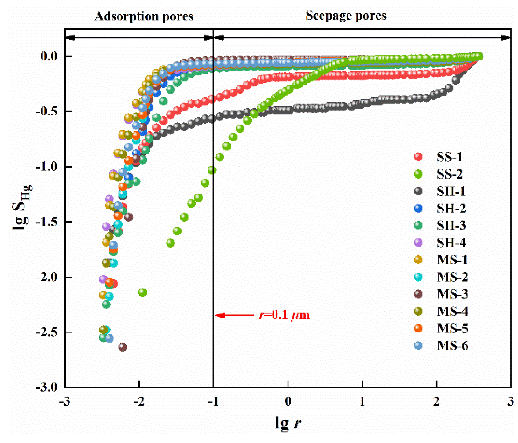
Figure 5 shows the double logarithmic relationship of all rock samples between r and SHg, in which an apparent difference exists in the tangent slope between the two sides of the experimental sample at r = 0.1 μm. Hence, the curves are divided into two parts, and the multifractal dimensions of the adsorption pore (r < 0.1 μm) and seepage pore (r ≥ 0.1 μm) are calculated. Table 5 shows the correlation effect of multifractal dimensions and that the average correlation coefficients of the adsorption pore and seepage pore are 0.843 and 0.871, respectively. The relationship between DMIP is DA < Df < DS, which has prominent multifractal characteristics. The values of DA (r < 0.1 μm) range from 2.045 (SH-3) to 2.540 (SH-1), with an average of 2.274. Meanwhile, those of DS (r ≥ 0.1 μm) are between 2.877 (SH-1) and 2.907 (SH-2), with an average of 2.933.

3.6. Fractal Dimension Calculation Using NMR

The total relaxation mechanism of NMR can be simplified as follows [43]:
1 T 2 1 T 2 S = ρ S V = ρ F s r
where T2S refers to the transverse volume relaxation, ms; ρ refers to the T2 surface intensity, μm/ms; and F refers to where ρ, F, and r refer to the T2 surface intensity, μm/ms, the pore shape factor and the pore radius, μm, respectively.
The Sv (cumulative pore volume fraction) can be expressed as:
S v = V < r V = r 3 D r m i n 3 D r m a x 3 D r m i n 3 D
When rmin << rmax, Equation (7) can be simplified as:
S v = r r m a x 3 D
Then, Equation (8) can be converted as:
S v = T 2 m a x T 2 D 3
Equation (10) can be expressed as:
lg S v = 3 D lg T 2 + D 3 lg T 2 m a x
where T2max, D, and SV refer to the maximal transverse relaxation, the fractal dimension, and the cumulative pore volume fraction, respectively.
As shown in Figure 6, the double logarithmic relationship of all rock samples between T2 and SV, in which an apparent difference exists in the tangent slope between the two sides of the experimental sample at T2 = 1 ms. Hence, the pore size is divided into two parts: T2 < 1 ms and T2 ≥ 1 ms, and the multifractal dimensions of the adsorption pore (T2 < 1 ms) and seepage pore (T2 ≥ 1 ms) are calculated, respectively.
As shown in Table 6, the DA is in the range of 2.036 (MS-3) to 2.456 (MS-1), with an average of 2.133. The DS is between 2.881 (MS-6) and 2.998 (SH-4), with an average of 2.933, denoting the prominent multifractal characteristics of the core samples. Moreover, the relationship of DNMR is DA (adsorption pore) < Df (full-size pore) < DS (seepage pore), Meanwhile, multifractal dimensions are calculated, in which the DA is close to 2, as well as the DS is close to 3. This implies that the adsorption pores have good homogeneity, while the seepage pore structure is relatively complex and has strong heterogeneity. Moreover, SS-2 has a wide distribution of the T2 spectrum with a bit of change in the correlation curve of the full pore size, indicating that the fractal dimension of the sandstone sample SS-2 shows good continuity.
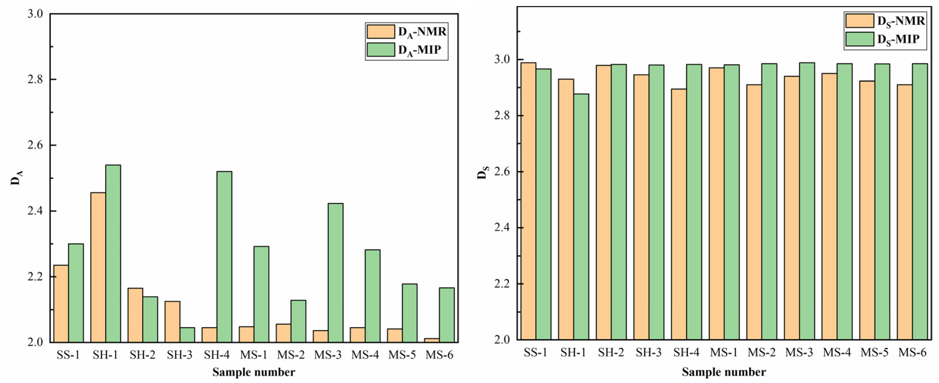
A comparison of the calculated results of DMIP and DNMR is shown in Figure 7. As mentioned above, both DMIP and DNMR exhibit obvious multifractal characteristics, in which the DA is substantially lower than DS, demonstrating great homogeneity of adsorption pores. The results of DMIP and DNMR are comparable when calculating the fractal dimension of seepage pores, although the DA estimated by the MIP experiment is typically higher than that determined by the NMR experiment. This may be due to the principle of MIP experiments. The measuring aperture and mercury injection pressure in MIP testing are inversely related. The mercury injection pressure used to evaluate the seepage pores is minimal and will not cause damage to the pore structure. However, the pore size of the adsorption pore is typically tiny, and the mercury injection pressure required for measurement is significant. The pore structure will be harmed by the excessive mercury injection pressure, and the pore structure’s increased heterogeneity will lead to measurement inaccuracies. The NMR experiment has the characteristic of being non-destructive, so the results are fairly precise when determining the adsorption pore’s size.

4. Discussion

The experimental rock samples present apparent multifractal characteristics on the basis of the calculation results of fractal dimensions. This section analyzes the influencing factors of the multifractal dimensions of 10 clay rock samples. Because the MIP experiment is affected by the mercury inlet pressure when measuring micropores, this section selects the DNMR for discussion [23].

4.1. Correlation between Physical Properties and Multifractal Dimensions

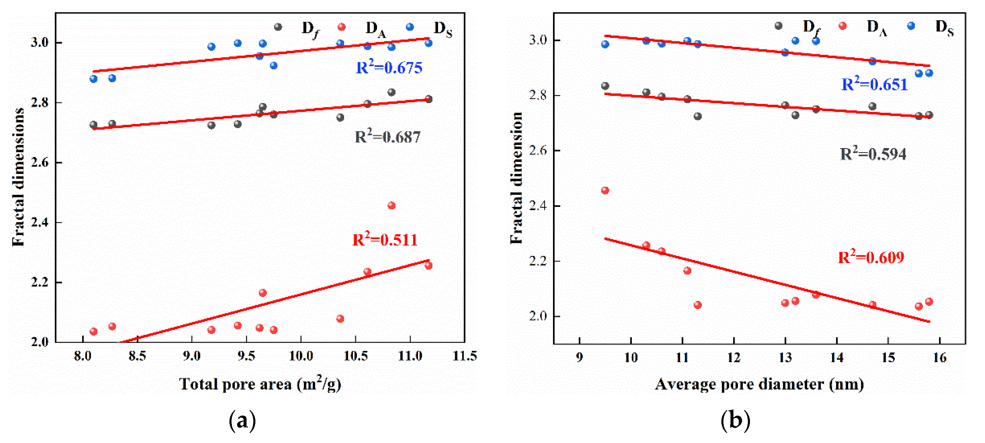
To explore the role of physical properties on the fractal features of the experiment samples, the correlation between multifractal dimensions, and the total pore area, the average pore diameter is plotted in Figure 8.
As can be observed, the numbered samples possess remarkable multifractal characteristics, which are affected considerably by petrophysical properties. The Df, DA, DS, and total pore area show a positive correlation, and the correlation coefficients are 0.687, 0.675, and 0.511, respectively. The relationship of multifractal dimensions is DA < DS, indicating that the seepage pore has a more heterogonous pore structure. Meanwhile, both DA and DS increase with increasing total pore area, which may be due to the fact that the change in the pore space and specific surface area leads to the rise in the heterogeneity of the pore structure [48]. The correlation of DA with the total pore area is better than that of DS with the total pore area, which is consistent with previous studies [19,23,49]. For adsorption pores, DA mainly reflects the surface structure of micropores, greatly influenced by the roughness and specific surface area of the pore surface.
Additionally, the Df, DA, and DS negatively correlate with the average pore diameter, with correlation coefficients of 0.597, 0.609, and 0.651, respectively. Both DA and DS increase with decreasing average pore diameters, indicating that rock samples with tiny average pore diameters may comprise more throats and micropores, resulting in increasing heterogeneity and more complex pore structures. The DS shows a better correlation with average pore diameters than DA. For seepage pores, DS may best depict the pore structure fractal dimension, which is more susceptible to the average pore diameter than DA. Multifractal dimensions can better characterize the heterogeneity of rock samples, in which DA reflects the surface structure of micropores, while DS represents the pore structure of macropores.

4.2. Correlation between Minerals and Multifractal Dimensions

The correlations between the multifractal dimensions and the TOC content, clay minerals, quartz, chlorite, and illite are shown in Figure 9. The Df and DA positively correlate with TOC content with correlation coefficients of 0.646 and 0.845, respectively, whereas the DS inconspicuously correlates with TOC content. TOC content reflects the abundance of organic matter. Rock samples with high TOC content have more micropores, which leads to a more complex pore structure of adsorption pores, resulting in an increase in DA [50].
Due to the interaction of multiple factors, i.e., depositional environment, pore type, and the type and content of mineralogical compositions, different mineralogical compositions have diverse mechanical properties and chemical stability, which have various effects on the fractal dimensions. The content of quartz and kaolinite have no apparent correlation with fractal dimensions for some reason, whereas the content of the clay mineral composition shows a negative correlation with the Df and DA but an inconspicuous correlation with DS. Moreover, the content of chlorite and illite shows no apparent correlation with Df and DS but a negative correlation with DA. Clay minerals, including chlorite and illite, mainly contained in the adsorption pores with relatively small pore sizes, are the main factors affecting the pore structure of reservoir rocks. Hence, the influence of clay mineral composition on the adsorption pores is significant. After reacting with the fluid, the surface of the clay minerals becomes uniform, and the pore heterogeneity is weakened, leading to a decrease in fractal dimensions.

4.3. Fractal Dimension Mathematical Model

As shown in Figure 3 and Figure 4, the experimental samples exhibit prominent multifractal characteristics. The multifractal dimensions are affected by many factors with complex mechanisms. In this paper, the mechanism of action is simplified in order to quantitatively study the effects of various factors on the multifractal dimension. It is assumed that each linearly independent factor linearly correlates with the fractal dimension. Based on correlation analysis, total pore area, and average pore diameter, the content of TOC and clay minerals are selected as the main factor affecting multifractal dimensions. A multivariate linear regression analysis of fractal dimensions was carried out using SPSS, a statistical software for multivariate statistics, to establish the corresponding multivariable linear regression model.
(1) The multivariable linear regression model is established as Equation (11):
D = a + b S + c r + d 1 C T + d 2 C C
where D is the multifractal dimension; a is const; b, c, d 1 ,   a n d   d 2 are the weighting factors of total pore area, average pore diameter, and the content of TOC and clay minerals, respectively; S is the total pore area; r is the average pore diameter; and C T   a n d   C C are the absolute content ratios of TOC and clay minerals, respectively.
(2) Entering the parameters after the standardization process, a multivariate linear regression analysis of multifractal dimensions was conducted using SPSS to establish the corresponding multivariable linear regression model, which is shown in Table 7.
The parameters of the multivariable linear regression models are presented in Table 6, in which R2 is the correlation coefficient, F is the statistical value of F-test, and a is const. In this section, ten rock samples from the Daqiang Coal Mine are selected for analysis, and the relationship between the total pore area, average pore diameter, TOC content, clay mineral content, and multifractal dimensions is discussed. Therefore, the number of samples and independent variables is 10 and 4, respectively. Moreover, the degree of freedom is determined by the number of samples and independent variables, and 0.05 is selected as the level of significance in a significance test.
(1) Firstly, the significance test of the whole regression model should be conducted before the significance test of each regression coefficient, which is the joint hypotheses test (F-test). The F-test is used to determine whether the model has a regression relationship. After determining the number of independent variables, degrees of freedom, and the level of a significance test, the critical value of the F-test can be obtained by the critical table, in which F 0.05 4,5 = 5.192. When F F 0.05 ( 4,5 ) , there is a linear regression relationship between influence factors and multifractal dimensions. Moreover, it can be found that there is a linear regression relationship between Df, DA, DS, and influence factors.
(2) Secondly, a Student’s t-test (t-test) is carried out on the model parameters of each factor to judge the significance of each independent variable to the dependent variable, of which the degree of freedom and the level of a significance test are 5 and 0.05, respectively. After determining the degree of freedom, and the level of a significance test, the critical value of the T-test can be obtained using the critical table. The critical table of the t-test shows that t 5 0.05 = 2.571; when t t 5 ( 0.05 ) , the influence factor has a significant effect on multifractal dimensions.
(3) Finally, the influencing factors are quantitatively analyzed. The larger the absolute value of the influencing weight coefficient is, the greater the prestige of the factor on the fractal dimension is, and vice versa.
As can be observed, the correlation effect of the multivariate linear regression model is excellent. On the one hand, for adsorption pores, the total pore area, and the contents of TOC and clay minerals have significant effects on DA, while the average pore size has no apparent effect. On the other hand, for seepage pores, only the average pore size has a significant effect on the DS, while the total pore area, the contents of TOC and clay mineral composition have no apparent effect, which confirms the above analysis. The multivariate linear regression model of fractal dimension can be expressed as follows:
D A = 1.648 + 0.413 S + 0.578 C T 0.390 C c D S = 3.148 1.154 r

5. Conclusions

In this paper, the pore structure and fractal features of three typical coal-measure sedimentary rocks from the Daqiang and Jiahe Coal Mines in China were investigated using MIP and NMR experiments. The following conclusions were obtained:
(1)
The investigated samples are dominated by clay minerals and quartz, with TOC contents ranging from 0.03% to 10.81%. The main composition of clay minerals is kaolinite and chlorite.
(2)
The pore structure features of the investigated samples reveal obvious deviations. All the PSD curves are unimodal distributions. The sandstone (SS-1), mudstone, and shale are mainly with nanopores of 0.01–1 μm, while the sandstone (SS-2) is dominated by mesopores and macropores of 1–100 μm.
(3)
The pore structures of the investigated samples show prominent multipartite characteristics using MIP and NMR tests. Multifractal dimensions can better characterize the heterogeneity of rock samples, in which DA reflects the surface structure of micropores, while DS represents the pore structure of macropores.
(4)
Multifractal dimensions are affected by many factors, in which DA is greatly influenced by the pore surface features and mineral components and DS by average pore diameters. Moreover, the multivariate linear regression model of adsorption pores and seepage pores were established, respectively, which has a better correlation effect on the multifractal dimensions.

Author Contributions

Conceptualization, N.Z.; Methodology, S.W.; Software, X.X.; Validation, H.W.; Resources, X.S. and M.H.; Writing—original draft, S.W. All authors have read and agreed to the published version of the manuscript.

Funding

Funding for this study was provided by the National Natural Science Foundation of China (42277195), the Innovation Fund Research of China University of Mining & Technology, Beijing (SKLGDUEK202218), the Fundamental Research Funds for the Central Universities of China (2021YJSSB10), and the Undergraduate Innovation Program of China University of Mining & Technology, Beijing (C202006968).

Data Availability Statement

Not applicable.

Conflicts of Interest

The authors declare no conflict of interest.

References

  1. Zou, C.N.; Yang, Z.; Tao, S.Z.; Yuan, X.J.; Zhu, R.K.; Hou, L.H.; Wu, S.T.; Sun, L.; Zhang, G.S.; Bai, B.; et al. Continuous hydrocarbon accumulation over a large area as a distinguishing characteristic of unconventional petroleum: The Ordos Basin, North-Central China. Earth-Sci. Rev. 2013, 126, 358–369. [Google Scholar] [CrossRef] [Green Version]
  2. Wang, Y.; Liu, L.; Cheng, H. Gas Adsorption Characterization of Pore Structure of Organic-rich Shale: Insights into Contribution of Organic Matter to Shale Pore Network. Nat. Resour. Res. 2021, 30, 2377–2395. [Google Scholar] [CrossRef]
  3. Xu, S.; Hu, E.; Li, X.; Xu, Y. Quantitative Analysis of Pore Structure and Its Impact on Methane Adsorption Capacity of Coal. Nat. Resour. Res. 2020, 30, 605–620. [Google Scholar] [CrossRef]
  4. Loucks, R.G.; Reed, R.M.; Ruppel, S.C.; Hammes, U. Spectrum of pore types and networks in mudrocks and a descriptive classification for matrix-related mudrock pores. Aapg. Bull. 2012, 96, 1071–1098. [Google Scholar] [CrossRef] [Green Version]
  5. Clarkson, C.R.; Solano, N.; Bustin, R.M.; Bustin, A.M.M.; Chalmers, G.R.L.; He, L.; Melnichenko, Y.B.; Radliński, A.P.; Blach, T.P. Pore structure characterization of North American shale gas reservoirs using USANS/SANS, gas adsorption, and mercury intrusion. Fuel 2013, 103, 606–616. [Google Scholar] [CrossRef]
  6. Jiang, J.; Yang, W.; Cheng, Y.; Zhao, K.; Zheng, S. Pore structure characterization of coal particles via MIP, N2 and CO2 adsorption: Effect of coalification on nanopores evolution. Powder Technol. 2019, 354, 136–148. [Google Scholar] [CrossRef]
  7. Yang, X.; Guo, S. Comparative analysis of shale pore size characterization methods. Pet. Sci. Technol. 2020, 38, 793–799. [Google Scholar] [CrossRef]
  8. Shi, X.; Xu, H.; Che, M.; Xiao, C.; Ni, H.; Gao, Q. Investigations of fracture behavior and pore structure change in pulse fracturing for cement block. Int. J. Rock. Mech. Min. Sci. 2023, 166, 105366. [Google Scholar] [CrossRef]
  9. Zhao, Y.; Sun, Y.; Yuan, L.; Xu, Q. Impact of nanopore structure on coal strength: A study based on synchrotron radiation nano-CT. Results Phys. 2020, 17, 103029. [Google Scholar] [CrossRef]
  10. Liu, S.; Huang, Z. Exploration of microstructure characteristics and mechanical behaviors of thermal-damaged argillaceous sandstone via LF-NMR and µ-CT technologies. Geomech. Geophys. Geo 2023, 9, 27. [Google Scholar] [CrossRef]
  11. Zhang, N.; Zhao, F.; Guo, P.; Li, J.; Gong, W.; Guo, Z.; Sun, X. Nanoscale Pore Structure Characterization and Permeability of Mudrocks and Fine-Grained Sandstones in Coal Reservoirs by Scanning Electron Microscopy, Mercury Intrusion Porosimetry, and Low-Field Nuclear Magnetic Resonance. Geofluids 2018, 2018, 2905141. [Google Scholar] [CrossRef]
  12. He, J.; Ding, W.; Li, A.; Sun, Y.; Dai, P.; Yin, S.; Chen, E.; Gu, Y. Quantitative microporosity evaluation using mercury injection and digital image analysis in tight carbonate rocks: A case study from the Ordovician in the Tazhong Palaeouplift, Tarim Basin, NW China. J. Nat. Gas. Sci. Eng. 2016, 34, 627–644. [Google Scholar] [CrossRef]
  13. Yang, R.; He, S.; Yi, J.; Hu, Q. Nano-scale pore structure and fractal dimension of organic-rich Wufeng-Longmaxi shale from Jiaoshiba area, Sichuan Basin: Investigations using FE-SEM, gas adsorption and helium pycnometry. Mar. Pet. Geol. 2016, 70, 27–45. [Google Scholar] [CrossRef]
  14. Fu, H.; Yan, D.; Yao, C.; Su, X.; Wang, X.; Wang, H.; Li, Y. Pore Structure and Multi-Scale Fractal Characteristics of Adsorbed Pores in Marine Shale: A Case Study of the Lower Silurian Longmaxi Shale in the Sichuan Basin, China. J. Earth Sci. 2022, 33, 1278–1290. [Google Scholar] [CrossRef]
  15. Song, Z.; Liu, G.; Yang, W.; Zou, H.; Sun, M.; Wang, X. Multi-fractal distribution analysis for pore structure characterization of tight sandstone—A case study of the Upper Paleozoic tight formations in the Longdong District, Ordos Basin. Mar. Pet. Geol. 2018, 92, 842–854. [Google Scholar] [CrossRef]
  16. Wang, T.; Tian, F.; Deng, Z.; Hu, H.; Xie, Z.; Liu, D. The Pore Structure of Marine to Continental Transitional Shales in the Permian Shanxi Formation on the East Margin of the Ordos Basin, China. Geofluids 2022, 2022, 5601862. [Google Scholar] [CrossRef]
  17. Zhang, N.; Wang, S.; Zhao, F.; Sun, X.; He, M. Characterization of the Pore Structure and Fluid Movability of Coal-Measure Sedimentary Rocks by Nuclear Magnetic Resonance (NMR). ACS Omega 2021, 6, 22831–22839. [Google Scholar] [CrossRef]
  18. Wu, B.; Xie, R.; Wang, X.; Wang, T.; Yue, W. Characterization of pore structure of tight sandstone reservoirs based on fractal analysis of NMR echo data. J. Nat. Gas Sci. Eng. 2020, 81, 103483. [Google Scholar] [CrossRef]
  19. Li, Z.; Shen, X.; Qi, Z.; Hu, R. Study on the pore structure and fractal characteristics of marine and continental shale based on mercury porosimetry, N2 adsorption and NMR methods. J. Nat. Gas Sci. Eng. 2018, 53, 12–21. [Google Scholar] [CrossRef]
  20. Al-Yaseri, A.Z.; Lebedev, M.; Vogt, S.J.; Johns, M.L.; Barifcani, A.; Iglauer, S. Pore-scale analysis of formation damage in Bentheimer sandstone with in-situ NMR and micro-computed tomography experiments. J. Pet. Sci. Eng. 2015, 129, 48–57. [Google Scholar] [CrossRef] [Green Version]
  21. Xie, R.; Wu, Y.; Liu, K.; Liu, M.; Xiao, L. De-noising methods for NMR logging echo signals based on wavelet transform. J. Geophys. Eng. 2014, 11, 035003. [Google Scholar] [CrossRef]
  22. Xie, W.; Yin, Q.; Zeng, J.; Wang, G.; Feng, C.; Zhang, P. Fractal-Based Approaches to Pore Structure Investigation and Water Saturation Prediction from NMR Measurements: A Case Study of the Gas-Bearing Tight Sandstone Reservoir in Nanpu Sag. Fractal Fract. 2023, 7, 273. [Google Scholar] [CrossRef]
  23. Guo, X.; Huang, Z.; Zhao, L.; Han, W.; Ding, C.; Sun, X.; Yan, R.; Zhang, T.; Yang, X.; Wang, R. Pore structure and multi-fractal analysis of tight sandstone using MIP, NMR and NMRC methods: A case study from the Kuqa depression, China. J. Pet. Sci. Eng. 2019, 178, 544–558. [Google Scholar] [CrossRef]
  24. Wang, G.; Han, D.; Qin, X.; Liu, Z.; Liu, J. A comprehensive method for studying pore structure and seepage characteristics of coal mass based on 3D CT reconstruction and NMR. Fuel 2020, 281, 118735. [Google Scholar] [CrossRef]
  25. Wang, J.; Cao, Y.; Liu, K.; Gao, Y.; Qin, Z. Fractal characteristics of the pore structures of fine-grained, mixed sedimentary rocks from the Jimsar Sag, Junggar Basin: Implications for lacustrine tight oil accumulations. J. Pet. Sci. Eng. 2019, 182, 106363. [Google Scholar] [CrossRef]
  26. Ramia, M.E.; Martín, C.A. Sedimentary rock porosity studied by electromagnetic techniques: Nuclear magnetic resonance and dielectric permittivity. Appl. Phys. A 2014, 118, 769–777. [Google Scholar] [CrossRef]
  27. Gao, H.; Li, H. Determination of movable fluid percentage and movable fluid porosity in ultra-low permeability sandstone using nuclear magnetic resonance (NMR) technique. J. Pet. Sci. Eng. 2015, 133, 258–267. [Google Scholar] [CrossRef]
  28. Li, X.; Kang, Y.; Haghighi, M. Investigation of pore size distributions of coals with different structures by nuclear magnetic resonance (NMR) and mercury intrusion porosimetry (MIP). Measurement 2018, 116, 122–128. [Google Scholar] [CrossRef]
  29. Wang, F.; Yang, K.; Cai, J. Fractal characterization of tight oil reservoir pore structure using nuclear magnetic resonance and mercury intrusion porosimetry. Fractals 2018, 26, 1840017. [Google Scholar] [CrossRef] [Green Version]
  30. Wang, F.; Yang, K.; You, J.; Lei, X. Analysis of pore size distribution and fractal dimension in tight sandstone with mercury intrusion porosimetry. Results Phys. 2019, 13, 102283. [Google Scholar] [CrossRef]
  31. Yan, J.; Zhang, S.; Wang, J.; Hu, Q.; Wang, M.; Chao, J. Applying Fractal Theory to Characterize the Pore Structure of Lacustrine Shale from the Zhanhua Depression in Bohai Bay Basin, Eastern China. Energy Fuels 2018, 32, 7539–7556. [Google Scholar] [CrossRef]
  32. Lai, J.; Wang, G. Fractal analysis of tight gas sandstones using high-pressure mercury intrusion techniques. J. Nat. Gas Sci. Eng. 2015, 24, 185–196. [Google Scholar] [CrossRef]
  33. Turlapati, V.Y.; Prusty, B.K.; Bakshi, T. Detailed Pore Structure Study of Damodar Valley and Upper Assam Basin Shales Using Fractal Analysis. Energy Fuels 2020, 34, 14001–14011. [Google Scholar] [CrossRef]
  34. Zhang, J.; Li, X.; Wei, Q.; Sun, K.; Zhang, G.; Wang, F. Characterization of Full-Sized Pore Structure and Fractal Characteristics of Marine–Continental Transitional Longtan Formation Shale of Sichuan Basin, South China. Energy Fuels 2017, 31, 10490–10504. [Google Scholar] [CrossRef]
  35. Zhou, L.; Kang, Z. Fractal characterization of pores in shales using NMR: A case study from the Lower Cambrian Niutitang Formation in the Middle Yangtze Platform, Southwest China. J. Nat. Gas Sci. Eng. 2016, 35, 860–872. [Google Scholar] [CrossRef]
  36. Yao, Y.; Liu, D.; Che, Y.; Tang, D.; Tang, S.; Huang, W. Petrophysical characterization of coals by low-field nuclear magnetic resonance (NMR). Fuel 2010, 89, 1371–1380. [Google Scholar] [CrossRef]
  37. Fan, X.; Wang, G.; Li, Y.; Dai, Q.; Linghu, S.; Duan, C.; Zhang, C.; Zhang, F. Pore structure evaluation of tight reservoirs in the mixed siliciclastic-carbonate sediments using fractal analysis of NMR experiments and logs. Mar. Pet. Geol. 2019, 109, 484–493. [Google Scholar] [CrossRef]
  38. Zhao, Y.; Zhu, G.; Dong, Y.; Danesh, N.N.; Chen, Z.; Zhang, T. Comparison of low-field NMR and microfocus X-ray computed tomography in fractal characterization of pores in artificial cores. Fuel 2017, 210, 217–226. [Google Scholar] [CrossRef]
  39. ISO/WD 19990:2023; Natural Stones—Determination of Water Absorption, Density, Porosity, and Bulk Specific Gravity. International Organization for Standardization: Geneva, Switzerland, 2023.
  40. ISO 10370:2014; Petroleum Products—Determination of Carbon Residue—Micro Method. International Organization for Standardization: Geneva, Switzerland, 2014.
  41. ISO 14689:2017; Geotechnical Investigation and Testing—Identification, Description and Classification of Rock. Organization for Standardization: Geneva, Switzerland, 2017.
  42. ISO 15901-1:2016; Evaluation of Pore Size Distribution and Porosity of Solid Materials by Mercury Porosimetry and Gas Adsorption—Part 1: Mercury Porosimetry. International Organization for Standardization: Geneva, Switzerland, 2016.
  43. Lai, J.; Wang, G.; Fan, Z.; Chen, J.; Wang, S.; Zhou, Z.; Fan, X. Insight into the Pore Structure of Tight Sandstones Using NMR and HPMI Measurements. Energy Fuels 2016, 30, 10200–10214. [Google Scholar] [CrossRef]
  44. Zhang, P.; Lu, S.; Li, J.; Chen, C.; Xue, H.; Zhang, J. Petrophysical characterization of oil-bearing shales by low-field nuclear magnetic resonance (NMR). Mar. Pet. Geol. 2018, 89, 775–785. [Google Scholar] [CrossRef]
  45. ASTM E2977-14; Standard Practice For Measuring and Reporting Performance Of Fourier-Transform Nuclear Magnetic Resonance (FT-NMR) Spectrometers For Liquid Samples. American Society for Testing and Materials: West Conshohocken, PA, USA, 2014.
  46. Cheng, M.; Li, C.; Zhou, L.; Feng, L.; Algeo, T.J.; Zhang, F.; Romaniello, S.; Jin, C.; Ling, H.; Jiang, S. Transient deep-water oxygenation in the early Cambrian Nanhua Basin, South China. Geochim. Cosmochim. Acta 2017, 210, 42–58. [Google Scholar] [CrossRef]
  47. Ge, X.; Fan, Y.; Zhu, X.; Li, R. Determination of nuclear magnetic resonance T-2 cutoff value based on multifractal theory—An application in sandstone with complex pore structure. Geophysics 2015, 80, D11–D21. [Google Scholar] [CrossRef]
  48. Li, J.; Wang, S.; Lu, S.; Zhang, P.; Cai, J.; Zhao, J.; Li, W. Microdistribution and mobility of water in gas shale: A theoretical and experimental study. Mar. Pet. Geol. 2019, 102, 496–507. [Google Scholar] [CrossRef]
  49. Shao, X.; Pang, X.; Li, H.; Zhang, X. Fractal Analysis of Pore Network in Tight Gas Sandstones Using NMR Method: A Case Study from the Ordos Basin, China. Energy Fuels 2017, 31, 10358–10368. [Google Scholar] [CrossRef]
  50. Ji, W.; Song, Y.; Jiang, Z.; Meng, M.; Liu, Q.; Chen, L.; Wang, P.; Gao, F.; Huang, H. Fractal characteristics of nano-pores in the Lower Silurian Longmaxi shales from the Upper Yangtze Platform, south China. Mar. Pet. Geol. 2016, 78, 88–98. [Google Scholar] [CrossRef]
Figure 1. The X-ray fluorescence measurements of the rock samples (a) SH-1 clay minerals. (b) SH-2 clay minerals. (c) MS-1 clay minerals. (d) MS-2 clay minerals. (e) SS-1 clay minerals [17].
Energies 16 03812 g001
Figure 2. Pore size distribution curves of MIP.
Energies 16 03812 g002
Figure 3. The T2 distributions of the investigated samples.
Energies 16 03812 g003
Figure 4. Pore size distribution curves of NMR.
Energies 16 03812 g004
Figure 5. Double logarithmic relationship between r and SHg of MIP.
Energies 16 03812 g005
Figure 6. Double logarithmic relationship between T2 and Sv of NMR.
Energies 16 03812 g006
Figure 7. Comparative analysis of DMIP and DNMR.
Energies 16 03812 g007
Figure 8. Correlations between (a) total pore area, (b) average pore diameter, and multifractal dimensions.
Energies 16 03812 g008
Figure 9. Correlations between the content of (a) TOC content, (b) clay minerals, (c) quartz, (d) kaolinite, (e) chlorite, (f) illite, and multifractal dimensions.
Energies 16 03812 g009
Table 1. Macromorphological characteristics of the rock samples.
Sample No.LithologyPetrographic Characteristics
SS-1sandstoneCaesious sandstone with good gradation and fine psephicity of grains
SS-2Grey–white fine sandstone with good gradation of detrital grains
SH-1shaleBrown laminar shale with noticeable rhyolitic structure
SH-2Dark-grey laminated silty shale
SH-3Grey laminated silty shale
SH-4Brown laminated silty shale
MS-1mudstoneGray massive mudstone
MS-2Gray massive mudstone
MS-3Yellow–gray massive mudstone
MS-4Gray massive mudstone
MS-5Dark-gray massive mudstone
MS-6Gray massive mudstone
Table 2. Total organic carbon (TOC) contents and mineralogical compositions of the investigated rock samples [11].
Sample No.TOC (%)Minerals (%)Clay Minerals (%)
Clay MineralsQuartzK-FeldsparPlagioclaseKaoliniteChloriteIlliteI/S%S
SS-10.03304381977941030
SS-20.06125411178963225
SH-110.812652513601072325
SH-25.183424365523111125
SH-32.924341412492092225
SH-43.292516124623131825
MS-11.735535373426142620
MS-20.8449354124425121925
MS-30.6242394124526101925
MS-40.735038393925132325
MS-51.0847374124523131925
MS-61.2144413124425121925
Table 3. Petrophysical properties of the investigated rock samples [11].
Sample No.Density
(g/cm3)
Total Porosity
φ (%)
Permeability
K (mD)
Total Pore Area
(m2/g)
Average Pore Diameter (nm)
SS-12.573.700.004610.6110.60
SS-22.1717.502.41335.20230.40
SH-12.531.400.000310.839.5
SH-22.646.300.00069.6511.1
SH-32.368.200.00159.1811.3
SH-42.5113.400.002811.1710.3
MS-12.444.700.00189.6213
MS-22.465.800.00489.4213.2
MS-32.406.400.00088.115.6
MS-42.407.700.001710.3613.6
MS-52.408.300.00129.7514.7
MS-62.3710.200.00268.2715.8
Table 4. Pore size distributions of the investigated rock samples.
Sample No.Semaphore Amplitude/a.u.
Adsorption PoresSeepage Pores
R ≤ 0.1 μmProportion/%0.1 μr < r ≤ 1 μmProportion/%R > 1 μmProportion/%
SS-11724.8978.93%256.9811.76%203.389.31%
SS-2127.321.11%3562.9930.98%7811.4267.92%
SH-11008.9882.14%63.925.20%155.5112.66%
SH-22440.4753.63%2047.1144.99%62.641.38%
SH-32208.5649.31%1992.9644.50%2776.19%
SH-46739.3156.14%5166.5543.04%98.140.82%
MS-13167.7945.49%3796.2354.51%00.00%
MS-23800.5577.68%1033.7221.13%58.471.20%
MS-32947.8244.62%3638.855.08%19.360.29%
MS-43073.6143.69%3891.4255.32%69.540.99%
MS-53305.648.37%3528.6351.63%00.00%
MS-62793.1138.87%4392.1561.13%00.00%
Table 5. Calculation results of DMIP.
Sample No.Porosity
φ (%)
Permeability
K (mD)
TOC
(%)
DfCorrelation CoefficientDA (r ≤ 0.1 μm)Correlation CoefficientDS (r > 0.1 μm)Correlation Coefficient
SS-13.700.00460.032.8210.3912.3010.7732.9660.758
SS-217.502.41330.062.5070.766////
SH-11.400.000310.812.7710.4252.540.5632.8770.694
SH-26.300.00065.182.7930.4232.1390.8712.9790.94
SH-38.200.00152.922.7240.482.0450.9132.9450.758
SH-413.400.00283.292.6630.3912.520.8412.8950.929
MS-14.700.00181.732.7640.4252.2920.8772.970.998
MS-25.800.00480.842.6820.4362.1280.8892.910.833
MS-36.400.00080.622.7250.4512.4230.8872.940.998
MS-47.700.00170.732.7530.442.2820.8892.950.765
MS-58.300.00121.082.7620.4352.1780.8882.9230.998
MS-610.200.00261.212.6830.4462.1660.882.910.905
Table 6. Calculation results of DNMR.
Sample No.Porosity
φ (%)
Permeability
K (mD)
TOC
(%)
DfCorrelation CoefficientDA (T2 ≤ 1 ms)Correlation CoefficientDS (T2 > 1 ms)Correlation Coefficient
SS-13.700.00460.032.7950.4252.2350.842.9880.812
SS-217.502.41330.062.5070.766////
SH-11.400.000310.812.8340.3912.4560.7732.9850.817
SH-26.300.00065.182.7860.4232.1650.8712.9970.45
SH-38.200.00152.922.7240.482.0410.9132.9860.509
SH-413.400.00283.292.8110.3912.2560.8412.9980.794
MS-14.700.00181.732.7640.4252.0480.8772.9550.793
MS-25.800.00480.842.7280.4362.0560.8892.9980.33
MS-36.400.00080.622.7250.4512.0360.8872.8790.905
MS-47.700.00170.732.750.442.0780.8892.9970.41
MS-58.300.00121.082.760.4352.0410.8882.9230.89
MS-610.200.00261.212.7290.4462.0530.882.8810.88
Table 7. Parameters of multivariable linear regression models.
ModelR2FaSrTOCClay Minerals
BetatBetatBetatBetat
DA0.98131.4541.6480.4132.8770.2741.5300.5783.642−0.390−2.906
DS0.9338.3713.1480.2821.068−1.154−3.491−0.474−1.6220.3021.221
Disclaimer/Publisher’s Note: The statements, opinions and data contained in all publications are solely those of the individual author(s) and contributor(s) and not of MDPI and/or the editor(s). MDPI and/or the editor(s) disclaim responsibility for any injury to people or property resulting from any ideas, methods, instructions or products referred to in the content.