2.1. Esterification of Bagasse
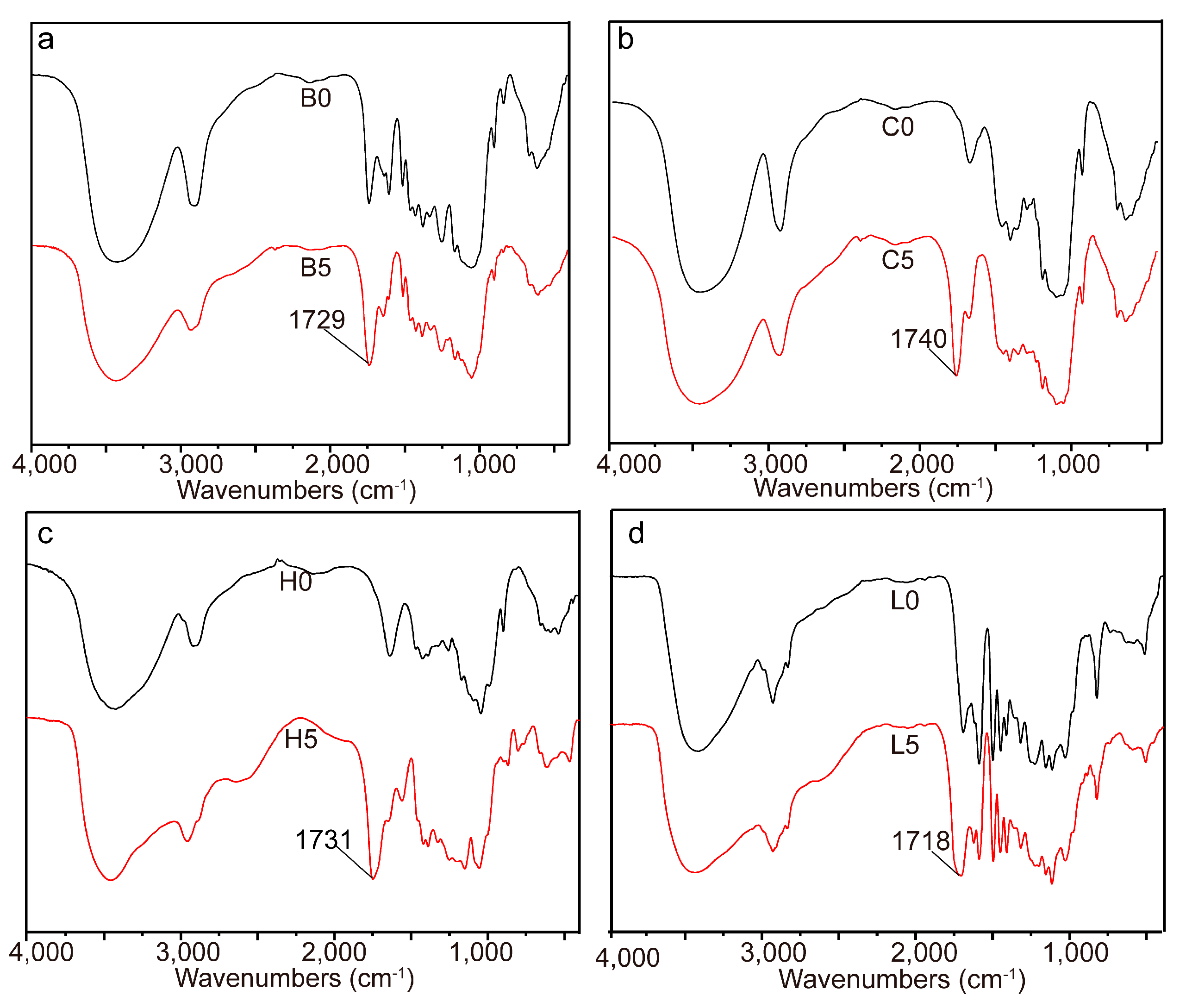
The homogeneous esterification of bagasse with glutaric anhydride was confirmed by FT-IR studies. The intensity of the band at 1729 cm
−1 (
Figure 1a), corresponding to C=O stretching, increased due to the esterification of bagasse [
21]. Due to the complex components and complicated linkages in bagasse, it was very difficult to identify the exact changes of bagasse during the homogeneous esterification. Therefore, the three main components (cellulose, hemicelluloses, and lignin) were isolated from bagasse and esterified with glutaric anhydride under the same conditions as bagasse in AmimCl to elucidate the possible structural changes of bagasse.
Compared with that of unmodified cellulose, the FT-IR spectrum of the esterified cellulose presented a new band at 1740 cm
−1 (
Figure 1b), indicating the formation of cellulose ester in the homogeneous system [
16]. Similarly, in the FT-IR spectrum of the esterified hemicelluloses (
Figure 1c), the new absorption at 1731 cm
−1 suggested the esterification of hemicelluloses with glutaric anhydride. By comparison with unmodified lignin, the intensity of the absorbance at 1718 cm
−1 in the esterified lignin (
Figure 1d) increased obviously, indicating the attachment of glutaryl groups onto lignin. These results indicated that cellulose, hemicelluloses, and lignin were all involved in the esterification with glutaric anhydride during the homogenous esterification of bagasse in AmimCl.
2.2. PS of Bagasse
The decrease in the hydroxyl content of esterified sample was due to the substitution of glutaryl groups. As shown in
Table 1, raising the dosage of glutaric anhydride from 10 to 40 mmol/g resulted in an increase in the substituted hydroxyl content of bagasse samples from 1.64 (B1) to 4.48 (B4) mmol/g, corresponding to a PS increase from 11.47% to 31.33%. A further increase in the dosage of glutaric anhydride from 40 to 50 mmol/g resulted in an unexpected decrease in the substituted hydroxyl content from 4.48 (B4) to 3.96 (B5) mmol/g, corresponding to a PS decrease from 31.33% to 27.69%. These results implied that changing glutaric anhydride dosage could regulate PS of bagasse samples, consistent with the previous study [
22]. As shown in
Table 2, unmodified bagasse was insoluble in water and common organic solvents, while glutaric anhydride could be dissolved in the selected solvents. After modification, the esterified bagasse samples were insoluble, swollen or soluble in water or organic solvents, especially showing the good solubility in DMSO. These changes of solubility were probably caused by the substitution of hydroxyls in bagasse (with different DS) with glutaryl group, indicating the occurrence of the esterification between bagasse and glutaric anhydride. However, it is impossible to obtain the PS changes of each component during the homogeneous esterification of bagasse because of the complicated components and complex linkages. Therefore, the isolated fractions were esterified with glutaric anhydride at different dosages (10–50 mmol/g) as bagasse to comparatively study the correlation between PS of each component and the dosage of glutaric anhydride.
With the increment of glutaric anhydride dosage from 10 to 50 mmol/g, the substituted hydroxyl content of cellulose samples increased from 1.30 (C1) to 4.52 (C5) mmol/g, corresponding to an increase of PS from 7.02% to 24.41%. These results were probably due to the fact that the increased dosage of glutaric anhydride would enhance the interaction between glutaric anhydride and cellulose [
16].
An increase in the dosage of glutaric anhydride from 10 to 50 mmol/g also led to the increased substituted hydroxyl contents of hemicelluloses samples from 1.13 (H1) to 4.54 (H5) mmol/g, corresponding to the PS increase from 7.46% to 29.97%. Considering the similarity between the anhydroglucose units (AGU) and anhydroxylose units (AXU), these increases were also resulted from the enhanced interaction between hemicelluloses and glutaric anhydride.
As shown in
Table 1, improving the dosage of glutaric anhydride from 10 to 20, 30, 40, and 50 mmol/g, the substituted hydroxyl contents of lignin samples reached 2.41 (L1), 2.27 (L2), 2.48 (L3), 3.15 (L4), and 2.62 (L5) mmol/g, respectively, and the corresponding PS changed from 47.53% to 44.77%, 48.92%, 62.13%, and 51.68%, respectively. It was interesting to observe the PS fluctuation of lignin samples with the increment of glutaric anhydride dosage. These results indicated that the PS changes of lignin were distinct from those of carbohydrates (cellulose and hemicelluloses). The detailed content changes of lignin hydroxyl groups during the homogeneous esterification are listed in
Table 3. Clearly, the aliphatic and phenolic hydroxyl content decreased from 3.96 (L0) to 1.91 (L1), 2.08 (L2), 1.94 (L3), 1.48 (L4), and 2.05 (L5) mmol/g, and from 1.11 (L0) to 0.75 (L1), 0.72 (L2), 0.65 (L3), 0.44 (L4), and 0.40 (L5) mmol/g, respectively. These results indicated both aliphatic and phenolic hydroxyls were esterified with glutaric anhydride during the homogeneous esterification [
23]. The ratio of aliphatic hydroxyls to phenolic hydroxyls obviously decreased from 3.57 (L0) to 2.55 (L1), indicating the prior esterification of aliphatic hydroxyls at very low dosage of glutaric anhydride(10 mmol/g). With the increment of glutaric anhydride dosage from 10 to 50 mmol/g, the ratio gradually increased from 2.55 (L1) to 5.13 (L5), which was due to the increased esterification of phenolic hydroxyls.
2.3. Reactive Sites of Bagasse
1D (
1H,
13C) and 2D NMR (HSQC) spectroscopy are powerful technologies to confirm the occurrence of esterification, and analyze the detailed reactive sites of bagasse during the homogeneous esterification. In the
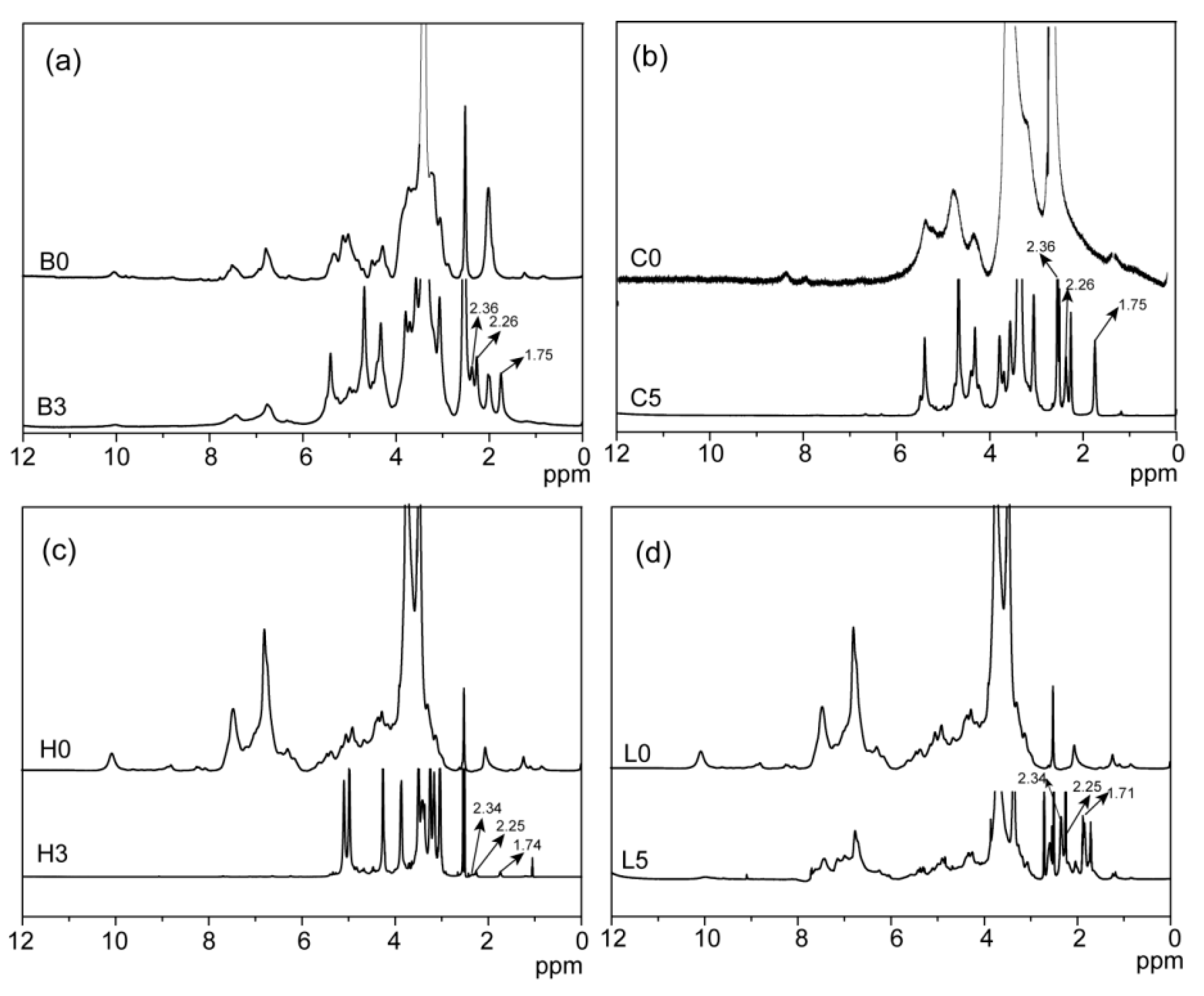
1H NMR spectrum of the esterified bagasse (
Figure 2a), peaks at 1.75, 2.26, and 2.36 ppm for glutaryl protons proved the esterification of bagasse with glutaric anhydride. Compared with that of unmodified bagasse, peaks at 20.34, 33.14, 173.02, and 174.43 ppm in
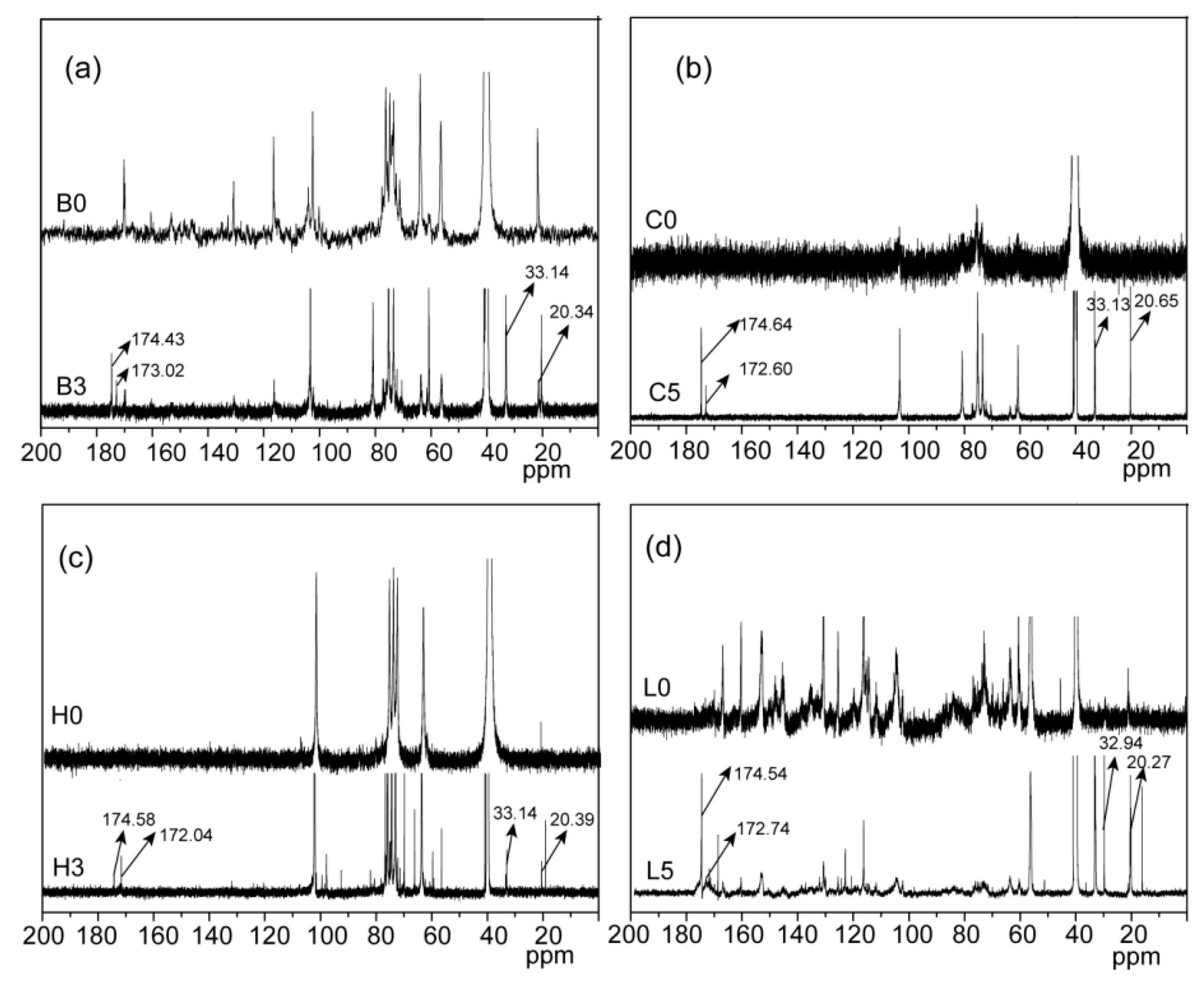
13C NMR spectrum of the esterified bagasse B3 (
Figure 3a) correspond to glutaryl carbons, further confirming the esterification between bagasse and glutaric anhydride, consistent with the FT-IR analysis.
In the 2D HSQC NMR spectra of bagasse samples (
Figure 4), cross-peaks from carbohydrate polymers (cellulose and hemicelluloses) were seriously overlapped, and the partial cross-peaks from lignin were recognizable. The poor distinguishability of the correlation signals in the HSQC spectra of bagasse samples made it inaccurate to analyze the detailed esterification information of reactive sites in each component during the homogenous esterification. Therefore, the detailed esterification information of reactive sites in each component was further analyzed with the HSQC spectra of the esterified fractions.
Compared with that of unmodified cellulose, proton peaks from the glutaryl group at 1.75, 2.26, and 3.36 ppm in the
1H NMR spectrum of the esterified cellulose (
Figure 2b) were clearly identified. Besides, carbon signals from glutaryl group in the
13C NMR spectrum of the esterified cellulose (
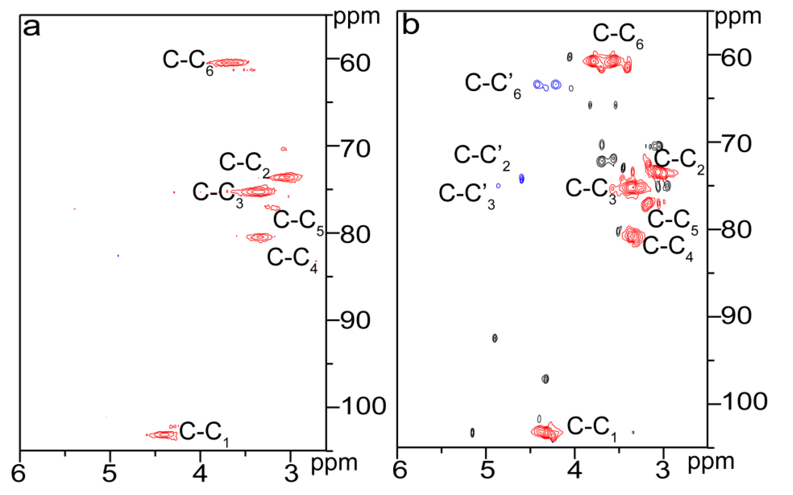
Figure 3b) at 20.65, 33.13, 172.60, and 174.64 ppm were also well resolved. These results confirmed the attachment of glutaryl group onto cellulose during the homogeneous esterification. The HSQC spectra of unmodified (C0, a) and esterified (C5, b) cellulose in DMSO-
d6 are shown in
Figure 5. The primary polysaccharide correlation peaks were well assigned based on their 1D (
1H and
13C) NMR spectra and the previous literature [
24,
25]. In the HSQC spectrum of cellulose sample C5, the correlation peaks at δ
C/δ
H 63.40/4.42 and 63.43/4.21 ppm correspond to C’
6/H’
6 from substituted C
6 (C–C’
6) in AGU, and the cross-peaks at δ
C/δ
H 74.24/4.60 and 74.99/4.86 ppm are for C’
2/H’
2 and C’
3/H’
3 from substituted C
2 (C–C’
2) and C
3 (C–C’
3) in AGU, respectively. The successful esterification of hydroxyls at C
2, C
3, and C
6 positions of AGU during the homogeneous derivatization was also observed in the esterification of cellulose with succinic anhydride in DMSO/BmimCl [
26]. The PS of C
2–OH, C
3–OH, and C
6–OH of AGU could be easily evaluated upon the integral area of the substituted and unsubstituted characteristic correlations [
25]. The results showed that the PS of hydroxyls at C
2, C
3, and C
6 positions of AGU were 2.2%, 0.7% and 10.7%, respectively. Therefore, the reactivity of hydroxyls in AGU during the homogeneous esterification followed the order of C
6–OH > C
2–OH > C
3–OH.
Considering the short-branched chain structure consisting of various sugar units in hemicelluloses [
27], the reactivity of main-chains and side-chains of hemicelluloses during the homogeneous esterification was further evaluated. According to the contents of different monosaccharides, main-chains of hemicelluloses were xylan, and side-chains were consisted of the other monosaccharides. As shown in
Table 4, the monosaccharide content of side-chains obviously decreased from 12.17% (H0) to 6.17% (H1), suggesting the prior esterification of side-chains at a very low dosage of glutaric anhydride (10 mmol/g), which was probably due to the more reactive primary hydroxyl groups on side chains [
25]. Further increasing glutaric anhydride dosage from 10 to 40 mmol/g led to an increase in the monosaccharide content of side-chains from 6.17% (H1) to 8.63% (H4), indicating the primary esterification of secondary hydroxyls on main-chains, which was probably due to the abundance of main-chain sugars. An increase in the dosage of glutaric anhydride from 40 to 50 mmol/g resulted in a decrease in the monosaccharide content of side-chains from 8.63% (H4) to 5.73% (H5) and an increase in the content of xylose from 91.37% (H4) to 94.27% (H5). These results indicated that secondary hydroxyls on side-chains were more easily esterified than those on main-chains, which was probably due to the lower steric hindrance of side-chains.
Three peaks at 1.74, 2.25, and 2.34 ppm in the
1H NMR spectrum of the esterified hemicelluloses (
Figure 2c) are assigned to the glutaryl protons, and signals at 20.29, 33.13, 172.04, and 174.58 ppm in the
13C NMR spectrum of the esterified hemicelluloses (
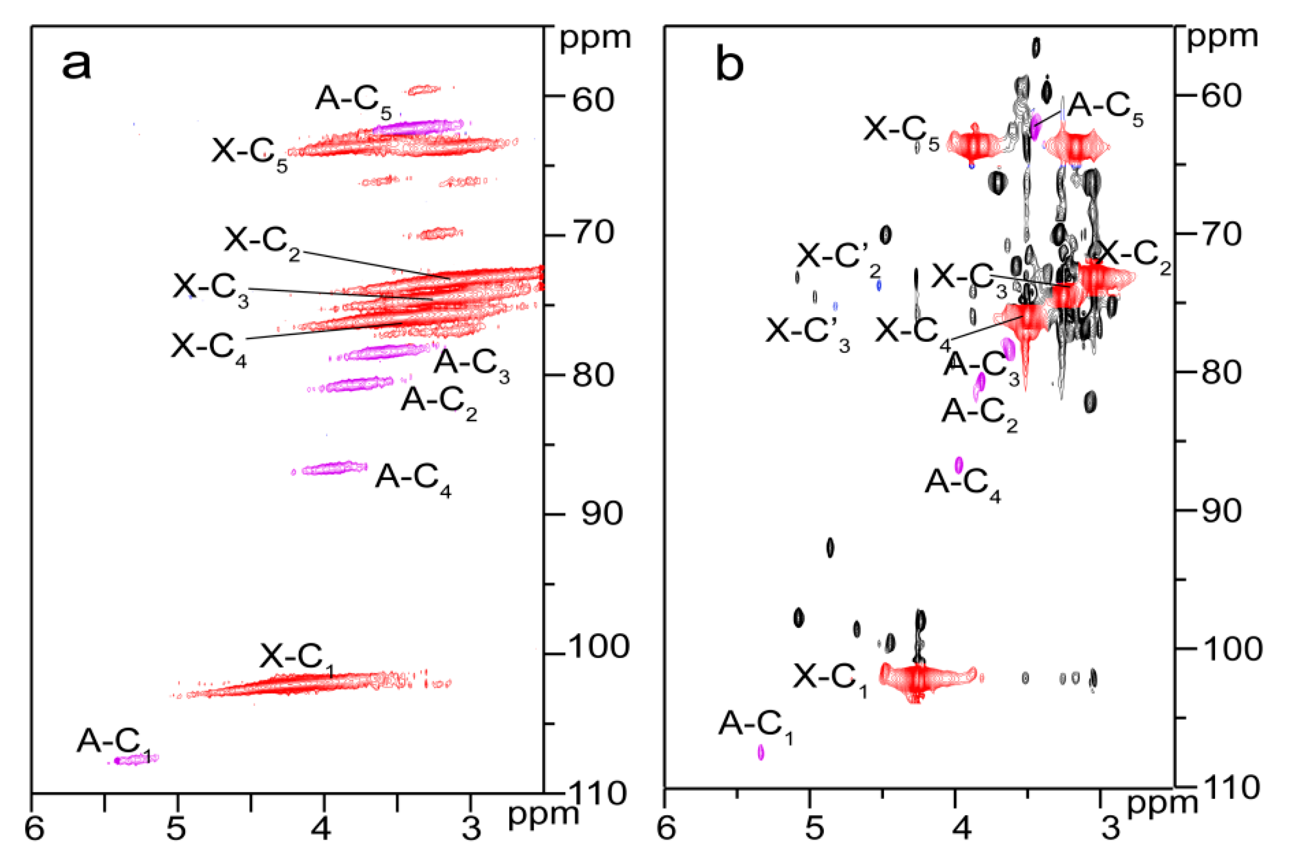
Figure 3c) are originated from the carbons of glutaryl group. These confirmed the occurrence of esterification between hemicelluloses and glutaric anhydride in AmimCl. The reactive sites of AXU from xylan during the homogeneous esterification were explored with 2D HSQC NMR in DMSO-
d6, as shown in
Figure 6. The characteristic cross-peaks for xylan and arabinose were well resolved in the spectra of hemicelluloses samples (H0 and H3), based on their 1D (
1H and
13C) NMR spectra and the previous study [
28]. The correlation peaks at δ
C/δ
H 74.21/3.87 and 76.01/4.25 ppm are associated with C’
2/H’
2 and C’
3/H’
3 from the substituted hydroxyls at C
2 (X-C’
2) and C
3 (X-C’
3) positions of AXU. By integrating the correlations assigned to substituted and unsubstituted C
2 and C
3 in AXU, the PS of hydroxyls at C
2 and C
3 positions of AXU were 1.7% and 1.6%, indicating the similar reactivity of secondary hydroxyls of AXU.
By comparison, three signals at 1.71, 2.25, and 2.34 ppm in the
1H NMR spectrum of the esterified lignin (
Figure 2d) correspond to the glutaryl protons, and four carbon signals at 20.27, 32.94, 172.74, and 174.54 ppm from the
13C NMR spectrum of the esterified lignin (
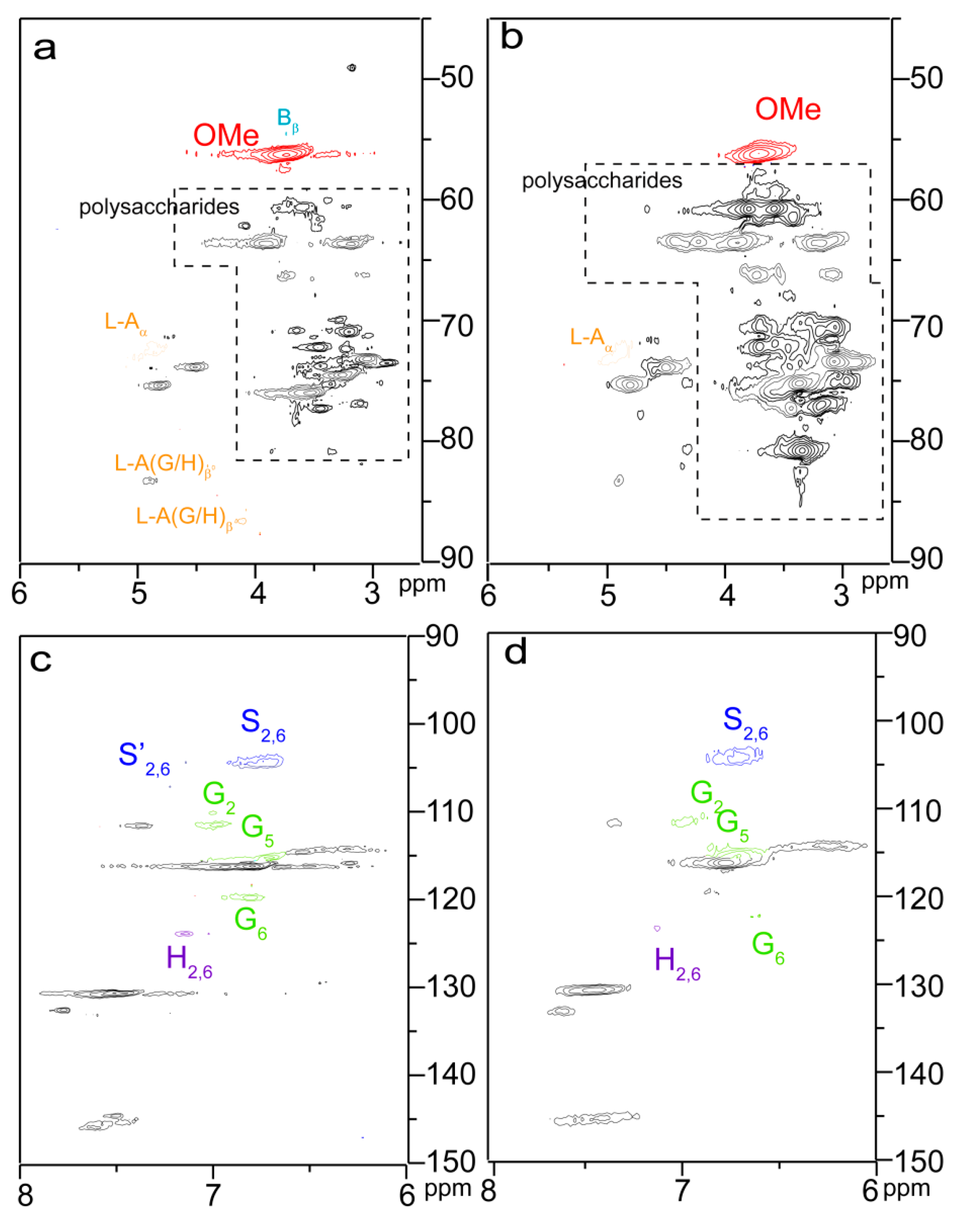
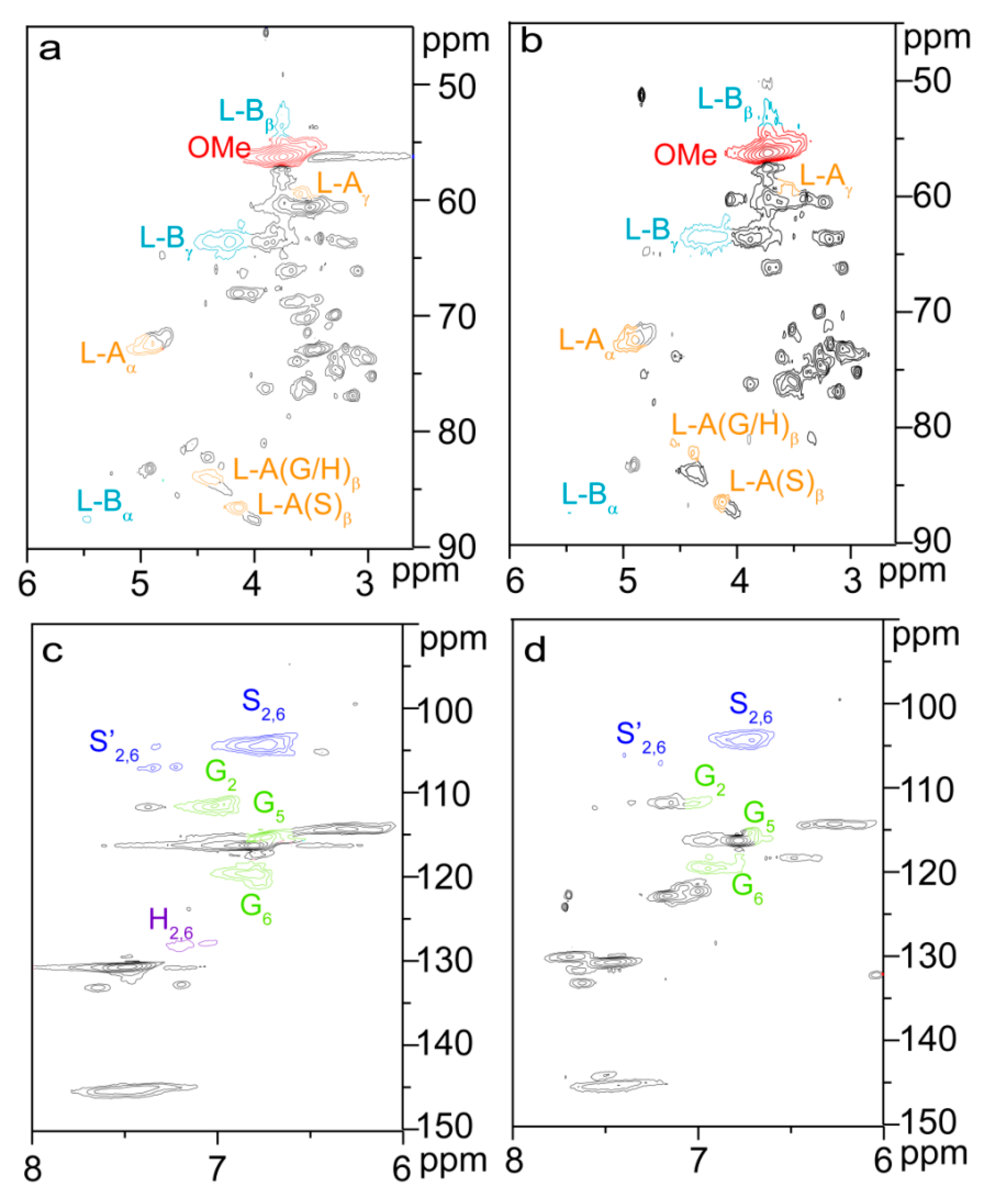
Figure 3d) relate to the carbons of glutaryl group. The presence of these signals confirmed the esterification between lignin and glutaric anhydride under the selected conditions. Semiquantitative 2D HSQC analysis of lignin samples were used to reveal the content changes of lignin substructures, according to the previous literature [
24,
29]. The HSQC spectra of unmodified (L0,
a and
c) and esterified (L5,
b and
d) lignin are shown in
Figure 7, and the substructures and aromatic units identified in lignin are depicted in
Figure 8. By semiqualitative analysis, the content of primary lignin substructures is listed in
Table 5. The content of β–
O–4′ aryl ether decreased from 45.6/100Ar (L0) to 44.4/100Ar (L5), suggesting the cleavage of β–
O–4′ during the homogeneous esterification [
30]. After esterification, cross-peaks from phenylcoumaran (β-5′) and H-units could not be distinguished under the present contour level, indicating the degradation of β-5′ and H-units during the homogeneous esterification. Besides, the syringyl to guaiacyl (S/G) ratio slightly increased from 1.11 (L0) to 1.16 (L5), which was probably resulted from the slight degradation of G-units during the homogeneous esterification. Semiquantitative 2D HSQC analysis revealed that the cleavage of lignin side-chains and degradation of aromatic units simultaneously occurred during the homogeneous esterification. The cleavage of lignin side-chain linkages and the degradation of aromatic units were probably due to the presence of glutaric acid, which was released from glutaric anhydride in AmimCl [
31].
2.5. Degradation of Esterified Bagasse
The degradation degree of bagasse during the homogeneous esterification was further investigated with GPC analysis. The weight-average molecular weight (
Mw) decreased from 42,129 (B0) to 35,511 (B3) g/mol (
Table S1), suggesting the degradation of bagasse after the homogeneous esterification. Besides, compared with that of unmodified bagasse, some signals disappeared in the HSQC spectrum of the esterified bagasse B3 (
Figure 4), confirming the degradation of bagasse during the homogeneous esterification. However, because of the complicated components and complex linkages in bagasse, it was impossible to directly obtain the detailed degradation information of each component. Therefore, the detailed degradation information of each component was illuminated with the GPC analysis of the esterified fractions.
The noticeably increased correlation peaks from low-molecular fractions in the HSQC spectrum of cellulose sample C5 indicated the significant degradation of cellulose upon esterification in AmimCl. As shown in
Table S1, the
Mw of cellulose samples obviously decreased from 57,787 (C0) to 35,511 (C5) g/mol, confirming the degradation of cellulose during the homogeneous esterification. Compared with unmodified hemicelluloses H0, signals from low-molecular fractions also obviously increased in the HSQC spectrum of esterified hemicelluloses, suggesting the degradation of hemicelluloses during the homogeneous esterification. GPC results showed that an improvement in the dosage of glutaric anhydride from 0 to 40 mmol/g also led to the
Mw decrease from 34,116 (H0) to 24,493 (H4) g/mol, affirming the degradation of hemicelluloses samples. The degradation of carbohydrates (cellulose and hemicelluloses) was probably caused by glutaric acid formed upon esterification in AmimCl [
25], which was similar to the previous report [
33]. After esterification, the
Mw of lignin samples increased from 15,105 (L0) to 29,554 (L5) g/mol, confirming the successful introduction of glutaryl groups into lignin [
34]. The
Mw of lignin sample L5 was much higher than the theoretical value calculated from the PS of esterified lignin, which was possibly due to the diesterification of lignin at high glutaric anhydride dosage.


