Comparative Surface Morphology, Chemical Composition, and Cytocompatibility of Bio-C Repair, Biodentine, and ProRoot MTA on hDPCs
Abstract
1. Introduction
2. Materials and Methods
2.1. Material Extracts
2.2. Cell Isolation and Culture
2.3. Scanning Electron Microscopy (SEM): Sample Visualization, Element Analysis, and Cell Attachment
2.4. Cell Viability Assay
2.5. Cell Morphology Analysis
2.6. Statistical Analysis
3. Results
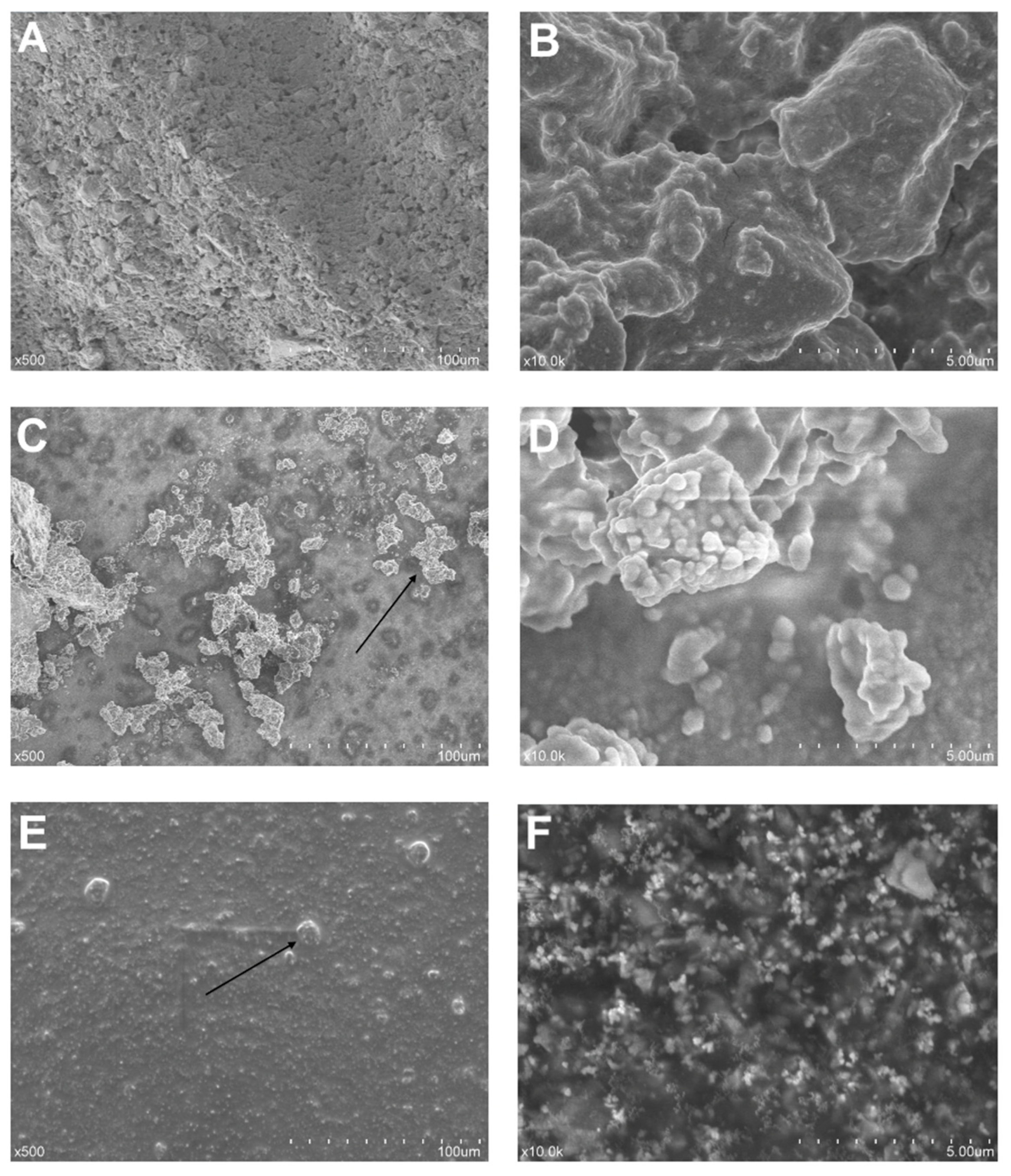
3.1. Sample Morphology
3.2. Element Analysis
3.3. Cell Attachment Analysis
3.4. Cell Viability Assay
3.5. Cell Morphology Analysis
4. Discussion
5. Conclusions
Author Contributions
Funding
Conflicts of Interest
References
- Shah, D.; Lynd, T.; Ho, D.; Chen, J.; Vines, J.; Jung, H.-D.; Kim, J.-H.; Zhang, P.; Wu, H.; Jun, H.-W.; et al. Pulp–Dentin Tissue Healing Response: A Discussion of Current Biomedical Approaches. J. Clin. Med. 2020, 9, 434. [Google Scholar] [CrossRef] [PubMed]
- Hashemi-Beni, B.; Khoroushi, M.; Foroughi, M.R.; Karbasi, S.; Khademi, A. Tissue engineering: Dentin – pulp complex regeneration approaches (A review). Tissue Cell 2017, 49, 552–564. [Google Scholar] [CrossRef] [PubMed]
- Shi, X.; Mao, J.; Liu, Y. Pulp stem cells derived from human permanent and deciduous teeth: Biological characteristics and therapeutic applications. Stem Cells Transl. Med. 2020, 9, 445–464. [Google Scholar] [CrossRef] [PubMed]
- DClinDent, B.K.; Ricucci, D.; Saoud, T.M.; Sigurdsson, A.; Kahler, B. Vital pulp therapy of mature permanent teeth with irreversible pulpitis from the perspective of pulp biology. Aust. Endod. J. 2019, 46, 154–166. [Google Scholar] [CrossRef]
- Spagnuolo, G.; Codispoti, B.; Marrelli, M.; Rengo, C.; Rengo, S.; Tatullo, M. Commitment of Oral-Derived Stem Cells in Dental and Maxillofacial Applications. Dent. J. 2018, 6, 72. [Google Scholar] [CrossRef]
- Kingshott, P.; Andersson, G.G.; McArthur, S.L.; Griesser, H. Surface modification and chemical surface analysis of biomaterials. Curr. Opin. Chem. Biol. 2011, 15, 667–676. [Google Scholar] [CrossRef]
- De Caluwé, T.; Vercruysse, C.; Ladik, I.; Convents, R.; Declercq, H.; Martens, L.; Verbeeck, R. Addition of bioactive glass to glass ionomer cements: Effect on the physico-chemical properties and biocompatibility. Dent. Mater. 2017, 33, e186–e203. [Google Scholar] [CrossRef]
- Schmalz, G.; Widbiller, M.; Galler, K. Material Tissue Interaction—From Toxicity to Tissue Regeneration. Oper. Dent. 2016, 41, 117–131. [Google Scholar] [CrossRef]
- Raghavendra, S.S.; Jadhav, G.R.; Gathani, K.M.; Kotadia, P. Bioceramics in endodontics—A review. J. Istanb. Univ. Fac. Dent. 2017, 51, S128–S137. [Google Scholar] [CrossRef]
- Sanz, J.L.; Rodríguez-Lozano, F.J.; Llena, C.; Sauro, S.; Forner, L. Bioactivity of Bioceramic Materials Used in the Dentin-Pulp Complex Therapy: A Systematic Review. Materials 2019, 12, 1015. [Google Scholar] [CrossRef]
- Camilleri, J.; Ford, T.P. Mineral trioxide aggregate: A review of the constituents and biological properties of the material. Int. Endod. J. 2006, 39, 747–754. [Google Scholar] [CrossRef] [PubMed]
- Parirokh, M.; Torabinejad, M.; Dummer, P.M.H. Mineral trioxide aggregate and other bioactive endodontic cements: An updated overview—Part I: Vital pulp therapy. Int. Endod. J. 2017, 51, 177–205. [Google Scholar] [CrossRef] [PubMed]
- Ha, W.N.; Nicholson, T.M.; Kahler, B.; Walsh, L.J. Mineral Trioxide Aggregate—A Review of Properties and Testing Methodologies. Materials 2017, 10, 1261. [Google Scholar] [CrossRef] [PubMed]
- Žižka, R.; Šedý, J.; Gregor, L.; Voborná, I. Discoloration after Regenerative Endodontic Procedures: A Critical Review. Iran. Endod. J. 2018, 13, 278–284. [Google Scholar] [CrossRef] [PubMed]
- Prati, C.; Gandolfi, M.G. Calcium silicate bioactive cements: Biological perspectives and clinical applications. Dent. Mater. 2015, 31, 351–370. [Google Scholar] [CrossRef]
- Rodríguez-Lozano, F.J.; López-García, S.; García-Bernal, D.; Lloret, M.R.P.; Guerrero-Gironés, J.; Pecci-Lloret, M.P.; Lozano, A.; Llena, C.; Spagnuolo, G.; Forner, L. In Vitro Effect of Putty Calcium Silicate Materials on Human Periodontal Ligament Stem Cells. Appl. Sci. 2020, 10, 325. [Google Scholar] [CrossRef]
- Bortoluzzi, E.A.; Niu, L.-N.; Palani, C.D.; El-Awady, A.R.; Hammond, B.D.; Pei, D.-D.; Tian, F.-C.; Cutler, C.W.; Pashley, D.H.; Tay, F.R. Cytotoxicity and osteogenic potential of silicate calcium cements as potential protective materials for pulpal revascularization. Dent. Mater. 2015, 31, 1510–1522. [Google Scholar] [CrossRef]
- Rajasekharan, S.; Martens, L.C.; Cauwels, R.G.E.C.; Anthonappa, R.P.; Verbeeck, R.M.H. Biodentine™ material characteristics and clinical applications: A 3 year literature review and update. Eur. Arch. Paediatr. Dent. 2018, 19, 1–22. [Google Scholar] [CrossRef]
- Koutroulis, A.; Kuehne, S.A.; Cooper, P.R.; Camilleri, J. The role of calcium ion release on biocompatibility and antimicrobial properties of hydraulic cements. Sci. Rep. 2019, 9, 19019. [Google Scholar] [CrossRef]
- Tomás-Catalá, C.J.; Collado-González, M.; García-Bernal, D.; Sánchez, R.E.O.; Forner, L.; Llena, C.; Lozano, A.; Moraleda, J.M.; Rodríguez-Lozano, F.J. Biocompatibility of New Pulp-capping Materials NeoMTA Plus, MTA Repair HP, and Biodentine on Human Dental Pulp Stem Cells. J. Endod. 2018, 44, 126–132. [Google Scholar] [CrossRef]
- Agrafioti, A.; Taraslia, V.; Chrepa, V.; Lymperi, S.; Panopoulos, P.; Anastasiadou, E.; Kontakiotis, E.G. Interaction of dental pulp stem cells with Biodentine and MTA after exposure to different environments. J. Appl. Oral Sci. 2016, 24, 481–486. [Google Scholar] [CrossRef] [PubMed]
- Benetti, F.; Queiroz Índia, O.D.A.; Cosme-Silva, L.; Conti, L.C.; De Oliveira, S.H.P.; Cintra, L.T.A. Cytotoxicity, Biocompatibility and Biomineralization of a New Ready-for-Use Bioceramic Repair Material. Braz. Dent. J. 2019, 30, 325–332. [Google Scholar] [CrossRef] [PubMed]
- Tomás-Catalá, C.J.; Collado-González, M.; García-Bernal, D.; Oñate-Sánchez, R.E.; Forner, L.; Llena, C.; Lozano, A.; Castelo-Baz, P.; Moraleda, J.M.; Rodríguez-Lozano, F.J. Comparative analysis of the biological effects of the endodontic bioactive cements MTA-Angelus, MTA Repair HP and NeoMTA Plus on human dental pulp stem cells. Int. Endod. J. 2017, 50, e63–e72. [Google Scholar] [CrossRef] [PubMed]
- Húngaro-Duarte, M.A.; Marciano, M.; Vivan, R.R.; Filho, M.T.; Guerreiro-Tanomaru, J.M.; Camilleri, J. Tricalcium silicate-based cements: Properties and modifications. Braz. Oral Res. 2018, 32, 70. [Google Scholar] [CrossRef]
- Youssef, A.-R.; Emara, R.; Taher, M.M.; Al-Allaf, F.A.; Almalki, M.; Almasri, M.A.; Siddiqui, S.S. Effects of mineral trioxide aggregate, calcium hydroxide, biodentine and Emdogain on osteogenesis, Odontogenesis, angiogenesis and cell viability of dental pulp stem cells. BMC Oral Health 2019, 19, 133. [Google Scholar] [CrossRef]
- ISO 10993-5:2009 Biological Evaluation of Medical Devices—Part 5: Tests for In Vitro Cytotoxicity. 2009. Available online: https://www.iso.org/standard/36406.html (accessed on 10 January 2020).
- Collado-González, M.; Lloret, M.R.P.; Tomás-Catalá, C.J.; García-Bernal, D.; Sánchez, R.E.O.; Llena, C.; Forner, L.; Rosa, V.; Rodríguez-Lozano, F.J. Thermo-setting glass ionomer cements promote variable biological responses of human dental pulp stem cells. Dent. Mater. 2018, 34, 932–943. [Google Scholar] [CrossRef]
- Peters, C.I. Research that matters-biocompatibility and cytotoxicity screening. Int. Endod. J. 2013, 46, 195–197. [Google Scholar] [CrossRef]
- Voicu, G.; Didilescu, A.C.; Stoian, A.B.; Dumitriu, C.; Greabu, M.; Andrei, M. Mineralogical and Microstructural Characteristics of Two Dental Pulp Capping Materials. Materials 2019, 12, 1772. [Google Scholar] [CrossRef]
- Mandava, J.; Arikatla, S.K.; Chalasani, U.; Yelisela, R.K. Interfacial adaptation and penetration depth of bioceramic endodontic sealers. J. Conserv. Dent. 2018, 21, 373–377. [Google Scholar] [CrossRef]
- Camilleri, J.; Sorrentino, F.; Damidot, D. Investigation of the hydration and bioactivity of radiopacified tricalcium silicate cement, Biodentine and MTA Angelus. Dent. Mater. 2013, 29, 580–593. [Google Scholar] [CrossRef]
- Jiménez-Sánchez, M.D.C.; Segura-Egea, J.J.; Díaz-Cuenca, A.; Jimenez-Sanchez, M. Physicochemical parameters - hydration performance relationship of the new endodontic cement MTA Repair HP. J. Clin. Exp. Dent. 2019, 11, e739–e744. [Google Scholar] [CrossRef] [PubMed]
- López-García, S.; Lloret, M.R.P.; Guerrero-Gironés, J.; Pecci-Lloret, M.P.; Lozano, A.; Llena, C.; Rodríguez-Lozano, F.J.; Forner, L. Comparative Cytocompatibility and Mineralization Potential of Bio-C Sealer and TotalFill BC Sealer. Materials 2019, 12, 3087. [Google Scholar] [CrossRef] [PubMed]
- Singh, G.; Gupta, I.; ElShamy, F.M.M.; Boreak, N.; Homeida, H.E. In vitro comparison of antibacterial properties of bioceramic-based sealer, resin-based sealer and zinc oxide eugenol based sealer and two mineral trioxide aggregates. Eur. J. Dent. 2016, 10, 366–369. [Google Scholar] [CrossRef] [PubMed]
- Khashaba, R.M.; Chutkan, N.B.; Borke, J.L. Comparative study of biocompatibility of newly developed calcium phosphate-based root canal sealers on fibroblasts derived from primary human gingiva and a mouse L929 cell line. Int. Endod. J. 2009, 42, 711–718. [Google Scholar] [CrossRef]
- Cintra, L.T.A.; Benetti, F.; de Azevedo Queiroz, Í.O.; de Araújo Lopes, J.M.; de Oliveira, S.H.P.; Araújo, G.S.; Gomes-Filho, J.E. Cytotoxicity, Biocompatibility, and Biomineralization of the New High-plasticity MTA Material. J. Endod. 2017, 43, 774–778. [Google Scholar] [CrossRef]
- Yazdi, K.A.; Ghabraei, S.; Bolhari, B.; Kafili, M.; Meraji, N.; Nekoofar, M.H.; Dummer, P.M.H. Microstructure and chemical analysis of four calcium silicate-based cements in different environmental conditions. Clin. Oral Investig. 2018, 23, 43–52. [Google Scholar] [CrossRef]
- López-García, S.; Lozano, A.; García-Bernal, D.; Forner, L.; Llena, C.; Guerrero-Gironés, J.; Moraleda, J.M.; Murcia, L.; Rodríguez-Lozano, F.J. Biological Effects of New Hydraulic Materials on Human Periodontal Ligament Stem Cells. J. Clin. Med. 2019, 8, 1216. [Google Scholar] [CrossRef]
- Sarkar, N.; Caicedo, R.; Ritwik, P.; Moiseyeva, R.; Kawashima, I. Physicochemical basis of the biologic properties of mineral trioxide aggregate. J. Endod. 2005, 31, 97–100. [Google Scholar] [CrossRef]
- Vallittu, P.; Boccaccini, A.R.; Hupa, L.; Watts, D.C. Bioactive dental materials—Do they exist and what does bioactivity mean? Dent. Mater. 2018, 34, 693–694. [Google Scholar] [CrossRef]
- Sequeira, D.B.; Seabra, C.M.; Palma, P.J.; Cardoso, A.L.; Peça, J.; Santos, J.M. Effects of a New Bioceramic Material on Human Apical Papilla Cells. J. Funct. Biomater. 2018, 9, 74. [Google Scholar] [CrossRef]
- Song, J.-S.; Mante, F.K.; Romanow, W.J.; Kim, S. Chemical analysis of powder and set forms of Portland cement, gray ProRoot MTA, white ProRoot MTA, and gray MTA-Angelus. Oral Surg. Oral Med. Oral Pathol. Oral Radiol. Endodontol. 2006, 102, 809–815. [Google Scholar] [CrossRef] [PubMed]
- Diomede, F.; Marconi, G.; Guarnieri, S.; D’Attilio, M.; Cavalcanti, M.F.X.B.; Mariggiò, M.A.; Pizzicannella, J.; Trubiani, O. A Novel Role of Ascorbic Acid in Anti-Inflammatory Pathway and ROS Generation in HEMA Treated Dental Pulp Stem Cells. Materials 2019, 13, 130. [Google Scholar] [CrossRef] [PubMed]
- Olcay, K.; Taşli, P.N.; Güven, E.P.; Ülker, G.M.Y.; Öğüt, E.E.; Çiftçioğlu, E.; Kiratli, B.; Şahin, F. Effect of a novel bioceramic root canal sealer on the angiogenesis-enhancing potential of assorted human odontogenic stem cells compared with principal tricalcium silicate-based cements. J. Appl. Oral Sci. 2020, 28, e20190215. [Google Scholar] [CrossRef] [PubMed]
- D’Antò, V.; Di Caprio, M.P.; Ametrano, G.; Simeone, M.; Rengo, S.; Spagnuolo, G. Effect of Mineral Trioxide Aggregate on Mesenchymal Stem Cells. J. Endod. 2010, 36, 1839–1843. [Google Scholar] [CrossRef] [PubMed]
- Mds, A.M.Z.E.; Hamama, H.H.; Msc, M.A.A.E.; Mds, M.E.G.; Mahmoud, S.H.; Neelakantan, P. The effect of four materials on direct pulp capping: An animal study. Aust. Endod. J. 2020. [Google Scholar] [CrossRef]
- Dahake, P.T.; Panpaliya, N.P.; Kale, Y.J.; Dadpe, M.V.; Kendre, S.B.; Bogar, C. Response of stem cells from human exfoliated deciduous teeth (SHED) to three bioinductive materials—An in vitro experimental study. Saudi Dent. J. 2019, 32, 43–51. [Google Scholar] [CrossRef]
- Akbulut, M.B.; Arpaci, P.U.; Eldeniz, A.U. ‘Effects of novel root repair materials on attachment and morphological behaviour of periodontal ligament fibroblasts: Scanning electron microscopy observation’. Microsc. Res. Tech. 2016, 79, 1214–1221. [Google Scholar] [CrossRef]
- Lv, F.; Zhu, L.; Zhang, J.; Yu, J.; Cheng, X.; Peng, B. Evaluation of the in vitro biocompatibility of a new fast-setting ready-to-use root filling and repair material. Int. Endod. J. 2016, 50, 540–548. [Google Scholar] [CrossRef]
- Poggio, C.; Ceci, M.; Dagna, A.; Beltrami, R.; Colombo, M.; Chiesa, M. In vitro cytotoxicity evaluation of different pulp capping materials: A comparative study. Arhiv za Higijenu Rada i Toksikologiju 2015, 66, 181–188. [Google Scholar] [CrossRef]


| Element | Sample 1 | Sample 2 | Sample 3 | Average |
|---|---|---|---|---|
| Carbon | 25.25% | 15.88% | 13.10% | 18.08% |
| Oxygen | 30.39% | 31.97% | 34.84% | 32.40% |
| Aluminium | 0.23% | 0.31% | 0.16% | 0.23% |
| Silicon | 4.67% | 4.61% | 4.19% | 4.49% |
| Calcium | 37.46% | 44.45% | 44.19% | 42.03% |
| Zirconium | 2.02% | 2.78% | 3.53% | 2.78% |
| Element | Sample 4 | Sample 5 | Sample 6 | Average |
|---|---|---|---|---|
| Carbon | 16.76% | 13.29% | 12.42% | 14.16% |
| Oxygen | 47.32% | 41.80% | 35.63% | 41.58% |
| Aluminium | 0.91% | 0.69% | 1.50% | 1.03% |
| Silicon | 4.01% | 4.89% | 6.46% | 5.12% |
| Calcium | 29.55% | 37.77% | 41.78% | 36.37% |
| Zirconium | 1.44% | 1.55% | 2.21% | 1.73% |
| Element | Sample 7 | Sample 8 | Sample 9 | Average |
|---|---|---|---|---|
| Carbon | 33.61% | 30.85% | 39.98% | 34.81% |
| Oxygen | 30.48% | 34.92% | 38.14% | 34.51% |
| Aluminium | 1.01% | 0.75% | 0.60% | 0.79% |
| Silicon | 3.20% | 3.08% | 2.00% | 2.76% |
| Calcium | 15.56% | 14.71% | 9.60% | 13.29% |
| Zirconium | 16.14% | 15.69% | 9.67% | 13.83% |
© 2020 by the authors. Licensee MDPI, Basel, Switzerland. This article is an open access article distributed under the terms and conditions of the Creative Commons Attribution (CC BY) license (http://creativecommons.org/licenses/by/4.0/).
Share and Cite
Ghilotti, J.; Sanz, J.L.; López-García, S.; Guerrero-Gironés, J.; Pecci-Lloret, M.P.; Lozano, A.; Llena, C.; Rodríguez-Lozano, F.J.; Forner, L.; Spagnuolo, G. Comparative Surface Morphology, Chemical Composition, and Cytocompatibility of Bio-C Repair, Biodentine, and ProRoot MTA on hDPCs. Materials 2020, 13, 2189. https://doi.org/10.3390/ma13092189
Ghilotti J, Sanz JL, López-García S, Guerrero-Gironés J, Pecci-Lloret MP, Lozano A, Llena C, Rodríguez-Lozano FJ, Forner L, Spagnuolo G. Comparative Surface Morphology, Chemical Composition, and Cytocompatibility of Bio-C Repair, Biodentine, and ProRoot MTA on hDPCs. Materials. 2020; 13(9):2189. https://doi.org/10.3390/ma13092189
Chicago/Turabian StyleGhilotti, James, José Luis Sanz, Sergio López-García, Julia Guerrero-Gironés, María P. Pecci-Lloret, Adrián Lozano, Carmen Llena, Francisco Javier Rodríguez-Lozano, Leopoldo Forner, and Gianrico Spagnuolo. 2020. "Comparative Surface Morphology, Chemical Composition, and Cytocompatibility of Bio-C Repair, Biodentine, and ProRoot MTA on hDPCs" Materials 13, no. 9: 2189. https://doi.org/10.3390/ma13092189
APA StyleGhilotti, J., Sanz, J. L., López-García, S., Guerrero-Gironés, J., Pecci-Lloret, M. P., Lozano, A., Llena, C., Rodríguez-Lozano, F. J., Forner, L., & Spagnuolo, G. (2020). Comparative Surface Morphology, Chemical Composition, and Cytocompatibility of Bio-C Repair, Biodentine, and ProRoot MTA on hDPCs. Materials, 13(9), 2189. https://doi.org/10.3390/ma13092189


