Review

Local Interactions of Atmospheric Oxygen with MoS2 Crystals

by
Robert Szoszkiewicz
Faculty of Chemistry, Biological and Chemical Research Centre, University of Warsaw, Żwirki i Wigury 101, 02-089 Warsaw, Poland
Materials 2021, 14(20), 5979; https://doi.org/10.3390/ma14205979
Submission received: 11 August 2021 / Revised: 29 September 2021 / Accepted: 7 October 2021 / Published: 11 October 2021

Abstract

:
Thin and single MoS2 flakes are envisioned to contribute to the flexible nanoelectronics, particularly in sensing, optoelectronics and energy harvesting. Thus, it is important to study their stability and local surface reactivity. Their most straightforward surface reactions in this context pertain to thermally induced interactions with atmospheric oxygen. This review focuses on local and thermally induced interactions of MoS2 crystals and single MoS2 flakes. First, experimentally observed data for oxygen-mediated thermally induced morphological and chemical changes of the MoS2 crystals and single MoS2 flakes are presented. Second, state-of-the-art mechanistic insight from computer simulations and arising open questions are discussed. Finally, the properties and fate of the Mo oxides arising from thermal oxidation are reviewed, and future directions into the research of the local MoS2/MoOx interface are provided.

1. Introduction

MoS2 belongs to a class of transition metal dichalcogenides (TMDCs). TMDCs share a common formula MeX2, where Me is a transition metal element from group four (Ti, Zr, Hf), five (V, Nb or Ta) or six (Mo, W), and X is a chalcogen (S, Se or Te). Their crystalline structure comprises an inner Me layer sandwiched by two X layers. Owing to two types of the Mo coordination and various arrangements of the S-Mo-S layers, there are three MoS2 polymorphs found in nature, with the 2H MoS2 structure being the most abundant (see Figure 1).
Initially, microscopic and naturally occurring MoS2 crystals, aka molybdenites, were used as solid lubricants in various encapsulated devices, such as low Earth orbit (LEO) satellites [2]. This is thanks to their low friction and propensity to peel off easily, because of any two adjacent layers interacting weakly via the van der Waals forces [3,4,5]. Later in time, microscopic MoS2 crystallites and their composites found usage in batteries, photovoltaic devices and catalysts for the hydrogen evolution reaction (HER) and other catalytic processes [6,7,8,9,10]. In recent years, interest in single MoS2 crystals has surged in the context of nanoelectronics, particularly on flexible substrates. According to the Web of Science, a number of published papers with the “MoS2” keyword increased slowly from 164 papers in 2000 to 220 papers in 2010, but then it started to grow exponentially from 288 papers in 2011 to reach 5549 papers in 2019 and level off to 5661 manuscripts published in 2020. Coincidently, year 2011 marked appearance of the first published reports about single-layer MoS2 transistors [11].
Most of the current research with MoS2 crystals has been performed on the 2H MoS2 polymorph. Moreover, 2H MoS2 has been shown to be a 2D semiconductor with high electron mobilities of up to 200 cm2 V−1 s−1 and a bulk 1.2 eV indirect bandgap, which increases and changes its character with decreasing number of the MoS2 layers, so that the 2H MoS2 monolayer has a 1.8 eV direct bandgap [3]. These properties make single 2H MoS2 flakes of variable thickness an ideal material for tightly packed nanoscale transistors and devices with variable bandgap. Recently, single-layer MoS2 based transistors, as well as their assemblies in the form of logic circuits [12,13,14] and in-memory computing devices with room-temperature current on/off ratios of 1 × 10⁸ and ultralow standby power dissipation in single layers of MoS2, have been developed [15,16]. At the same time, great progress has been made into synthesis of wafer-scale polycrystalline MoS2 monolayers [17] and large-domain (≈500 μm) single-crystalline MoS2 monolayers by chemical vapor deposition [14,18], as well as the wafer-scale transfer and stacking of monolayer MoS2 for heterogeneous integrations [19,20]. Due to discovery of large photoluminescence quantum efficiency of the chemically treated MoS2 crystals [21], next-generation flexible nanoelectronics devices based on single MoS2 flakes appear already in sensing, optoelectronics and energy harvesting [3,22,23]. Furthermore, the presence of various conduction mechanisms beyond electronic currents, such as spin currents, polariton currents, valley and other topological currents within single MoS2 layers and/or their heterostructures with other 2D materials, has opened new vistas in the MoS2 based nanoelectronics [3,24,25].
Each microelectronic device heats up while working, mostly due to Joule heating, and some MoS2 based transistors have been measured recently to locally reach 250 °C and more [26]. In addition, exposure to ambient conditions has been shown to reversibly reduce the on-state current in back-gated bilayer MoS2 based FETs by up to two orders of magnitude [27]. However, other studies on thin MoS2 FET transistors have shown up to three-fold drops in carrier mobilities in air with respect to vacuum, which did not recover upon vacuum annealing [28]. Therefore, it is important to investigate the chemical surface reactivity of single MoS2 flakes in ambient conditions above room temperature. Furthermore, the impact of relative humidity cannot be neglected due to capillary condensation at local lengths [29,30].
From the time of seminal works of Ross et al. [31] on Mo polycrystalline powders in the 1950s till about the 2010s MoS2 crystals have been considered inert to oxidation till 600 °C in air [32]. Substantial progress has been made in this field, particularly after 2013, when several seminal papers about local oxidative etching have been published [33,34,35]. A whole zoo of the surface reactions of 2H MoS2 crystals with oxygen, much beyond oxidative etching, has been discovered by using high-resolution tools, such as AFMs, SEMs and TEMs. These reactions involve physio and chemical oxygen adsorption, oxygen dissociative reactions, and other direct and non-direct oxidation mechanisms, as well as oxygen penetration between the stacked MoS2 layers [33,35,36,37,38,39,40,41,42,43]. However, despite substantial efforts interactions of oxygen with single MoS2 flakes and crystals are still not well understood, particularly in terms of several competing thermal etching mechanisms. Finally, it is also important to learn what kinds of Mo oxides are produced and in what morphologies they remain on the MoS2 surface, e.g., clusters/particles, dendrites, islands or even complete layers.
Consequently, this review summarizes the current state of the field and focuses on thermally induced interactions with oxygen of both thick and thin 2H MoS2 crystallites, as well as their thin flakes, down to the single 2H monolayers and nanosheets. The research performed in ambient and humid environments is reviewed together with the behavior of the MoS2 in selected liquids. The emphasis is on the structural data, if these are available in the original studies. Phenomenological observations and the mechanisms of these processes obtained mostly via computer simulations are detailed upon in Section 2 and Section 3, respectively. Various MoOx oxides, their derivatives and properties as well as future directions of research in the MoS2/MoOx interface are reviewed in Section 4. It is an interesting interface due to semiconducting character of the involved species, tunable MoS2 bandgap depending on its thickness and yet unclear semiconducting properties of the thin Mo oxide layers.

2. Phenomenological Observations of Thermal MoS2 Oxidation in Air and in Water

To start with, one must acknowledge major differences between MoS2 powders, bulk MoS2 crystals and single MoS2 flakes. Pulverized MoS2 powders comprise mostly the 2H MoS2 polymorph, and their physicochemical properties have been studied macroscopically. Bulk MoS2 crystals have been studied on the macro- and micro-scales, but mostly computationally. Single microscopic and exclusively 2H MoS2 crystalline flakes of various thickness from one monolayer (ML) to much thicker structures have been studied computationally, as well as experimentally. Research in these different MoS2 forms and such different length scales have provided certain amount of information at each length scale, which is often not directly transferable to a different length scale.
Early experimental studies of polycrystalline MoS2 powders have shown that Mo oxide layers present on the surface form slowly and act as passivating layers till at least 100 °C [31,35]. However, freshly exfoliated single MoS2 flakes did not show any direct manifestations of the protective oxide layers [44]. They displayed electronic density shifts within the MoS2 above 200 °C [35]. At temperatures between 320 and 400 °C oxidative thermal etching regime has been observed in the case of single MoS2 flakes [33,34,35,40]. Above 400–410 °C substantial oxidation involving decrease of the flakes’ volume has been observed in air [41,45]. It confirmed predictions about particularly fast oxidation along the crystalline edges [35,41,42,46]. Oxidation and oxidative etching in some instances produced visible MoO3 deposits (in various forms) appearing on the single MoS2 flakes/nanosheets [42,47,48] or even full MoS2 transformation [35] into single MoO3 crystals. The phenomenological aspects related to the MoOx formation on globally heated basal planes within the MoS2 flakes in dry air are summarized in Figure 2.
Finally, additions of appreciable amounts of water vapors and/or water liquids have been shown to change everything and produce larger amounts of Mo oxides and their various forms then oxidation in air [31,36,42]. Below we discuss the aforementioned phenomena in more detail, as well as other related phenomena that are seen at various temperatures.

2.1. Interactions of the MoS2 Samples with Oxygen till 300 °C

MoS2 polycrystalline powders are slowly passivated by Mo oxides at temperatures up to at least 100 °C. Already in 1953–1955, Ross et al. published several reports on the existence of the MoO3 at low heating temperatures on powdered and pulverized macroscopic MoS2 samples originating from crystalline molybdenites [31,49]. Initially, Ross et al. used only potentiometric detection of surface acidity resulting both from Mo oxides and sulfates, since both sulfur and the molybdenum atoms can be oxidized. Later, to reduce interference from sulfates, they produced colorimetric detection tests of molybdenum thiocyanate complex derived from MoO3. The acidity measurements suggested the existence of MoO3 being only about one monolayer thick. Furthermore, no measurable increase of surface acidity in MoS2 samples kept at room temperature for 20 months was observed. This suggested stable MoS2 passivation at room temperature, achieved with a very thin MoO3 passivation layer.
For the powdered MoS2 samples, which were chemically stripped from a protective oxide layer due to a prolonged incubation in concentrated ammonia, Ross et al. measured their macroscopic oxidation rates at 100 °C in dry air and at 85 °C in humid air [31,49]. Humid air was produced with water vessel placed within the oven, but its actual relative humidity at 85 °C was not measured. A maximum percentage of such produced oxide was at most corresponding to about 1/3 of the MoO3 monolayer for oxidation times of up to ca. 200 h. They also observed a 10-fold increase of the MoS2 surface acidity for samples heated at 110 °C in air for 45 days, which corresponded to creation of up to several MoO3 monolayers. They reported as well that humid air oxidized the samples several times faster than air. In either case, the MoO3 layer formed very slowly, particularly at room temperature.
From the experiments on the single MoS2 flakes (see respective paragraphs below) the MoO3 presence already at room and low heating temperatures can be understood only as appearing mostly due to abounding presence of easily oxidized edges and defects within the MoS2 powdered crystallites. However, stable MoO3 passivation is understandable due to stable and dense structure of the most stable α-MoO3 (molybdite) comprising two well-interconnected layers of the MoO6 octahedra (see Figure 19, in Section 4) [50,51]. Such exceptional stability of the protective MoO3 layer at ambient conditions has been confirmed by other non-directly related studies. First, it has been observed that transparent MoO3 crystals change into yellow and grayish blue, only when oxygen defects are forcefully introduced, e.g., via stimulated hydrogen adsorption [50]. Second, XPS and XRD experiments on thin Mo films have shown that all defective MoO3 species with Mo5+ and Mo4+ oxidized to MoO3 already at 5% of oxygen content in reactive gases [52].
Thermodynamic calculations show that bulk MoS2 crystals converts exclusively into MoO3 in dry air, but other Mo oxide species appear in humid air. By performing thermodynamic calculations and using enthalpies of bulk reactions, Walter et al. obtained that within a closed system all of the MoS2 is expected to transform into the MoO3 species already at room temperature. Same outcome was obtained at any temperature above room temperature until a sublimation temperature of bulk MoO3, which was about 470 °C (see Figure 3a) [36]. Above 470 °C, volatile (MoO3)x (x > 3) species started to appear. At 525 °C none of the MoO3 was predicted to stay on the oxidized MoS2 samples.
Somewhat different MoS2 oxidation outcome has been obtained by Walter et al. in the presence of humid air (see Figure 3b). For calculations therein, partial pressure of water vapors was set to the saturated water vapor pressure at room temperature, or to 100% relative humidity at 25 °C (298 K). Due to a saturated water pressure being a steeply increasing function above 200 °C, this corresponds to almost zero relative humidity above 300 °C. Nevertheless, what apparently mattered were initially added water molecules, which yielded additional Mo oxidized species. Not only MoO3, but also volatile molybdenum (VI) hydroxy-oxides, MoO2(OH)2(g), were predicted to appear above 300 °C in the calculations of Walter et al. in water vapors. It is not clear whether MoO2(OH)2 originated directly from water-mediated oxidation of the MoS2 crystals or rather from water reacting with the MoO3 adsorbed already on the MoS2 crystals, which is also likely, although has not been proven [42].
Smolik et al. reported experimental data and thermodynamical calculations on oxidation, volatilization and re-deposition of molybdenum oxide species formed from a certain molybdenum alloy heated between 400 and 800 °C in flowing dry and humid air [53]. Their conclusions agreed with the simulations of Walter et al., as well as their own simulations based on similar data as the ones of Walter et al. The Mo oxide species on Mo alloy underwent volatilization, which was dominated by the appearance of MoO3 above 550 °C and by the appearance of the MoO2(OH)2, formed from the small ingress of water vapor, at temperatures below 550 °C. All of these products were measured in the exhaust gases.
In conclusion, bulk thermodynamic simulations predict a full conversion of the MoS2 crystals into MoO3(g) as well as appearance of some polyoxides in dry air. However, existence of other volatile Mo species, such as MoO2(OH)2, has been predicted and observed in humid air. These results provide solid basis for discussion of the MoS2 oxidation, particularly at elevated temperatures.
Single non-defective MoS2 flakes do not oxidize spontaneously. There have been at least several studies tackling aging of single MoS2 flakes for a prolonged time in air. For example, Mirabelli et al. did not observe any aging over 27 days for mechanically exfoliated geological 2H MoS2 in air [54]. Their AFM topography results yielded pretty constant RMS roughness values of 0.2–0.4 nm throughout the 27 days after exfoliation. However, no other techniques, such as XPS or photoluminescence, have been used therein to confirm the lack of any passivating oxide layer on their samples. Similar conclusions have been reached by Jo et al. in their experimental and simulation study of mechanically cleaved geological MoS2 microflakes [55]. Experimentally, Jo et al. wanted to quantify the oxide growth rate using spectroscopic ellipsometry (SE) and found that their pristine MoS2 surface did not spontaneously oxidize in air within their experimental time scale of 6 days. Their SE modeling and data analysis predicted a constant oxide thickness of ca. 1 Å, which is much less than thickness of a single MoO3 layer. Such an outcome was obtained 5 h after cleaving, and it remained constant for the time of measurements, which was about 6 days. In addition, they did XPS studies on MoS2 crystals stored at ambient conditions after a year from its initial mechanical cleavage and did not detect any MoO3 species either, i.e., the Mo6+ states have been missing in their Mo 3d XPS spectra.
In contrast, Gao et al. [56] showed that chemical vapor deposition (CVD)-grown MoS2 monolayers were very air-sensitive. Their XPS and Auger electron spectroscopy studies conducted for about a year revealed gradual oxidation with time, which initiated from the crystal edges and propagated into the basal planes. In particular, their calculated percentage of the Mo6+ states within the XPS spectra of the Mo 3d electronic states increased from 14% for the 6-month-old sample to 35% for the 1-year-old sample. Next, they used photoluminescence studies to carry out “accelerated aging tests”, i.e., 20 min heating in 80 °C and at relative humidity of 65%. They compared mechanically exfoliated MoS2 monolayers and CVD-grown MoS2 monolayers, both on SiO2/Si substrates. The results indicated that mechanically exfoliated MoS2 samples showed only slight signs of ageing when compared to the CVD-grown MoS2 samples. This suggested that a large presence of S vacancies along the grain boundaries in the CVD-grown samples played a crucial role in oxidation.
Room temperature interactions of basal MoS2 planes with oxygen produce defects, which can be oxidized, but not towards MoO3. While computational approaches to MoS2 oxidation are presented later on, their main conclusions are that O2 molecules in contrast to molecular oxygen do not want to react with S atoms on a pristine, non-defective MoS2 surface [43,57,58,59]. Nevertheless, a defect-free character of single MoS2 crystals in ambient conditions results has been questioned by Peto et al. [60], who showed single oxidation events and high molecular oxygen affinity to the omnipresent sulfur vacancies on the MoS2 basal planes. Their freshly prepared 2H geological MoS2 crystals contained a native point defect density in the range of 1 × 10¹¹ to 1 × 1012 cm−2. After a month of ambient exposure their STM measurements revealed formation of new point defects in basal planes, increasing the defect concentration 10-fold into the 3 × 10¹² to 2 × 1013 cm−2 range. After a year of ambient exposure, they obtained yet another 10-fold increase in the defect concentration, now to 5 × 10¹³ to 1 × 1014 cm−2. The studies of Peto et al. did not find any direct evidence for the MoO3 present on such samples. They found that defects reacted readily with atomic and molecular oxygen to form substitutional oxygen species, i.e., oxygen atoms directly connected to Mo atoms.
Computational studies suggest that MoO3 can appear on crystal edges already at room temperature. The oxidation at room temperature has been suggested to proceed easily on MoS2 edges, as well as on grain boundaries and under-coordinated atomic sites via either oxygen substitutional doping (therein: barrier-less direct bonding with Mo atoms via Mo dangling bonds) [57] or via low-barrier dissociative oxygen adsorption (therein: O-S bonds formation with DFT calculated activation energy of only 0.3 eV) [58]. Martincová et al. [58] suggested that dissociative oxygen molecule splitting (viewed as catalyzed by an MoS2 edge) leads to substitutional binding of oxygen atoms to Mo atoms via a mechanism involving several steps and in consequence formations of the structures resembling MoO3 structures. Details of their proposed mechanism are discussed in Section 3 of this review. Chemical inertness of the basal MoS2 flakes towards interactions with oxygen and postulated relative easiness for such reactions at the crystal edges resemble the conclusions obtained in the case of HER catalyzed by MoS2. HER studies revealed that only crystalline edges were active, while the basal planes were inert [8].
Thin and defected MoS2 nanosheets dissolve in water at room temperate above pH of 2 and below concentrations of 2 mM. Wang et al. [61] compared very defective liquid-exfoliated from powder (ce-MoS2) nanosheets with averaged lateral dimensions of 250 nm with less defective, ultrasonically exfoliated from powder (ue-MoS2) nanosheets. In both cases the edge effects played a crucial role due to small lateral dimensions of the nanosheets of 100–150 nm vs. several microns in typical mechanically exfoliated geological MoS2 flakes. Furthermore, the ce-MoS2 nanosheets aggregated visibly in water below pH of 1.9 and in concentration of Mo species of 210 ppm Mo (highest out of the three concentrations they prepared), which corresponded to 2.2 mM solution of Mo. In contrast, the ue-MoS2 nanosheets remained at least partially dissolved in all solutions (see Figure 4a–e).
Wang et al. found out that in pH of 7 the ue-MoS2 nanosheets dissolved only by 5% (of their mass) over 7 days as compared to about 50% mass loss in the case of more defective ce-MoS2 nanosheets (see Figure 4g). Thus, single non-defective MoS2 flakes should not dissolve in tap/fresh/aerated water, which has a typical pH between 5 and 6 due to dissolution of CO2 from air. This outcome has been indeed observed in Reference [42]. However, Zhang et al. [62] obtained needlelike protrusions observed by AFM topography on geological mechanically exfoliated MoS2 already after more than 1 h of their incubation in DI water with initial pH of 5.65. The authors have associated them with MoO3 hydrates crystallizing from the solution containing molybdate ions. While the appearance of the molybdates is possible a priori, other studies [63,64] have shown that eventual crystallization of insoluble MoO3 hydrates occurs at highly acidic conditions, at which, in turn, the MoS2 does not want to dissolve. This puzzle needs more studies due to complicated chemistry of molybdates, since the appearance of even monomeric molybdate ions, MoO42−, leads to various polymolybdate species depending on the total concentration of Mo and pH. For example, Oyerindea et al. [65] studied solutions of molybdate ions as a function of pH and concentration. At a pH of more than 6, MoO42− ions were dominated at all Mo concentrations. At a pH < 6, several polymeric molybdates appeared. At high Mo concentrations, hepta-, hexa- and octamolybdates became dominant successively as the pH was lowered. Mononuclear species remained dominant below 1 mM. Similarly, Piquemal et al. noted that that monomeric molybdates are predominant at pH > 8 and low concentrations, but oligomeric non-peroxidic species prevail at high concentrations and low pH [66]. Finally, local oxidation of the MoS2 crystals has been studied by oxidation scanning probe lithography and suggested their subsequent dissolution in water [67]. In the light of results of Wang et al. [61], water-soluble molybdate ions could have been created therein via electrochemical etching of the MoS2 crystals.
Overall, according to the results obtained on defective MoS2 nanosheets, their dissolution in water is possible at small concentrations of Mo species and at a pH more than 2. This dissolution decreases substantially with the increasing quality of the MoS2 sample, i.e., smaller number of basal plane defects. Following that trend, single non-defective MoS2 flakes have been found by some researchers not to dissolve in water [42]. Nevertheless, other researchers reported the opposite effect, i.e., dissolving of mechanically exfoliated MoS2 crystals in water [61]. Thus, the subject needs more quantitative research, particularly that various molybdate ions form during the dissolving of the Mo oxide species [68].
In conclusion, regarding oxidation of the MoS2 samples at ambient conditions, very slow growth of the passivating Mo oxide layer has been observed on the MoS2 powders, likely due to their rough surface with many crystalline edges and defects [31,49]. Furthermore, the appearance of Mo oxides at room temperature, mostly in the form of MoO3 (with other oxide forms below a detection threshold), has been confirmed on the CVD-grown defective MoS2 monolayers [56]. However, in contrast to the MoS2 powders and CVD-grown MoS2 samples, no oxidation nor aging of the geological MoS2 crystalline flakes has been observed at room temperature. The lack of the passivating Mo oxide layers on single mechanically exfoliated MoS2 flakes has been confirmed even in the samples left in air for a year. Single MoS2 flakes, however, have been shown to gradually increase their number of sulfur defects when left in air, which, in turn, became oxidized towards substitutional Mo species, but not MoO3 [60]. However, some variations in MoS2 stoichiometry between even the same kind of mechanically exfoliated samples might also take its toll [69]. Finally, Yamamoto et al. [35] showed—via local micro-Raman studies—that thin MoS2 flakes start to show any electronic density changes only above 200 °C. In the light of the aforementioned studies, this might mean that accumulation of defects and oxygenated S vacancies start to become detectable microscopically only above 200 °C.
To complement the aforementioned findings in air, the studies in water at ambient conditions have provided the following conclusions. Defective MoS2 nanosheets have been found to partially dissolve in water, particularly above pH = 2 and at concentrations of total Mo of less than 2 mM. Their dissolution products were various kinds of molybdate ions, with monomolybdates prevailing above pH of 6–8, depending on the report. Single non-defective MoS2 flakes have been found by some researchers to be stable in water, while other reports showed needlelike protrusions on MoS2 crystals left in water for more than 1 h. Thus, more research is needed to resolve this issue.

2.2. Microscopic Oxidative Etching between 300 and 400 °C

In contrary to no oxidation of the pristine MoS2 basal planes below 200 °C, a thermally induced oxidative etching regime has been established in dry air at temperatures between 300 and 400 °C. Such conditions have produced characteristic microscopic triangular etch pits within the MoS2 basal planes, as has been observed mostly via AFM. When single MoS2 crystals were heated for a short time, the etch pits on basal planes were associated with virtually no change in thickness and lateral dimension of the studied MoS2 crystals. Figure 5 summarizes the main results of three seminal papers published in 2013 that were first to report on oxidative etching in thin 2H MoS2 flakes heated above 300 °C [33,34,35]. These results were supplemented later by similar one-monolayer deep triangular etch pits on thick 2H MoS2 flaked heated above 320 °C [40].
Based on the AFM topography images an onset for formation of the triangular etch pits has been observed at 345 ± 10 °C in air [33], 330 ± 30 °C also in air [34] and at 320 ± 20 °C in mixture O2/Ar. Temperature errors were estimated by the author of this review not from the precision of the temperature control, which was likely very high in all of these studies, but from the temperature difference between reported data points. A great majority of etch pits were only one MoS2 layer deep and originated on single MoS2 flakes of 1–9 ML in thickness. Furthermore, they changed orientations in between the subsequent layers (see Figure 5j,k). The same effects at similar temperatures have been observed on thick (more than 10 ML) MoS2 flakes (see Figure 6) [40].
Some variability of the growth speeds for triangular etch pits has been observed between MoS2 flakes, which suggested thermally activated processes being at the origin of oxidative etching [35,40]. As it is discussed later, Ukegbu et al. have exploited this observation further to calculate activation energy of the oxidative etching process from the growth rate of the triangular etch pits [40]. The triangular shape of the pits was related to the hexagonal symmetry of the Mo planes within the MoS2 crystal lattice, and several mechanisms for their creation have been proposed [33,35,40,70], as is explained later in the review. Furthermore, etching events on crystalline edges were observed by using S-TEM (see Figure 7). Thanks to S-TEM studies, a zigzag Mo (ZZ-Mo) edge was assigned as the dominant termination of the triangular etch pits.
Raman methods in detecting oxidative etching. Further details on oxidative etching and triangular etch pits formation have been found using local Raman studies. Current local (usually confocal) Raman setups are easy to use and offer small laterally probed areas with diameters of several microns and less. Furthermore, MoS2 flakes display strong Raman bands at 384 cm−1 (E12g mode) and 408 cm−1 (A1g mode). The mode E12g originates from vibrations of S and Mo atoms within their respective crystal planes, while the mode A1g probes the out-of-plane vibrations of S atoms along the c-axis in a crystal [39]. Chakraborty et al. [71] calculated and measured that any increase of the electron density within single monolayer thick MoS2 flakes associated with its oxidation is expected to result in (i) decrease of the linewidth, (ii) increase of intensity and (iii) increase in frequency (blueshift) of the A1g band. They have also reported similar, but smaller changes for the E12g band. Yamamoto et al. [35] observed a decrease of the linewidth and a blueshift of the A1g band upon progressive formation of triangular etch pits within thin MoS2 flakes (see Figure 8). At the same time, they observed smaller, but exactly opposite effects for the E12g mode. Their results for the E12g mode were, however, in opposition to the original theory and experiments by Chakraborty et al. Indeed, Yamamoto et al. claimed that the behavior of E12g band upon progressive oxidative etching is unclear. Thus, following the trend set by the behavior of the A1g mode, they attributed observed results to an electron density withdrawal from the MoS2 layer and consequently its p-type doping upon progressive oxidative etching.
Similar trends of the A1g and E12g Raman bands were observed by Zhou et al. [33] while heating MoS2 flakes in air (see Figure 9). They also claimed that the observed effects may originate from the oxygen-induced hole doping, rather than any MoS2 lattice deformation induced by thermal etching. The same has been suggested by Wu et al. [34]. However, the hypothesis of the p-type doping of the MoS2 layer is complementary to the hypothesis for the formation of small fragments of the p-type MoO3 islands on a pristine n-type MoS2 surface. In addition, Yamamoto et al., as well as Zhou et al., have shown that frequency shifts of the A1g and E12g modes upon oxidation become negligible for MoS2 flakes being thicker than four MoS2 monolayers, i.e., 2.5 nm thick. This effect has been explained by the very local nature of the expected changes, where only the basal MoS2 monolayer is affected, while at least several MoS2 layers are probed by Raman beam. Nevertheless, the authors in Reference [40] performed very careful measurements of the A1g and E12g modes on several MoS2 flakes thicker than 10 ML. They observed some differences, but did not collect enough data to explain the observed miniscule differences in the position and width of the collected A1g and E12g modes.
In conclusion, oxidative etching has been so far studied carefully at microscopic scales using AFM methods, S-TEM and Raman spectroscopy. These studies confirmed appearance of the well-formed equilateral triangular etch pits, which grew with temperature and heating time. The also confirmed that such etch pits grew preferential along a zigzag Mo edge termination. Raman studies suggested that within such oxidative regime the MoS2 layer became p-doped, which was explained by the oxygen-induced hole doping, but does not exclude formation of the p-doped Mo oxide patches/clusters. Thus, more research is needed to address the origins of the p-doping observed during MoS2 oxidative etching.
Experimentally obtained activation energies for oxidative etching. There are several relevant experimental studies, which obtained activation energies of the oxidative etching. First, an apparent activation energy, Ea, for triangular etch pits formation in microscopic MoS2 flakes has been experimentally obtained by Rao et al. [37] for thin 1–2 ML MoS2 flakes. They obtained Ea = 0.54 ± 0.14 eV using the rate of disappearance of the characteristic A1g and E12g Raman peaks associated with the MoS2 crystals, which clearly vanish once MoS2 crystals disappear. Ukegbu et al. studied thick MoS2 flakes [40]. They obtained Ea = 1.15 ± 0.25 eV using AFM investigations of the etch pits growth rate obtained at various oxidation temperatures and reaction times. Both of these reports have some shortages. On one hand, some outliers in the data of Rao et al. might be due to non-Arrhenius corrections to the reaction progress from local accumulates of the MoO3 species. By accounting on them, an activation energy of 1.09 eV was obtained [40]. On the other hand, Ukebgu et al. estimated the value of Ea from an averaged growth of the side lengths of triangular etch pits. However, they did not study etching reactions originating from one and the same node, i.e., one given Mo atom.
Additional processes competitive to and/or following up oxidative etching. In addition to etching two other kinds of interactions with molecular oxygen, which can also follow up oxidative etching, have been observed. In particular, the presence of small MoOx clusters, identified as mostly the MoO3 clusters, as well as presence of small MoO3 patches [34,42]. These are discussed in detail in the fourth section of this review. Furthermore, other processes have been spotted, such as oxygen diffusion into the freshly exposed etch pits [72] and oxygen incorporation in between the MoS2 sheets, which based on the XRD results swells the MoS2 crystals (see Figure 10 and References [9,10]). Finally, other kinds of the MoS2 etching have been observed via either XeF2 [73], He+ [74], N2 [45] or laser irradiation [75,76].

2.3. Microscopic Oxidation above 400 °C

Several research groups have shown that thin MoS2 flakes deposited on silica/silicon either convert (at least partially) into the MoO3 crystals or vanish when heated above 400 °C [35,37,42]. In the case of thick MoS2 flakes, such oxidation has been associated with substantial crystal volume loss [35,36,40]. Figure 11 presents several examples of such rapid volumetric oxidation.
Oxidation at temperatures of more than 500 °C has been shown to be detrimental even to the thick MoS2 flakes and led to formation of deep indents and likely Mo oxide patches with messy topographies (see Figure 12) [41,45]. Those were surface morphologies without any clear pit pattern (observed previously in oxidative etching), but rather with many surface pits of various depths and various forms of surface islands with frayed edges. Often, some dendritic structures were observed, as in Figure 12e. The resulting Mo oxides have been shown to leave the substrate surface, but often not entirely if oxidation was not too rapid [35,36,40].
There are clear differences between topographic outcome on the MoS2 flakes in the regime of oxidative etching (clean flakes with triangular etch pits; see Figure 5 and Figure 6) and in the regime of vigorous oxidation (messy surface with large topographical variations; see Figure 11 and Figure 12). Thus, one might expect different reaction mechanisms at each of these oxidation regimes. However, based on the recently published literature, we suggest that the messy-looking MoS2 surface after its vigorous oxidation can originate from physical blockage of oxidation along particular spatial directions due to locally formed accumulations of the surface-adsorbed Mo oxides and MoOx clusters [42,47,48]. Furthermore, it has been suggested that locally observed dendritic structures, observed on the MoS2 samples heated above 500 °C (see Figure 12e) might originate from Mo oxides forming along the directions of the largest concentrations of the initially pre-adsorbed surface oxygen [41]. Nevertheless, in the light of thermodynamic calculations Walter et al. [36] those dendritic structures are rather to originate from rapid surface disappearance of the MoO3 oxides above 500 °C due to their conversion of into volatile Mo polymeric oxides, here: Mo3O9(g) and Mo4O12(g), which leave rapidly the MoS2 crystal surface. Consequently, visible topographic differences between oxidative etching and vigorous oxidations, do not necessarily correspond to a switch between different oxidation mechanisms at these two oxidation regimes [40,41,42]. Those conjectures, however, together with the role of the surface adsorbed oxygen, need stronger experimental proofs as well as relevant theoretical/simulation data.
“Rapid” oxidation at humid conditions and in water. One of the first glimpses into the effects of relative humidity on rapid thermal oxidation of single microscopic flakes at local scales have been provided by Walter et al. [36]. They studied bulk single-crystal synthetic MoS2 mechanically exfoliated by the Scotch-tape method onto 300 nm SiO2/p+ Si. Oxygen and oxygen and water vapor (introduced via passing of oxygen through a water bubbler before reaching a furnace) were used to oxidize such MoS2 flakes at temperatures from 25 to 550 °C. At a 450 °C heating temperature, Walter et al. calculated very fast etching rates of 40 nm/min for the samples oxidized in oxygen, and up to several times more for analogous samples oxidized in the presence of water vapors. Oxidation at even higher temperature of 550 °C for 20 min resulted in formation of a condensed molybdenum oxide phases containing both MoO3 and MoO2, as confirmed by Raman spectra. However, those Raman spectra did not show any Raman peaks from the MoS2 flakes, thus suggesting that all of the MoS2 ions got oxidized within the investigated area, and only the remaining Mo oxide crystals have been spotted. Consequently, no chemical coexistence of Mo oxides and MoS2 flakes was demonstrated therein. In another study, Ukegbu and Szoszkiewicz compared percentage of the oxide content onto thick MoS2 flakes oxidized progressively from 270 to 500 °C in dry and humid air and using EDS measurements [40]. Moisture has been delivered by placing an open water container present within a heating oven. As expected, the results showed more oxygen and, in consequence, more Mo oxide on MoS2 samples oxidized in humid compared to dry conditions. In both cases, however, the likely Mo oxide layers were several nm thick.
According to our knowledge, two other thermal oxidation studies at truly elevated relative humidity have been reported so far. Therein, an enclosed chemical reactor with a controlled pressure of water vapors was used. The first study discussed the coexistence of the Mo oxides onto MoS2 flakes oxidized at high relative humidity of 80 ± 7% and at a mean temperature of only 205 °C [42]. The second study discussed a regime of low relative humidity of 9.2 ± 2.2% and heating temperature of 221 ± 8 °C [48]. In both studies, the MoO3 layer of thickness of ca. 2 nm has been detected via AFM, KPFM and mechanical scratch tests, as well as Auger spectroscopy. However, a systematic relative humidity-temperature study is needed to validate the hypothesis that oxidation at locally increased relative humidity produces dense MoO3 layers onto the MoS2 substrates.
It has been suggested that heating above 400 °C and in high relative humidity result in an accelerated production of the MoO3 species and their partial conversion to volatile MoO2(OH)2 both for the MoS2 powders and single microscopic MoS2 flakes [42]. First, Smolik et al. reported experimental data and thermodynamical calculations on oxidation, volatilization and re-deposition of molybdenum oxide species formed from a certain molybdenum alloy heated between 400 and 800 °C in flowing humid air [53]. The Mo oxide species on the alloy’s surface underwent volatilization, which was detected in the exhaust fumes by the appearance of the MoO3 above 550 °C and by the appearance of the MoO2(OH)2 below 550 °C. Second, Walter et al. calculated that MoO2(OH)2 is produced in thermal oxidation of the bulk MoS2 crystals already at 300 °C (see Figure 3b) [36]. Third, Wang et al. [61] have recently detected that small MoS2 nanosheets in water convert partially already at room temperature into the molybdate ions (MoO42−), which is also known to be a main product of reaction of MoO3 with water and can reconvert into other Mo species [65,66]. It is worth noting that temperatures above 200 °C and high local relative humidity mimic conditions for a hydrothermal MoO3 synthesis. Therein, various Mo species have been found to coexist in solution and the outcome was very pH dependent [68]. Consequently, more quantitative research is needed to address the MoS2 oxidation at elevated temperatures in humid conditions.
In conclusion, temperatures above 400 °C have been shown to be particularly detrimental to the MoS2 flakes. In the case of thin flakes, such oxidation leads to their fast disappearance. In the case of thick flakes, messy surface topographies and various indents, which do not resemble triangular etch pits, observed in oxidative etching have been produced. Water vapors enhance these effects and lead to complete layers of the Mo oxides loosely bound to MoS2 flakes. There are still outstanding questions, which have not been resolved, such as origins of dendritic structures appearing on the rapidly oxidized MoS2 flakes, and more generally microscopic mechanistic differences between oxidative etching and rapid/vigorous oxidation regimes. Some of them are tackled in the next sections. Finally, understanding of the results for the MoS2 oxidation in water and high relative humidity, particularly at a high temperature, is still elusive due to various forms of Mo species known to exist at such conditions, depending on the local pH and large density of defects within such MoS2 samples.

3. Mechanistic Details of the MoS2 Oxidative Processes

In this section, we start with listing the most probable stoichiometry leading to oxidative etching/oxidation of the MoS2 flakes in air, humid air and in water. Later, we discuss their mechanistic details based on the published computer calculations, which are mostly local DFT calculations. Apparently only dry oxidation, i.e., without water molecules included, has been computationally studied so far. When available, we provide computationally obtained activation energies (kinetics) and binding energy gains (thermodynamics), as well as compare simulations with existing experimental observations.

3.1. Stoichiometric Considerations

Ross et al. suggested that the following overall MoS2 oxidation reaction occurs for MoS2 powder in humid air:
2 MoS 2 + 9 O 2 + 4 H 2 O 2 MoO 3 + 4 H 2 SO 4
Since H2SO4 is known to be highly hygroscopic, this reaction is expected to be shifted strongly towards the products in water vapors due to local water condensation [29,30]. Indeed, Ross et al. observed more “acidity” than predicted by Equation (1), but attributed it to experimental issues and creation of addition molybdic acid from MoO3.
Observations of the 2H MoS2 nanosheets in water, together with detection of the arising species, have been performed recently by Wang et al. [61]. They detected that molybdate ion (MoO42−) was a main product of the MoS2 dissolution in such conditions, according to the following:
2 MoS 2 + 9 O 2 + 6 H 2 O   2 MoO 4 2 + 4 SO 4 2 + 12 H +
Equation (2) points towards partial dissolution of the MoS2, which in fact agrees with Equation (1), since molybdic acid was detected originally by Ross et al., and such an acid is the product of the reaction of MoO3 with water. However, in light of Equation (1), it becomes questionable whether MoO3 is formed first, or rather a direct mechanism of the MoS2 conversion into MoO42− in water occurs, Equation (2). It seems that both pathways are possible.
Ross et al. did not provide a formula for an oxidation reaction in the lack of water, but since they discussed only the appearance of the MoO3 species, one supposes the following reaction:
2 MoS 2 + 7 O 2   2 MoO 3 + 4 SO 2
The SO2 is known to convert into SO3, particularly at elevated temperatures, which produces a more likely alternative to Equation (3):
2 MoS 2 + 9 O 2   2 MoO 3 + 4 SO 3
Consequently, Equations (3) and (4) are the most likely oxidation outcomes in dry air, particularly that MoO3 is the most stable of all of the Mo oxides [50]. However, different stoichiometries are also possible. These would lead to the MoOx, (MoO3)y and SOx species [36]. A good selection of atomistic processes leading to such outcomes is discussed below, with particular stress on their multistep nature. More exotic final products, and in particular, Mo hydrates and Mo hydroxy-oxides, have been suggested to be produced in water [42,62] or when oxidation is catalyzed by water vapors [36,42]. Such Mo hydrates and hydroxy-oxides have not been detected so far on single MoS2 flakes, and, according to our knowledge, atomistic simulation approaches leading to these species are currently lacking in the literature.

3.2. Computer Simulations

A great majority of mechanistic approaches used atomistic calculations based on the local DFT approaches, which were a primary tool for investigating defect formation energies, reaction energy barriers, reaction mechanisms and the electronic structure properties of the considered MoS2 atomistic structures. Typical modeling packages (VASP and/or Quantum-Expresso) differed on the choice of pseudopotentials, periodic supercells, plane-waves, modern exchange and correlation energy functionals, and efficient algorithms to optimize electronic structure, ionic positions and lattice geometries. To compute transition states, the nudged-elastic-band methods were used [78]. They were implemented in the aforementioned packages. Sometimes, ab initio molecular dynamics studies were used to learn about molecular evolution of the pre-obtained intermediate structures. In addition, local functionals and generalized gradient approximations often underestimate reaction barriers due to self-interaction errors [58]. Therefore, obtained reactions barriers should be treated with caution when comparing simulations and experimental details.
Oxidation studies on the MoS2 basal planes. In the case of the pristine basal MoS2 planes, it has been established that initially physisorbed oxygen prefers to sit above sulfur atoms, leading to marginally stable physisorbed oxygen states with an energy gain of only about 0.1 eV, which is comparable with thermal energy of oxygen molecules, kBT, being 0.05 eV at room temperature [41,43,59,79].
Regarding engaging into a chemical reaction, the O2 molecules, in contrast to molecular oxygen, have been computationally shown to be non-reactive towards the S atoms. A high kinetic activation energy barrier of 1.59 eV was obtained for dissociative adsorption of the O2 molecule on 1 ML thick MoS2 (see Figure 13a). Dissociative oxygen adsorption leads to adsorbed oxygen atoms in the form of two stable oxygen-terminated sulfurs. This can be considered already oxidation or a step leading to Mo oxides in the meaning of Equations (1)–(4). It is denoted here as follows:
O 2 + MoS 2   Mo ( S O ads ) 2
Not only is the kinetic barrier for dissociative O2 adsorption large, but the process is thermodynamically not efficient, because its binding energy of about 0.8 eV is much less than the kinetic barrier of 1.59 eV [43,57]. Therefore, it is not expected to propagate, and single O2 dissociative events are rare.
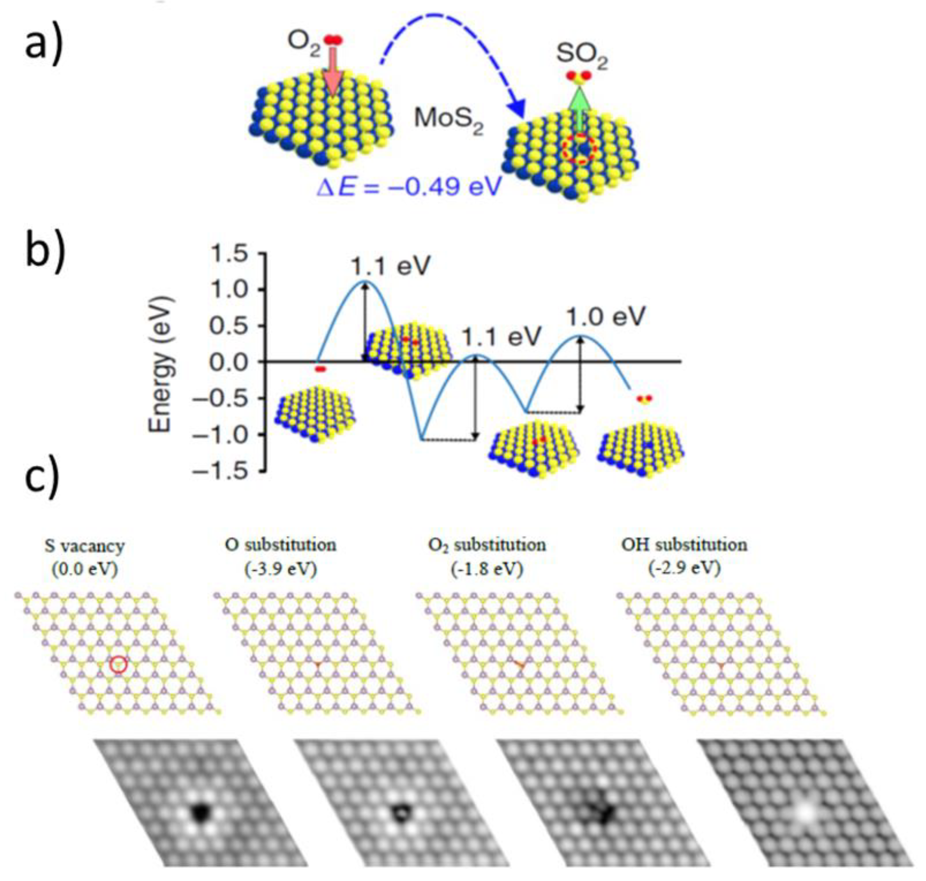
The findings of Santosh et al. contribute well to explaining the exceptional stability of the pristine, non-defective MoS2 flakes against oxidation. Direct oxygen binding to Mo atoms is impossible on pristine basal planes due to steric reasons. However, other oxidative processes have been found to exist on pristine and non-defective MoS2 basal planes. For example, oxygen-induced single sulfur vacancy (SSV) creation was observed by Peto et al. [60], who—due to advances in local scale imaging—supplemented their DFT calculations with experimental observations. To start with, they showed an SSV creation by oxygen with enthalpy of such a reaction or formation energy of −0.49 eV (see Figure 14a):
O 2   SSV + SO 2
Despite being thermodynamically favorable, the reaction had to proceed through several stages, but their details were not provided (see Figure 14b). Furthermore, Peto et al. did not find that a created sulfur vacancy would eventually lead to any Mo oxides. However, they suggested that SSV will react easily with atomic and molecular oxygen to form substitutional oxygen species, i.e., oxygen atoms filling the vacancy and becoming directly connected to Mo atoms. The observed process was explained via preserving the original crystal lattice, with the Mo sites in a trigonal prismatic configuration, but coordinated by five S atoms and a substitutional O atom. The mechanistic details for such substitutions were not provided, but the corresponding formation energy for the oxidized vacancy of ~4 eV (see Figure 14c) suggested single oxygen atom substitutions, in accordance with their atomic-resolution STM data. As related, Grønborg et al. studied oxygen exchange on epitaxially grown MoS2 single-layer nanosheets [80]. Using a combination of STM and ambient pressure XPS performed in a function of temperature and pressure, they found that such an O exchange was an activated process with an energy barrier of ~0.79 ± 0.20 eV. Atom-resolved STM images reveal O as single defects located on isolated positions on the upper S lattice of MoS2, almost in accordance with the results of single sulfur vacancies and their oxidations obtained by Peto et al. [60], who got an apparent activation energy barrier of 1–1.1 eV.
It is noteworthy that the speed for the SSV creation onto the MoS2 basal plane under ambient conditions was found by Peto et al. [60] to be of the order of one defect per min per μm2. Using the data of Ross et al. obtained for Mo powders in the case of their prolonged oxidation at 100 °C in dry air, one obtains an oxidation speed of about 160 oxidized Mo atoms per minute per μm2 [31]. This clearly illustrates different kinds of oxidation mechanisms occurring in these different kinds of the MoS2 samples and/or further suggests that macroscopic oxidation of the MoS2 powders did not occur on the pristine, non-defected MoS2 basal planes.
Recently, a more comprehensive scenario for initial events associated with oxidative etching has been proposed by Farigliano et al. [72] for the non-defective MoS2 basal plane (see Figure 15). They used NEB calculations at 0 K temperature to find out crucial intermediates within the processes and then proceeded with ab initio molecular-dynamics simulations at higher temperatures to show decomposition pathways of the key intermediate in the processes. The key initial intermediate consisted of an O atom adsorbed on top of an S atom (Oads) with a second O atom inserted (Oin) into the S−Mo bond, i.e., the Oads−S−Oin−Mo (OSOMo) moiety. Such an intermediate was obtained via two pathways (see Figure 15b). The first pathway proceeded via a dissociative oxygen adsorption pathway on neighboring sulfur atoms with Ea of 2.16 eV and its further surface reorganization. Second pathway was a direct O2 adsorption on the same sulfur atom with Ea of 1.93 eV and its further surface reorganization. However, Ea for both pathways are at least 0.4 eV more than obtained via Santosh et al. [43] for dissociative oxygen adsorption onto pristine MoS2, which suggests that the trajectory proposed by Farigliano et al. [72] less probable or that there were some major differences in the DFT implementations between these studies. Nevertheless, the results of Farigliano et al. provide an important link between the dissociative oxygen adsorption and oxidative etching. This is because, in the subsequent steps, Farigliano et al. [72] have found out that the OSOMo intermediate decomposes either via direct SO2 desorption to generate a single sulfur vacancy, or via SO desorption, leaving substitutional oxygen on the surface.
Defective basal MoS2 planes. It has been established that single sulfur vacancies in the basal sulfur planes are the most frequent and important from the point of view of an initial oxygen attack [70,81,82]. Therefore, beyond pristine non-defected basal MoS2 surface, similar surfaces, but with SSV, have been computationally considered.
The process of dissociative oxygen adsorption occurring on the S-defective sites within an MoS2 basal planes has led the calculations of Santosh et al. to a substitutional oxygen, Os, bound to Mo atoms, as well as an adsorbed oxygen bound to a sulfur atom [43], Oads (see Figure 13b,c for more details). The corresponding reaction could be denoted:
O 2 + Mo ( S ) ( SSV ) Mo ( O ) S SO ads
Santosh et al. obtained an energy barrier of 0.8 eV for a process characterized by Equation (7), which is only half of the value obtained on pristine MoS2 basal plane. Nevertheless, a much larger value of the obtained binding energy with respect to dissociative adsorption on pristine basal planes, i.e., 1.7 eV energy gain after the reaction, provides a viable thermodynamic argument for this process. In other words, it is still not very probable, but once it occurs, the resulting energy can accelerate the process when other SSVs are in the close neighborhood. Consequently, this could also explain why S vacancies at the grain boundaries in polycrystalline samples accelerate the oxidation process in CVD samples as compared to the monocrystalline non-defected MoS2 basal planes. However, dissociative oxygen adsorption does not produce MoO3, which has been detected on the mentioned earlier CVD-grown MoS2 samples. It can be seen, however, as a first step towards the creation of the MoO3 species, as is explained later on in the review.
Another take on the dissociative oxygen adsorption occurring on the S-defective sites within an MoS2 basal planes has been published by Nan et al. [79], who, similarly to Santosh et al. [43], used a nudged elastic band (NEB) method implemented within their DFT calculations. They obtained an oxygen molecule substituting two neighboring Mo atoms (see Figure 16). The apparent activation energy barrier of the entire process (1.05 eV) was associated with a binding energy gain of 2.25 eV, which made the process thermodynamically favorable, similar to the results of Santosh et al., who had, however, obtained a much lower energy barrier, and thus provided a more likely reaction pathway.
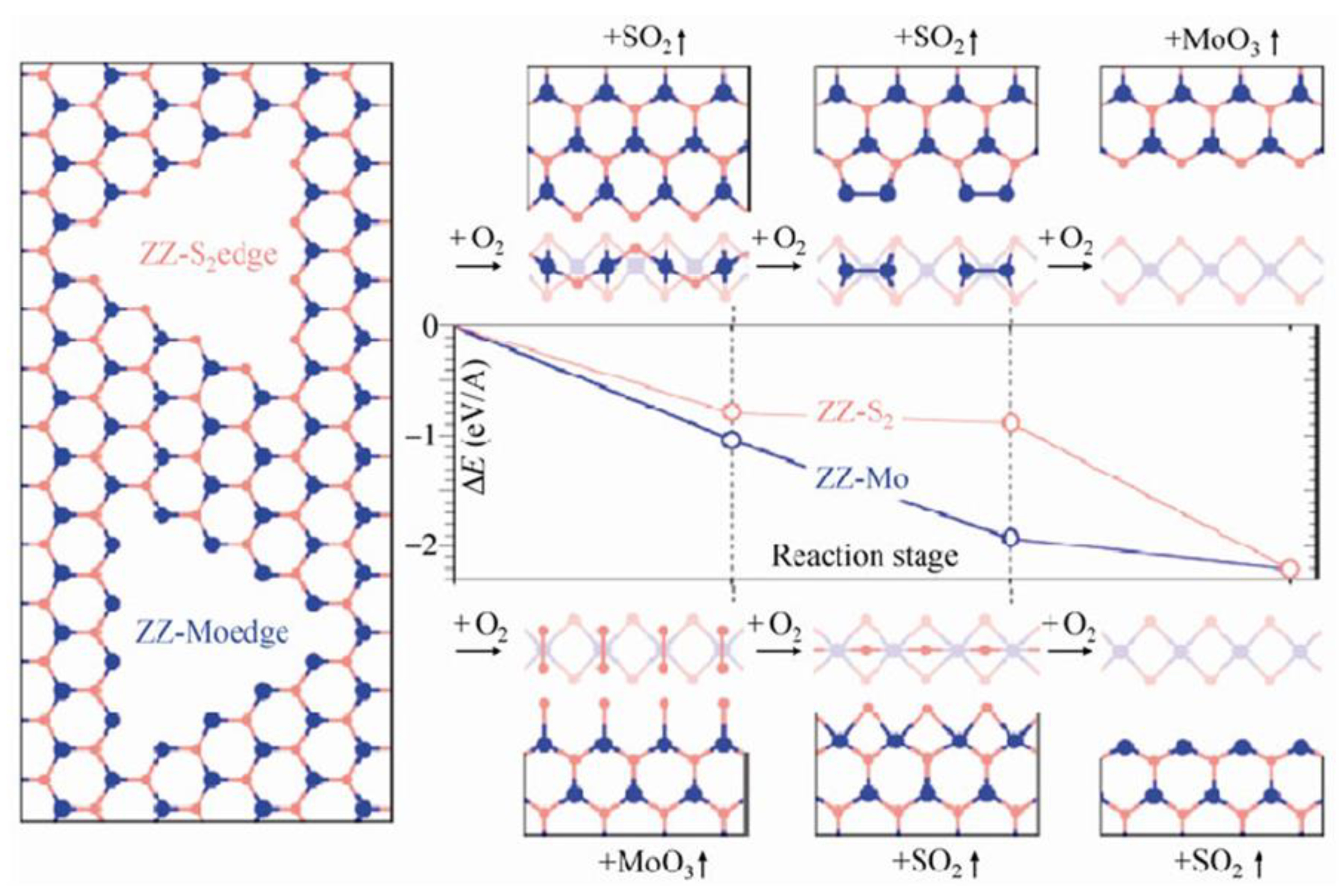
A direct take on oxidative etching, without involving any dissociative oxygen adsorption, but with an initially defective MoS2 crystalline edge has been proposed by Zhou et al. [33]. Their first-principles computations identified the most exothermic reaction path, which proceeded along three independent zigzag directions created by honeycomb assembly of the Mo atoms. Similar conclusions and explanations were provided by experimental works of Lv et al. [70] from high-resolution S-TEM studies (see Figure 7).
Zhou et al. presented their mechanistic considerations, along with respective binding energies, Eb, obtained in each step of the processes they described (see Figure 17). Unfortunately, the values of activation energies for each stage were not provided. Thus, a direct kinetic comparison with the aforementioned models is not possible. The studies of Zhou et al. considered an already formed pit within the basal MoS2 plane, which by itself to form is a very costly process with an activation energy per edge atom between 1.59 and 2.19 eV, as provided by Santosh et al. [43] and Farigliano et al. [72], respectively. From such a starting point, the results of Zhou et al. provided a tempting explanation of the oxidative etching. The proposed mechanism fulfilled a generally accepted reaction presented in Equation (3), but involved three stages and significant surface reconstructions after each step. First, an oxygen molecule had to react with unsaturated Mo atoms. A molecule of the MoO3 was produced, as well as Mo vacancies with exposed S atoms. Second, the unsaturated and S terminated layer yielded two SO2 molecules via two subsequent reaction steps with O2 molecules. The reaction was expected to propagate along the C3 symmetry within the basal planes of the MoS2 crystals to create macroscopically observed triangular etch pits. Since there are two possible starting configurations, i.e., either ZZ-S or ZZ-Mo edges, the authors investigated the two of them and concluded that the reaction progressing along the ZZ-Mo edges is preferential in terms of Eb drops along the process.
The results of Zhou et al. can be also applied to defective MoS2 basal planes. In addition, other kinds of interactions of oxygen with crystalline edges and grain boundaries in polycrystalline MoS2 samples have been studied. Longo et al. [57] have studied oxygen adsorption onto Mo atoms within the MoS2 nanoribbons. They considered two different scenarios: armchair and zigzag nanoribbons, with adsorption in the metal sites only, since metal–oxygen bonds are much more thermodynamically favorable than sulfur–oxygen bonds, due to the larger difference in respective electronegativity. They claimed that such oxygen chemisorption was a barrierless process, where Mo dangling bonds were a strong thermodynamic and kinetic driving force. Their results did not refer directly to dissociative oxygen splitting or to oxidative etching.
Martincová et al. have studied oxidative etching directly on “simplified edges” of the MoS2 crystals [46,58]. The edge was modelled as a nanostripe cut out from a monolayer of 2H-MoS2. It was constructed from S-Mo-S supercells that were only four atoms wide in X and Y directions and long yielding a total of 16 × 3 = 48 atoms. The initial state was taken to be the one in which the O2 molecule is in the edge vicinity. The final state of the dissociative splitting was taken to be the one with two separate oxygen atoms adsorbed on adjacent sulfur atoms at an edge. Several dissociation pathways were considered, differing mainly in an initial position of the O2 molecule. The most favorable pathway yielded a very low O2 dissociation barrier of 0.31 eV (see Figure 18). However, several other pathways yielded similar barriers, all not exceeding 0.5 eV. Such results still do not suggest immediate dissociative O2 splitting onto MoS2 edges; however, they clearly produce much lower Ea values than 0.8 eV obtained by Santosh et al. for the same process on a defective MoS2 basal plane.
Very small values of the activation energies obtained by Martincová et al. in the case of edge initiated oxidative etching are expected to produce single reaction events occurring at room temperature over a time scale of μs according to Equation (8) obtained from the transition state theory:
t   [ s ]   ~   10 12   exp ( 11604 × E a   [ eV ]   /   T [ K ] )
Using Equation (8), one can determine that energy barriers of ca. 1 eV—obtained experimentally in the case of oxidative etching—would correspond to a time scale of a month at room temperature, which is according to what has been obtained for oxidative etching both from simulations and experiments. However, the link between dissociative oxygen adsorption and oxidative etching is not clear. Martincová et al. used a thermodynamic argument that an energy gain of about 2.5 eV in the rate limiting dissociative adsorption was large enough to allow all subsequent reactions to happen. They suggested that after an oxygen molecule dissociated, any adsorbed O atoms (Oads) reorganized and started to bind to previously unoxidized sulfur atoms. However, an energy barrier for this process was not provided. Then, in a subsequent step the double substituted sulfur would leave in a form of the SO2 molecule with a calculated energy barrier of 0.35 eV for leaving. Exposed Mo atoms would then interact barrierlessly with another oxygen molecule, as in the aforementioned works of Longo et al. [57]. Finally, further oxidation (with energy barriers not provided) would occur through formation of one-dimensional chain-like structures resembling the in-plane (longer) and out-of-plane (shorter) connections between Mo and O atoms in the β–MoO3. Such Mo-O structures were bound to respective MoS2 edges and facilitated the spread of oxidation onto the surface. Interestingly, the stress associated with the misfit between the MoS2 and MoO3 lattices made the resulting MoO3 structures amorphous [83]. Despite some missing energy barriers, the results of Martincová et al. provide a very good qualitative explanation for the fast oxidative etching rates on edges and defects as compared with basal MoO3 planes. They also explain formation of the MoO3 and MoOx species on very defective MoS2 flakes already at room temperature.
Overall, there are several successes of the presented above theoretical approaches. First, chemical inertness for reactions with oxygen of the non-defective MoS2 basal planes at room temperature has been explained in terms of high activation barrier for a dissociative oxygen adsorption onto such planes with an energy barrier of at least 1.6 eV. Second, according to experimental results, oxygen-induced single sulfur vacancy (SSV) creation and its later oxidation have been shown to slowly introduce defects within the MoS2 flakes, even at room temperatures, thus making them prone to oxidation via decreasing the energy barrier down to 0.8 eV on SSV and to 0.3 eV on edges/grain boundaries [60]. These results agree qualitatively with experimentally obtained activation energies for oxidative etching which range from 0.6 to ca. 1 eV for pristine MoS2 samples, where single oxidation events according to the transition state theory would occur on the time scales of months (see Equation (8)). Third, other oxygen related processes of similar apparent activation energies to oxidation/etching events have been simulated as well. For example, oxygen diffusion into the freshly exposed etch pits [72] or in between the MoS2 layers [9,10], which swell the MoS2 flakes. However, the experimentally obtained presence of the MoO3 species on defective MoS2 samples at room temperature was only qualitatively explained [46,58]. Same with faster oxidation/etching on the edges of the MoS2 crystals as compared to the basal planes at elevated heating temperatures [35,41,42,46]. Consequently, there are several outstanding questions arising from the theoretical approaches. First, a unified picture for oxidative etching is still elusive. Some models and theoretical simulations refer to dissociative oxygen adsorption on sulfur atoms as an initial limiting step, i.e., studies by Martincová et al. [46], as well as by Farigliano et al. [72], while others refer to direct reaction of oxygen with Mo atoms exposed due to omnipresent single sulfur vacancies, i.e., Lv et al. [70], Santosh et al. [43] and Zhou et al. [33]. In addition, the role of initially physically adsorbed oxygen in thermal etching is still not clear [41], since its diffusion to the reaction centers can compete with direct reactions with atmospheric oxygen. Next, the role of other etching mechanisms such as pure thermal etching without a need for oxygen molecules also needs further theoretical insight [45]. Finally, due to subtle differences within the DFT approaches and varying size and geometries (mostly on edges) of the chosen simulation supercells, there is still room for improvements and new results to explain oxidative etching, as well as the tentative existence of the MoOx species onto various places within the MoS2 crystals.

4. Mo Oxides and Their Derivatives in MoS2 Oxidation

Thermodynamic calculations agree with experimental data that α-MoO3 is the most expected MoS2 oxidation product in dry and humid air [36,50,51]. However, recent findings confirm that that thicknesses of the MoS2 and MoO3 single layers are similar [42], which brings up a related question: whether MoO3 are present on oxidatively etched MoS2 flakes at all, and if yes, then in which form? Figure 2 has summarized current and related phenomenological findings. More details are presented herein.
Figure 19 provides some structural information about Mo oxide species. First of all, an orthorhombic α-MoO3 is a 2D material, similarly as the 2H-MoS2, with layered structure held together by van der Waals forces. Each α-MoO3 layer comprises well-connected MoO6 octahedra stacked at two height levels. Within each level the MoO6 octahedra are connected by shared lateral ends only. Connections between upper and ground levels octahedra are realized by sharing a common edge (see Figure 19b) [51]. Second, Mo hydrates can be seen as derivative species, where water molecules (either in the form of coordinated water molecules or crystal water molecules) have penetrated and destroyed the α-MoO3 structure (see Figure 19d). Finally, in the Mo hydroxyoxides (not shown) coordinated waters from hydrates are substituted by –OH groups.
During oxidative etching (300 to 400 °C), which occurs at isolated spots, the resulting MoO3 species are expected to leave into the gas phase. Therefore, their surface presence is not expected. Nevertheless, Gronberg et al. [80] claimed to detect MoO3 species on epitaxially created MoS2 nanosheets already at about 280 °C. Furthermore, Park et al. demonstrated that CVD-grown MoS2 monolayers contain intrinsic MoOx and are quickly oxidized at 100 °C (3 vol% O2/He) [47]. In addition, nanomechanical detection of the tentative MoOx species or highly defected MoS2 layers was developed on single, non-defective MoS2 flakes thermally etched in air at 320–410 °C [42,85]. Finally, very recently, direct chemical proofs for microscale presence of the MoO3 nanoparticles and small patched on single MoS2 flakes in air during thermal oxidative etching, particularly between 350 and 390 °C, have been provided in separate two reports (one on geological another on CVD-grown MoS2) via a combination of local AFM, XPS, TEM, SEM and XAS studies [47,48]. In particular, Park et al. [48] showed that such small MoO3 nanoparticles promote oxidation on CVD-grown MoS2.
Several reports focused on creation of the MoOx layers under dry and humid conditions as well as in water [41,42,62,67]. However, so far, several contradicting hypotheses for the presence of the Mo oxygenated species in water have been suggested, hinging upon the possibility of spotting various kinds of molybdate ions, molybdic acids, Mo hydrates and Mo hydroxy-oxides [53]. In the case of more vigorous oxidation above 410 °C, some the of the produced MoO3 has been found to stay on the surface in the form of patches and layers (see Figure 12), which are very thin and defective due to geometrical mismatch with an underlying MoS2 layer [33,34,42]. Finally, the MoO3 species have been shown by other, unrelated reports to have a strong tendency to sublimate above 470 °C in air [36] or even at lower temperatures in humid air [42], which should also limit their surface presence. Their thin and likely unstructured counterparts might do so at a lower temperature, and it has been suggested to happen already above 410 °C, in air, for very thin MoOx patches [42].
In order to appreciate the aforementioned studies, one must understand that detection of small amounts of the MoO3/MoOx species together with their various morphologies is difficult. The α-MoO3 crystals are optically transparent and of similar thickness to single MoS2 layers [42,50]. Their presence is easily observed by XPS results [36,42,45,83], but typical XPS studies are not local and report on the MoO3 adsorbed on the substrate together with many single MoS2 flakes. Furthermore, a vast majority of XPS studies collected on ensembles of single thermally oxidized MoS2 flakes at a temperature above 370 °C have exclusively detected the MoO3 oxides without any traces of other Mo oxides [42,45,83]. This observation, however, is somewhat expected when Mo oxides are present, since a majority of the XPS studies detect only Mo6+ and Mo4+ based on Mo 3d electrons. The Mo6+ states originate exclusively from the most expected MoO3 species. However, the Mo4+ states can originate from both MoS2 and tentative MoO2. Thus, in order to detect MoO2 one would need to detect less S2− and more Mo4+ as compared to pristine samples. Nevertheless, less sulfur can also be observed when MoO3 starts to cover basal MoS2 planes limiting electrons originating from sulfur, which reach the XPS detectors. Therefore, MoO2 when present at small quantities in relation to large quantities of simultaneously present MoO3 will likely be masked in typical XPS studies. Similarly, Micro-Raman studies also lack sensitivity in MoO3 detection [40]. According to our knowledge, micro-Raman measurements have not yet delivered any direct proof for the Mo oxide present on single MoS2 flakes/crystals as a result of their oxidative etching and oxidations. At best, some authors were able to report Raman shifts only for fully established MoO3 crystals [35], while the majority of others could not see any Raman signal from the MoO3 (expected at 158, 285, 666, 820 and 995 cm−1 in the case of non-resonant Raman excitation) [40,42,48,86]. In addition, both thick MoS2 and MoO3 are non-magnetic [34]. However, very thin MoS2 layers show in-plane magnetism altering in signs between subsequent layers, which produce magnetic contrast between MoO3 onto thin MoS2, depending on the number of the MoS2 layers [34,87,88].
Taking it all together, while MoO3 species are locally detected via a combination of several techniques, such as KPFM, nanomechanical studies, XAS, XAS and STEM. However, proper local Mo speciation and surface distribution of other eventual Mo oxides and their derivatives such as hydrates and hydroxy-oxides have not been performed yet.
The MoS2/MoOx interface. Due to various possible MoOx forms arising on the MoS2 surface at various oxidative conditions, the issue of creating and controlling their geometrical dimensions, as well as their chemical composition, becomes intriguing and timely. Pristine 2H MoS2 exhibits n-type conduction independent of the metal contact due to a strong Fermi level pinning near the conduction band, promoted especially by sulfur vacancies [89]. However, chemical modification of the n-type MoS2 towards p-type MoS2 has been reported by using, for example, AuCl3 solution [90]. Pristine α-MoO3 also exhibits n-type conduction, but with a wide bandgap (>2.7 eV). Nonstoichiometric-reduced MoOx (2 < x < 3) oxides are more conductive, and eventually MoO2 is semi-metallic [50]. In addition, thin Mo oxide layers are expected to differ in their electrical properties, depending on their thickness and degree of crystallinity [50,91], and small amounts of α-MoO3 can induce p-type doping [92,93]. Therefore, thin Mo oxide patches, if present on the MoS2 crystals, are expected to modify their electrical properties and even help in creation electronic p-n junctions in situ, which are nanoscopic in size. A priori eventual boron diffusion from within the typical p-doped boron Si wafers to the Si/SiO2/MoS2 interface could interfere therein. However, any meaningful boron diffusion to that interface is expected only above 850 °C [94], which is above predicted sublimation of even thick MoO3 layers. Finally, a promising new approach to locally create the MoS2/MoOx interface would be to use nanolithographic techniques, such as the thermochemical nanolithography (TCNL) [95], local oxidation nanolithography [96] or other lithographic approaches [97,98]. Such methods are expected to produce locally MoO3 nanoparticles/thin patches and to chemically reduce any earlier-created MoO3 layers. They also have the potential to locally produce active MoOx-MoS2 junctions of various geometries and chemistries.

5. Summary

This review focused on the thermally induced interactions of 2H MoS2 crystals with oxygen molecules in ambient conditions. Experimental (Section 2) and computational (Section 3.2) studies investigating oxygen-induced changes to the MoS2 structure at various thermal regimes were presented and discussed. Next, the properties and fate of the Mo oxides and other related Mo species arising from thermal oxidation were reviewed (Section 4), along with the properties and tentative ways of creating and modifying the MoS2/MoOx interface.

Funding

This work was supported by the National Science Center, Poland, grant no. 2017/27/B/ST4/00697 (PI: RSz), as well as by the UW Statuary Funds.

Institutional Review Board Statement

Not applicable.

Informed Consent Statement

Not applicable.

Data Availability Statement

Data sharing not applicable.

Acknowledgments

Figure 1 was prepared using Vesta software (Amakubo, Tsukuba-shi, Japan). Copyright (©) 2006-2021, Koichi Momma and Fujio Izumi, https://jp-minerals.org/vesta/en/ (accessed on 24 September 2021).

Conflicts of Interest

The author declares no conflict of interest.

References

  1. Kadantsev, E.S.; Hawrylak, P. Electronic structure of a single MoS2 monolayer. Solid State Commun. 2012, 152, 909–913. [Google Scholar] [CrossRef]
  2. Tagawa, M.; Muromoto, M.; Hachiue, S.; Yokota, K.; Ohmae, N.; Matsumoto, K.; Suzuki, M. Hyperthermal atomic oxygen interaction with MoS2 lubricants and relevance to space environmental effects in low earth orbit—Effects on friction coefficient and wear-life. Tribol. Lett. 2005, 18, 437–443. [Google Scholar] [CrossRef]
  3. Wang, Q.H.; Kalantar-Zadeh, K.; Kis, A.; Coleman, J.; Strano, M.S. Electronics and optoelectronics of two-dimensional transition metal dichalcogenides. Nat. Nanotechnol. 2012, 7, 699–712. [Google Scholar] [CrossRef] [PubMed]
  4. Fleischauer, P.D.; Lince, J. A comparison of oxidation and oxygen substitution in MoS2 solid film lubricants. Tribol. Int. 1999, 32, 627–636. [Google Scholar] [CrossRef]
  5. Lince, J.; Frantz, P.P. Anisotropic oxidation of MoS2 crystallites studied by angle-resolved X-ray photoelectron spectroscopy. Tribol. Lett. 2001, 9, 211–218. [Google Scholar] [CrossRef]
  6. Imanishi, N.; Kanamura, K.; Takehara, Z. Synthesis of MoS2 thin film by chemical vapor deposition method and discharge characteristics as a cathode of the lithium secondary battery. J. Electrochem. Soc. 1992, 139, 2082–2087. [Google Scholar] [CrossRef]
  7. Jäger-Waldau, A.; Lux-Steiner, M.; Bucher, E. MoS2, MoSe2, WS2 and WSe2 thin films for photovoltaics. Solid State Phenom. 1994, 37–38, 479–484. [Google Scholar] [CrossRef]
  8. Jaramillo, T.F.; Jorgensen, K.P.; Bonde, J.; Nilsen, J.H.; Horch, S.; Chorkendorff, I. Identification of active edge sites for electrochemical H2 evolution from MoS2 nanocatalysts. Science 2007, 317, 100–102. [Google Scholar] [CrossRef] [Green Version]
  9. Guo, J.; Li, F.; Sun, Y.; Zhang, X.; Tang, L. Oxygen-incorporated MoS2 ultrathin nanosheets grown on graphene for efficient electrochemical hydrogen evolution. J. Power Sources 2015, 291, 195–200. [Google Scholar] [CrossRef]
  10. Xie, J.; Zhang, J.; Li, S.; Grote, F.; Zhang, X.; Wang, R.; Lei, Y.; Pan, B.; Xie, Y. Controllable Disordered engineering in oxygen-incorporated MoS2 ultrathin nanosheets for efficient hydrogen evolution. J. Am. Chem. Soc. 2013, 135, 17881–17888. [Google Scholar] [CrossRef]
  11. Radisavljevic, B.; Radenovic, A.; Brivio, J.; Giacometti, V.; Kis, A. Single-layer MoS2 transistors. Nat. Nanotechnol. 2011, 6, 147–150. [Google Scholar] [CrossRef] [PubMed]
  12. Radisavljevic, B.; Whitwick, M.B.; Kis, A. Integrated circuits and logic operations based on single-layer MoS2. ACS Nano 2011, 5, 9934–9938. [Google Scholar] [CrossRef] [PubMed]
  13. Wachter, S.; Polyushkin, D.; Bethge, O.; Mueller, T. A microprocessor based on a two-dimensional semiconductor. Nat. Commun. 2017, 8, 14948. [Google Scholar] [CrossRef] [Green Version]
  14. Lee, J.; Pak, S.; Giraud, P.; Lee, Y.-W.; Cho, Y.; Hong, J.; Jang, A.R.; Chung, H.-S.; Hong, W.-K.; Jeong, H.Y.; et al. Thermodynamically stable synthesis of large-scale and highly crystalline transition metal dichalcogenide monolayers and their unipolar n-n heterojunction devices. Adv. Mater. 2017, 29, 1702206. [Google Scholar] [CrossRef] [PubMed]
  15. Tan, C.; Liu, Z.; Huang, W.; Zhang, H. Non-volatile resistive memory devices based on solution-processed ultrathin two-dimensional nanomaterials. Chem. Soc. Rev. 2015, 44, 2615–2628. [Google Scholar] [CrossRef] [Green Version]
  16. Marega, G.M.; Zhao, Y.; Avsar, A.; Wang, Z.; Tripathi, M.; Radenovic, A.; Kis, A. Logic-in-memory based on an atomically thin semiconductor. Nature 2020, 587, 72–77. [Google Scholar] [CrossRef]
  17. Kang, K.; Xie, S.; Huang, L.; Han, Y.; Huang, P.Y.; Mak, K.F.; Kim, C.J.; Muller, D.; Park, J. High-mobility three-atom-thick semiconducting films with wafer-scale homogeneity. Nature 2015, 520, 656–660. [Google Scholar] [CrossRef] [PubMed]
  18. Chang, M.C.; Ho, P.H.; Tseng, M.F.; Lin, F.Y.; Hou, C.H.; Lin, I.K.; Wang, H.; Huang, P.P.; Chiang, C.H.; Yang, Y.C.; et al. Fast growth of large-grain and continuous MoS2 films through a self-capping vapor-liquid-solid method. Nat. Commun. 2020, 11, 3682. [Google Scholar] [CrossRef]
  19. Shim, J.; Bae, S.-H.; Kong, W.; Lee, D.; Qiao, K.; Nezich, D.; Park, Y.J.; Zhao, R.; Sundaram, S.; Li, X.; et al. Controlled crack propagation for atomic precision handling of wafer-scale two-dimensional materials. Science 2018, 362, 665–670. [Google Scholar] [CrossRef] [Green Version]
  20. Kang, K.; Lee, K.H.; Han, Y.; Gao, H.; Xie, S.; Muller, D.A.; Park, J. Layer-by-layer assembly of two-dimensional materials into wafer-scale heterostructures. Nature 2017, 550, 229–233. [Google Scholar] [CrossRef]
  21. Amani, M.; Lien, D.H.; Kiriya, D.; Xiao, J.; Azcatl, A.; Noh, J.; Madhvapathy, S.R.; Addou, R.; Kc, S.; Dubey, M.; et al. Near-unity photoluminescence quantum yield in MoS2. Science 2015, 350, 1065–1068. [Google Scholar] [CrossRef] [PubMed] [Green Version]
  22. Gonzalez Marin, J.F.; Unuchek, D.; Watanabe, K.; Taniguchi, T.; Kis, A. MoS2 photodetectors integrated with photonic circuits. NPJ 2D Mater. Appl. 2019, 3, 14. [Google Scholar] [CrossRef] [Green Version]
  23. Gong, C.; Zhang, Y.; Chen, W.; Chu, J.; Lei, T.; Pu, J.; Dai, L.; Wu, C.; Cheng, Y.; Zhai, T.; et al. Electronic and optoelectronic applications based on 2D novel anisotropic transition metal dichalcogenides. Adv. Sci. 2017, 4, 1700231. [Google Scholar] [CrossRef] [PubMed]
  24. Unuchek, D.; Ciarrocchi, A.; Avsar, A.; Watanabe, K.; Taniguchi, T.; Kis, A. Room-temperature electrical control of exciton flux in a van der Waals heterostructure. Nature 2018, 560, 340–344. [Google Scholar] [CrossRef] [PubMed]
  25. Mak, K.F.; He, K.; Shan, J.; Heinz, T.F. Control of valley polarization in monolayer MoS2 by optical helicity. Nat. Nanotechnol. 2012, 7, 494–498. [Google Scholar] [CrossRef]
  26. Yalon, E.; McClellan, C.J.; Smithe, K.K.H.; Muñoz Rojo, M.; Xu, R.L.; Suryavanshi, S.V.; Gabourie, A.; Neumann, C.M.; Xiong, F.; Farimani, A.B.; et al. Energy dissipation in monolayer MoS2 electronics. Nano Lett. 2017, 17, 3429–3433. [Google Scholar] [CrossRef] [Green Version]
  27. Qiu, H.; Pan, L.; Yao, Z.; Li, J.; Shi, Y.; Wang, X.; Park, W.; Park, J.; Jang, J.; Lee, H.; et al. Electrical characterization of back-gated bi-layer MoS2 field-effect transistors and the effect of ambient on their performances. Appl. Phys. Lett. 2012, 100, 123104. [Google Scholar] [CrossRef]
  28. Park, W.; Park, J.; Jang, J.; Lee, H.; Jeong, H.; Cho, K.; Hong, S.; Lee, T. Oxygen environmental and passivation effects on molybdenum disulfide field effect transistors. Nanotechnology 2013, 24, 095202. [Google Scholar] [CrossRef]
  29. Li, T.-D.; Gao, J.; Szoszkiewicz, R.; Landman, U.; Riedo, E. Structured and viscous water in subnanometer gaps. Phys. Rev. B 2007, 75, 115415. [Google Scholar] [CrossRef] [Green Version]
  30. Szoszkiewicz, R.; Riedo, E. Nucleation time of nanoscale water bridges. Phys. Rev. Lett. 2005, 95, 135502. [Google Scholar] [CrossRef] [Green Version]
  31. Ross, S. Surface oxidation of molybdenum disulfide. J. Phys. Chem. 1955, 59, 889–892. [Google Scholar] [CrossRef]
  32. Ebrahimi Kahrizsangi, R.; Abbasi, M.H.; Saidi, A. Model-Fitting Approach to Kinetic Analysis of Non-Isothermal Oxidation of Molybdenite. Iran. J. Chem. Chem. Eng. 2007, 26, 119–123. [Google Scholar]
  33. Zhou, H.; Yu, F.; Liu, Y.; Zou, X.; Cong, C.; Qiu, C.; Yu, T.; Yan, Z.; Shen, X.; Sun, L.; et al. Thickness-dependent patterning of MoS2 sheets with well-oriented triangular pits by heating in air. Nano Res. 2013, 6, 703–711. [Google Scholar] [CrossRef]
  34. Wu, J.; Li, H.; Yin, Z.; Li, H.; Liu, J.; Cao, X.; Zhang, Q.; Zhang, H. Layer thinning and etching of mechanically exfoliated MoS2 nanosheets by thermal annealing in air. Small 2013, 9, 3314–3319. [Google Scholar] [CrossRef]
  35. Yamamoto, M.; Einstein, T.L.; Fuhrer, M.S.; Cullen, W.G. Anisotropic etching of atomically thin MoS2. J. Phys. Chem. C 2013, 117, 25643–25649. [Google Scholar] [CrossRef]
  36. Walter, T.N.; Kwok, F.; Simchi, H.; Aldosari, H.M.; Mohney, S.E. Oxidation and oxidative vapor-phase etching of few-layer MoS2. J. Vac. Sci. Technol. B 2017, 35, 21203. [Google Scholar] [CrossRef]
  37. Rao, R.; E Islam, A.; Campbell, P.M.; Vogel, E.M.; Maruyama, B. In situ thermal oxidation kinetics in few layer MoS2. 2D Mater. 2017, 4, 025058. [Google Scholar] [CrossRef]
  38. Tang, J.; Wei, Z.; Wang, Q.; Wang, Y.; Han, B.; Li, X.; Huang, B.; Liao, M.; Liu, J.; Li, N.; et al. In situ oxygen doping of monolayer MoS2 for novel electronics. Small 2020, 16, 2–9. [Google Scholar] [CrossRef]
  39. Wang, G.; Pandey, R.; Karna, S.P. Physics and chemistry of oxidation of two-dimensional nanomaterials by molecular oxygen. Wiley Interdiscip. Rev. Comput. Mol. Sci. 2017, 7, e1280. [Google Scholar] [CrossRef]
  40. Ukegbu, U.; Szoszkiewicz, R. Microscopic kinetics of heat-induced oxidative etching of thick MoS2 crystals. J. Phys. Chem. C 2019, 123, 22123–22129. [Google Scholar] [CrossRef]
  41. Spychalski, W.L.; Pisarek, M.; Szoszkiewicz, R. Microscale Insight into Oxidation of Single MoS2 Crystals in Air. J. Phys. Chem. C 2017, 121, 26027–26033. [Google Scholar] [CrossRef]
  42. Szoszkiewicz, R.; Rogala, M.; Dabrowski, P. Surface-bound and volatile mo oxides produced during oxidation of single MoS2 crystals in air and high relative humidity. Materials 2020, 13, 3067. [Google Scholar] [CrossRef] [PubMed]
  43. KC, S.; Longo, R.C.; Wallace, R.M.; Cho, K. Surface oxidation energetics and kinetics on MoS2 monolayer. J. Appl. Phys. 2015, 117, 135301. [Google Scholar] [CrossRef]
  44. Krawczyk, M.; Pisarek, M.; Szoszkiewicz, R.; Jablonski, A. Surface characterization of MoS2 atomic layers mechanically exfoliated on a Si substrate. Materials 2020, 13, 3595. [Google Scholar] [CrossRef] [PubMed]
  45. Jia, F.; Liu, C.; Yang, B.; Song, S. Microscale control of edge defect and oxidation on molybdenum disulfide through thermal treatment in air and nitrogen atmospheres. Appl. Surf. Sci. 2018, 462, 471–479. [Google Scholar] [CrossRef]
  46. Martincova, J.; Otyepka, M.; Lazar, P. Oxidation of metallic two-dimensional transition metal dichalcogenides: 1T-MoS2 and 1T-TaS2. 2D Mater. 2020, 7, 045005. [Google Scholar] [CrossRef]
  47. Park, S.; Garcia-Esparza, A.T.; Abroshan, H.; Abraham, B.; Vinson, J.; Gallo, A.; Nordlund, D.; Park, J.; Kim, T.R.; Vallez, L.; et al. Operando study of thermal oxidation of monolayer MoS2. Adv. Sci. 2021, 8, 2002768. [Google Scholar] [CrossRef]
  48. Rogala, M.; Sokołowski, S.; Ukegbu, U.; Mierzwa, A.; Szoszkiewicz, R. Direct identification of surface bound MoO3 on single MoS2 flakes heated in dry and humid air. Adv. Mater. Interfaces 2021, 2100328. [Google Scholar] [CrossRef]
  49. Ballou, E.V.; Ross, S. The adsorption of benzene and water vapors by molybdenum disulfide. J. Phys. Chem. 1953, 57, 653–657. [Google Scholar] [CrossRef]
  50. De Castro, I.A.; Datta, R.S.; Ou, J.Z.; Castellanos-Gomez, A.; Sriram, S.; Daeneke, T.; Kalan-tar-Zadeh, K. Molybdenum oxides—From fundamentals to functionality. Adv. Mater. 2017, 29, 1–31. [Google Scholar] [CrossRef]
  51. Åsbrink, S.; Kihlborg, L.; Malinowski, M. High-pressure single-crystal X-ray diffraction studies of MoO3. I. Lattice parameters up to 7·4 GPa. J. Appl. Crystallogr. 1988, 21, 960–962. [Google Scholar] [CrossRef]
  52. Bihn, J.H.; Park, J.; Kang, Y.C. Synthesis and characterization of mo films deposited by RF sputtering at various oxygen ratios. J. Korean Phys. Soc. 2011, 58, 509–514. [Google Scholar] [CrossRef]
  53. Smolik, G.R.; Petti, D.A.; Schuetz, S.T. Oxidation and volatilization of TZM alloy in air. J. Nucl. Mater. 2000, 283-287, 1458–1462. [Google Scholar] [CrossRef] [Green Version]
  54. Mirabelli, G.; McGeough, C.; Schmidt, M.; McCarthy, E.K.; Monaghan, S.; Povey, I.M.; McCarthy, M.M.; Gity, F.; Nagle, R.E.; Hughes, G.; et al. Air sensitivity of MoS2, MoSe2, MoTe2, HfS2, and HfSe2. J. Appl. Phys. 2016, 120, 125102. [Google Scholar] [CrossRef] [Green Version]
  55. Jo, S.S.; Singh, A.; Yang, L.; Tiwari, S.C.; Hong, S.; Krishnamoorthy, A.; Sales, M.G.; Oliver, S.M.; Fox, J.; Cavalero, R.L.; et al. Growth kinetics and atomistic mechanisms of native oxidation of ZrSxSe2–x and MoS2 crystals. Nano Lett. 2020, 20, 8592–8599. [Google Scholar] [CrossRef] [PubMed]
  56. Gao, J.; Li, B.; Tan, J.; Chow, P.; Lu, T.-M.; Koratkar, N. Aging of transition metal dichalcogenide monolayers. ACS Nano 2016, 10, 2628–2635. [Google Scholar] [CrossRef]
  57. Longo, R.C.; Addou, R.; Santosh, K.C.; Noh, J.Y.; Smyth, C.M.; Barrera, D.; Zhang, C.; Hsu, J.W.P.; Wallace, R.M.; Cho, K. Intrinsic air stability mechanisms of two-dimensional transition metal dichalcogenide surfaces: Basal versus edge oxidation. 2D Mater. 2017, 4, 025050. [Google Scholar] [CrossRef]
  58. Martincová, J.; Otyepka, M.; Lazar, P. Is single layer MoS2 stable in the air? Chem. A Eur. J. 2017, 23, 13233–13239. [Google Scholar] [CrossRef]
  59. Sen, H.S.; Sahin, H.; Peeters, F.M.; Durgun, E. Monolayers of MoS2 as an oxidation protective nanocoating material. J. Appl. Phys. 2014, 116, 083508. [Google Scholar] [CrossRef] [Green Version]
  60. Pető, J.; Ollár, T.; Vancsó, P.; Popov, Z.I.; Magda, G.Z.; Dobrik, G.; Hwang, C.; Sorokin, P.B.; Tapasztó, L. Spontaneous doping of the basal plane of MoS2 single layers through oxygen substitution under ambient conditions. Nat. Chem. 2018, 10, 1246–1251. [Google Scholar] [CrossRef]
  61. Wang, Z.; Bussche, A.V.D.; Qiu, Y.; Valentin, T.M.; Gion, K.; Kane, A.B.; Hurt, R.H. Chemical dissolution pathways of MoS2 nanosheets in biological and environmental media. Environ. Sci. Technol. 2016, 50, 7208–7217. [Google Scholar] [CrossRef] [Green Version]
  62. Zhang, X.; Jia, F.; Yang, B.; Song, S. Oxidation of molybdenum disulfide sheet in water under in situ atomic force microscopy observation. J. Phys. Chem. C 2017, 121, 9938–9943. [Google Scholar] [CrossRef]
  63. Seguin, L.; Figlarz, M.; Cavagnat, R.; Lassègues, J.C. Infrared and Raman spectra of MoO3 molybdenum trioxides and MoO3 xH2O molybdenum trioxide hydrates. Spectrochim. Acta Part A Mol. Biomol. Spectrosc. 1995, 51, 1323–1344. [Google Scholar] [CrossRef]
  64. Cruywagen, J.J.; Heyns, J.B.B. Solubility of yellow molybdenum(VI) oxide dihydrate (Mo03.2H20) in 3,0M-sodium perchlorate at 25 °C. S. Afr. J. Chem. 1981, 34, 118–120. [Google Scholar]
  65. Oyerinde, O.F.; Weeks, C.L.; Anbar, A.D.; Spiro, T.G. Solution structure of molybdic acid from Raman spectroscopy and DFT analysis. Inorganica Chim. Acta 2008, 361, 1000–1007. [Google Scholar] [CrossRef]
  66. Piquemal, J.-Y.; Manoli, J.-M.; Beaunier, P.; Ensuque, A.; Tougne, P.; Legrand, A.-P.; Brégeault, J.-M. Using inorganic silicate precursor/molybdenum peroxo complexes/onium salt interfaces in aqueous acidic media to design mesoporous silica with high molybdenum content and high dispersion. Microporous Mesoporous Mater. 1999, 29, 291–304. [Google Scholar] [CrossRef]
  67. Espinosa, F.M.; Ryu, Y.K.; Marinov, K.; Dumcenco, D.; Kis, A.; Garcia, R. Direct fabrication of thin layer MoS2 field-effect nanoscale transistors by oxidation scanning probe lithography. Appl. Phys. Lett. 2015, 106, 103503. [Google Scholar] [CrossRef] [Green Version]
  68. Fang, L.; Shu, Y.; Wang, A.A.; Zhang, T. Green synthesis and characterization of anisotropic uniform single-crystal α-MoO3 nanostructures. J. Phys. Chem. C 2007, 111, 2401–2408. [Google Scholar] [CrossRef]
  69. Addou, R.; McDonnell, S.; Barrera, D.; Guo, Z.; Azcatl, A.; Wang, J.; Zhu, H.; Hinkle, C.L.; Quevedo-Lopez, M.; Alshareef, H.N.; et al. Impurities and electronic property variations of natural MoS2 crystal surfaces. ACS Nano 2015, 9, 9124–9133. [Google Scholar] [CrossRef]
  70. Lv, D.; Wang, H.; Zhu, D.; Lin, J.; Yin, G.; Lin, F.; Zhang, Z.; Jin, C. Atomic process of oxidative etching in monolayer molybdenum disulfide. Sci. Bull. 2017, 62, 846–851. [Google Scholar] [CrossRef] [Green Version]
  71. Chakraborty, B.; Bera, A.; Muthu, D.V.S.; Bhowmick, S.; Waghmare, U.V.; Sood, A.K. Symmetry-dependent phonon renormalization in monolayer MoS2 transistor. Phys. Rev. B 2012, 85, 2–5. [Google Scholar] [CrossRef] [Green Version]
  72. Farigliano, L.M.; Paredes-Olivera, P.A.; Patrito, E.M. Initial steps of oxidative etching of MoS2 basal plane induced by O2. J. Phys. Chem. C 2020, 124, 13177–13186. [Google Scholar] [CrossRef]
  73. Huang, Y.; Wu, J.; Xu, X.; Ho, Y.; Ni, G.; Zou, Q.; Koon, G.K.W.; Zhao, W.; Castro Neto, A.H.; Eda, G.; et al. An innovative way of etching MoS2: Characterization and mechanistic investigation. Nano Res. 2013, 6, 200–207. [Google Scholar] [CrossRef] [Green Version]
  74. Maguire, P.; Jadwiszczak, J.; O’Brien, M.; Keane, D.; Duesberg, G.S.; McEvoy, N.; Zhang, H. Defect-moderated oxidative etching of MoS2. J. Appl. Phys. 2019, 126, 164301. [Google Scholar] [CrossRef]
  75. Castellanos-Gomez, A.; Barkelid, M.; Goossens, A.M.; Calado, V.E.; van der Zant, H.S.J.; Steele, G.A. Laser-thinning of MoS2: On demand generation of a single-layer semiconductor. Nano Lett. 2012, 12, 3187–3192. [Google Scholar] [CrossRef] [PubMed] [Green Version]
  76. Sunamura, K.; Page, T.R.; Yoshida, K.; Yano, T.A.; Hayamizu, Y. Laser-induced electrochemical thinning of MoS2. J. Mater. Chem. C 2016, 4, 3268–3273. [Google Scholar] [CrossRef]
  77. Ukegbu, U.K. Microscale Etching/Oxidation of Thick MoS2 Flakes. Master’s Thesis, University of Warsaw, Warsaw, Poland, 2018. [Google Scholar]
  78. Henkelman, G.; Uberuaga, B.P.; Jónsson, H. A climbing image nudged elastic band method for finding saddle points and minimum energy paths. J. Chem. Phys. 2000, 113, 9901–9904. [Google Scholar] [CrossRef] [Green Version]
  79. Nan, H.; Wang, Z.; Wang, W.; Liang, Z.; Lu, Y.; Chen, Q.; He, D.; Tan, P.; Miao, F.; Wang, X.; et al. Strong photoluminescence enhancement of MoS2 through defect engineering and oxygen bonding. ACS Nano 2014, 8, 5738–5745. [Google Scholar] [CrossRef] [PubMed] [Green Version]
  80. Grønborg, S.S.; Thorarinsdottir, K.; Kyhl, L.; Rodríguez-Fernández, J.; E Sanders, C.; Bianchi, M.; Hofmann, P.; Miwa, J.; Ulstrup, S.; Lauritsen, J.V. Basal plane oxygen exchange of epitaxial MoS2 without edge oxidation. 2D Mater. 2019, 6, 045013. [Google Scholar] [CrossRef]
  81. Hong, J.; Hu, Z.; Probert, M.; Li, K.; Lv, D.; Yang, X.; Gu, L.; Mao, N.; Feng, Q.; Xie, L.; et al. Exploring atomic defects in molybdenum disulphide monolayers. Nat. Commun. 2015, 6, 6293. [Google Scholar] [CrossRef] [Green Version]
  82. Zhou, W.; Zou, X.; Najmaei, S.; Liu, Z.; Shi, Y.; Kong, J.; Lou, J.; Ajayan, P.M.; Yakobson, B.I.; Idrobo, J.C. Intrinsic structural defects in monolayer molybdenum disulfide. Nano Lett. 2013, 13, 2615–2622. [Google Scholar] [CrossRef]
  83. Ko, T.Y.; Jeong, A.; Kim, W.; Lee, J.; Kim, Y.; Lee, J.E.; Ryu, G.H.; Park, K.; Kim, D.; Lee, Z.; et al. On-stack two-dimensional conversion of MoS2 into MoO3. 2D Mater. 2016, 4, 014003. [Google Scholar] [CrossRef]
  84. Kuzmin, A.; Purans, J. Dehydration of the molybdenum trioxide hydrates MoO3·nH2O: In situ x-ray absorption spectroscopy study at the Mo K edge. J. Phys. Condens. Matter 2000, 12, 1959–1970. [Google Scholar] [CrossRef] [Green Version]
  85. Rice, R.H.; Mokarian-Tabari, P.; King, W.P.; Szoszkiewicz, R. Local thermomechanical analysis of a microphase-separated thin lamellar PS-b-PEO film. Langmuir 2012, 28, 13503–13511. [Google Scholar] [CrossRef] [PubMed]
  86. Zhu, H.; Qin, X.; Cheng, L.; Azcatl, A.; Kim, J.; Wallace, R.M. Remote plasma oxidation and atomic layer etching of MoS2. ACS Appl. Mater. Interfaces 2016, 8, 19119–19126. [Google Scholar] [CrossRef] [PubMed]
  87. Li, H.; Qi, X.; Wu, J.; Zeng, Z.; Wei, J.; Zhang, H. Investigation of MoS2 and graphene nanosheets by magnetic force microscopy. ACS Nano 2013, 7, 2842–2849. [Google Scholar] [CrossRef] [Green Version]
  88. Lavini, F.; Calò, A.; Gao, Y.; Albisetti, E.; Li, T.D.; Cao, T.; Li, G.; Cao, L.; Aruta, C.; Riedo, E. Friction and work function oscillatory behavior for an even and odd number of layers in polycrystalline MoS2. Nanoscale 2018, 10, 8304–8312. [Google Scholar] [CrossRef]
  89. Kim, C.; Moon, I.; Lee, D.; Choi, M.S.; Ahmed, F.; Nam, S.; Cho, Y.; Shin, H.-J.; Park, S.; Yoo, W.J. Fermi level pinning at electrical metal contacts of monolayer molybdenum dichalcogenides. ACS Nano 2017, 11, 1588–1596. [Google Scholar] [CrossRef]
  90. Choi, M.S.; Qu, D.; Lee, D.; Liu, X.; Watanabe, K.; Taniguchi, T.; Yoo, W.J. Lateral MoS2 p–n junction formed by chemical doping for use in high-performance optoelectronics. ACS Nano 2014, 8, 9332–9340. [Google Scholar] [CrossRef]
  91. Inzani, K.; Nematollahi, M.; Vullum-Bruer, F.; Grande, T.; Reenaas, T.W.; Selbach, S.M. Electronic properties of reduced molybdenum oxides. Phys. Chem. Chem. Phys. 2017, 19, 9232–9245. [Google Scholar] [CrossRef]
  92. Chuang, S.; Battaglia, C.; Azcatl, A.; McDonnell, S.; Kang, J.S.; Yin, X.; Tosun, M.; Kapadia, R.; Fang, H.; Wallace, R.M.; et al. MoS2 P-type transistors and diodes enabled by high work function MoOx contacts. Nano Lett. 2014, 14, 1337–1342. [Google Scholar] [CrossRef] [PubMed] [Green Version]
  93. Xing, K.; Xiang, Y.; Jiang, M.; Creedon, D.L.; Akhgar, G.; Yianni, S.A.; Xiao, H.; Ley, L.; Stacey, A.; McCallum, J.C.; et al. MoO3 induces p-type surface conductivity by surface transfer doping in diamond. Appl. Surf. Sci. 2020, 509, 144890. [Google Scholar] [CrossRef] [Green Version]
  94. Kessler, M.A.; Ohrdes, T.; Wolpensinger, B.; Harder, N.-P. Charge carrier lifetime degradation in Cz silicon through the formation of a boron-rich layer during BBr3diffusion processes. Semicond. Sci. Technol. 2010, 25, 055001. [Google Scholar] [CrossRef]
  95. Szoszkiewicz, R.; Okada, T.; Jones, S.C.; Li, T.D.; King, W.P.; Marder, S.R.; Riedo, E. high-speed, sub-15 nm Feature size thermochemical nanolithography. Nano Lett. 2007, 7, 1064–1069. [Google Scholar] [CrossRef] [PubMed]
  96. Martínez, R.V.; Martínez, J.; Garcia, R. Silicon nanowire circuits fabricated by AFM oxidation nanolithography. Nanotechnology 2010, 21, 245301. [Google Scholar] [CrossRef] [PubMed]
  97. Chen, L.; Wen, J.; Zhang, P.; Yu, B.; Chen, C.; Ma, T.; Lu, X.; Kim, S.H.; Qian, L. Nanomanufacturing of silicon surface with a single atomic layer precision via mechanochemical reactions. Nat. Commun. 2018, 9, 1542. [Google Scholar] [CrossRef] [Green Version]
  98. Garcia, R.; Knoll, A.; Riedo, E. Advanced scanning probe lithography. Nat. Nanotechnol. 2014, 9, 577–587. [Google Scholar] [CrossRef] [Green Version]
Figure 1. Crystalline structures of the MoS2 polymorphs. Each MoS2 layer is a stack comprising an S plane on a Mo plane on yet another S plane. (a) The most common 2H polymorphic structure with a hexagonal symmetry, two layers per repeat unit and Mo trigonal prismatic coordination; (b) 3R structure with rhombohedral symmetry, three layers per repeat unit and Mo trigonal prismatic coordination; (c) 1T structure with tetragonal symmetry, one layer per repeat unit and Mo octahedral coordination (same as in the most stable oxides and molybdates). The lattice constant “a” is 0.31 nm [1], and the thickness of a single MoS2 layer is between 0.65 and 0.7 nm, depending on the particular substrate.
Materials 14 05979 g001
Figure 2. Phenomenological aspects of the MoOx formation onto MoS2 basal planes during their heating in dry air. (a) Above ca. 320 °C, triangular 1 ML deep etch pits were observed [33,34,35,40]. (b) Nucleated growth of the etch pits was supplemented by appearance of small sub-nm size MoOx clusters detected already at ca. 350 °C via a combination of various experimental techniques [47]. (c) At temperatures between 370 and 390 °C, the largest amounts of Mo oxides and their derivatives accumulate on the MoS2 surface [42]. Some of it forms irregular patches (marked in violet) [34,42]. (d) Above 410 °C, less oxide has been detected indirectly and locally via AFM techniques, as well as chemically and globally via XPS studies. In addition, in this regime, substantial surface defects have been observed. (e) Unpublished analysis based on the XPS results (from Reference [42]) collected on Si substrates with sparsely distributed and mechanically exfoliated 2H MoS2 flakes. It suggests the largest amount of the surface presents MoO3 oxides in the vicinity of 370 °C.
Materials 14 05979 g002
Figure 3. Thermodynamic calculations by Walter et al. showing thermal oxidation products for a fixed volume of the MoS2 crystal placed within a closed container at partial pressure of oxygen, similar to air, (a) and with water vapors (b). Reprinted from Reference [36], with permission. Copyright 2017, American Vacuum Society.
Materials 14 05979 g003
Figure 4. Very defective liquid-exfoliated nanosheets from the MoS2 powder (ce-MoS2) formed colloidal solution and dissolved in water, when at pH > 2 and at Mo concentrations below 2.2 mM. Less defective, ultrasonically exfoliated nanosheets from the MoS2 powder (ue-MoS2) dissolved much less. (a) Dissolution of ce-MoS2 nanosheets at low [Mo] of 8.4 ppm corresponding to 0.09 mM Mo in both air-saturated and deoxygenated water. Measured solution pH values are provided in parentheses for each data point. Upon ce-MoS2 dissolution pH decreases. (b) Same, at [Mo] of 42 ppm (0.44 mM Mo). Larger pH decrease was observed upon ce-MoS2 dissolution than in (a). (c) Photographs showing that ce-MoS2 ions were colloidally stable in all dispersions after 105-day incubation, except at the highest starting Mo concentrations (210 ppm or 2.2 mM) in O2-containing water. Aggregation of the ce-MoS2 below pH of 1.9 was confirmed by measurements of zeta potentials (d) and hydrodynamic sizes (e) at various pH values. Dynamic light scattering was used to measure the change, and the pH of the solution was adjusted by addition of HCl or NaOH to ce-MoS2 solution (∼10 ppm of Mo). (f) pH-dependent dissolution rates obtained by using various buffer solutions. The lines showed results of an empirical kinetic law. (g) Dissolution rates for chemically vs. ultrasonically exfoliated MoS2 nanosheets in HEPES buffers (pH 7) at [Mo] of 8.4 ppm. Both kinds of MoS2 nanosheets underwent continuous dissolution, but the kinetics was much slower for the ultrasonically exfoliated samples. Reprinted from Reference [61], with permission. Copyright 2016 American Chemical Society.
Materials 14 05979 g004
Figure 5. Three seminal studies from 2013 detailing oxidative thermal etching onto basal planes of thin 2H MoS2 crystals with emphasis on the formed triangular etch pits. (ad) Typical AFM height images showing growth of the equilateral triangular etch pits on a single MoS2 flake at sample temperatures of 325, 345, 355 and 365 °C, respectively (each heated for 2 h) [33]. (eh) Simultaneous etching and thinning of 2L MoS2 nanosheet [34]. (e,f) Optical image of the flake before (e) and after (f) thermal annealing at 330 °C for 15 h. AFM height (g) and phase (h) images of the thinned MoS2 nanosheet. (i) AFM topography image of a thin MoS2 flake annealed at 320 °C for 3 h [35], (j,k) close-ups on two areas surrounded by dashed lines in the panel (i) and differing in the number of MoS2 monolayers: 1 L in (j) and 4 L in (k). They show alternating triangular etch pits between odd and even number of layers. (l) Schematic molecular structures of the suggested etch pits. The structure with Mo-edges exposed was suggested to be more stable. (m) Height profiles along the dashed lines passing through selected pits confirming their preferential depth of 1 MoS2 layer. Parts (ad) are reprinted from Reference [33], with permission from Springer Nature Customer Service Centre GmbH: Springer Nature, Nano Research (2013); parts (eh) are reprinted from Reference [34], with permission. Copyright ©2013 WILEY-VCH Verlag GmbH & Co. KGaA Weinheim. Parts (im) are reprinted in part from Reference [35], with permission. Copyright 2013 American Chemical Society.
Materials 14 05979 g005
Figure 6. Growth of local triangular etch pits on thick MoS2 crystals due to their heating. AFM investigations as a function of heating time on ~30 nm thick single MoS2 flake. Scale bar 1 μm. For better visualization, only one-way LFM (∼friction) images are shown. Images from (ae) were obtained after heating 10, 15, 20, 25 and 30 min at 370 °C, respectively. Reprinted from Reference [40], with permission. Copyright 2019 American Chemical Society.
Materials 14 05979 g006
Figure 7. Etching on crystalline edges of the 2H MoS2 crystals. (ac) ADF-STEM images of etched grain boundaries in as-grown MoS2 monolayers. The residual edges are marked with blue, red and green lines, representing zigzag (ZZ) Mo edges, ZZ S edges and ZZ S-Mo edges, respectively. Different grains are labelled by different colors, which are also marked with solid circles in the corresponding FFT patterns presented in insets. Reprinted from Reference [70], Copyright (2017), with permission from Elsevier.
Materials 14 05979 g007
Figure 8. Raman studies of A1g and E12g bands on single-layer MoS2 as a function of heating temperature. (a) Raman shifts and (b) Linewidths of the investigated modes, respectively. Following the trend set by the behavior of the A1g mode, the authors of Reference [35] claimed to observe an electron density withdrawal from the MoS2 layer upon oxidative etching. Reprinted from Reference [35], with permission. Copyright 2013 American Chemical Society.
Materials 14 05979 g008
Figure 9. (a,b) Comparison of the Raman spectra of 1L and 2L MoS2 heated in air at different temperatures. (c,d) The corresponding frequency differences (frequency of the A1g mode minus frequency of the E12g mode) and intensity ratios (A1g mode to E12g mode) for 1L (black) and 2L (red) MoS2. Increasing ratio of the intensities in (d) corresponds to decreasing ratio of the linewidths. Reprinted by permission from Springer Nature Customer Service Centre GmbH: Springer Nature, Nano Research [33] (2013).
Materials 14 05979 g009
Figure 10. Structural models of (a) the oxygen-incorporated MoS2 with enlarged interlayer spacing and (b) the pristine 2H-MoS2. Reprinted from Reference [10], with permission. Copyright 2013 American Chemical Society.
Materials 14 05979 g010
Figure 11. Substantial shrinking of the single MoS2 flakes after rapid thermal oxidation in air above 400 °C. (Color online) (ac) Optical images of the before (left) and after (right) examples of oxidized flakes previously mechanically exfoliated on Si substrates and annealed in Ar to remove scotch tape contaminants, all flakes heated for 20 min at O2. (a) At 350 °C, not much etching/oxidation was observed. (b) At 450 °C. (c) At 550 °C. In each image, the darker regions of each flake are 5–10 layers of MoS2 (3.2–6.5 nm), which etch at similar rates. All scale bars are 10 µm. (dg) 5 µm × 5 µm AFM LFM images (for better contrast) of a single ca. 20 nm–thick MoS2 flake on silica/silicon substrate [77]. The images were collected at room temperature after progressive heating of the sample in air at temperatures and times provided: (d) 370 °C at 10 min, (e) 400 °C at 5 min, (f) 410 °C at 5 min and (g) 420 °C at 5 min. Parts (ac) are reprinted from Reference [36], with permission. Copyright 2017, American Vacuum Society; parts (dg) are adapted from Reference [77].
Materials 14 05979 g011
Figure 12. AFM topographical characterization of vigorous oxidation of single MoS2 flakes above 500 °C. (Color online) (a,b) 500 °C in air for 10 min and 1 h, respectively; (c,d) 500 °C in air for 2 h; (e) after several heating rounds of 5–10 min each in temperatures 270–470 °C, followed by a last round at 500 °C. Lateral image dimension of 5 μm. Inset: EDS spectrum after a final round of heating. Visible dendritic structures are present on the MoS2 surface. See their discussion in text. Parts (ad) are reprinted from Reference [45], Copyright (2017), with permission from Elsevier. Part (e) was reprinted from Reference [41], with permission. Copyright 2017 American Chemical Society.
Materials 14 05979 g012
Figure 13. DFT calculations of oxygen dissociative adsorption on MoS2 basal plane according to Santosh et al. [43]. (a) Pristine (non-defective) MoS2 surface resulting in two adsorbed (Oads) oxygen atoms on neighboring sulfur atoms; (b) S-deficient MoS2 surface (SSV) resulting in one adsorbed Oads and one substitutional oxygen (Os). (c) Close-up on the final state from (b) to show atomic positions of Oads and Os. Reprinted from Reference [43], with the permission of AIP Publishing.
Materials 14 05979 g013
Figure 14. Energetics and kinetics of single sulfur vacancy creation and its later O substitution within the MoS2 basal plane obtained by Peto et al. [60]. (a) The process of chalcogenide atom vacancy formation through oxidation is characterized by a negative oxidation enthalpy of −0.49 eV. (b) Corresponding kinetic energy barriers calculated by the NEB model revealed barriers of ~1 eV. (c) O saturation of the SSV yields the ~4 eV energy gain, indicating the highly favorable nature of the O substitution process as a next step, in accordance with experimental STM findings. Reprinted by permission from Springer Nature Customer Service Centre GmbH: Springer Nature, Nature Chemistry [60] (2018).
Materials 14 05979 g014
Figure 15. Initial events in oxidative MoS2 etching according to Farigliano et al. [72]. (a) Initially adsorbed O2 molecule (in red) has interacted with the pristine MoS2 basal plane to produce the key intermediate consists of an O atom adsorbed on top of an S atom (in yellow) with a second O atom inserted (Oin) into the S−Mo bond, giving rise to a stable Oads−S−Oin−Mo (OSOMo) moiety. From the OSOMo intermediate, SO2 may desorb directly generating a single sulfur vacancy on the surface, while its decomposition leads to the desorption of SO and leaves substitutional oxygen on the surface. (b) Activation energies to transition to each intermediate referenced to the reacting O2 molecule adsorbed on top of an S atom of MoS2 calculated via the cNEB method. The arrows correspond to elementary reaction steps, and the number near each arrow is the activation energy barrier in eV. The green arrows correspond to processes that were observed in the ab initio MD simulations. Reprinted from Reference [72], with permission. Copyright 2020 American Chemical Society.
Materials 14 05979 g015
Figure 16. Oxygen dissociative adsorption on MoS2 basal with SSV according to Nan et al. [79]. The reaction path consists of two steps. In the first step, the physisorbed O2 approaches MoS2 sheet and the oxygen atom at the lower end forms chemical bonding with the unsaturated Mo atoms. In the second step, it takes 0.8 eV to accommodate the O2 from the standing configuration to the lying-down configuration, which has an energy advantage of 0.09 eV. Reprinted from Reference [79], with permission. Copyright 2014 American Chemical Society.
Materials 14 05979 g016
Figure 17. Oxidative etching according to Zhou et al. [33]. An atomic mechanism showing the formation of triangular pits in MoS2 layers. A pit in MoS2 could generally expose various types of edges, including ZZ-Mo and ZZ-S2, as shown on the left. The Mo and S atoms are represented by large (blue) and small (red) circles, respectively. Right: The atomic edge structures (top and side views) during oxidation of ZZ-Mo are shown in the bottom panels, and those for ZZ-S2 are displayed in the top panels. ΔE is defined as the enthalpy change (normalized by the edge length) from one edge structure to another, by incorporating O2 and evaporating MoO3 or SO2 gas molecules. Starting from the ZZ-Mo edge (bottom panel), the oxygen reacts first with Mo and then S, while for ZZ-S2, the reaction is in the reverse order. Notably, significant structural reconstructions are found in both cases. During the etching of ZZ-Mo, the energy drops monotonically, while for ZZ-S2, there is a plateau, suggesting that the ZZ-S2 edge probably propagates more slowly than in the ZZ-Mo edge, and thus appears at the etched pit. Reprinted by permission from Springer Nature Customer Service Centre GmbH: Springer Nature, Nano Research [33] (2013).
Materials 14 05979 g017
Figure 18. Single sulfur vacancy creation via dissociative O2 splitting on the edge of the MoS2 crystal [58]. Energy profile for the dissociative splitting of the O2 molecule at the Mo-edge of MoS2 and the geometry of the transition state (inset). Further reactions leading to single sulfur vacancy are also provided after the transition state. Reprinted from Reference [58], with permission. Copyright 2017 WILEY-VCH Verlag GmbH & Co. KGaA Weinheim.
Materials 14 05979 g018
Figure 19. Selected Mo oxide and Mo hydrate species. (a) MoO6 octahedra as found in the thermodynamically stable α-MoO3 and higher order polymeric molybdate ions. They are composed of molybdenum and oxygen atoms. (b) α-MoO3 (molybdite). (c) Metastable monoclinic β-MoO3. (d) A schematic view of the most popular hydrates crystalline structures of MoO3·2H2O, β-MoO3·H2O and α-MoO3 [1,2,3,4,5]. Open circles are coordinated water molecules, whereas solid circles show crystal water molecules. Dehydration temperatures are also presented. (ac) Adapted from de Castro et al. [50] with permissions. Copyright ©2017 WILEY-VCH Verlag GmbH & Co. KGaA Weinheim. (d) Reprinted from Reference [84], with permission. Copyright 2000 IOP Publishing Ltd.
Materials 14 05979 g019
Publisher’s Note: MDPI stays neutral with regard to jurisdictional claims in published maps and institutional affiliations.