Abstract
Objective: Laser treatment has been recently introduced in many fields of implant dentistry. The systematic review tried to address the question: “How does laser modification of titanium surface influence fibroblast adhesion?”. Methods: An electronic search of the PubMed and Scopus databases was performed. The following keywords were used: (laser) AND (fibroblast) AND (titanium) AND (implant OR disc) AND (proliferation OR adhesion). Initially, 136 studies were found. Ten studies met the inclusion criteria and were included in the review. All studies chosen to be included in the review were considered to have a low risk of bias. Results: Studies included in the review varied with laser parameters or ways of observing fibroblast behavior. Studies showed that fibroblasts tend to take different shapes and create extensions on modified surfaces and that their metabolic activity is more intense. One study concentrated on laser application and showed that three-directional laser application is the most successful in terms of fibroblast adhesion. Studies which concentrated more on laser parameters showed that too low energy density (lower or equal to 0.75 J/cm2) does not influence fibroblast adhesion. Increasing the energy density over 0.75 J/cm2 causes better cell adhesion of fibroblasts to the laser-modified sample. One included study focused on increasing titanium surface wettability, which also positively influenced cell adhesion. Conclusion: The studies included in the review proved a positive effect of laser-modified titanium surfaces on fibroblast adhesion. However, the application of an appropriate laser energy dose is crucial.
1. Introduction
In recent decades, dental implants have become common prosthetic rehabilitation for missing teeth [1,2]. They effectively improved other prosthetic rehabilitation methods such as partial and complete dentures or teeth-supported prosthetic reconstruction [3,4,5]. This phenomenon is because the implant’s esthetic features and the comfort of using it are superior to the traditional prostheses [3]. However, dental implant rehabilitation is more expensive and demands surgical intervention [1] Moreover, the survival rate in compromised patients (diabetes, elderly) decreased thus the implants with the surface pronounced the bone osseointegration are necessary [6,7]. To eliminate potential negative consequences researchers have been searching for optimal methods and biomaterials to provide a high contact of the implant to the bone.
A first-choice material in dental implants is titanium [8] It exhibits properties such as resistance to corrosion by fluids and acids produced by the human body or by oxygen, light weight, strength (titanium can withstand the forces of chewing) and biocompatibility [8] Titanium and its alloys are well tolerated by surrounding living tissue which promotes osseointegration around the implant [9]. However good bone regeneration is not enough for the successful treatment. In subsequent years after prosthetic reconstruction supported on implants, titanium is surrounded with peri-implant mucosal tissue which includes epithelial cells and fibroblasts [10]. The growing tissue creates a narrow connection between the material and the surrounding tissue [11]. This phenomenon protects from bacterial penetration into the sulcus which can lead to peri-implatitis and implant loss [12]. Titanium can be easily modified by varied treatments (chemical or mechanical) such as laser treatment, sandblasting, or acid etching which increases its beneficial features [2,6]. Both properties of titanium (micro and macroscopic) and quantity and quality of the bone are responsible for the treatment success [13].
Surface modification of the implant stimulates the growth of the fibroblasts which improves the healing and implantation process [12,14]. Cell adhesion to the implant surface is determined by the immune system response which consists of creating a thin layer of cells such as thrombocyte [15]. Due to contact with water contained in physiological fluids a layer of specific proteins which can affect the abundant surface (ex. integrins, cadherines, etc.) is created [16]. The proteins stimulate epithelial cells and fibroblast cells to adhere to the implant. Once they adhere, they adjust to the surface by changing the shape and growing extensions [17]. Fibroblast adhesion is a crucial phenomenon in successful treatment with dental implants. This capability is responsible for creating a proper gingival attachment between soft tissue and titanium implant surface [18].
Laser application in implant dentistry has been widely introduced [19,20,21,22,23]. Laser therapy found usage broadly in periodontology and surgery but also in endodontic treatment or cavity preparation [24,25,26,27,28,29,30,31]. Laser irradiation can be used to modify the surface roughness changing its topography. Scanning electron microscope (SEM) analysis shows a huge difference of the implant structure after the laser modification. Non-treated samples are smooth, while laser treatment generates grooves and pores. This therapy seems to increase the adhesive area and increase surface wettability and abutment integration and the chance of successful implant treatment [32].
Depending on the stage of peri-implantitis, various lasers can be applied to remove debris and granulation tissue covering the implant tissue at different levels (collar part, mid-high level). The use of lasers with high power can change the structure of the implant surface [32]. From a clinical point of view, it is essential to answer the question; does titanium surface which was changed (modified) with lasers have a high ability for the fibroblasts adhesion to the implant surface, which is the crucial factor responding for the fibroblasts’ adhesion to the implant surface and gingival peri-implant pocket reduction.
The systematic review aimed to investigate how titanium surface modification using different wavelengths influences fibroblast behavior (proliferation) and whether the laser application can increase the implant to soft tissue contact.
2. Materials and Methods
2.1. Protocol and Focused Question
A detailed description of text selection in the review was structured in accordance with PRISMA Statement [33]. The protocol is included in the paper (Figure 1).
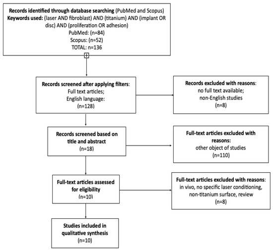
Figure 1.
PRISMA flowchart presenting the criteria for the included studies.
Focused question:
In this paper, the researchers focused on a question—“How laser modification of titanium surface influences fibroblast adhesion?”
2.2. Eligibility Criteria
Only studies that met the criteria below were included in the review:
- detailed laser conditioning—specified type and parameters
- titanium samples (titanium alloys were also included)
- fibroblast cells used as proliferating cells
- in vitro studies
- studies with a control group
- studies in English
- Reviewers agreed to exclude the following criteria:
- studies without a control group
- samples not made of pure titanium or its alloys
- non-English studies
- clinical reports
- review articles
- reviews
- meta-analysis
No restrictions were applied regarding the year of publication.
2.3. Information Sources, Search Strategy, and Study Selection
The literature review in PubMed and Scopus databases was conducted in September 2021 to find articles that related to laser’s influence on fibroblast adhesion to titanium surface. A specific search term (laser) AND (fibroblast) AND (titanium) AND (implant OR disc) AND (proliferation OR adhesion) was applied to find. The reviewers limited the search only to in vitro studies that related to eligibility criteria. Inclusion criteria consisted of in vitro studies where a titanium material was conditioned with a laser treatment. Only English studies and the ones available in full-text version were included. Criteria such as no laser conditioning, non-titanium material used or non-English papers were excluded. Meta-analysis and other review articles were not scanned.
2.4. Data Collection Process, Data Items
Data from papers that met the inclusion criteria was extracted by the two reviewers independently. The following data were used: first author, year of publication, study design, article title, laser type and specific changes in cell adhesion before and after laser modification and results. Extracted data were enrolled into an Excel spreadsheet created for this research.
2.5. Risk of Bias in Individual Studies
At the preliminary study selection each reviewer screened titles and abstracts separately to minimize the potential bias Cohen k test was used as a tool to determine the level of agreement between researchers [34]. In the case of any difference in opinion on the study inclusion or exclusion was resolved by discussion between the authors.
2.6. Quality Assessment
Two blinded reviewers screened the studies separately and independently to evaluate the quality of each included study. To establish study design, implementation and analysis the following criteria was used: sample quantity, indicated incubation time, full titanium sample specification, surface characteristics after laser application, fibroblast cells origin, laser type and laser parameters. The studies were graded on a scale from 0 to 9 points. The higher score indicated higher quality of the study. Any disagreements were resolved through discussion until reaching consensus.
2.7. Risk of Bias across Studies
The scores of each study were calculated and an overall estimate risk of bias (low, moderate, high) was made for each included study, as recommended in the Cochrane Handbook for Systematic Reviews of Interventions [35].
3. Results
3.1. Study Selection
The initial database search recognized 136 articles which were potentially applicable for the review. First title and abstract screening allowed to exclude 110 articles as not focused on the reviewed subject (no laser treatment used, other cells than fibroblasts for example bacteria, different sample material). Eighteen studies were selected for full-text screening, from which 8 were excluded due to not meeting defined inclusion criteria [36,37,38,39,40,41,42,43]. Ten papers were included for qualitative synthesis [37,38,39,40,41,42,43,44,45,46]. The flowchart below presents the main reasons for exclusion (Table 1.)

Table 1.
Reasons for exclusion of studies.
3.2. General Characteristics of the Included Studies
Ten studies were included in this review. In each study different lasers were used with different parameters that allowed to compare the influence of these devices on fibroblast adhesion to titanium surfaces. The general characteristics of each study and materials used are presented in Table 2.
Table 2.
General characteristic of the included studies.
In included studies titanium was the only material used; however, in each study the samples’ surface was prepared in a specific way—sandblasted, acid-etched, polished, autoclaved, washed in ultrasonic bath, cleaned with alcohol, acetone or water. Depending on the study the way of abundant decontamination was slightly different. Titanium was treated with different lasers i.e., Er:YAG [49], erbium fiber laser [52], ytterbium fiber laser [53], Ti:Sapphire laser fs-system laser [44,46], Nd:YAG [47], Nd:YVO4 [51], CO2 laser [48], KrF excimer laser [45], diode laser [50]. In eleven studies laser was the only treatment applied. In a study by Cao et al. [49] other kinds of surface modification were used to compare the adhesion. The methods applied in Cao et al. [49] study were stainless steel curette modification, ultrasonic system with straight carbon fiber tip and metal tip and rotating titanium brush. However, the elements that did not correspond to the review were omitted.
3.3. Subjects of the Study
The full-text articles review found heterogeneity in the papers. All the studies included in the review concerned fibroblasts—whether human cells from biopsies [46,49,50,51] or cell culture [52,53] or mouse embryonic cells [44,47,48]. In one study, cell origin was not defined [45]. In each case the cells were properly prepared to increase adhesion and proliferation features. The most common method was cell supplementation with fetal bovine/calf serum, antibiotic therapy and trypsinization. In a study by Lee DW et al. [46] L-glutamine was applied as an additional supplementation. Similarly in study by Chikarakara et al. [48] amphotericin B was added. The authors of one study did not precise the preparation procedure [45] (Table 3).
Table 3.
Characteristics of cells used in the studies.
The studies varied in kind of laser type and the lasing parameters. The surface modification consisted of creating microgrooves and microroughness on the disc which widened contact surface between the sample and the cells. Thanks to this modification the fibroblasts were prompted to grow and to form pseudopods which could influence better adhesion. The lasers used in included papers could be divided into the following groups: Nd:YAG [47], Nd:YVO4 [51], Er:YAG [49], erbium fiber laser [52], ytterbium fiber laser [52], Ti:Sapphire laser [44,46], diode laser [49], KrF excimer laser [45] and CO2 laser [48] (Table 4).
Table 4.
Characteristics of lasers used for treatments.
3.4. Main Study Outcomes
The studies included in the review were not homogenic in terms of laser treatment. They varied with laser wavelength, energy density and power output. Some did not precise any lasing parameters [51]. The researchers decided to have a more general look on the modification and how it influences fibroblast behavior. The main study outcome is that despite differences in laser wavelength and parameters, the cell adhesion increased. It could be concluded by observing fibroblasts’ morphology changes [46,50,51,52,53] or by examining changes in producing proteins responsible for adhesion [46,49,53]. The most common method of examining cell morphology was using SEM [46,50,51,52,53]. Fibroblasts showed various shapes on modified surfaces [50], denser growth [52], generating microstructures such as long extensions (pseudopodia) [44,46,51]. These structures followed the grooves or created bridges if the holey structures were prepared on the samples [45]. Certain studies analyzed the problem in microscale—adherent protein expression was measured [49,53]. Studies concentrated on proteins such as vinculin [46,53], FAK (focal adhesion kinase) [49,51], ITGB1 (integrin-beta1) [49] or integrin-beta4 [46] showed that protein expression was more intense on laser-modified specimens which proves the increased adhesion.
3.5. Quality Assessment
The articles included in the review were qualified as high-quality scoring 7/9 [44,46,48,49,53], 8/9 [47,52] or 9/9 points [50]. None of the studies was obtained and excluded as low-quality or moderate paper, but some of the studies were qualified as a moderate risk of bias scoring 6/9 points [45,51] (Table 5). However, missing information was not crucial for the research results.
Table 5.
Quality assessment of the included studies.
4. Discussion
Most studies that met the inclusion criteria and were considered in the review proved that laser therapy can increase cell adhesion to modified surfaces. The included papers could be divided into seven groups depending on what laser type was used—Nd:YAG [47], Nd:YVO4 [51], Er:YAG [49], erbium fiber laser [52], ytterbium fiber laser [53], Ti:Sapphire laser [44,46], diode laser [50], KrF excimer laser [45] and CO2 laser [48]. All these applied lasers have ability to alter the surface of the titanium which promoted the adhesion of the fibroblasts regardless of the type of fibroblast line and the grade of titanium alloy.
In studies by Vignesh et al. [47] and by Baltriukiene D et al. [51] authors used different neodynium lasers—sNd:YAG [54] and Nd:YVO4 [55]—to modify titanium discs. However, the results reached in the studies were similar. After SEM analysis in both cases a specific surface landscape was observed presenting characteristic roughness similar to troughed and holey structures [47,51]. Moreover, laser treatment keeps the purity of the disc, not contaminating it with additional compounds such as by-products of mechanical treatment [47]. In each study different fibroblasts cells were used—from biopsies [51] or from cell culture [47]; however, the result of these two studies was the same. Cells were observed under the SEM minimum 24 h after seeding. Low magnification showed a huge variety of cell shapes and extensions they grew. More careful observation at high magnification showed that the extensions grow into microgrooves and pores created by laser beam. Similar results were reached in other included studies [44,52] where SEM analysis proved the presence of pseudopodia and lamellipodia in laser-dimpled areas. All papers reported better cell adhesion to modified Ti-implant.
Certain studies examined cell viability by measuring cell metabolic activity. In their study Chikarakara et al. [48] showed that roughness induces cells activity as the percentage of reduced resazurin was higher. Studies conducted by Baltriukiene et al. [51] and Khadra et al. [50] also examined cell viability. However, in these cases Nd:YVO4 and diode laser treatment do not seem to increase this feature as the viability was the same in all samples.
Er:YAG laser was used in the study by Cao J et al. [49]. This study did not concentrate on cell morphology but considered gene expression responsible for adhesion (FAK and ITGB1) and ECM synthesis (COL1A1 and FN1). Results showed that all treatments applied in the study increased HGF’s adhesive strength but only on sandblasted surfaces. Samples cleaned only with acetone and alcohol showed reduction of the strength. The SAE (sandblasted) samples showed better conditions for gene expression, especially on SAE and laser-modified surfaces where a significant increase of COL1A1 expression was noticed. However, the positive results of laser treatment were only observed in SAE samples. Laser treatment did not increase surface roughness parameters in the case of other samples [49].
Study based on erbium fiber laser use proved that cell attachment depends on topography itself [52]. That means that the shape, direction and crosshatching of the grooves is crucial for good adhesion. The best evidence for that is the study where each group was laser-modified in different ways—unidirectional application, two-directional and three-directional crosshatching. Three-directional modification was the most successful titanium treatment in the context of fibroblast adhesion. Thus, the crosshatching not only increased cell adhesion but also contributed to better fibroblast spread on the examined surface [52].
The laser type seems not to be crucial in enhancing cell response. More important are the parameters and dosage. Laser energy density must be set for particular parameters, because too low energy density will not cause any difference in cell reaction compared to the control sample [50]. After reaching the lower limit, the increase of the energy dose influences surface roughness (the distance between the nanostructures is bigger) which entails better cell growth (longer extensions) [44]. Similar reaction is going to be reached by exposing the sample to multiple laser doses instead of one dose [45]. Multiple doses or increasing the number of pulses increase surface roughness which causes better cell adhesion however the difference in cell adhesion can be only noticed compared to control trials [45,50].
Except for the parameters described above, some studies considered the difference of wettability of the control and treated samples [44,53] cases showed that laser therapy increases surface wettability as reducing contact (wettability) angle. Therefore, the contact of the titanium implant with physiological fluids is better and protein adsorption increases what is a key to successful cell adhesion to the abundant and fast peri-implant tissue healing process [46]. However, some laser therapies can reduce contact angle too much (below 10 grades). This effect can lead to cell adhesion disorders or unsuccessful alignment what results in implantation treatment failure [53].
5. Conclusions
From the included studies, it can be concluded that ytterbium fiber laser, erbium fiber laser, Nd:YVO4, Er:YAG, CO2, Nd:YAG, KrF excimer laser, GaAlAs diode laser and Ti:Sapphire laser therapy positively influences fibroblast cells adhesion to the modified titanium surface. The review shows that most of laser type treatment increases surface roughness stimulating the cells to adhere and proliferate. However crucial is laser density and multiple exposures.
Author Contributions
Conceptualization, J.M., M.D.; methodology, J.M., J.K.; formal analysis, R.W., K.G.-L., data curation, J.K., R.W., K.G.-L.; writing—original draft preparation, J.K., J.M.; writing—review and editing, M.D., J.M.; supervision and funding acquisition, M.D., J.M. All authors have read and agreed to the published version of the manuscript.
Funding
This research did not receive any specific grant from funding agencies in the public, commercial, or not-for-profit sectors.
Institutional Review Board Statement
Not applicable.
Informed Consent Statement
Not applicable.
Data Availability Statement
The data presented in this study are available on request from the corresponding authors.
Conflicts of Interest
The authors declare no conflict of interest.
References
- Dominiak, M.; Matys, J. Assessment of pain when uncovering implants with Er:YAG laser or scalpel for second stage surgery. Adv. Clin. Exp. Med. 2016, 25, 1179–1184. [Google Scholar] [CrossRef] [PubMed] [Green Version]
- Matys, J.; Botzenhart, U.; Gedrange, T.; Dominiak, M. Thermodynamic effects after Diode and Er:YAG laser irradiation of grade IV and v titanium implants placed in bone-an ex vivo study. Preliminary report. Biomed. Tech. 2016, 61, 499–507. [Google Scholar] [CrossRef]
- Buser, D.; Sennerby, L.; De Bruyn, H. Modern implant dentistry based on osseointegration: 50 years of progress, current trends and open questions. Periodontol. 2000 2017, 73, 7–21. [Google Scholar] [CrossRef] [PubMed]
- Deeb, J.G.; Bencharit, S.; Dalal, N.; Abdulmajeed, A.; Grzech-Leśniak, K. Using Er:YAG laser to remove lithium disilicate crowns from zirconia implant abutments: An in vitro study. PLoS ONE 2019, 14, e0223924. [Google Scholar] [CrossRef] [PubMed] [Green Version]
- Grzech-Leśniak, K.; Bencharit, S.; Bs, N.D.; Mroczka, K.; Deeb, J.G. In vitro examination of the Use of Er:YAG laser to retrieve lithium disilicate crowns from titanium implant abutments. J. Prosthodont. 2019, 28, 672–676. [Google Scholar] [CrossRef] [PubMed]
- Compton, S.; Clark, D.; Chan, S.; Kuc, I.; Wubie, B.; Levin, L. Dental Implants in the Elderly Population: A Long-Term Follow-up. Int. J. Oral Maxillofac. Implant. 2017, 32, 164–170. [Google Scholar] [CrossRef] [PubMed]
- Perry, R.K.; Thomas, J.H. How do smoking, diabetes, and periodontitis affect outcomes of implant treatment? PubMed. Int. J. Oral Maxillofac. Implant. 2007, 22, 173–205. [Google Scholar]
- Greger, M.; Widomská, M.; Snášel, V. Structure and Properties of Dental Implants. In Proceedings of the METAL 2012—Conference Proceedings, 21st International Conference on Metallurgy and Materials, Brno, Czech Republic, 23–25 May 2012; Volume 120, pp. 434–439. [Google Scholar]
- Bosshardt, D.D.; Chappuis, V.; Buser, D. Osseointegration of titanium, titanium alloy and zirconia dental implants: Current knowledge and open questions. Periodontol. 2000 2017, 73, 22–40. [Google Scholar] [CrossRef] [PubMed]
- Chai, W.L.; Razali, M.; Ngeow, W.C. Dimension and Structures of Biological Seal of Peri-Implant Tissues. In Dental Implantology and Biomaterial; InTech: London, UK, 2016. [Google Scholar]
- Sculean, A.; Bosshardt, D.; Gruber, R. Soft tissue wound healing around teeth and dental implants. J. Clin. Periodontol. 2014, 41 (Suppl. 15), S6–S22. [Google Scholar] [CrossRef] [Green Version]
- Świder, K.; Dominiak, M.; Grzech-Leśniak, K.; Matys, J. Effect of different laser wavelengths on periodontopathogens in peri-implantitis: A review of in vivo studies. Microorganisms 2019, 7, 189. [Google Scholar] [CrossRef] [PubMed] [Green Version]
- Smeets, R.; Stadlinger, B.; Schwarz, F.; Beck-Broichsitter, B.; Jung, O.; Precht, C.; Kloss, F.; Gröbe, A.; Heiland, M.; Ebker, T. Impact of dental implant surface modifications on osseointegration. BioMed Res. Int. 2016, 2016, 1–16. [Google Scholar] [CrossRef] [PubMed] [Green Version]
- Sterczała, B.; Grzech-Leśniak, K.; Michel, O.; Trzeciakowski, W.; Dominiak, M.; Jurczyszyn, K. Assessment of human gingival fibroblast proliferation after laser stimulation in vitro using different laser types and wavelengths (1064, 980, 635, 450, and 405 nm)—Preliminary report. J. Pers. Med. 2021, 11, 98. [Google Scholar] [CrossRef] [PubMed]
- Pivodovaa, V.; Frankovaa, J.; Ulrichovaa, J. Osteoblast and gingival fibroblast markers in dental implant studies. Biomed. Pap. 2011, 155, 109–116. [Google Scholar] [CrossRef] [PubMed] [Green Version]
- Bernhardt, A.; Schneider, J.; Schroeder, A.; Papadopoulous, K.; Lopez, E.; Brückner, F.; Botzenhart, U. Surface conditioning of additively manufactured titanium implants and its influence on materials properties and in vitro biocompatibility. Mater. Sci. Eng. C 2021, 119, 111631. [Google Scholar] [CrossRef] [PubMed]
- Miura, S.; Takebe, J. Biological behavior of fibroblast-like cells cultured on anodized-hydrothermally treated titanium with a nanotopographic surface structure. J. Prosthodont. Res. 2012, 56, 178–186. [Google Scholar] [CrossRef] [Green Version]
- Smeets, R.; Henningsen, A.; Jung, O.; Heiland, M.; Hammächer, C.; Stein, J.M. Definition, etiology, prevention and treatment of peri-implantitis—A review. Head Face Med. 2014, 10, 34. [Google Scholar] [CrossRef] [PubMed] [Green Version]
- Matys, J.; Świder, K.; Flieger, R. Laser instant implant impression method: A case presentation. Dent. Med Probl. 2017, 54, 101–106. [Google Scholar] [CrossRef] [Green Version]
- Matys, J.; Romeo, U.; Mroczka, K.; Grzech-Leśniak, K.; Dominiak, M. Temperature Changes and SEM effects of three different implants-abutment connection during debridement with Er:YAG laser: An ex vivo study. Materials 2019, 12, 3748. [Google Scholar] [CrossRef] [PubMed] [Green Version]
- Matys, J.; Flieger, R.; Tenore, G.; Grzech-Leśniak, K.; Romeo, U.; Dominiak, M. Er:YAG laser, piezosurgery, and surgical drill for bone decortication during orthodontic mini-implant insertion: Primary stability analysis—An animal study. Lasers Med. Sci. 2018, 33, 489–495. [Google Scholar] [CrossRef] [Green Version]
- Matys, J.; Flieger, R.; Świder, K.; Gedrange, T.; Hutchings, G.; Dyszkiewicz-Konwińska, M.; Kempisty, B.; Nammour, S.; Dominiak, M.; Grzech-Leśniak, K. A clinical trial of photobiomodulation effect on orthodontic microscrews stability using a 635 nm red laser light. Photobiomodulation Photomed. Laser Surg. 2020, 38, 607–613. [Google Scholar] [CrossRef] [PubMed]
- Matys, J.; Flieger, R.; Dominiak, M. Effect of diode lasers with wavelength of 445 and 980 nm on a temperature rise when uncovering implants for second stage surgery—An ex-vivo study in pigs. Adv. Clin. Exp. Med. 2017, 26, 687–693. [Google Scholar] [CrossRef] [Green Version]
- Nammour, S.; Mobadder, M.; Maalouf, E.; Namour, M.; Namour, A.; Rey, G.; Matamba, P.; Matys, J.; Zeinoun, T.; Grzech-Leśniak, K. Clinical evaluation of diode (980 nm) laser-assisted non-surgical periodontal pocket therapy: A randomized comparative clinical trial and bacteriological study. Photomed. Laser Surg. 2020, 39, 10–22. [Google Scholar] [CrossRef] [PubMed]
- Zakrzewski, W.; Dobrzynski, M.; Kuropka, P.; Matys, J.; Malecka, M.; Kiryk, J.; Rybak, Z.; Dominiak, M.; Grzech-Lesniak, K.; Wiglusz, K.; et al. Removal of composite restoration from the root surface in the cervical region using Er: YAG laser and drill—In vitro study. Materials 2020, 13, 3027. [Google Scholar] [CrossRef]
- Deeb, J.G.; Grzech-Leśniak, K.; Weaver, C.; Matys, J.; Bencharit, S. Retrieval of glass fiber post using Er:YAG laser and conventional endodontic ultrasonic method: An in vitro study. J. Prosthodont. 2019, 28, 1024–1028. [Google Scholar] [CrossRef] [PubMed]
- Deeb, J.G.; Smith, J.; Belvin, B.R.; Lewis, J.; Grzech-Leśniak, K. Er:YAG Laser irradiation reduces microbial viability when used in combination with irrigation with sodium hypochlorite, chlorhexidine, and hydrogen peroxide. Microorganisms 2019, 7, 612. [Google Scholar] [CrossRef] [Green Version]
- Matys, J.; Grzech-Leśniak, K. Dental aerosol as a hazard risk for dental workers. Materials 2020, 13, 5109. [Google Scholar] [CrossRef]
- Grzech-Leśniak, K.; Matys, J. The effect of Er:YAG lasers on the reduction of aerosol formation for dental workers. Materials 2021, 14, 2857. [Google Scholar] [CrossRef] [PubMed]
- Lang, N.P.; Lindhe, J. Clinical periodontology and implant dentistry, 2 Volume Set. In Clinical Periodontology and Implant Dentistry, 6th ed.; Niklaus, P.L., Lindhe, J., Eds.; Wiley-Blackwell: Hoboken, NJ, USA, 2015; pp. 256–269. ISBN 978-0-470-67248-8. [Google Scholar]
- Grzech-Leśniak, K.; Gaspirc, B.; Sculean, A. Clinical and microbiological effects of multiple applications of antibacterial pho-todynamic therapy in periodontal maintenance patients. A randomized controlled clinical study. Photodiagnosis Photodyn. Ther. 2019, 27, 44–50. [Google Scholar] [CrossRef]
- Borcherding, K.; Marx, D.; Gätjen, L.; Specht, U.; Salz, D.; Thiel, K.; Wildemann, B.; Grunwald, I. Impact of laser structuring on medical-grade titanium: Surface characterization and in vitro evaluation of osteoblast attachment. Materials 2020, 13, 2000. [Google Scholar] [CrossRef]
- Schulz, K.F.; Altman, D.G.; Moher, D. CONSORT 2010 statement: Updated guidelines for reporting parallel group randomised trials. Int. J. Surg. 2010, 340, 698–702. [Google Scholar]
- Watson, P.; Petrie, A. Method agreement analysis: A review of correct methodology. Theriogenology 2010, 73, 1167–1179. [Google Scholar] [CrossRef] [PubMed] [Green Version]
- Higgins, J.P.; Thomas, J.; Chandler, J.; Cumpston, M.; Li, T.; Page, M.J.; Welch, V.A. Cochrane Handbook for Systematic Reviews of Interventions; John Wiley & Sons: Hoboken, NJ, USA, 2019. [Google Scholar]
- Schaeske, J.; Fadeeva, E.; Schlie-Wolter, S.; Deiwick, A.; Chichkov, B.N.; Ingendoh-Tsakmakidis, A.; Stiesch, M.; Winkel, A. Cell type-specific adhesion and migration on laser-structured opaque surfaces. Int. J. Mol. Sci. 2020, 21, 8442. [Google Scholar] [CrossRef]
- Lee, H.J.; Lee, J.; Lee, J.T.; Hong, J.S.; Lim, B.S.; Park, H.J.; Kim, Y.K.; Kim, T. Il microgrooves on titanium surface affect pe-ri-implant cell adhesion and soft tissue sealing; An in vitro and in vivo study. J. Periodontal Implant. Sci. 2015, 45, 120–126. [Google Scholar] [CrossRef] [PubMed] [Green Version]
- Corvino, E.; Pesce, P.; Mura, R.; Marcano, E.; Canullo, L. Influence of modified titanium abutment surface on peri-implant soft tissue behavior: A systematic review of in vitro studies. Int. J. Oral Maxillofac. Implant. 2020, 35, 503–519. [Google Scholar] [CrossRef]
- Zhang, Q.; Dong, J.; Peng, M.; Yang, Z.; Wan, Y.; Yao, F.; Zhou, J.; Ouyang, C.; Deng, X.; Luo, H. Laser-induced wettability gradient surface on NiTi alloy for improved hemocompatibility and flow resistance. Mater. Sci. Eng. C 2020, 111, 110847. [Google Scholar] [CrossRef]
- Khosroshahi, M.E.; Mahmoodi, M.; Saeedinasab, H. In vitro and in vivo studies of osteoblast cell response to a titanium-6 aluminium-4 vanadium surface modified by neodymium:yttrium–aluminium–garnet laser and silicon carbide paper. Lasers Med. Sci. 2009, 24, 925–939. [Google Scholar] [CrossRef] [PubMed]
- Abrahamsson, I.; Zitzmann, N.U.; Berglundh, T.; Linder, E.; Wennerberg, A.; Lindhe, J. The mucosal attachment to titanium implants with different surface characteristics: An experimental study in dogs. J. Clin. Periodontol. 2002, 29, 448–455. [Google Scholar] [CrossRef]
- Pérez-Díaz, L.; DeDavid, B.A.; Gehrke, S.A. Evaluation of Fibroblasts cells viability and adhesion on six different titanium surfaces: An in vitro experimental study. Recent Pat. Biotechnol. 2018, 12, 145–153. [Google Scholar] [CrossRef]
- Gheisarifar, M.; Thompson, G.A.; Drago, C.; Tabatabaei, F.; Rasoulianboroujeni, M. In vitro study of surface alterations to polyetheretherketone and titanium and their effect upon human gingival fibroblasts. J. Prosthet. Dent. 2021, 125, 155–164. [Google Scholar] [CrossRef]
- Aliuos, P.; Fadeeva, E.; Badar, M.; Winkel, A.; Mueller, P.P.; Warnecke, A.; Chichkov, B.; Lenarz, T.; Reich, U.; Reuter, G. Evaluation of single-cell force spectroscopy and fluorescence microscopy to determine cell interactions with femtosecond-laser microstructured titanium surfaces. J. Biomed. Mater. Res. Part A 2013, 101A, 981–990. [Google Scholar] [CrossRef]
- Heinrich, A.; Dengler, K.; Koerner, T.; Haczek, C.; Deppe, H.; Stritzker, B. Laser-modified titanium implants for improved cell adhesion. Lasers Med. Sci. 2008, 23, 55–58. [Google Scholar] [CrossRef]
- Lee, D.-W.; Kim, J.-G.; Kim, M.-K.; Ansari, S.; Moshaverinia, A.; Choi, S.-H.; Ryu, J.-J. Effect of laser-dimpled titanium surfaces on attachment of epithelial-like cells and fibroblasts. J. Adv. Prosthodont. 2015, 7, 138–145. [Google Scholar] [CrossRef] [PubMed]
- Vignesh, N.S.; Bhuminathan, M.; Santhosh, S. Comparative evaluation of the three different surface treatments-conventional, laser and Nano technology methods in enhancing the surface characteristics of commercially pure titanium discs and their effects on cell adhesion: An in vitro study. J. Pharm. Bioallied Sci. 2015, 7, 89–91. [Google Scholar] [CrossRef]
- Chikarakara, E.; Fitzpatrick, P.; Moore, E.; Levingstone, T.; Grehan, L.; Higginbotham, C.; Vazquez, M.; Bagga, K.; Naher, S.; Brabazon, D. In vitro fibroblast and pre-osteoblastic cellular responses on laser surface modified Ti–6Al–4V. Biomed. Mater. 2015, 10, 015007. [Google Scholar] [CrossRef] [PubMed] [Green Version]
- Jie, C.; Wang, T.; Yinfei, P.; Zhihui, T.; Huanxin, M. Influence on proliferation and adhesion of human gingival fibroblasts from different titanium surface decontamination treatments: An in vitro study. Arch. Oral Biol. 2018, 87, 204–210. [Google Scholar]
- Khadra, M.; Kasem, N.; Lyngstadaas, S.P.; Haanæs, H.R.; Mustafa, K. Laser therapy accelerates initial attachment and sub-sequent behaviour of human oral fibroblasts cultured on titanium implant materia: A scanning electron microscopic and his-tomorphometric analysis. Clin. Oral Implants Res. 2005, 16, 168–175. [Google Scholar] [CrossRef] [PubMed]
- Baltriukienė, D.; Sabaliauskas, V.; Balčiūnas, E.; Melninkaitis, A.; Liutkevičius, E.; Bukelskienė, V.; Rutkūnas, V. The effect of laser-treated titanium surface on human gingival fibroblast behavior. J. Biomed. Mater. Res. Part A 2014, 102, 713–720. [Google Scholar] [CrossRef]
- Çelebi, H.; Arpacı, P.; Çelik, I.; Akman, S. The Effects of laser with different parameters and crosshatching on fibroblast adhesion and proliferation to implant surfaces. Int. J. Oral Maxillofac. Implant. 2017, 32, 1231–1240. [Google Scholar] [CrossRef] [Green Version]
- Aktas, O.C.; Metzger, W.; Haidar, A.; Açil, Y.; Gülses, A.; Wiltfang, J.; Sacramento, C.M.; Nothdurft, F.P. Enhancing adhesion and alignment of human gingival fibroblasts on dental implants. J. Cranio Maxillofac. Surg. 2019, 47, 661–667. [Google Scholar] [CrossRef]
- Tezel, A.; Kara, C.; Balkaya, V.; Orbak, R. An evaluation of different treatments for recurrent aphthous stomatitis and patient perceptions: Nd:YAG laser versus medication. Photomed. Laser Surg. 2009, 27, 101–106. [Google Scholar] [CrossRef] [PubMed]
- Khadra, M.; Lyngstadaas, S.P.; Haanæs, H.R.; Mustafa, K. Effect of laser therapy on attachment, proliferation and differenti-ation of human osteoblast-like cells cultured on titanium implant material. Biomaterials 2005, 26, 3503–3509. [Google Scholar] [CrossRef] [PubMed]
Publisher’s Note: MDPI stays neutral with regard to jurisdictional claims in published maps and institutional affiliations. |
© 2021 by the authors. Licensee MDPI, Basel, Switzerland. This article is an open access article distributed under the terms and conditions of the Creative Commons Attribution (CC BY) license (https://creativecommons.org/licenses/by/4.0/).