Article

Synthesis of Rare-Earth-Doped Strontium Tungstate Phosphor at Room Temperature and Applied Flexible Composite

1
Department of Smart Manufacturing Engineering, Changwon National University, Changwon 51140, Republic of Korea
2
Research and Business Development Foundation, Engineering Building, Silla University, Busan 45985, Republic of Korea
3
Department of Mechatronics Convergence Engineering, Changwon National University, Changwon 51140, Republic of Korea
*
Authors to whom correspondence should be addressed.
Materials 2022, 15(24), 8922; https://doi.org/10.3390/ma15248922
Submission received: 26 October 2022 / Revised: 5 December 2022 / Accepted: 12 December 2022 / Published: 13 December 2022

Abstract

:
In this study, we successfully synthesized rare-earth-doped crystalline SrWO4 at room temperature by co-precipitation. The results from the X-ray diffraction analysis showed a main diffraction peak related to the (112) plane. Phosphors doped with either Dy3+ or Sm3+ ions showed strong light absorption in the UV region and blue-yellow and red light emission. To synthesize a white light phosphor, Dy3+ and Sm3+ ions were co-doped to produce a SrWO4:[Sm3+]/[Dy3+] phosphor. When the Sm3+ ion concentration was increased and the Dy3+ concentration was maintained, the red light intensity increased while the blue-yellow light intensity decreased. The composites were combined with polydimethylsiloxane (PDMS), and a flexible composite material was fabricated. The composite exhibited various luminescence properties under UV and visible light, which suggested its potential for use as an LED color filter.

1. Introduction

Phosphors doped with rare earth (RE) tungstates have attracted significant research interest because they can be used as white-light-emitting diodes, solid-state lighting, displays, and in various lighting industries [1,2]. Tungstates such as barium tungstate (BaWO4), strontium tungstate (SrWO4), and calcium tungstate (CaWO4) are chemically stable, have a broad absorption wavelength in the UV–vis region, and are capable of excellent energy transfer to rare earth ions [3,4,5]. Manufacturing a phosphor that emits a variety of colors with high luminescence intensity requires a thermally and chemically stable host structure doped with rare earth ions such as terbium (Tb3+), europium (Eu3+), dysprosium (Dy3+), and samarium (Sm3+), which possess many energy levels in the visible light region [6,7]. Among these rare earth ions, Dy3+ emits blue light via magnetic dipole transition (4F9/26H15/2) and yellow light via electric dipole transition (4F9/26H13/2) [8]. Sm3+ emits orange light via magnetic dipole (4G5/26H7/2) transition and red light via electric dipole transition (4G5/26H9/2) [9]. Hence, Dy3+ and Sm3+ are both compelling doping candidates for use in phosphor materials. Yu et al. synthesized a Ca3−xLix(PO4)2−x(SO4)x:Dy3+, Sm3+ phosphor by the sol–gel method and investigated the effect of Sm3+ ion concentration on the crystal structure and luminescence intensity. The authors found that the co-doping of Dy3+ and Sm3+ emitted warm white light and proposed that it was an effective method to implement [10]. Jung et al. synthesized BaWO4 and CaWO4 using the co-precipitation method and doped Tb3+ and Eu3+ rare earth ions to produce green and red phosphors that can impart luminescent properties to the host [11,12]. Tan et al. synthesized a NaLa(WO3)2 phosphor doped with Dy3+ and Sm3+ using the thermal decomposition method. They found that when the doping concentration of Sm3+ increased, the intensity of the light emitted by Dy3+ decreased as energy was transferred from Dy3+ to Sm3+ [13]. Sun et al. also reported a color change due to energy transfer by the co-doping of Dy3+ and Sm3+ into Ca3TeO6 host crystals using the solid-state reaction method [14].
Herein, we report a successful synthesis of SrWO4 by a co-precipitation method at room temperature. We investigated the effect of doping SrWO4 with either Dy3+ or Sm3+, and co-doping SrWO4 with both Dy3+ and Sm3+ as well as the effect of altering the Sm3+ concentration. This study also characterized the luminescence intensity, crystal structure, and particle shape of the synthesized phosphors. Finally, a flexible LED filter was produced by combing the synthesized phosphor with a polymer.

2. Materials and Methods

2.1. Synthesis of the Crystalline SrWO4 and SrWO4:RE3+ by Co-Precipitation

Starting materials: Strontium acetate ((CH3CO2)Sr, Sigma-Aldrich), sodium tungstate (Na2WO4·2H2O, Sigma-Aldrich), dysprosium nitrate (Dy(NO3)3·xH2O, Dy3+, Sigma-Aldrich), and samarium nitrate (Sm(NO3)3·6H2O, Sm3+, Sigma-Aldrich).
A total of 1 mmol of (CH3CO2)Sr and 1 mmol of Na2WO4·2H2O were placed in beakers ‘A’ and ‘B’, respectively, and each beaker was combined with 100 mL of distilled water. The solutions in each beakers ‘A’ and ‘B’ were completely dissolved until the mixtures were transparent in color. Next, the two solutions were combined and stirred for 30 min at ambient temperature. After mixing, the final mixture changed to an opaque white color and formed a powder precipitate. The mixture was centrifuged at 4000 rpm to recover the powder. The recovered powder was washed twice with distilled water to remove side products and unreacted compounds and then centrifuged again. Finally, the powder was dried at 80 °C in an oven for 16 h (Figure 1). The phosphor was synthesized by adding 0.25 mmol each of Dy3+ and Sm3+ to beaker ‘A’, and the remaining steps were performed as above. White light phosphor was synthesized by co-doping Dy3+ and Sm3+ and fixing the amount of Dy3+ while altering the amount of Sm3+. The doping percentages for all samples are shown in Table 1.

2.2. Fabricated Flexible Color Composite

To prepare the composite, 0.1 g of the synthesized powder, 2 g of polydimethylsiloxane (PDMS) polymer, and 0.2 g of curing agent were mixed until homogenous. Next, the mixture was poured into a square mold and placed in an oven at 80 °C for 1 h to form the composite. The composite was then irradiated with an ultraviolet lamp to observe any color change.

2.3. Characterization

X-ray diffraction analysis (XRD) (X’Pert PRO MPD, 40 kV, 30 mA, Cu–Kα radiation (wavelength: 1.5406 Å)) was used to characterize the crystal structure of the synthesized phosphor powder. The XRD analysis was carried out at a scan rate of 4° per minute at a diffraction angle of 10° to 70°. The crystal grains’ size and microscopic surface shape were characterized with a scanning electron microscope (CZ, MIRA I LMH, TESCAN), and a fluorescence photometer (FS-2, Scinco) with a xenon lamp as a light source was used for emission and absorption characteristics.

3. Results & Discussion

3.1. Characteristics of SrWO4 and Single Doped SrWO4

Figure 2a shows the XRD peaks of SrWO4, SrWO4:Dy3+, and SrWO4:Sm3+. SrWO4 showed a tetragonal (a = 5.400 Å, b = 5.400 Å, c = 11.910 Å) structure that was in good agreement with ICDD # 01-089-2568. The single-doped samples used 0.25 mmol of rare earth ions. The XRD analysis showed a main diffraction peak on the (112) plane. Likewise, the RE-doped SrWO4 samples exhibited a strong (112) main peak.
Figure 2b shows the lattice constant change with and without RE doping along the (112) plane. The lattice constant of the (112) plane significantly changed due to the RE doping (SrWO4: 0.290 nm, SrWO4:Dy3+: 0.2891 nm, SrWO4:Sm3+: 0.2891 nm). This change in the crystal lattice of SrWO4 by RE doping was attributed to the relatively large ionic radius (Sr: 1.18 Å, W: 0.66 Å, Dy: 1.07 Å, Sm: 1.22 Å) of the RE dopants [15]. Figure 3 shows the absorption and emission spectra of each sample. The host SrWO4 showed absorption from 220 nm to 340 nm and peaked at 277 nm. When the sample was excited at the highest peak (277 nm), a spectrum with a range of 350–650 nm peaking at 492 nm in a blue-white emission spectrum was observed (Figure 3a). Figure 3b shows the absorption and emission spectra of the SrWO4:Dy3+ phosphor powder. In the absorption spectrum controlled with an emission wavelength of 572 nm, the charge transfer band (CTB) absorption signal generated between the Dy3+ cations and O2- anions peaked at 253 nm. Moreover, several absorption narrow bands generated within the 4f-4f electron arrangement of Dy3+ ions were observed [16]. Among them, the absorption wavelength at 351 nm had the strongest absorption intensity and signaled the 6H15/26P7/2 transition of Dy3+. The absorption wavelengths at 325, 364, and 386 nm, which had relatively weaker absorptions, were identified as 6H15/26P3/2 transitions. These absorption signals were generated by the 6H15/26P5/2 and 6H15/24I13/2 transitions [17]. After excitation at 253 nm, the SrWO4:Dy3+ phosphor powder exhibited a yellow emission spectrum with a peak at 572 nm due to the 4F9/26H13/2 electric dipole transition of Dy3+ ions. Furthermore, the blue emission band belonging to the host was also observed. Since the emission peak was strong due to the electric dipole transition, the Dy3+ ion doped in the SrWO4 host lattice was in a non-inversion symmetric site [18]. For the phosphor doped with Sm3+ ions, when the emission wavelength was controlled at 643 nm, a CT band with a peak at 248nm and an absorption band with a peak at 297nm were observed. This absorption signal originated from the Sm3+ ions located within the host lattice. When the synthesized sample was excited at 248 nm, blue light emission by the SrWO4 matrix, green light emission at 560 nm (4G5/26H15/2), orange light emission at 599 nm (4G5/26H9/2), and red light emission signals at 643 nm (4G5/26H11/2) were generated [19]. Since the intensity of the red emission caused by electric dipole transition was about 1.1 times stronger than that caused by magnetic dipole transition, the Sm3+ ions in the SrWO4 host crystal were located in non-inversion symmetric sites [20]. In addition, as in the undoped SrWO4 sample, blue light emission was observed in the doped specimen due to absorption and emission by the matrix in a wide range of light absorptions.
The FE-SEM images of all samples showed that they all had long cylindrical shapes. For SrWO4, the particle size was ~6.2 µm and ~1.92 µm in the longitudinal and transverse direction, respectively (Figure 4a). For the RE-doped SrWO4:Dy3+, the particle size averaged ~5.09 µm (longitudinal) and ~1.49 µm (transverse) (Figure 4b), and for SrWO4:Sm3+, the particles measured ~5.61 µm (longitudinal) and 1.49 µm (transverse) (Figure 4c).

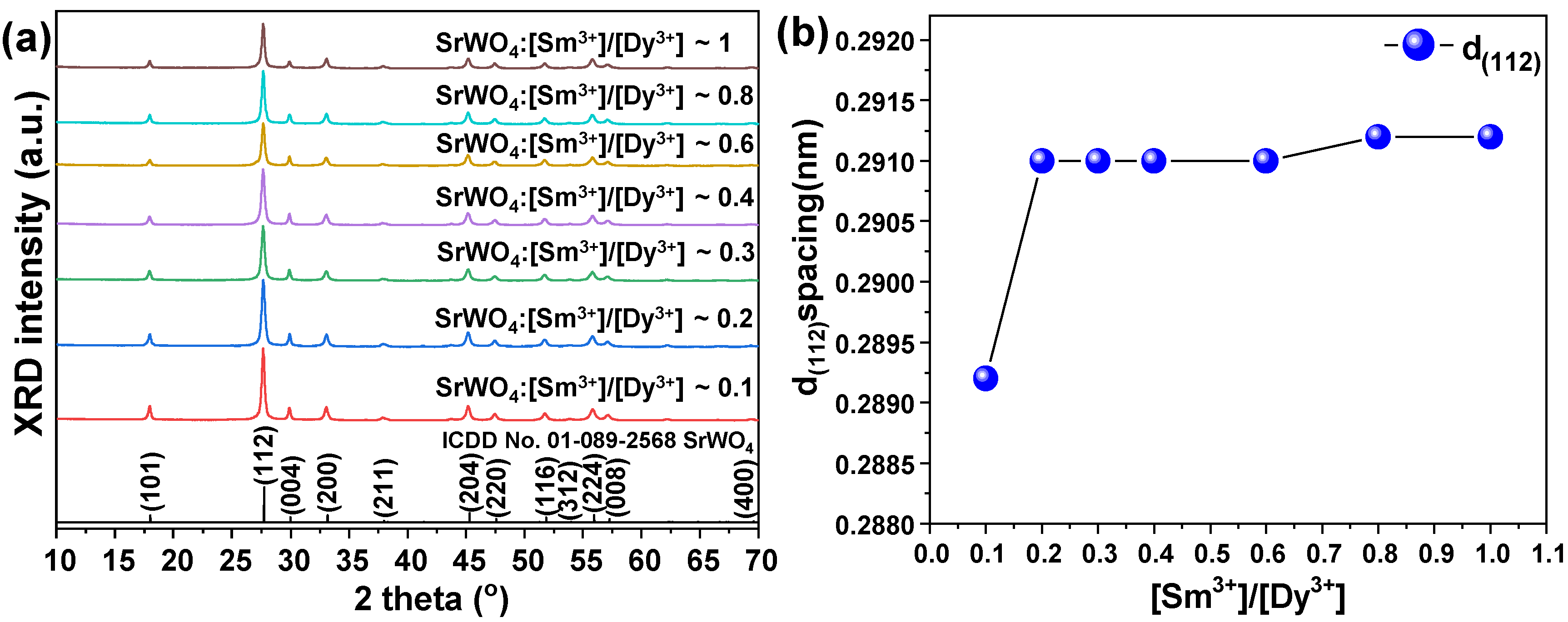
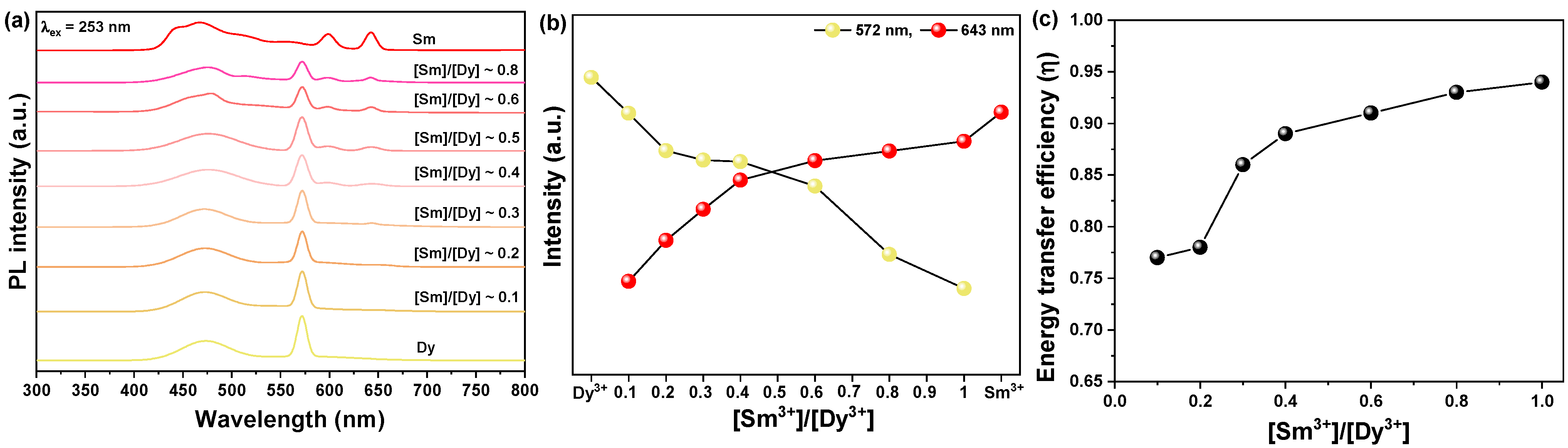
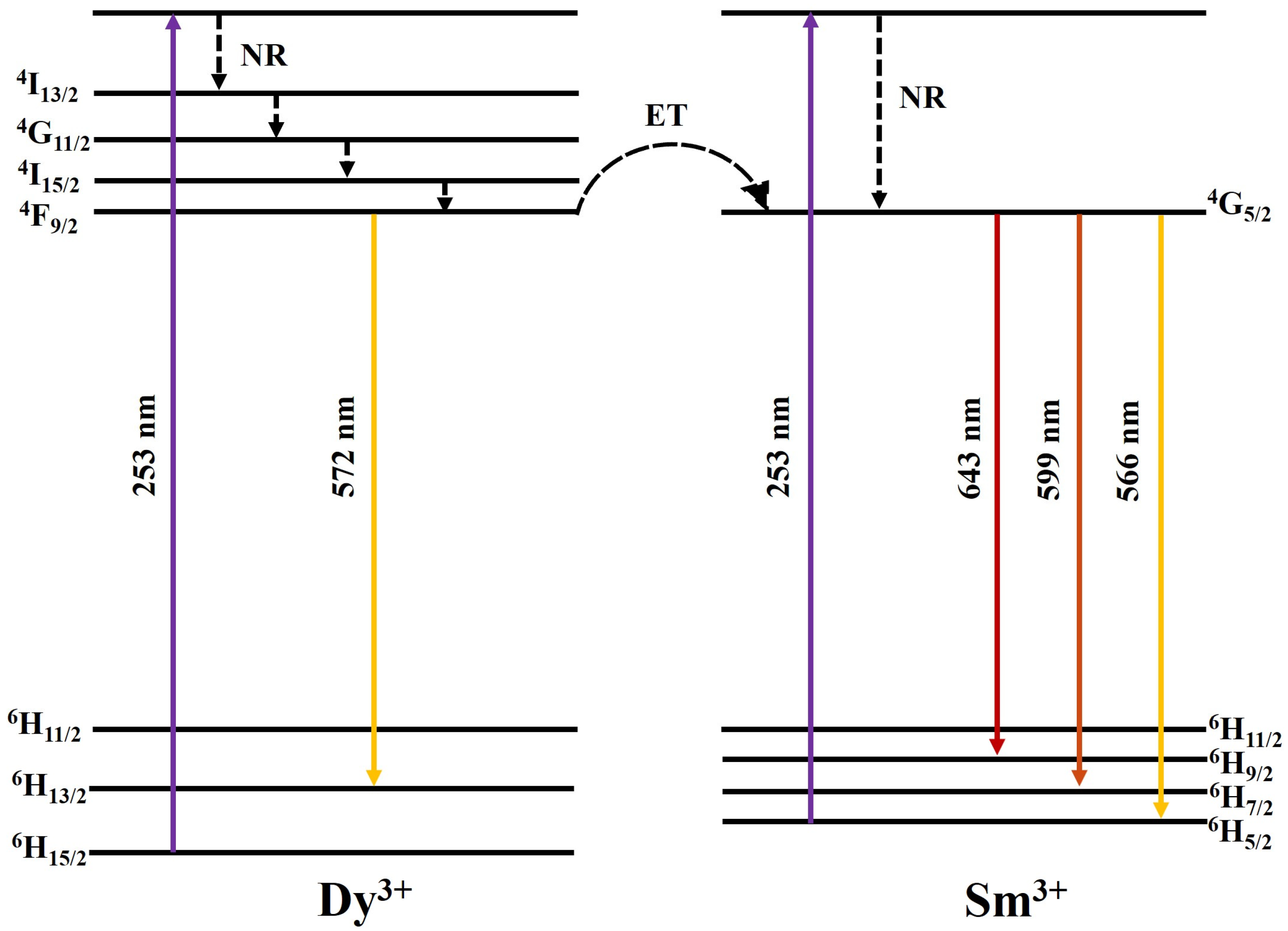
3.2. Characteristics of [Sm3+]/[Dy3+] Co-Doped SrWO4

Figure 5a shows the XRD peaks of SrWO4 co-doped with the rare earth ions Dy3+ and Sm3+ for synthesis as a white-light-emitting phosphor. The XRD pattern did not indicate a secondary phase caused by the RE doping but exhibited the diffraction signal of the main peak (112). Figure 5b shows the lattice constant change in the (112) plane, which was the main peak of the RE-co-doped SrWO4:[Sm3+]/[Dy3+] samples. As reported above, the lattice constants of the SrWO4:Dy3+ and SrWO4:Sm3+ samples decreased compared to the un-doped sample. However, as seen in Figure 5b, the lattice constants of the SrWO4:[Sm3+]/[Dy3+] sample increased. The higher lattice constants could have been caused by crystal lattice distortion or structural change induced by the additional amount of RE ions having a rather large ionic radius.
Figure 6 shows the FE-SEM image and EDS mapping component analysis of the synthesized SrWO4:[Sm3+]/[Dy3+] phosphor. The particle shape grew in the longitudinal direction of a cylindrical shape and resembled a dumbbell, with an estimated size of 5.26 µm and 2.33 µm in the longitudinal and transverse directions, respectively. The EDS component analysis showed the presence of Sr, W, O, Dy, and Sm, which confirmed that the RE ions were doped (Figure 6b,c).
Figure 7a shows the emission spectrum of the SrWO4:[Sm3+]/[Dy3+] phosphor powder co-doped while changing the Sm3+ ion concentration (the Dy3+ ion concentration was fixed). When excited at 253 nm and as the doping concentration of Sm3+ increased, the following were simultaneously observed: blue light at 492 nm, yellow light at 572 nm, orange light at 599 nm, and red light at 643 nm. As the concentration of Sm3+ ions increased, the intensity of yellow light emission by the Dy3+ ions decreased. The lower yellow light emission intensity meant that the emission energy was converted from the Dy3+ ions in the host lattice to the Sm3+ ions (Figure 7b). The energy transfer efficiency from the Dy3+ to Sm3+ ions can be expressed by Equation (1) [21].
    η = 1 I / I 0
where I is the emission intensity of Dy3+ ions in SrWO4:[Sm3+]/[Dy3+] phosphors and I0 is the emission intensity of Dy3+ ions in the SrWO4:Dy3+ phosphors. As shown in Figure 7c, the energy transfer efficiency tended to increase as the amount of Sm3+ ions added increased. However, as the emission intensity decreased, a concentration-quenching phenomenon due to excessive rare earth doping was observed [22].
The electrons located at the ground state, 6H15/2, of the Dy3+ ions absorbed energy under a 253 nm excitation energy and later jumped to the excited state. Since the high energy level was unstable, these electrons dropped successively to the lower-energy excited state, 4F9/2, by non-radiative transition (NR). With the populated 4F9/2 level, the radiative transitions of Dy3+ occurred with yellow emissions due to 4F9/26H13/2 transitions, respectively. In the interim, partial electrons located at the 4F9/2 level of Dy3+ were relaxed to the 6G5/2 level of Sm3+ by the resonance between the two levels, which ultimately gave rise to the characteristic emissions of Sm3+ (Figure 8).

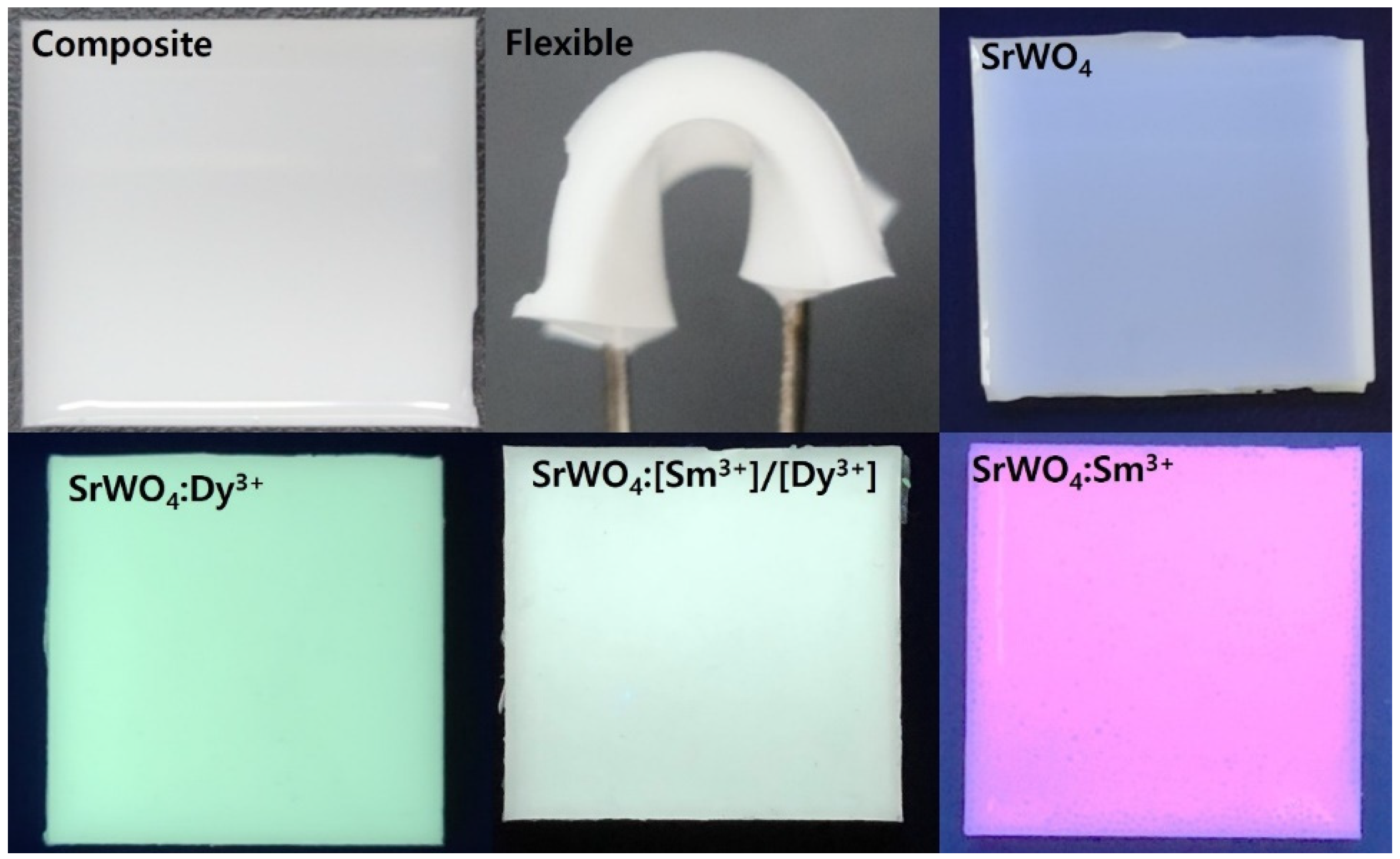
3.3. Application in a Flexible Composite LED Filter

To explore the applicability of the synthesized phosphor, herein referred to as an LED color filter, a flexible composite was fabricated by mixing the phosphor with a PDMS polymer, as explained in detail in Section 2.2. The manufactured composite showed characteristics of blue-white, blue-yellow, and main red light in response to UV light. The composite was flexible by hand and appeared suitable for use as an LED color filter (Figure 9).

4. Conclusions

We successfully synthesized crystalline SrWO4 at room temperature using the co-precipitation method. When doped with Dy3+ and Sm3+, yellow and red phosphors were obtained. The crystal structure of the synthesized phosphor was tetragonal, and a change in the lattice constant was observed due to the Dy3+ and Sm3+ dopants. The synthesized phosphor possessed a cylindrical shape, as confirmed by the FE-SEM images. For the SrWO4:[Sm3+]/[Dy3+] phosphor, the intensity of blue-yellow light emitted by the Dy3+ ions decreased as the concentration of Sm3+ ions increased. We also fabricated a flexible composite by mixing the synthesized phosphor with a PDMS polymer to demonstrate the potential applicability of the RE-doped phosphor produced in this study. The composite showed blue-white, blue-yellow, and red light emissions under UV and visible light, suggesting its potential application as an LED color filter.

Author Contributions

Conceptualization, J.-Y.J. and S.-G.P. methodology, J.-Y.J.; software, J.-Y.J. and J.-H.W.; validation, J.-Y.J., J.-H.W. and S.-G.P.; formal analysis, J.-Y.J.; investigation, J.-Y.J. and S.-G.P.; resources, J.-Y.J. and S.-G.P.; data curation, J.-Y.J. and J.-H.W.; writing—original draft preparation, J.-Y.J., J.-H.W. and S.-G.P. writing—review and editing, J.-Y.J. and S.-G.P.; visualization, J.-Y.J. and J.-H.W.; supervision, J.-Y.J. and S.-G.P.; project administration, J.-Y.J.; funding acquisition, S.-G.P. All authors have read and agreed to the published version of the manuscript.

Funding

This research is funded by the Financial Program for Self-Directed Research Capacity in 2022.

Institutional Review Board Statement

Not applicable.

Informed Consent Statement

Not applicable.

Data Availability Statement

The data presented in this study are available on request from the corresponding author.

Conflicts of Interest

The authors declare no conflict of interest.

References

  1. Al-Waisawy, S.; George, A.F.; Jadwisienczak, W.M.; Rahman, F. Preparation of balanced trichromatic white phosphors for solid-state white lighting. Luminescence 2017, 32, 791–799. [Google Scholar] [CrossRef] [PubMed]
  2. Ahn, Y.N.; Kim, K.D.; Anoop, G.; Kim, G.S.; Yoo, J.S. Design of highly efficient phosphor-converted white light-emitting diodes with color rendering indices (R1–R15) ≥ 95 for artificial lighting. Sci. Rep. 2019, 9, 16848. [Google Scholar] [CrossRef] [PubMed] [Green Version]
  3. Wlodarczyk, D.; Bulyk, L.; Berkowski, M.; Glowacki, M.; Kosyl, K.M.; Kaczmarek, S.M.; Kowalski, Z.; Wittlin, A.; Przybylinska, H.; Zhydachevskyy, Y.; et al. High-Pressure Low-Temperature Optical Studies of BaWO4:Ce, Na Crystals. Inorg. Chem. 2019, 58, 5617. [Google Scholar] [CrossRef] [PubMed]
  4. Dirany, N.; McRae, E.; Arab, M. Morphological and structural investigation of SrWO4 microcrystals in relationship with the electrical impedance properties. CrystEngComm 2017, 19, 5008–5021. [Google Scholar] [CrossRef]
  5. Koukou, V.; Martini, N.; Valais, I.; Bakas, A.; Kalyvas, N.; Lavdas, E.; Fountos, G.; Kandarakis, I.; Michail, C. Resolution Properties of a Calcium Tungstate (CaWO4) Screen Coupled to a CMOS Imaging Detector. J. Phys. Conf. Ser. 2017, 931, 12027. [Google Scholar] [CrossRef] [Green Version]
  6. Sun, X.; Sun, X.; Li, X.; He, J.; Wang, B. Synthesis and Luminescence of BaWO4:Ln3+ (Ln = Eu, Tb, and Dy) Powders. J. Electron. Mater. 2014, 43, 3534–3538. [Google Scholar] [CrossRef]
  7. Huang, J.; Lu, W.; Yue, D.; Wang, Y.; Wang, Z.; Jin, L.; Zhang, L.; Li, Z. Controllable synthesis of multi-morphological SrWO4:Ln3+ (Ln = Eu, Tb) hierarchical structures and their luminescence properties. CrystEngComm 2019, 21, 6482–6490. [Google Scholar] [CrossRef]
  8. Tian, B.; Chen, B.; Tian, Y.; Sun, J.; Li, X.; Zhang, J.; Zhong, H.; Cheng, L.; Hua, R. Concentration and temperature quenching mechanisms of Dy3+ luminescence in BaGd2ZnO5 phosphors. J. Phys. Chem. Solids 2012, 73, 1314–1319. [Google Scholar] [CrossRef]
  9. Ofelt, G.S. Intensities of crystal spectra of rare-earth ions. J. Chem. Phys. 1962, 37, 511–520. [Google Scholar] [CrossRef]
  10. Yu, M.; Xu, X.; Zhang, W.; Chen, X.; Zhang, P.; Huang, Y. The effect of Sm3+ co-doping on the luminescence properties of Ca2·85Li0·15(PO4)1·85(SO4)0.15: Dy3+ white-emitting phosphors. J. Alloys Compd. 2020, 817, 152761. [Google Scholar] [CrossRef]
  11. Jung, J.; Kim, J.; Shim, Y.; Hwang, D.; Son, C.S. Structure and Photoluminescence Properties of Rare-Earth (Dy3+, Tb3+, Sm3+)-Doped BaWO4 Phosphors Synthesized via Co-Precipitation for Anti-Counterfeiting. Materials 2020, 13, 4165. [Google Scholar] [CrossRef] [PubMed]
  12. Yi, S.; Jung, J. Calcium Tungstate Doped with Rare Earth Ions Synthesized at Low Temperatures for Photoactive Composite and Anti-Counterfeiting Applications. Crystals 2021, 11, 1214. [Google Scholar] [CrossRef]
  13. Tan, X.; Wang, Y.; Zhang, M. Solvothermal synthesis, luminescence and energy transfer of Dy3+ and Sm3+ doped NaLa(WO4)2 nanocubes. J. Photochem. Photobiol. A Chem. 2018, 353, 65–70. [Google Scholar] [CrossRef]
  14. Sun, X.; Huang, Z.; Fu, X.; Xu, L.; Liu, K.; Yuan, H. Generation of warm white light by doping Sm3+ in Ca3TeO6:Dy3+ fluorescent powders. Ceram. Int. 2020, 46, 14252–14256. [Google Scholar] [CrossRef]
  15. Van Orman, J.A.; Grove, T.L.; Shimizu, N. Rare earth element diffusion in diopside: Influence of temperature, pressure, and ionic radius, and an elastic model for diffusion in silicates. Contrib. Mineral. Petrol. 2001, 141, 687–703. [Google Scholar] [CrossRef]
  16. Diaz-Torres, L.A.; De la Rosa, E.; Salas, P.; Romero, V.H.; Angeles-Chávez, C. Efficient photoluminescence of Dy3+ at low concentrations in nanocrystalline ZrO2. J. Solid State Chem. 2008, 181, 75–80. [Google Scholar] [CrossRef]
  17. Yasaka, P.; Kaewkhao, J. White emission materials from glass doped with rare Earth ions: A review. AIP Conf. Proc. 2016, 1719, 020002. [Google Scholar] [CrossRef] [Green Version]
  18. Selvalakshmi, T.; Sellaiyan, S.; Uedono, A.; Chandra Bose, A. Investigation of defect related photoluminescence property of multicolour emitting Gd2O3:Dy3+ phosphor. RSC Adv. 2014, 4, 34257. [Google Scholar] [CrossRef]
  19. Stojadinović, S.; Vasilić, R. Orange–red photoluminescence of Nb2O5:Eu3+, Sm3+ coatings formed by plasma electrolytic oxidation of niobium. J. Alloys Compd. 2016, 685, 881–889. [Google Scholar] [CrossRef]
  20. Gupta, S.K.; Pathak, N.; Kadam, R.M. An efficient gel-combustion synthesis of visible light emitting barium zirconate perovskite nanoceramics: Probing the photoluminescence of Sm3+ and Eu3+ doped BaZrO3. J. Lumin. 2016, 169, 106–114. [Google Scholar] [CrossRef]
  21. Yang, L.; Mi, X.; Su, J.; Zhang, X.; Bai, Z.; Wang, N.; Lin, J. Tunable luminescence and energy transfer properties in YVO4:Bi3+, Ln3+ phosphors prepared by microwave sintering method. J. Mater. Sci. Mater. Electron. 2018, 29, 7941. [Google Scholar] [CrossRef]
  22. Ayvacikli, M.; Kaynar, Ü.H.; Karabulut, Y.; Canimoglu, A.; Bakr, M.; Akca, S.; Can, N. Synthesis and photoluminescence characteristics of Dy incorporated MoO3 phosphor: Suppression concentration quenching. Appl. Radiat. Isot. 2020, 164, 109321. [Google Scholar] [CrossRef] [PubMed]
Figure 1. Schematic of co-precipitation procedure.
Materials 15 08922 g001
Figure 2. (a) XRD patterns of crystalline SrWO4 and SrWO4:RE3+ and (b) d(112) spacing of samples.
Materials 15 08922 g002
Figure 3. (a) Luminescence spectra of SrWO4, (b) SrWO4:Dy3+, and (c) SrWO4:Sm3+.
Materials 15 08922 g003
Figure 4. FE-SEM images of (a) SrWO4, (b) SrWO4:Dy3+, and (c) SrWO4:Sm3+ powders.
Materials 15 08922 g004
Figure 5. (a) XRD patterns of crystalline SrWO4:[Sm3+]/[Dy3+] and (b) d(112) spacing of samples.
Materials 15 08922 g005
Figure 6. (a) FE-SEM image of SrWO4:[Sm3+]/[Dy3+], (b) EDS layered image, and (c) elemental mapping analysis.
Materials 15 08922 g006
Figure 7. (a) PL spectra of SrWO4:[Sm3+]/[Dy3+] under 253 nm, (b) change in PL intensity at 572 nm and 643 nm, and (c) energy transfer efficiency.
Materials 15 08922 g007
Figure 8. Schematic of energy level structure of the Dy3+ and Sm3+ ions.
Materials 15 08922 g008
Figure 9. Images of flexible composite under daylight and UV light.
Materials 15 08922 g009
Table 1. Reagents and moles used in the synthesis.
SrWO4 Up-Conversion Phosphor Synthesis
Reagents(CH3CO2)SrNa2WO4·2H2ODy(NO3)3·xH2OSm(NO3)3·6H2O
Molecular weight (g/mol)205.93329.85348.51444.47
Used mole (mmol)110.250.025~0.25
[Dy3+]/[Sm3+] Ratio
Reagents(CH3CO2)SrNa2WO4·2H2ODy(NO3)3·xH2OSm(NO3)3·6H2O
Used mole (mmol)110.250.025
110.250.05
110.250.075
110.250.1
110.250.15
110.250.2
110.250.25
Publisher’s Note: MDPI stays neutral with regard to jurisdictional claims in published maps and institutional affiliations.

