Article

The Mutual Incorporation of Mg2+ and CO32− into Hydroxyapatite: A DFT Study

by
Olga N. Makshakova
1,
Marat R. Gafurov
2 and
Margarita A. Goldberg
3,*
1
Kazan Institute of Biochemistry and Biophysics, Federal Research Center Kazan Scientific Center of Russian Academy of Sciences, 420111 Kazan, Russia
2
Institute of Physics, Kazan Federal University, 420008 Kazan, Russia
3
A.A. Baikov Institute of Metallurgy and Materials Science, Russian Academy of Sciences, 119334 Moscow, Russia
*
Author to whom correspondence should be addressed.
Materials 2022, 15(24), 9046; https://doi.org/10.3390/ma15249046
Submission received: 31 October 2022 / Revised: 10 December 2022 / Accepted: 15 December 2022 / Published: 17 December 2022
(This article belongs to the Special Issue X-ray Diffraction(XRD)for Hydroxyapatite)

Abstract

:
Hydroxyapatite (HA) with a stoichiometry composition of Ca10(PO4)6(OH)2 is widely applied for various biomedical issues, first of all for bone defect substitution, as a catalyst, and as an adsorbent for soil and water purification. The incorporation of foreign ions changes the acid–base relation, microstructure, porosity, and other properties of the HA materials. Here, we report the results of calculations of the density functional theory and analyze the possibility of two foreign ions, CO32− and Mg2+, to be co-localized in the HA structure. The Na+ was taken into account for charge balance preservation. The analysis revealed the favorable incorporation of CO32− and Mg2+ as a complex when they interact with each other. The energy gain over the sole ion incorporation was pronounced when CO32− occupied the A position and Mg2+ was in the Ca(2) position and amounted to -0.31 eV. In the most energy-favorable complex, the distance between Mg2+ and the O atom of carbonate ion decreased compared to Mg…O distances to the surrounding phosphate or hydroxide ions, and amounted to 1.98 Å. The theoretical calculations agree well with the experimental data reported earlier. Understating the structure–properties relationship in HA materials varying in terms of composition, stoichiometry, and morphology paves the way to rational designs of efficient bio-based catalytic systems.

Graphical Abstract

1. Introduction

1.1. Hydroxyapatite’s Applications

Hydroxyapatite (HA) is one of the most important bio-ceramics, which gains more attention in the form of porous scaffolds [1], granules [2], the component of composite materials [3,4,5], coatings [6], and nanoparticles [7,8]. Due to its derived surface with acid–base properties, HA has been widely used in organic synthesis and heterogeneous catalysis [9,10,11,12]. The wide range of reactions includes the formation of C–C, C–O, C–N, and C–S bonds, and reactions of oxidation, reduction, cycloadditions, etc. The stoichiometric composition of HA is Ca10(PO4)6(OH)2. At the stoichiometric Ca/P ratio, HA behaves as a basic catalyst despite the acid site presence. At a low non-stoichiometric ratio between Ca and P amounting to 1.50, HA behaves as an acid catalyst, despite the basic site presence [13].
The HA properties and morphology could be modified by tuning their composition and stoichiometry via the inclusion of ‘foreign’ ions [13,14]. Typical trace elements in HA of bones and tooth enamel are ~4–8 wt.% of carbonate; ~0.4–0.9 wt.% of sodium and magnesium; and others such as potassium, fluoride, and chloride are less than 0.1 wt.% [15]. Additionally, the catalytic properties could be improved by the introduction of different transitional metals such as Ni [16] or Fe [17]. The morphology, stability, solubility, mechanical properties, and biological behaviors of HA synthesized in vitro are also modulated by the incorporation of foreign ions. According to their size and charge, cations, such as Na+, Zn2+, Mg2+, and Fe3+ substitute Ca2+ [18,19,20,21]. In contrast, anions, such as CO32−, SiO44−, F, and Cl can theoretically substitute PO43− (B position) or OH (A position) (Figure 1). It is important to note, the size of the anion is, apparently, decisive, i.e., comparably large SiO44- was reported to be found only in the B position [22,23,24].

1.2. Carbonate Substitutions

Since carbonated HA is a major component of bone and dental enamel, it has been extensively studied experimentally [25,26,27,28,29,30,31,32]. There are also numerous computational studies of the inclusion of CO32− into HA cells [33,34,35]. The in silico studies were conducted using the ab initio crystal-field method [33], density functional theory (DFT) [35], and molecular dynamics (MD) [34]. Both natural and synthetic samples present different concentrations of type A and type B defects; however, their distribution within the lattice is not clear [36]. The MD simulations revealed that upon the carbonate uptake from the solution into the HA crystal, the type A defect was energetically preferred, followed by type AB, and then type B [34]. The DFT calculations using generalized gradient approximation and Perdew–Burke–Ernzerhof (PBE) exchange-correlation potential showed that the most energetically stable substitution was rather type AB, followed by type A, and then type B [35].
The analysis by FTIR spectroscopy was capable of distinguishing the A and B positions of CO32− characteristic for bone and enamel: (1) apatite channel, oriented with two oxygen atoms close to the c-axis (type A1); (2) close to a sloping face of the PO4 tetrahedron (type B). The third position in a stuffed channel (type A2) was a feature of high-pressure [30]. There was a report claiming that type B substitution was more favorable than type A substitution, revealed by FTIR spectra and the computed crystal energy [37]. In our recent study, the trace amount of CO32− in synthesized HA was of the A-B substitutional type [38].
The introduction of CO32− requires a charge compensation. For the charge balance in type B carbonated HA, the Na+ co-substitution was experimentally identified as the most likely case [39]. Sodium ion was observed in biological apatites, and the Na+ radius was close to that of Ca2+ ion [36]. In contrast, Astala and Stott [40] found the most favorable charge compensation scheme for the B type by adding a hydrogen atom linked to a neighboring phosphate.
The values of the HA specific surface area (SSA) could be controlled by the CO32− anions’ introduction in the structure. Thus, as CO32− anions were introduced, the SSA value grew from 79 to 107 m2/g [41]. When Na+ and CO32− ions were added during the synthesis, the powders with the HA structure with a slight change in the a and c parameters were obtained. These materials were characterized by the presence of both acidic and basic properties, which could also be controlled by the variation of the Ca/P ratio. While the existence of HPO4 ions led to the increase in acid sites, the addition of the CO32− anions resulted in the growth of the basicity of HAp. At the same time, the presence of Na+ resulted in the complementary basic sites’ increase. Thus, the obtaining of the Ca-deficient HA with the introduction of both Na+ and CO32− ions led to the enhancement and adjustment of the acidic and basic surface properties. The presented Na+ and CO32− co-doped HA could be applied as the efficient catalysts in the thiolation reaction of methanol [41].

1.3. Magnesium Substitutions

The Mg2+ contents in enamel, bone, and dentin are about 0.44, 0.72, and 1.23 wt.%, respectively [42,43]. It plays a key role in bone metabolism, in particular, during the early stages of osteogenesis and when magnesium-depleted bones are fragile [44,45]. When magnesium was introduced in the HA structure, the decrease in the degree of crystallinity, thermal stability, and strong distortions of the HA lattice was observed. This was due to the distinction of the ionic radius of Mg2+ (0.72 Å) compared to Ca2+ (1.04 Å) [46]. According to [47], the introduction of Mg2+ led to the growth of parameter a with the absence of the noticeable changes in parameter c, when Mg-HA mesoporous powders were obtained by the microwave-assistance synthesis [47]. Laurencin et al. [42] demonstrated that the introduction of Mg2+ resulted in a fall of parameter a, regardless of the substituted position. The magnesium and carbonated co-doped powders were characterized by a similar tendency when materials were synthesized by the wet synthesis after the drying at 60 °C [48].
According to the Rietveld and DFT ab-initio calculations, the magnesium cations could occupy the Ca(1) in the HA structure [46,49]. At the same time, the substitution Ca(2) position by the Mg2+ was supported by multinuclear solid-state NMR and X-ray absorption spectroscopy [42]. Upon Mg2+ incorporation, the environment of the anions was disordered. The calculations showed the shortening of Mg…O distances and modifications of the HA lattice [38,42].
The SSA of Mg2+-doped HA could be lower and higher compared to HA, depending on the synthesis route and magnesium amount [38,50,51]. The existence of magnesium cations, which were characterized by the more acidic sites, compared to calcium cations could increase the final yield of the Biginelli product. At the same time, the leaching of magnesium during the catalytic process is a challenge up to today, which has led to limitations of the application of MgHAp as a catalyst [13].

1.4. Magnesium and Carbonate Co-Substitutions

The incorporation of magnesium could facilitate the presence of carbonate. The carbonated Mg-substituted HA is closer to native human bone tissue. The type of carbonate substitution is linked with the synthesis route. Lala et al. [52] synthesized materials via the ball milling of the MgO, CaCO3, and CaHPO4·2H2O, and presented the preferential introduction of Mg2+ in the Ca(2) position for the type A carbonated HA according to the Rietveld’s refinement for XRD and FTIR analysis. The experiments on the HAp powders’ precipitation from the simulated body fluid solution showed that Mg2+ and CO32− ions coming together were favorably incorporated in the powders [53]. These may imply that the co-substitution of the ions is favorable in HAp [54,55] Our recent work revealed that the introduction of Mg2+ results in the more predictable formation of A type CO32−, unlikely to carbonate HA where a mixed A-B type of CO32− incorporation occurs. Additionally, the type of carbonate substitution is affected by the heat treatment, and A is energetically more favorable in the high-temperature environment, whereas the B type is preferred to the A type in the aqueous solution environment.
In the current work, we present the DFT study of HA co-doped with CO32− and Mg2+. The substitution of divalent Ca2+ by Mg2+ was performed in both Ca(1) and Ca(2) positions. For the incorporation of CO32−, the charge compensation was as follows: (1) when the carbonate ion was in the A position it replaced two OH ions [35], (2) when the carbonate ion was in the B position, one Ca2+ was replaced with Na+ [36]. Then, the combination of positions for all components was thoroughly analyzed. The energy gain upon the co-incorporation of CO32− and Mg2+ has been calculated again as the energy gain from the sole ion CO32− or Mg2+ incorporation into HA. The positions of the foreign ions’ incorporation and the scheme of charge compensation would crucially affect the local acid/base properties of doped HA and their use in catalysis. The results of the modelling would create a platform for the rational design of such materials and provide important parameters, as cell dimensions, to verify the ions’ incorporation at given experiment conditions.

2. Methods

The 1 × 1 × 1 monoclinic HA (88 atoms in the cell) was applied for calculations. The initial unit–cell parameters were taken from [56]. Previously, for both pure HA and HA containing foreign ions, it was shown that one monoclinic cell (space group P21/b) was sufficient to reproduce the g-factors and A-hyperfine values for various paramagnetic species [57,58] and phonon spectral density [59]. It was possible to adequately describe HA systems containing foreign cations, such as aluminum, iron, and magnesium cations [18,38,60,61]. It is important to note, the descriptions of monoclinic and hexagonal cells (usually found in the experiment) use different axis notation which should not lead to confusion, as demonstrated by our results [60].
The foreign ions were incorporated as follows: cations substituted Ca2+ in two positions, and carbonate anion substituted anions in a position of orthophosphate or hydroxide ions. To respect the principle of electroneutrality, the inclusion of divalent carbonate in the position of the OH- group required an additional hydroxide ion elimination. Then, CO32− was incorporated into the PO43− position, and one of the neighboring calcium ions (in the Ca(1) or Ca(2) position) was replaced with a sodium cation.
The optimization of cell geometry was carried out at the DFT level. A plane-wave basis was used with the ultrasoft pseudopotentials of Vanderbilt [62]. The generalized-gradient approximation was used in the scheme of Perdew–Wang (GGA PW91) to describe the exchange–correlation functional. [63]. For the smooth part of the electron wave functions, the 45 Ry kinetic energy cutoff was taken. For the augmented electron density, the cutoff value was 300 Ry. All calculations were performed using the Quantum ESPRESSO program package [64].
In the course of geometry optimization, both atomic positions and cell geometry were allowed to change. The convergence on forces was achieved with the threshold value 10−3 a.u. The Monkhorst-Pack scheme was used to sample the Brillouin zone with 2 × 2 × 1 k-point mesh [65].
The energy change upon the incorporation of a pair of foreign ions was calculated as follows (1):
ΔE = ΔEHA+Mg+CO3 − ΔEHA+Mg − ΔEHA+CO3 + ΔEHA
where ΔEHA+Mg+CO3 stands for the HA system containing both magnesium and carbonate ions; ΔEHA+Mg–HA containing magnesium foreign ion only; ΔEHA+CO3–HA containing carbonate foreign ion only; ΔEHA–the energy of pure HA.

3. Results and Discussion

3.1. Mg2+ Incorporation into HA

Divalent magnesium may substitute Ca2+ in HA without other compositional alternations since no charge compensation is needed. However, due to the large size difference (according to the Pauling scale, Mg2+ is about 0.28 Å smaller than Ca2+), the substitution leads to the contraction of the cell and distortion of the HA lattice [42]. At an atomistic level, it relates to a local shift of the ions coordinating Mg2+ [38].
In the cell of HA, there are two positions of Ca2+ calcium ions (Figure 1). In the first position, referred to as Ca(1), coordination of Ca2+ occurs via 6 oxygen atoms from 4 orthophosphates. The first coordination sphere of calcium in the Ca-2 position includes 7 oxygen atoms: 6 from four orthophosphates and 1 hydroxide. In the literature, the Mg2+ substitution was hypothesized in both Ca(1) [35,66] and Ca(2) positions [42,54]. The quantum chemical energy estimations at the GGA PBE level of theory revealed both the preference of the Ca(1) substitution by 0.012 eV [46] and Ca(2) substitution by 0.25 eV [42] and by ~0.1 eV [67]. Here, we present our own calculations to resolve the ambiguity. Furthermore, the energy and cell parameters of the Mg-doped HA cell are crucial to deduce the influence of carbonate ion upon their mutual incorporation into HA, which will be analyzed in the following chapter.
Some parameters of the cell geometry upon Mg2+ inclusion and relative values for pure HA are given in Table 1. According to the Ca…O distances in the first coordination shell, the surrounding of Ca(2) forms tighter contacts with the minimal distance to the calcium ion, amounting to 2.36Å (Figure 2). In the Ca(1) position, the shortest Ca…O distance is larger at 2.39 Å. The geometric parameters are close to those calculated at the GGA PBE level of theory [18]. When Mg2+ is incorporated into a calcium position, the first coordination shell overcomes a notable perturbation. The contraction in the first coordination shell results in the decrease in the Mg2+…O distances. In the Ca(1) position, the minimal distance between the magnesium ion and the closest oxygen atom of orthophosphate decreases to 2.15 Å. In the Ca(2) position, the closest oxygen atom (the hydroxide one) appears at a distance of 2.09 Å. One might anticipate that a more noticeable local condensation of ions would lead to a greater compression of the lattice cell. In contrast, despite the most pronounced shortening of the Mg2+…O distances in the Ca(2) position, the contraction of the cell lattice is more pronounced when Mg2+ is in the Ca(1) position. To form shorter contacts in the coordination sphere, Mg2+ that resides in the Ca(2) position is shifted towards phosphate groups by 0.29 Å from the related position of Ca2+. The OH ion is shifted towards Mg2+ by 0.3Å so that it declines from the axis of the anion channel. This distortion of the anion channel is supported by the FTIR data [38].
In addition, our results of energy estimation support the inclusion of Mg2+ into the Ca(2) position with a slight preference: the energy gain into Ca(2) against Ca(1) amounts to 0.042 eV (Table 1).
Despite the fact that the energy preference is not largely pronounced, our results are rather supportive of the results of the Laurencin et al. [42] and Saito et al. [67] reports, rather than the Ren et al. one [35].

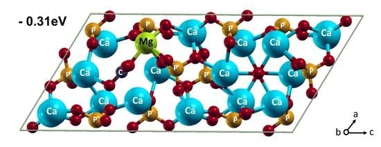
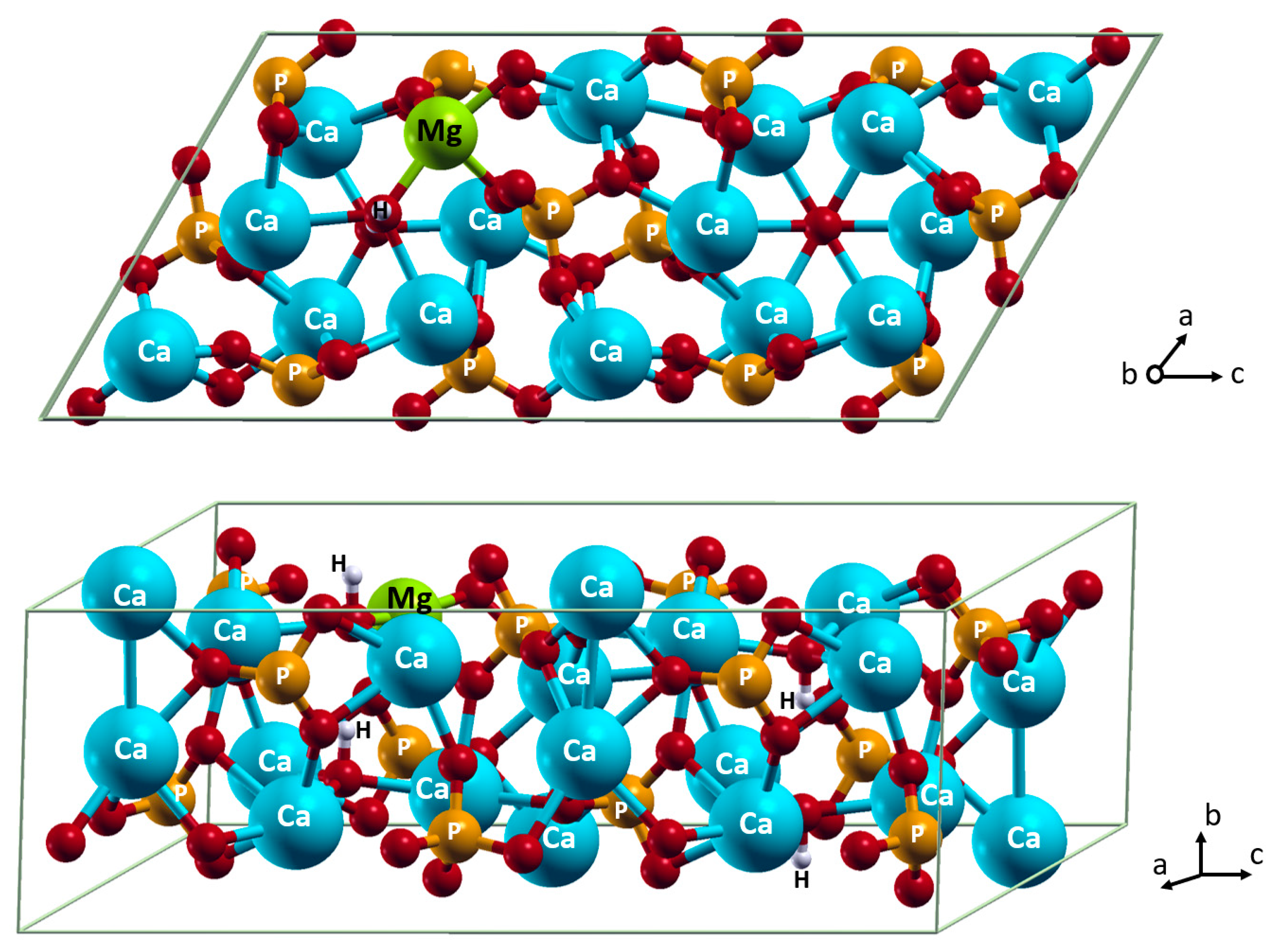
3.2. Mg2+ and CO32− Co-Incorporation into HA

3.2.1. Carbonate in the OH (A) position

When HA carbonated in the A position (Figure 3) and magnesium is incorporated into the position of the Ca(2), it has the most favorable energy, −0.308 eV (Table 2, Figure 4). This implies the incorporation of Mg2+ and CO32 happens more beneficially as a complex than as sole ions. Indeed, the combination of the carbonate positioning in the anion channel and the magnesium positioning in the layer surrounding the anion channel allows the two ions to interact at the Mg…O distance of 1.98 Å. This distance is shorter than the Mg…O distances to orthophosphate or hydroxide ions, at least by 0.11 Å. In contrast, when Mg2+ is incorporated in the Ca(1) position, which is remote from the anion channel, the magnesium and carbonate ions cannot interact with each other. Such incorporation as sole ions does not lead to any energy change.
When CO32− incorporates in the A position, the cell slightly expands. The following incorporation of magnesium cation into the Ca(1) position results in an additive cell contraction. Interestingly, there are no cell size changes observed upon the following incorporation of magnesium into the Ca(2).

3.2.2. Carbonate in the PO43− (B) position

The incorporation of CO32− into the B position requires a compositional alteration for the charge compensation. The most commonly used scheme is the substitution of one Ca2+ with one Na+ ion. This is due to their comparable size and the fact that sodium impurity is one of the most commonly present factors in natural HA, and simultaneously with carbonate [36]. The skeleton contains 99% of the calcium of the body, 35% of the body’s sodium, 80% of the body’s carbonates, and 60% of the magnesium [68], and additional trace elements such as K, Cr, Zn, and Sr [69]. The carbonate content of bone mineral is shown to vary depending on the age of the individual [70,71], and type B carbonate is the most abundant species in the bones of young humans, with increasing A type in the bones of old people [72]. Thus, in the current section, we explore the panel of possible combinations of the inclusion of both Na+ and Mg2+ in the Ca(1) and Ca(2) positions, keeping CO32− in the B position, similar to in the native human bone [73].
The incorporation of CO32− together with Na+ results in a contraction of the cell by 0.6–0.7%, irrespective of the Na+ position. The following incorporation of Mg2+ leads to further cell contraction by 0.7% and 1.0%, respectively, for the Ca(2) and Ca(1) substitution, which are in agreement with the result of Section 3.1.
All variations of the Mg2+ and Na+ positioning show the distances between Mg2+ and CO32− as long as in the Mg2+-doped HA, which is lacking CO32− in the B position. The mutual orientation of Mg2+ and the carbonate at such distances does not bring any notable energy gain upon the mutual incorporation of the ions to HA (Table 2). Tighter interactions with the carbonate ion are not possible because of forces acting on the Mg2+ from the surrounding ions in the cell. There is no additional impact from Na+ on the interactions between Mg2+ and CO32−. Previously, the synthesis of Mg-doped carbonate-apatite containing 6 wt.% carbonate was performed by dropping sodium hydrogen carbonate (NaHCO3) solution and H3PO4 into the suspension of MgCl2·6H2O and Ca(OH)2. The FTIR results confirm the (B) position of carbonate and Rietveld method was applied to evaluate that Mg2+ substitutes essentially in the Ca (1) site; at the same time, the presence of Na+ ions was not discussed in the paper but could be assumed as a trace element [73].
Interestingly, at DFT level of calculations the incorporation of CO32− into the B position occurs as least favorable, irrespective of the scheme for the charge compensation used [67].

4. Conclusions

The results of our calculations at the DFT level revealed that the co-doping of HA by CO32− and Mg2+ can be more efficient than the doping of sole carbonate or magnesium ions. This is due to the energy gain from direct interactions between CO32− and Mg2+ ions. The complex is formed more eagerly in the tight anion channel, namely when CO32− replaces hydroxide ions, and Mg2+ resides in one of the Ca(2) positions close to the carbonate. When carbonate replaces PO43− ions, its interactions with Mg2+ residing in the proximity, at either Ca(1) or Ca(2) positions, are not beneficial. The theoretical results support the change in the FTIR spectra, published earlier, showing the increase in the carbonate population in the A position when Mg2+ is co-doped. The obtained results contribute to the understating of the doped HA structure and reveal the energy efficiency of a paired carbonate magnesium incorporation. Altogether, it paves the way to rational designs of bio-based catalytic systems.

Author Contributions

Conceptualization, O.N.M. and M.R.G.; methodology, O.N.M.; validation, O.N.M. and M.R.G.; formal analysis, O.N.M.; data curation, M.R.G. and M.A.G.; writing—original draft preparation, O.N.M.; general discussion, O.N.M.; writing—review and editing, M.R.G. and M.A.G. All authors have read and agreed to the published version of the manuscript.

Funding

This research was funded by the Russian Science Foundation, grant number 22-79-10293. O.N.M. is thankful for partial financial support from the government assignment for the FRC Kazan Scientific Centre of RAS in part of the general discussion and for providing the access to the electronic database of the scientific publications. The computer simulations were carried out using the equipment of the shared research facilities of HPC computing resources at Lomonosov Moscow State University and the Joint Supercomputer Centre of the Russian Academy of Sciences.

Institutional Review Board Statement

Not applicable.

Informed Consent Statement

Not applicable.

Data Availability Statement

Not applicable.

Conflicts of Interest

The authors declare no conflict of interest.

References

  1. Barinov, S.M.; Komlev, V.S. Approaches to the Fabrication of Calcium Phosphate-Based Porous Materials for Bone Tissue Regeneration. Inorg. Mater. 2016, 52, 339–346. [Google Scholar] [CrossRef]
  2. Komlev, V.S.; Barinov, S.M.; Koplik, E.V. A Method to Fabricate Porous Spherical Hydroxyapatite Granules Intended for Time-Controlled Drug Release. Biomaterials 2002, 23, 3449–3454. [Google Scholar] [CrossRef] [PubMed]
  3. Goldberg, M.A.; Smirnov, V.V.; Kutsev, S.V.; Shibaeva, T.V.; Shvorneva, L.I.; Sergeeva, N.S.; Sviridova, I.K.; Barinov, S.M. Hydroxyapatite-Calcium Carbonate Ceramic Composite Materials. Inorg. Mater. 2010, 46, 1269–1273. [Google Scholar] [CrossRef]
  4. Rajendran, A.; Barik, R.C.; Natarajan, D.; Kiran, M.S.; Pattanayak, D.K. Synthesis, Phase Stability of Hydroxyapatite-Silver Composite with Antimicrobial Activity and Cytocompatability. Ceram. Int. 2014, 40, 10831–10838. [Google Scholar] [CrossRef]
  5. Kaygili, O.; Tatar, C.; Yakuphanoglu, F.; Keser, S. Nano-Crystalline Aluminum-Containing Hydroxyapatite Based Bioceramics: Synthesis and Characterization. J. Solgel Sci. Technol. 2013, 65, 105–111. [Google Scholar] [CrossRef]
  6. Shamray, V.F.; Sirotinkin, V.P.; Smirnov, I.V.; Kalita, V.I.; Fedotov, A.Y.; Barinov, S.M.; Komlev, V.S. Structure of the Hydroxyapatite Plasma-Sprayed Coatings Deposited on Pre-Heated Titanium Substrates. Ceram. Int. 2017, 43, 9105–9109. [Google Scholar] [CrossRef]
  7. Othmani, M.; Aissa, A.; Grelard, A.; Das, R.K.; Oda, R.; Debbabi, M. Synthesis and Characterization of Hydroxyapatite-Based Nanocomposites by the Functionalization of Hydroxyapatite Nanoparticles with Phosphonic Acids. Colloids Surf. A Physicochem. Eng. Asp. 2016, 508, 336–344. [Google Scholar] [CrossRef]
  8. Kaygili, O.; Keser, S.; Kom, M.; Eroksuz, Y.; Dorozhkin, S.V.; Ates, T.; Ozercan, I.H.; Tatar, C.; Yakuphanoglu, F. Strontium Substituted Hydroxyapatites: Synthesis and Determination of Their Structural Properties, in Vitro and in Vivo Performance. Mater. Sci. Eng. C 2015, 55, 538–546. [Google Scholar] [CrossRef]
  9. Gruselle, M.; Tonsuaadu, K. Tunable calcium-apatites as solid catalysts for classical organic reactions. Curr. Org. Chem. 2017, 21, 688–697. [Google Scholar] [CrossRef] [Green Version]
  10. Kaneda, K.; Mizugaki, T. Design of High-Performance Heterogeneous Catalysts Using Apatite Compounds for Liquid-Phase Organic Syntheses. ACS Catal. 2017, 7, 920–935. [Google Scholar] [CrossRef]
  11. Gruselle, M. Apatites: A New Family of Catalysts in Organic Synthesis. J. Organomet. Chem. 2015, 793, 93–101. [Google Scholar] [CrossRef]
  12. Fihri, A.; Len, C.; Varma, R.S.; Solhy, A. Hydroxyapatite: A Review of Syntheses, Structure and Applications in Heterogeneous Catalysis. Coord. Chem. Rev. 2017, 347, 48–76. [Google Scholar] [CrossRef]
  13. Gruselle, M.; Tõnsuaadu, K.; Gredin, P.; Len, C. Apatites Based Catalysts: A Tentative Classification. Mol. Catal. 2022, 519, 112146. [Google Scholar] [CrossRef]
  14. Šupová, M. Substituted Hydroxyapatites for Biomedical Applications: A Review. Ceram. Int. 2015, 41, 9203–9231. [Google Scholar] [CrossRef]
  15. Dorozhkin, S.V. Calcium orthophosphates: Occurrence, properties, biomineralization, pathological calcification and biomimetic applications. Biomatter 2011, 1, 121–164. [Google Scholar] [CrossRef]
  16. İnce, T.; Kaygili, O.; Tatar, C.; Bulut, N.; Koytepe, S.; Ates, T. The Effects of Ni-Addition on the Crystal Structure, Thermal Properties and Morphology of Mg-Based Hydroxyapatites Synthesized by a Wet Chemical Method. Ceram. Int. 2018, 44, 14036–14043. [Google Scholar] [CrossRef]
  17. Iriarte-Velasco, U.; Ayastuy, J.L.; Boukha, Z.; Bravo, R.; Gutierrez-Ortiz, M.Á. Transition Metals Supported on Bone-Derived Hydroxyapatite as Potential Catalysts for the Water-Gas Shift Reaction. Renew. Energy 2018, 115, 641–648. [Google Scholar] [CrossRef]
  18. Makshakova, O.N.; Shurtakova, D.V.; Vakhin, A.V.; Grishin, P.O.; Gafurov, M.R. Incorporation of Iron(II) and (III) in Hydroxyapatite—A Theoretical Study. Crystals 2021, 11, 1219. [Google Scholar] [CrossRef]
  19. Shurtakova, D.V.; Grishin, P.O.; Gafurov, M.R.; Mamin, G.V. Using DFT to Calculate the Parameters of the Crystal Field in Mn2+ Doped Hydroxyapatite Crystals. Crystals 2021, 11, 1050. [Google Scholar] [CrossRef]
  20. Pathak, D.K.; Pandey, P.M. Evaluation of in Vitro Corrosion Behavior of Zinc–Hydroxyapatite and Zinc–Hydroxyapatite–Iron as Biodegradable Composites. J. Biomed. Mater. Res. B Appl. Biomater. 2021, 109, 436–450. [Google Scholar] [CrossRef]
  21. Barinov, S.M.; Fadeeva, I.V.; Ferro, D.; Rau, J.V.; Cesaro, S.N.; Komlev, V.S.; Fomin, A.S. Stabilization of Carbonate Hydroxyapatite by Isomorphic Substitutions of Sodium for Calcium. Russ. J. Inorg. Chem. 2014, 53, 164–168. [Google Scholar] [CrossRef]
  22. Liu, Z.; Zhang, L.; Wang, X.; Jiang, G.; Yang, M. A Comparative Study of the Dissolubility of Pure and Silicon Substituted Hydroxyapatite from Density Functional Theory Calculations. J. Mol. Model. 2018, 24, 1–9. [Google Scholar] [CrossRef] [PubMed]
  23. Palard, M.; Champion, E.; Foucaud, S. Synthesis of Silicated Hydroxyapatite Ca10(PO4)6 − x(SiO4)x(OH)2 − x. J. Solid State Chem. 2008, 181, 1950–1960. [Google Scholar] [CrossRef]
  24. Camaioni, A.; Cacciotti, I.; Campagnolo, L.; Bianco, A. Silicon-Substituted Hydroxyapatite for Biomedical Applications. In Hydroxyapatite (Hap) for Biomedical Applications; Woodhead Publishing: Sawston, UK, 2015; pp. 343–373. [Google Scholar] [CrossRef]
  25. Barralet, J.; Best, S.; Bonfield, W. Carbonate Substitution in Precipitated Hydroxyapatite: An Investigation into the Effects of Reaction Temperature and Bicarbonate Ion Concentration. J. Biomed. Mater. Res. 1998, 41, 79–86. [Google Scholar] [CrossRef]
  26. Penel, G.; Leroy, G.; Rey, C.; Bres, E. MicroRaman Spectral Study of the PO4 and CO3 Vibrational Modes in Synthetic and Biological Apatites. Calcif. Tissue Int. 1998, 63, 475–481. [Google Scholar] [CrossRef]
  27. Wilson, R.M.; Elliott, J.C.; Dowker, S.E.P. Rietveld Refinement of the Crystallographic Structure of Human Dental Enamel Apatites. Am. Mineral. 1999, 84, 1406–1414. [Google Scholar] [CrossRef]
  28. Dorozhkin, S.V.; Epple, M. Biological and Medical Significance of Calcium Phosphates. Angew. Chem. Int. Ed. 2002, 41, 3130–3146. [Google Scholar] [CrossRef]
  29. Elliott, J.C. Structure and Chemistry of the Apatites and Other Calcium Orthophosphates; Studies in Inorganic Chemistry; Elsevier: Amsterdam, The Netherlands, 1994; Volume 18, pp. 1–389. [Google Scholar]
  30. Fleet Michael, E.; Liu, X.; King Penelope, L. Accommodation of the Carbonate Ion in Apatite: An FTIR and X-ray Structure Study of Crystals Synthesized at 2–4 GPa. Am. Mineral. 2004, 89, 1422–1432. [Google Scholar] [CrossRef]
  31. Wilson, R.M.; Elliott, J.C.; Dowker, S.E.P.; Smith, R.I. Rietveld Structure Refinement of Precipitated Carbonate Apatite Using Neutron Diffraction Data. Biomaterials 2004, 25, 2205–2213. [Google Scholar] [CrossRef]
  32. Antonakos, A.; Liarokapis, E.; Leventouri, T. Micro-Raman and FTIR Studies of Synthetic and Natural Apatites. Biomaterials 2007, 28, 3043–3054. [Google Scholar] [CrossRef]
  33. Peeters, A.; de Maeyer, E.A.P.; van Alsenoy, C.; Verbeeck, R.M.H. Solids Modeled by Ab Initio Crystal-Field Methods. 12. Structure, Orientation, and Position of A-Type Carbonate in a Hydroxyapatite Lattice. J. Phys. Chem. B 1997, 101, 3995–3998. [Google Scholar] [CrossRef]
  34. Peroos, S.; Du, Z.; de Leeuw, N.H. A Computer Modelling Study of the Uptake, Structure and Distribution of Carbonate Defects in Hydroxy-Apatite. Biomaterials 2006, 27, 2150–2161. [Google Scholar] [CrossRef] [PubMed]
  35. Ren, F.; Lu, X.; Leng, Y. Ab Initio Simulation on the Crystal Structure and Elastic Properties of Carbonated Apatite. J. Mech. Behav. Biomed. Mater. 2013, 26, 59–67. [Google Scholar] [CrossRef] [PubMed]
  36. Ulian, G.; Valdrè, G.; Corno, M.; Ugliengo, P. DFT Investigation of Structural and Vibrational Properties of Type B and Mixed AB Carbonated Hydroxylapatite. Am. Mineral. 2014, 99, 117–127. [Google Scholar] [CrossRef]
  37. Yang, W.H.; Xi, X.F.; Li, J.F.; Cai, K.Y. Comparison of Crystal Structure between Carbonated Hydroxyapatite and Natural Bone Apatite with Theoretical Calculation. Asian J. Chem. 2013, 25, 3673–3678. [Google Scholar] [CrossRef]
  38. Goldberg, M.A.; Fomin, A.S.; Murzakhanov, F.F.; Makshakova, O.N.; Donskaya, N.O.; Antonova, O.S.; Gnezdilov, O.I.; Mikheev, I.V.; Knotko, A.V.; Kudryavtsev, E.A.; et al. The Improved Textural Properties, Thermal Stability, and Cytocompatibility of Mesoporous Hydroxyapatite by Mg2+ Doping. Mater. Chem. Phys. 2022, 289, 126461. [Google Scholar] [CrossRef]
  39. Miake, Y.; Aoba, T.; Shimazu, Y.; Taya, Y.; Soeno, Y.; Sato, K. Fluoride and Apatite Formation in Vivo and in Vitro. J. Electron Microsc. 2003, 52, 615–625. [Google Scholar] [CrossRef]
  40. Astala, R.; Stott, M.J. First Principles Investigation of Mineral Component of Bone: CO3 Substitutions in Hydroxyapatite. Chem. Mater. 2005, 17, 4125–4133. [Google Scholar] [CrossRef]
  41. Lamonier, C.; Lamonier, J.-F.O.; Aellach, B.; Ezzamarty, A.; Leglise, J. Specific Tuning of Acid/Base Sites in Apatite Materials to Enhance Their Methanol Thiolation Catalytic Performances. Catal. Today 2010, 164, 124–130. [Google Scholar] [CrossRef]
  42. Laurencin, D.; Almora-Barrios, N.; de Leeuw, N.H.; Gervais, C.; Bonhomme, C.; Mauri, F.; Chrzanowski, W.; Knowles, J.C.; Newport, R.J.; Wong, A.; et al. Magnesium Incorporation into Hydroxyapatite. Biomaterials 2011, 32, 1826–1837. [Google Scholar] [CrossRef] [Green Version]
  43. Cacciotti, I.; Bianco, A.; Lombardi, M.; Montanaro, L. Mg-Substituted Hydroxyapatite Nanopowders: Synthesis, Thermal Stability and Sintering Behaviour. J. Eur. Ceram. Soc. 2009, 29, 2969–2978. [Google Scholar] [CrossRef]
  44. Landi, E.; Tampieri, A.; Mattioli-Belmonte, M.; Celotti, G.; Sandri, M.; Gigante, A.; Fava, P.; Biagini, G. Biomimetic Mg- and Mg,CO3-Substituted Hydroxyapatites: Synthesis Characterization and in Vitro Behaviour. J. Eur. Ceram. Soc. 2006, 26, 2593–2601. [Google Scholar] [CrossRef]
  45. Rude, R.K. Magnesium Deficiency: A Cause of Heterogenous Disease in Humans. J. Bone Miner. Res. 1998, 13, 749–758. [Google Scholar] [CrossRef] [PubMed]
  46. Ren, F.; Leng, Y.; Xin, R.; Ge, X. Synthesis, Characterization and Ab Initio Simulation of Magnesium-Substituted Hydroxyapatite. Acta Biomater. 2010, 6, 2787–2796. [Google Scholar] [CrossRef]
  47. Karunakaran, G.; Cho, E.B.; Kumar, G.S.; Kolesnikov, E.; Janarthanan, G.; Pillai, M.M.; Rajendran, S.; Boobalan, S.; Sudha, K.G.; Rajeshkumar, M.P. Mesoporous Mg-Doped Hydroxyapatite Nanorods Prepared from Bio-Waste Blue Mussel Shells for Implant Applications. Ceram. Int. 2020, 46, 28514–28527. [Google Scholar] [CrossRef]
  48. Sader, M.S.; Lewis, K.; Soares, G.A.; LeGeros, R.Z. Simultaneous Incorporation of Magnesium and Carbonate in Apatite: Effect on Physico-Chemical Properties. Mater. Res. 2013, 16, 779–784. [Google Scholar] [CrossRef]
  49. Sprio, S.; Pezzotti, G.; Celotti, G.; Landi, E.; Tampieri, A. Raman and Cathodoluminescence Spectroscopies of Magnesium-Substituted Hydroxyapatite Powders. J. Mater. Res. 2005, 20, 1009–1016. [Google Scholar] [CrossRef]
  50. Kaygili, O.; Keser, S.; Bulut, N.; Ates, T. Characterization of Mg-Containing Hydroxyapatites Synthesized by Combustion Method. Phys. B Condens. Matter 2018, 537, 63–67. [Google Scholar] [CrossRef]
  51. Stipniece, L.; Stepanova, V.; Narkevica, I.; Salma-Ancane, K.; Boyd, A.R. Comparative Study of Surface Properties of Mg-Substituted Hydroxyapatite Bioceramic Microspheres. J. Eur. Ceram. Soc. 2018, 38, 761–768. [Google Scholar] [CrossRef]
  52. Lala, S.; Ghosh, M.; Das, P.K.; Das, D.; Kar, T.; Pradhan, S.K. Magnesium Substitution in Carbonated Hydroxyapatite: Structural and Microstructural Characterization by Rietveld’s Refinement. Mater. Chem. Phys. 2016, 170, 319–329. [Google Scholar] [CrossRef]
  53. Cüneyt Tas, A. Synthesis of Biomimetic Ca-Hydroxyapatite Powders at 37 °C in Synthetic Body Fluids. Biomaterials 2000, 21, 1429–1438. [Google Scholar] [CrossRef] [PubMed]
  54. Matsunaga, K. First-Principles Study of Substitutional Magnesium and Zinc in Hydroxyapatite and Octacalcium Phosphate. J. Chem. Phys. 2008, 128, 06B618. [Google Scholar] [CrossRef] [PubMed] [Green Version]
  55. Gafurov, M.; Biktagirov, T.; Mamin, G.; Klimashina, E.; Putlayev, V.; Kuznetsova, L.; Orlinskii, S. The Interplay of manganese and nitrate in hydroxyapatite nanoparticles as revealed by pulsed EPR and DFT. Phys. Chem. Chem. Phys. 2015, 17, 20331–20337. [Google Scholar] [CrossRef] [PubMed]
  56. Yashima, M.; Yonehara, Y.; Fujimori, H. Experimental Visualization of Chemical Bonding and Structural Disorder in Hydroxyapatite through Charge and Nuclear-Density Analysis. J. Phys. Chem. C 2011, 115, 25077–25087. [Google Scholar] [CrossRef]
  57. Biktagirov, T.; Gafurov, M.; Mamin, G.; Klimashina, E.; Putlayev, V.; Orlinskii, S. Combination of EPR Measurements and DFT Calculations to Study Nitrate Impurities in the Carbonated Nanohydroxyapatite. J. Phys. Chem. A 2014, 118, 1519–1526. [Google Scholar] [CrossRef] [PubMed]
  58. Goldberg, M.A.; Akopyan, A.V.; Gafurov, M.R.; Makshakova, O.N.; Donskaya, N.O.; Fomin, A.S.; Polikarpova, P.P.; Anisimov, A.V.; Murzakhanov, F.F.; Leonov, A.V.; et al. Iron-Doped Mesoporous Powders of Hydroxyapatite as Molybdenum-Impregnated Catalysts for Deep Oxidative Desulfurization of Model Fuel: Synthesis and Experimental and Theoretical Studies. J. Phys. Chem. C 2021, 125, 11604–11619. [Google Scholar] [CrossRef]
  59. Biktagirov, T.; Gafurov, M.; Iskhakova, K.; Mamin, G.; Orlinskii, S. Phonon Spectrum in Hydroxyapatite: Calculations and EPR Study at Low Temperatures. J. Low Temp. Phys. 2016, 185, 627–632. [Google Scholar] [CrossRef] [Green Version]
  60. Goldberg, M.; Gafurov, M.; Makshakova, O.; Smirnov, V.; Komlev, V.; Barinov, S.; Kudryavtsev, E.; Sergeeva, N.; Achmedova, S.; Mamin, G.; et al. Influence of Al on the Structure and in Vitro Behavior of Hydroxyapatite Nanopowders. J. Phys. Chem. B 2019, 123, 9143–9154. [Google Scholar] [CrossRef] [Green Version]
  61. Goldberg, M.A.; Gafurov, M.R.; Murzakhanov, F.F.; Fomin, A.S.; Antonova, O.S.; Khairutdinova, D.R.; Pyataev, A.V.; Makshakova, O.N.; Konovalov, A.A.; Leonov, A.V.; et al. Mesoporous Iron(Iii)-Doped Hydroxyapatite Nanopowders Obtained via Iron Oxalate. Nanomaterials 2021, 11, 811. [Google Scholar] [CrossRef]
  62. Vanderbilt, D. Soft Self-Consistent Pseudopotentials in a Generalized Eigenvalue Formalism. Phys. Rev. B 1990, 41, 7892. [Google Scholar] [CrossRef]
  63. Perdew, J.P.; Wang, Y. Pair-Distribution Function and Its Coupling-Constant Average for the Spin-Polarized Electron Gas. Phys. Rev. B 1992, 46, 12947–12954. [Google Scholar] [CrossRef]
  64. Giannozzi, P.; Baroni, S.; Bonini, N.; Calandra, M.; Car, R.; Cavazzoni, C.; Ceresoli, D.; Chiarotti, G.L.; Cococcioni, M.; Dabo, I.; et al. QUANTUM ESPRESSO: A Modular and Open-Source Software Project for Quantum Simulations of Materials. J. Phys. Condens. Matter 2009, 21, 395502. [Google Scholar] [CrossRef] [PubMed]
  65. Monkhorst, H.J.; Pack, J.D. Special points for Brillouin-zone integrations. Phys. Rev. B 1976, 13, 5188. [Google Scholar] [CrossRef]
  66. Andrés, N.C.; D’Elía, N.L.; Ruso, J.M.; Campelo, A.E.; Massheimer, V.L.; Messina, P.V. Manipulation of Mg2+-Ca2+ Switch on the Development of Bone Mimetic Hydroxyapatite. ACS Appl. Mater. Interfaces 2017, 9, 15698–15710. [Google Scholar] [CrossRef] [PubMed] [Green Version]
  67. Saito, T.; Yokoi, T.; Nakamura, A.; Matsunaga, K. Formation Energies and Site Preference of Substitutional Divalent Cations in Carbonated Apatite. J. Am. Ceram. Soc. 2020, 103, 5354–5364. [Google Scholar] [CrossRef]
  68. Boivin, G.; Meunier, A.P.J. The mineralization of bone tissue: A forgotten dimension in osteoporosis research. Osteoporos. Int. 2003, 14, 19–24. [Google Scholar] [CrossRef] [PubMed]
  69. Karaaslan, F.; Mutlu, M.; Mermerkaya, M.U.; Karaoǧlu, S.; Saçmaci, Ş.; Kartal, Ş. Comparison of Bone Tissue Trace-Element Concentrations and Mineral Density in Osteoporotic Femoral Neck Fractures and Osteoarthritis. Clin. Interv. Aging 2014, 9, 1375. [Google Scholar] [CrossRef] [PubMed] [Green Version]
  70. Burnell, J.M.; Teubner, E.J.; Miller, A.G. Normal Maturational Changes in Bone Matrix, Mineral, and Crystal Size in the Rat. Calcif. Tissue Int. 1980, 31, 13–19. [Google Scholar] [CrossRef]
  71. Rey, C.; Renugopalakrishman, V.; Collins, B.; Glimcher, M.J. Fourier Transform Infrared Spectroscopic Study of the Carbonate Ions in Bone Mineral during Aging. Calcif. Tissue Int. 1991, 49, 251–258. [Google Scholar] [CrossRef]
  72. Rey, C.; Collins, B.; Goehl, T.; Dickson, I.R.; Glimcher, M.J. The Carbonate Environment in Bone Mineral: A Resolution-Enhanced Fourier Transform Infrared Spectroscopy Study. Calcif. Tissue Int. 1989, 45, 157–164. [Google Scholar] [CrossRef]
  73. Tampieri, A.; Celotti, G.; Landi, E. From Biomimetic Apatites to Biologically Inspired Composites. Anal. Bioanal. Chem. 2005, 381, 568–576. [Google Scholar] [CrossRef] [PubMed]
Figure 1. Incorporation of foreign cations in Ca(1) and Ca(2) positions and anions in A and B positions. Color coding: Ca in Ca(1) position–magenta; Ca in Ca(2) position–cyan; P–orange; O–red; H–white. For clarity, all atoms except oxygen are named.
Materials 15 09046 g001
Figure 2. Incorporation of Mg2+ in Ca(2) position. Color coding: Ca–cyan; P–orange; O–red; H–white; Mg–light green. For clarity, all atoms except oxygen are named.
Materials 15 09046 g002
Figure 3. Incorporation of CO32− in A position. Color coding: Ca–cyan; P–orange; O–red; H–white; C–dark grey. For clarity, all atoms except oxygen are named.
Materials 15 09046 g003
Figure 4. Incorporation of Mg2+ in Ca(2) position and CO32− in A position. Color coding: Ca–cyan; P–orange; O–red; H–white; C–dark grey; Mg–light green. For clarity, all atoms except oxygen are named.
Materials 15 09046 g004
Table 1. Some parameters of the HA cell with formula Ca10(PO4)6(OH)2 and the cell with Mg2+ incorporation according to the formula Ca9,5Mg0.5(PO4)6(OH)2. The relative Ca–O bonds in pure HA are represented.
IncorporationΔE, eVCell Volume, Å3Mg–O, ÅCa–O, Å
HA 1068.2
Mg in Ca(1)0.0421056.92.192.48
2.192.42
2.162.39
2.222.45
2.232.61
3.022.52
Mg in Ca(2)01059.22.092.38
2.122.41
2.212.55
2.12.36
2.522.37
2.972.73
3.753.49
Table 2. Some parameters of the HA cell with CO32− incorporation with formula Ca9.5Na0.5(CO3)0.5(PO4)5.5(OH)2 and Ca10(CO3)0.5(PO4)6(OH)1, and the cell with Mg2+ incorporation according to the formula Ca9Na0.5Mg0.5(CO3)0.5(PO4)5.5(OH)2 and Ca9.5Mg0.5(CO3)0.5(PO4)6(OH)1. The relative Ca–O bonds in pure HA are represented.
IncorporationΔE, eVCell Volume, Å3Mg–CO32−, ÅNa–CO32−, Å
CO32− in A-1082.2--
Mg in Ca(1)−0.0051072.05.07-
Mg in Ca(2)−0.3081082.61.98-
CO32− in B, Na in Ca(1)-1062.2-4.08
Mg in Ca(1)−0.0761052.62.154.06
Mg in Ca(2)−0.0371056.22.253.87
CO32− in B, Na in Ca(2)-1060.2-2.86
Mg in Ca(1)−0.0551049.42.312.74
Mg in Ca(2)−0.0451053.12.122.92
Publisher’s Note: MDPI stays neutral with regard to jurisdictional claims in published maps and institutional affiliations.

