Abstract
Gd3+ and Sm3+ co-activation, the effect of cation substitutions and the creation of cation vacancies in the scheelite-type framework are investigated as factors influencing luminescence properties. AgxGd((2−x)/3)−0.3−ySmyEu3+0.3☐(1−2x)/3WO4 (x = 0.50, 0.286, 0.20; y = 0.01, 0.02, 0.03, 0.3) scheelite-type phases (AxGSyE) have been synthesized by a solid-state method. A powder X-ray diffraction study of AxGSyE (x = 0.286, 0.2; y = 0.01, 0.02, 0.03) shows that the crystal structures have an incommensurately modulated character similar to other cation-deficient scheelite-related phases. Luminescence properties have been evaluated under near-ultraviolet (n–UV) light. The photoluminescence excitation spectra of AxGSyE demonstrate the strongest absorption at 395 nm, which matches well with commercially available UV-emitting GaN-based LED chips. Gd3+ and Sm3+ co-activation leads to a notable decreasing intensity of the charge transfer band in comparison with Gd3+ single-doped phases. The main absorption is the 7F0 → 5L6 transition of Eu3+ at 395 nm and the 6H5/2 → 4F7/2 transition of Sm3+ at 405 nm. The photoluminescence emission spectra of all the samples indicate intense red emission due to the 5D0 → 7F2 transition of Eu3+. The intensity of the 5D0 → 7F2 emission increases from ~2 times (x = 0.2, y = 0.01 and x = 0.286, y = 0.02) to ~4 times (x = 0.5, y = 0.01) in the Gd3+ and Sm3+ co-doped samples. The integral emission intensity of Ag0.20Gd0.29Sm0.01Eu0.30WO4 in the red visible spectral range (the 5D0 → 7F2 transition) is higher by ~20% than that of the commercially used red phosphor of Gd2O2S:Eu3+. A thermal quenching study of the luminescence of the Eu3+ emission reveals the influence of the structure of compounds and the Sm3+ concentration on the temperature dependence and behavior of the synthesized crystals. Ag0.286Gd0.252Sm0.02Eu0.30WO4 and Ag0.20Gd0.29Sm0.01Eu0.30WO4, with the incommensurately modulated (3 + 1)D monoclinic structure, are very attractive as near-UV converting phosphors applied as red-emitting phosphors for LEDs.
1. Introduction
Light-emitting diodes (LEDs) have now mostly replaced the other light sources such as incandescent or fluorescent lamps. These new light sources are more reliable, last longer and are more environmentally friendly [1]. New light sources find interesting areas of application, for example, for architectural lighting, lighting in electronic technology, etc. [2].
Phosphors based on tungstates and molybdates with broad and intense absorption bands in the near UV region are widely used. Scheelite-type tungstates and molybdates doped with rare-earth elements are promising materials for WLED [3,4,5,6,7,8,9,10,11] and solid-state lasers [12,13,14,15]. For example, for a phosphor based on NaEu(WO4)2, the emission intensity at ~615 nm and the integral intensity are 8.5 and 5.0 times higher, respectively, than for commercial Y2O2S:Eu3+ phosphor (~626 nm) [4]. Such materials can determine temperature gradients with high accuracy, which is important for their application as thermographic phosphors [16,17,18,19].
In the scheelite-related phases AgxEu(2−x)/3☐(1−2x)/3WO4 and AgxGd(2−x)/3−0.3Eu0.3☐(1−2x)/3WO4, strong absorption occurs at 395 nm [20]. This absorption region is in good agreement with the absorption region of a commercial GaN-based LED chip emitting n–UV. The good agreement between the absorption regions of the studied tungstates and the GaN-based chip makes it possible to use these phosphors as emitters. Phosphor AgxGd((2−x)/3)−0.3Eu3+0.3☐(1−2x)/3WO4 is a more efficient emitter than AgxEu(2−x)/3☐(1−2x)/3WO4 [20].
In the present research, we have focused on AgxGd((2−x)/3)−0.3−ySmyEu3+0.30☐(1−2x−2y)/3WO4 scheelites to analyze the influence of Gd3+ and Sm3+ co-activation and the number of cation vacancies on the luminescent properties and structure. Sm3+ cation is usually used as a sensitizer to enhance the luminescence of Eu3+ cations [4,21,22,23]. By changing the concentration of Sm3+ cations, it is possible to increase the width of the excitation band and thereby increase the luminescence intensity of Eu3+ cations upon excitation of the blue LED or near UV.
2. Materials and Methods
2.1. Sample Preparation
AgxGd((2−x)/3)−0.3−ySmyEu3+0.30☐(1−2x−2y)/3WO4 (x = 0.5, 0.286, 0.2; y = 0.01–0.3) (AxGSyE—short entry) was synthesized from a stoichiometric mixture of R2/3☐1/3WO4 (R = Sm, Eu, Gd) at 833 K for 10 h, followed by annealing at 1283 K for 95 h by the solid phase method. Ag2WO4 was synthesized at 633 K for 12 h from stoichiometric mixtures of WO3 (99.99%) and AgNO3 (99.99%), followed by annealing at 833 K for 42 h. R2/3☐1/3WO4 tungstates were synthesized from stoichiometric mixtures WO3 and R2O3 (99.99%) at 883 K for 12 h, followed by annealing at 1133 K for 82 h. To study the luminescent properties of AgxR((2−x)/3)−0.3Eu3+0.30☐(1−2x)/3WO4 (R = Eu, Gd; x = 0.5, 0.286, 0.2), we used samples obtained earlier [19,20].
2.2. Characterization
X-ray diffraction (XRD) patterns of AxGSyE (x = 0.5, 0.286, 0.2; y = 0.01–0.3) were obtained on a Thermo ARL X’TRA powder diffractometer (Waltham, MA, USA) (CuKα radiation, λ = 1.5418 Å, Bragg–Brentano geometry, scintillation detector, at TR, the 5–70° 2θ range, steps 0.02°). Moreover, XRD patterns of AgxR((2−x)/3)−0.3Eu3+0.30☐(1−2x)/3WO4 (R = Eu, Gd; x = 0.5, 0.286, 0.2) were obtained on a Huber G670 Guinier diffractometer (Rimsting, Germany) (Cu Kα1 radiation, transmission mode, curved Ge(111) monochromator, image plate detector, at TR, HUBER Diffraktionstechnik GmbH & Co. (Rimsting, Germany), 4–100° 2θ range, steps 0.01°). The lattice parameters were refined by the Le Bail method [24] using JANA2006 software (version Jana2006 for Windows) [25].
Photoluminescence excitation (PLE) and emission (PL) spectra were obtained on a Varian Cary Eclipse fluorescence spectrometer (75 kW xenon light source, pulse τ = 2 μs, frequency ν = 80 Hz, resolution 0.5 nm; R928 PMT Hamamatsu, Hamamatsu Photonics, Hamamatsu, Japan). All spectrum measurements were performed at room temperature in a copper cell (∅ 10 mm × 20 mm) and corrected for the sensitivity of the spectrometer. The integral intensity (Iint) of the 5D0 → 7F2 transition in polycrystalline Ag0.50Eu0.50WO4 was taken as 100%. The integral intensities of the 5D0 → 7F2 transition for other samples were normalized to the integral intensity of this transition for Ag0.50Eu0.50WO4. The Perkin Elmer LS–55 fluorescence spectrometer (PerkinElmer, Inc., Waltham, MA, USA) [26] was used to study the effect of temperature (300 K–700 K) on luminescent properties. The PL spectra were obtained with excitation at 395 nm, measurement step 20 s, heating rate 4 K/min. Before measuring the temperature, the presence of thermostimulated luminescence was checked upon excitation at 395 nm. All measurements of the samples were carried out under the same conditions.
3. Results
3.1. Crystallography
Figure 1 shows the XRD patterns of the four investigated phases. The x = 0.5 compounds, Ag0.5Gd0.2−ySmyEu3+0.30WO4 and Ag0.5Eu0.2Eu0.3WO4 belong to the tetragonal structure (space group (SG) I41/a). The XRD patterns of AxGSyE (x = 0.286, 0.2; y = 0.01–0.3) and Ag0.2Eu0.3Eu3+0.3☐0.2WO4 show intense reflections of a scheelite-type superstructure and satellite reflections with low intensity. The patterns can be attributed to monoclinic incommensurately modulated (3 + 1)D symmetry (superspace group (SSG) I2/b(αβ0)00). The unit cell parameters of the AxGSyE samples, determined from X-ray diffraction patterns using the Le Bail decomposition in the SSG I2/b(αβ0)00 and the modulation vector q, are given in Table 1 together with reference data.
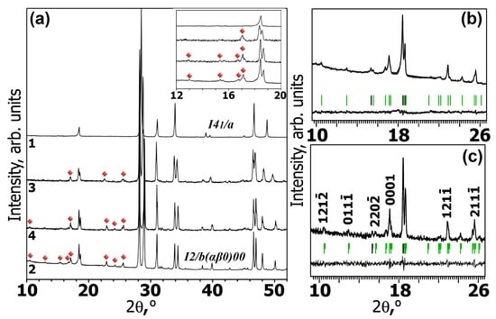
Figure 1.
(a) Parts of PXRD patterns of the Ag0.5Gd0.2Eu0.3WO4 (1), Ag0.2Gd0.3Eu3+0.3☐0.2WO4 (2) and AgxGd((2−x)/3)−0.33Sm0.03Eu3+0.30☐(1−2x−0.06)/3WO4 (x = 0.286 (3), 0.2 (4)) samples in 2θ ranges of 10–52°. The satellite reflections are indicated with red diamonds; (b,c) parts of experimental, calculated and difference XPD profiles in 2θ ranges of 9.6–26.3° after Le Bail decomposition of Ag0.2Gd0.3Eu3+0.3☐0.2WO4 ((b) Huber G670) and Ag0.2Gd0.27Sm0.03Eu3+0.30☐0.2WO4 ((c) Thermo ARL X’TRA, Thermo Fisher Scientific, Waltham, MA, USA). Black and green bars mark the positions of the main and satellite reflections, respectively. The indexation of some satellite reflections is shown.

Table 1.
Unit cell parameters and symmetry of AgxR((2−x)/3)−0.3Eu3+0.30☐(1−2x)/3WO4 (x = 0.5, 0.286, 0.2) solid solutions were determined from X-ray diffraction data.
3.2. Luminescence Properties
The photoluminescence emission (PL) and excitation (PLE) spectra of AxGSyE (x = 0.5, 0.286, 0.2; y = 0–0.03) tungstates are shown in Figure 2 and Figure 3. Figure 2a shows PLE spectra for the Eu3+ in Ag0.20Gd0.30−ySmyEu0.30☐0.20WO4 (y = 0–0.03) upon emission at 615 nm. The excitation spectra (PLE) contain a broad band (220–320 nm) and a group of narrow lines (320–550 nm). The luminescence spectra of AxGSyE (x = 0.5, 0.286, 0.2; y = 0–0.03) (λex = 395 nm) are shown in Figure 2b and Figure 3.
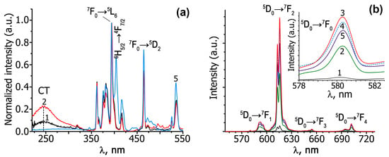
Figure 2.
Excitation (λem = 615 nm) (a) and luminescence (λex = 395 nm) (b) spectra of Ag0.20Eu0.60☐0.20WO4 (1) and Ag0.20Gd0.30−ySmyEu0.30☐0.20WO4 (y = 0 (2), 0.01 (3), 0.02 (4), 0.03 (5)) at room temperature. The luminescence spectra of the 5D0 → 7F0 transition for Eu3+ are shown in the inset (b).
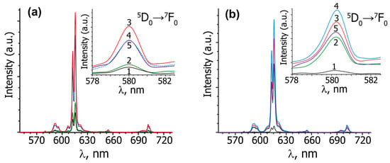
Figure 3.
The luminescence spectra (λex = 395 nm) of Ag0.5R0.20−ySmyEu0.30WO4 (a) and Ag0.286R0.271−ySmyEu0.30☐0.143WO4 (b) (R = Eu and y = 0 (1), R = Gd and y = 0 (2); R = Gd and y = 0.01 (3), 0.02 (4), 0.03 (5)) at room temperature. The luminescence spectra of the 5D0 → 7F0 transition for Eu3+ are shown in the inset (a,b).
The comparative integral intensity of the 5D0 → 7F2 luminescence for AgxEu3+(2−x)/3☐(1−2x)/3WO4 and AxGSyE scheelite-type phases is presented in Figure 4. The electric dipole transition 5D0 →7F2 at ~615 nm is the most intensive for all the luminescence spectra. The data on the effect of Gd3+ and Sm3+ co-activation on the 5D0 →7F2 transition luminescence intensity in AgxR((2−x)/3)−0.3−ySmyEu3+0.3☐(1−2x)/3WO4 (R = Eu, Gd, Sm; x = 0.50, 0.286, 0.20; y = 0–0.03) are summarized in Table S1 (Supporting Information). The comparison of the PL spectra and integral 5D0 → 7F2 emission intensity of Ag0.20Gd0.29Sm0.01Eu0.30WO4 and the commercially used Gd2O2S:Eu3+ red phosphor is shown in Figure 5.
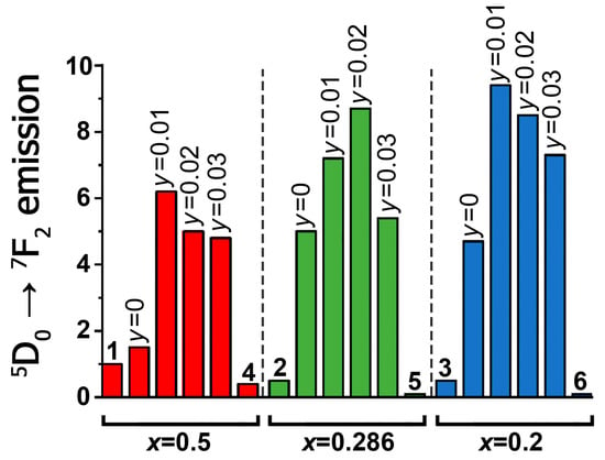
Figure 4.
Change in the integrated luminescence intensity of the 5D0 → 7F2 transition in Eu3+ (λex = 395 nm) for scheelite-related AgxEu3+(2−x)/3☐(1−2x)/3WO4 (x = 0.50 (1), 0.286 (2), 0.20 (3)), AgxSm(2−x)/3−0.3Eu0.3☐(1−2x)/3WO4 (x = 0.50 (4), 0.286 (5), 0.20 (6)) and AgxGd((2−x)/3)−0.3−ySmyEu3+0.3☐(1−2x)/3WO4 (x = 0.50, 0.286, 0.20; y = 0, 0.01, 0.02, 0.03). All intensities of the investigated phases are normalized to the value of the integral intensity of Ag0.50Eu0.50WO4 (1).
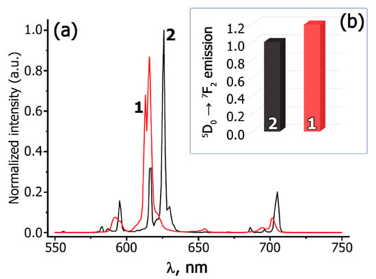
Figure 5.
Parts of room temperature PL (λex = 395 nm) spectra (a) and integral 5D0 → 7F2 emission intensity (b) of Ag0.20Gd0.29Sm0.01Eu0.30WO4 (1) and the commercially used phosphor Gd2O2S:Eu3+ (2). The luminescence (PL) spectra are normalized to the integral luminescence intensity of Gd2O2S:Eu3+.
To evaluate the behavior of thermal quenching, the PL spectra were studied at temperatures ranging from 300 to 700 K. Figure 6 and Figure 7 show the temperature-dependent PL spectra and the intensity of the 5D0 → 7F2 transition at ~615 nm.
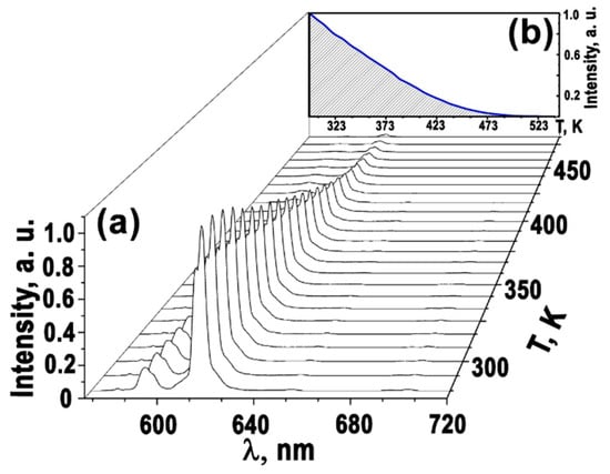
Figure 6.
Variation of PL spectra (a) and luminescence intensity of the 5D0 → 7F2 transition as a function of temperature—blue line (b) for Ag0.20Gd0.29Sm0.01Eu0.30☐0.20WO4 at ~615 nm.
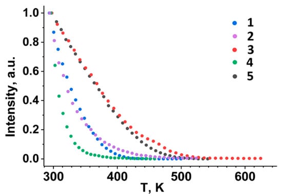
Figure 7.
The temperature dependences of the 5D0 → 7F2 luminescence intensity at ~615 nm of Ag0.5R0.20Eu0.30WO4 (R = Gd (1), Sm (2)), Ag0.20R0.30Eu0.30☐0.20WO4 (R = Gd (3), Sm (4)) and Ag0.20Gd0.29Sm0.01Eu0.30☐0.20WO4 (5).
4. Discussion
The present work is focused on the scheelite-type phases of AxGSyE (x = 0.5, 0.286, 0.2; y = 0.01, 0.02, 0.03, 0.3) with a variable composition. One can establish that the substitution of Ag+ by R3+ (R = Eu, Gd, Sm) leads to a noticeable distortion of the scheelite-like framework. The phase with x = 0.5 crystallizes in a tetragonal scheelite-like structure (I41/a). From the high-resolution synchrotron XRD data [20] for a sample with x = 0.5, it is found that there is no Ag+ and R3+ cations ordering.
We have previously analyzed that the formation of cationic vacancies in AgxGd((2−x)/3)−0.3Eu3+0.3☐(1−2x)/3WO4 [20] and AgxSm(2−x)/3☐(1−2x)/3WO4 [19] structures is accompanied by ordering of cations and vacancies with the formation of (3 + 1) D incommensurably modulated structures for AgxR(2−x)/3☐(1−2x)/3WO4 (R = Eu, Sm; x = 0.286, 0.2) phases. As revealed earlier for AgxGd((2−x)/3)−0.3Eu3+0.3☐(1−2x)/3WO4 (x = 0.286, 0.2) [20] and shown here for AxGSyE (x = 0.286, 0.2; y = 0.01, 0.02, 0.03), the structures of these scheelite-related phases have an incommensurately modulated character too. The AgxR(2−x)/3☐(1−2x)/3WO4 (R = Eu, Sm; x = 0.286, 0.2) aperiodic structures are built from two types of columns [… – (RO8/AgO8)–WO4 – …] and [… – ☐ – WO4 – …], which are extended along the c-axis. The structures of these phases are characterized by different distributions of Ag+/R3+ cations and vacancies in the A-cationic column of the scheelite structure. The specific coefficients α and β of the modulation vector q = αa* + βb* in incommensurately modulated structures, and the individual parameters of the atomic domains, define specific cations ordering for each modulated structure. In AgxR(2−x)/3☐(1−2x)/3WO4 (R = Eu, Sm; x = 0.286, 0.2) structures, two types of R–aggregates have been described: [R2O14] dimers and infinite [RO8]n chains from EuO8 polyhedra [19,20].
As an example, a portion of the Ag0.157Eu0.614☐0.229WO4 disordered structure is shown in the [001] projection in Figure 8a. [Eu2O14]-dimers are Eu-pairs surrounded by only WO4 tetrahedra and isolated from all other Eu3+ in the infinite [EuO8]n chains by vacancies and Ag+ cations (Figure 8b,c). The amount of [Eu2O14]-dimers in the AgxEu(2−x)/3☐(1−2x)/3WO4 framework depends on x. In the structures of AgxEu(2−x)/3☐(1−2x)/3WO4, the number of Eu3+ cations involved in the formation of [Eu2O14]-dimers increases from 15.56% to 28.33%, with decreasing x from 0.238 to 0.157 [20]. There is a relationship between the number of [Eu2O14]-dimers in the structure and the luminescence characteristics (total () and intrinsic () quantum yields and lifetimes of Eu3+ (5D0) (τobs)) [27]. The values of the luminescent characteristics increase with an increase in the number of Eu3+ dimers. However, the maximal of the luminescence characteristics is observed in the disordered structure of Na0.5Eu0.5MoO4.
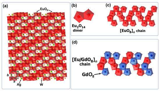
Figure 8.
ab-projection of a part of the supercell (8a × 10b × 1c) of the aperiodic structure Ag0.157Eu0.614☐0.229WO4 refined in Ref. [20] (a). The Ag, W and O atoms are shown in green, yellow and red, respectively. AgO8 polyhedra and WO4 tetrahedra are not shown. [Eu2O14] dimers are marked with white ellipses. Shown are [Eu2O14] dimers (b) and [EuO8]n chains (c) of EuO8 polyhedra in the Ag0.157Eu0.614☐0.229WO4 and [Eu/GdO8]n chains (d) in the AgxGd((2−x)/3)−0.3Eu3+0.3☐(1−2x)/3WO4 structure.
The phosphors AgxGd((2−x)/3)−0.3Eu3+0.3☐(1−2x)/3WO4 at the values of x (0.5–0) have better characteristics for emitters than AgxEu(2−x)/3☐(1−2x)/3WO4 [20]. In AgxGd((2−x)/3)−0.3Eu3+0.3☐(1−2x)/3WO4 the emission of Eu3+ (λex = 395 nm) increases with the Gd3+ content from 0.2 (x = 0.5) to 0.3 (x = 0.2) by a factor of 3 compared to the phosphors AgxEu(2−x)/3☐(1−2x)/3WO4 (Figure 4). The replacement of Eu3+ cations by Gd3+ is accompanied by the destruction of infinite [EuO8]n chains and the formation of [Eu2O14]-dimers and/or isolated EuO8 polyhedra inside them (Figure 8d).
The PLE spectra of AgxGd((2−x)/3)−0.3Eu3+0.3☐(1−2x)/3WO4 and AgxEu(2−x)/3☐(1−2x)/3WO4 are characterized by broad (220–320 nm) and narrow (320–500 nm) excitation bands. The broad excitation line peaking at ~240 nm is due to charge transfer (CT) from the 2p orbital of oxygen to the 3d orbital of tungsten in the WO42− tetrahedron [28,29] and coincides with the O2− → Eu3+ CT band. Co-activation with Gd3+ and Sm3+ ions leads to a noticeable decrease in intensity of CT bands in comparison with Gd3+ single-doped phosphors (Figure 2a). All the phases reveal characteristic lines in the spectral region from 320 to 500 nm, which originate from 4f–4f transitions of Eu3+ and Sm3+ (for Sm-containing compounds). The main absorption bands in the PLE spectra are associated with the 7F0 → 5L6 Eu3+ transition at 395 nm and the 6H5/2 → 4F7/2 Sm3+ transition at 405 nm.
In the PL spectra of AgxR((2−x)/3)−0.3Eu3+0.30☐(1−2x)/3WO4 (R = Eu, Gd; x = 0.5, 0.286, 0.2) and AxGSyE (x = 0.5, 0.286, 0.2; y = 0.01, 0.02, 0.03) in the region of 570–650 nm, the characteristic lines are observed for Eu3+ cations due to the 5D0 → 7FJ (J = 0, 1, 2) transitions (Figure 2b and Figure 3). In the luminescence spectra of all phases, the most intense electric dipole transition 5D0 → 7F2 is observed at ~615 nm, which indicates the absence of a center of symmetry in the polyhedra of Eu3+ cations [30,31]. The magnetic dipole transition 5D0 → 7F1 is observed at 590 nm. The intensity of this transition depends a little on the environment of the luminescent cation, since it has a magnetic dipole nature. The asymmetry coefficient (R/O), Iint(5D0 → 7F2)/Iint(5D0 → 7F1) [32] is usually used to estimate the symmetry of the Eu3+ polyhedron. The high values of the R/O ratio for the studied tungstates (Figure 2 and Figure 3) indicate a non-centrosymmetric local environment of Eu3+ in these phases. Further evidence of the non-centrosymmetric environment of europium cations in the PL structure of AxGSyE (x = 0.5, 0.286, 0.2; y = 0.01, 0.02, 0.03) is the presence of the 5D0 → 7F0 transition band at ~580 nm (insert in Figure 2b and Figure 3). In europium-containing phases, the 5D0 → 7F0 transition is forbidden for electric and magnetic dipole interactions. For this reason, the intensity of this transition is very low or not observed. The number of peaks in the region of the 5D0 → 7F0 transition indicates often the number of europium polyhedra in the compound. In the studied scheelites for all the compositions, only one peak is found in the region of the 5D0 → 7F0 transition (insert in Figure 2b and Figure 3). This is because the same environment of Eu3+ cations in the studied phases, and because europium cations occupy only one polyhedron [33].
In accordance with Table S1, the change of the symmetry from tetragonal for Ag0.5R0.2−ySmyEu3+0.3WO4 (R = Eu, Gd, Sm) to monoclinic for AxGSyE (x = 0.286, 0.20; y= 0.01, 0.02, 0.03) practically does not change the value of the R/O ratio and the position of the peaks in the region of 5D0 → 7F0 transitions (Figure 2b and Figure 3). Figure 4 shows the comparative integral luminescence intensity of the 5D0 → 7F2 transition (Eu3+) for AgxEu3+(2−x)/3☐(1−2x)/3WO4, AgxSm(2−x)/3−0.3Eu0.3☐(1−2x)/3WO4 and AxGSyE (x = 0.50, 0.286, 0.20; y = 0.01, 0.02, 0.03) (λex = 395 nm). According to Figure 4 and Table S1, the luminescence intensity of the 5D0 → 7F2 transition is increased from ~2 times (x = 0.2, y = 0.01 and x = 0.286, y = 0.02) to ~4 times (x = 0.5, y = 0.01) during Gd3+ and Sm3+ co-activation. The integrated emission intensity of Ag0.20Gd0.29Sm0.01Eu0.30WO4 in the 5D0 → 7F2 transition is ~20% higher than that of the Gd2O2S:Eu3+ phosphor, which is used in industry (Figure 5). This confirms that Ag0.286Gd0.252Sm0.02Eu0.30WO4 and Ag0.20Gd0.29Eu0.30Sm0.01WO4 with a (3 + 1)D monoclinic incommensurately modulated structure are more attractive for use as red-emitting phosphors for LEDs.
However, an increase in Sm3+ concentration leads to a noticeable decrease in Eu3+ luminescence intensity at the 5D0 → 7F2 transition. The intensity of the 5D0 → 7F2 transition of the AxGSyE (x = 0.5, 0.286, 0.2) phases is reduced almost 24 and 73 times with increasing y from 0.03 to 0.20 (x = 0.5) and 0.30 (x = 0.20), respectively. A further increase in the concentration of Sm3+ cations leads to lower europium luminescence intensity (Figure 4 and Table S1). The phosphors with a high concentration of Sm3+ are not related to the materials with a high luminescence efficiency [34,35] due to the large number of Sm3+ energy levels in the visible region of the spectrum (Figure 9). The presence of a large number of energy levels in the Sm3+ cation promotes energy transfer between the Sm3+ and Eu3+ cations. However, the opposite effect of energy transfer to the Sm3+ cation is also possible, with further luminescence quenching at defects or due to cross relaxation. On the contrary, when Gd3+ cations are introduced into the matrix, the reverse energy transfer to the Gd3+ cation is impossible due to the large energy difference (3.96 eV) between the ground and first excited states (Figure 9). Thus, when energy is transferred from Gd3+ to Eu3+, reverse transfer to the Gd3+ cation is impossible. For this reason, the introduction of Gd3+ cations into a matrix with europium is more efficient than the introduction of Sm3+.
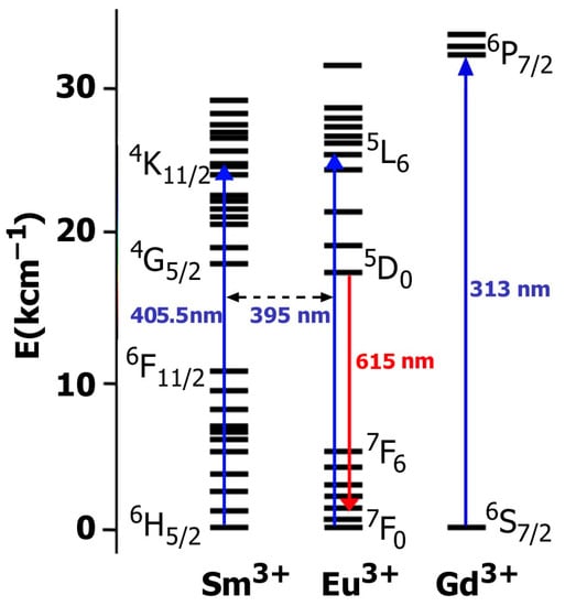
Figure 9.
Energy level of Sm3+, Eu3+ and Gd3+ ions. Blue arrows denote the transitions from the ground state to excited levels. Red arrow denotes radiative relaxation from the excited level to the ground state.
Figure 6 shows the luminescence spectra of Ag0.20Gd0.29Sm0.01Eu0.30☐0.20WO4 as a function of temperature. The luminescence intensity of the 5D0 → 7F2 transition in the region of ~615 nm decreases with increasing temperature and completely disappears at T = 473 K. As the temperature increases, the rapid quenching of the luminescence seems to be due to the nonradiative population of the excited states. The temperature dependencies of the luminescence intensity of the 5D0 → 7F2 transition at ~615 nm can be compared for the Ag0.20Gd0.29Sm0.01Eu0.30☐0.20WO4 and AgxR((2−x)/3)−0.3−ySmyEu3+0.3☐(1−2x)/3WO4 phases (Figure 7). The observed thermal quenching depends on both the crystal structure of the compounds and the Sm3+ concentration. The thermal behavior of PL in Ag0.20Gd((2−x)/3)−0.3−ySmyEu3+0.30☐0.20WO4 (y = 0, 0.01, 0.30) with the incommensurately modulated (3 + 1) D monoclinic structure differs from one of the Ag0.5R0.20Eu0.30WO4 (R = Gd, Sm) phases with the tetragonal I41/a scheelite structure. The red luminescence of Eu3+ (transition 5D0 → 7F2) in the monoclinic phases with y = 0 and y = 0.01 is more thermally stable than that of the tetragonal phases. However, the full substitution of Gd3+ by Sm3+ (y = 0.30) in Ag0.20R0.30Eu0.30WO4 leads to lower thermal stability of the 5D0 → 7F2 luminescence of Eu3+ cation.
The use of synthesized phosphors offers advantages over commercially available Gd2O2S:Eu3+ phosphor. The PL spectrum of Ag0.20Gd0.29Sm0.01Eu0.30WO4 slightly shifted towards a smaller wavelength (Figure 5). The calculated CIE coordinates (Figure 10) for Gd2O2S:Eu3+ are (0.6312; 0.3562), whereas for Ag0.20Gd0.29Sm0.01Eu0.30WO4 they are (0.6402; 0.3528). It can be observed from the CIE coordinates that the synthesized phosphor is closer to the red standard of the National Television Standard Committee (NTSC) system (0.67; 0.33). The color purity, calculated by the formula reported in [36], shows the values as 89% for Gd2O2S:Eu3+ and 92% for Ag0.20Gd0.29Sm0.01Eu0.30WO4. Based on these results, the synthesized phosphors have potential for use in UV-excited LED packages.
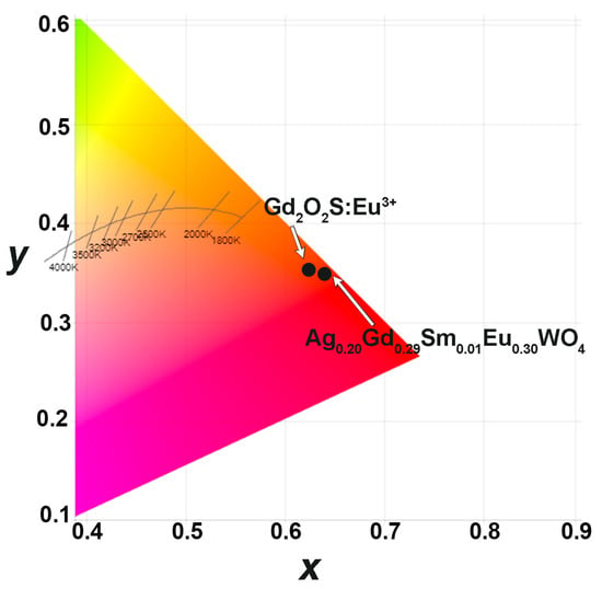
Figure 10.
CIE coordinated for Gd2O2S:Eu3+ and Ag0.20Gd0.29Sm0.01Eu0.30WO4.
5. Conclusions
Gd3+ and Sm3+ co-activation, the effect of cation substitutions and the formation of cation vacancies in AgxGd((2−x)/3)−0.3−ySmyEu3+0.30☐(1−2x−2y)/3WO4 (x = 0.5, 0.286, 0.2; y = 0.01, 0.02, 0.03) with a scheelite-type structure have been studied here as a factor for managing the luminescence properties. The XRD study of AgxGd((2−x)/3)−0.3−yEu3+0.30Smy☐(1−2x−2y)/3WO4 (x = 0.286, 0.2; y = 0.01, 0.02, 0.03) shows that its structures have incommensurately modulated character, as well as the structures of the other cation deficient scheelite-related phases. The strongest line at 395 nm in the PLE spectra of the synthesized phosphors is in good agreement with GaN LED chips emitting in the near-ultraviolet range. The electric dipole transition 5D0 → 7F2 at ~615 nm has the maximum intensity for all the PL spectra, and its intensity increases from ~2 times (x = 0.2, y = 0.01 and x = 0.286, y = 0.02) to ~4 times (x = 0.5, y = 0.01) when co-activation with Gd3+ and Sm3+ is implemented. Ag0.286Gd0.252Sm0.02Eu0.30WO4 and Ag0.20Gd0.29Sm0.01Eu0.30WO4 with a monoclinic incommensurately modulated (3 + 1)D monoclinic structure are very attractive as phosphor materials for converting near-UV radiation in LEDs. A considerable thermal quenching of the observed luminescence is revealed. The temperature dependence and behavior of the considered phases are influenced by the crystal structure of scheelite-type tungstates and the concentration of Sm3+ ions.
Supplementary Materials
The following supporting information can be downloaded at: https://www.mdpi.com/article/10.3390/ma16124350/s1, Table S1: Positions (λmax, nm) and integral intensities (Iint) of two maximum bands on PL spectra of AgxR((2−x)/3)−0.3−ySmyEu3+0.3☐(1−2x)/3WO4 (R = Eu, Gd, Sm; x = 0.50, 0.286, 0.20; y = 0, 0.01, 0.02, 0.03) scheelite-type phases. All samples are measured under the same conditions.
Author Contributions
Conceptualization, D.V.D. and V.A.M.; methodology, V.A.M. and E.G.K.; software, D.V.D. and I.I.L.; validation, D.V.D., I.I.L., A.V.I. and B.I.L.; formal analysis, D.V.D., I.I.L., A.V.I. and A.A.S.; investigation, D.V.D., I.I.L., A.V.I. and A.A.S.; resources, V.A.M., E.G.K. and B.I.L.; data curation, D.V.D., I.I.L., A.V.I. and A.A.S.; writing—original draft preparation, D.V.D. and V.A.M.; writing—review and editing, D.V.D., B.I.L., E.G.K., I.I.L. and V.A.M.; visualization, D.V.D. and V.A.M.; supervision, V.A.M. and B.I.L.; project administration, V.A.M. and B.I.L.; funding acquisition, V.A.M. and B.I.L. All authors have read and agreed to the published version of the manuscript.
Funding
The study was supported by the Development Program of the Interdisciplinary Scientific and Educational School of Lomonosov Moscow State University’s “The future of the planet and global environmental change”, and the state assignment of the Chemistry Department of Moscow State University (Agreement No. AAAA-A21-121011590086-0) and state of the Russian Federation (122011300125-2). A.A.S. and E.G.K. are grateful to the support from the Ministry of Science and Higher Education of the Russian Federation (project No. 0273-2021-0008). A.V.I. is grateful to the scientific project of the Ministry of Education and Science of the Russian Federation FEUZ–2023–0014. I.I.L. acknowledges the support from the Research Program No. AAAA–A19–119031890025–9 (ISSC UB RAS).
Institutional Review Board Statement
Not applicable.
Informed Consent Statement
Not applicable.
Data Availability Statement
Data will be available from the corresponding author on reasonable request.
Acknowledgments
Additional spectroscopic studies were carried out in the Research Center for Nanomaterials at the Ural Federal University (Ekaterinburg, Russia).
Conflicts of Interest
The authors declare no conflict of interest.
References
- Shur, M.S.; Zukauskas, A. Solid-state lighting: Toward superior illumination. Proc. IEEE. 2005, 93, 1691. [Google Scholar] [CrossRef]
- Yam, F.K.; Hassan, Z. Innovative advances in LED technology. Microelectr. J. 2005, 36, 129. [Google Scholar] [CrossRef]
- Haque, M.M.; Lee, H.-I.; Kim, D.-K. Luminescent properties of Eu3+-activated molybdate-based novel red-emitting phosphors for LEDs. J. Alloys Compd. 2009, 481, 792. [Google Scholar] [CrossRef]
- Shao, Q.; Li, H.; Wu, K.; Dong, Y.; Jiang, J. Photoluminescence studies of red-emitting NaEu (WO4)2 as a near-UV or blue convertible phosphor. J. Lumin. 2009, 129, 879. [Google Scholar] [CrossRef]
- Zhang, Y.; Jiao, H.; Du, Y. Luminescent properties of HTP AgGd1−xW2O8: Eux3+ and AgGd1−x(W1−yMoy)2O8: Eux3+ phosphor for white LED. J. Lumin. 2011, 131, 861–865. [Google Scholar] [CrossRef]
- Thangaraju, D.; Durairajan, A.; Balaji, D.; Moorthy Babu, S.; Hayakawa, Y. Novel KGd1−(x+y)EuxBiy(W1−zMozO4)2 nanocrystalline red phosphors for tricolor white LEDs. J. Lumin. 2013, 134, 244. [Google Scholar] [CrossRef]
- Wang, Z.; Zhong, J.; Jiang, H.; Wang, J.; Liang, H. Controllable Synthesis of NaLu(WO4)2:Eu3+ Microcrystal and Luminescence Properties for LEDs. Cryst. Growth Des. 2014, 14, 3767. [Google Scholar] [CrossRef]
- Zhang, W.; Li, J.; Wang, Y.; Long, J.; Qiu, K. Synthesis and luminescence properties of NaLa(MoO4)2−xAGx:Eu3+ (AG = SO42−, BO33−) red phosphors for white light emitting diodes. J. Alloys Compd. 2015, 635, 16. [Google Scholar] [CrossRef]
- Li, G.; Wei, Y.; Li, Z.; Xu, G. Synthesis and photoluminescence of Eu3+ doped CaGd2(WO4)4 novel red phosphors for white LEDs applications. Opt. Mater. 2017, 66, 253. [Google Scholar] [CrossRef]
- Li, W.; Yang, M.; Kang, F.; Sun, G. Color selection and red fluorescence enhancement through the controllable energy transfer in NaxCa1-2xWO4:Eux3+ phosphor for UV converted LEDs. Mater. Chem. Phys. 2018, 207, 396. [Google Scholar] [CrossRef]
- Lu, D.; Gong, X.; Chen, Y.; Huang, J.; Lin, Y.; Luo, Z.; Huang, Y. Synthesis and photoluminescence characteristics of the LiGd3(MoO4)5:Eu3+ red phosphor with high color purity and brightness. Opt. Mater. Exp. 2018, 8, 259–269. [Google Scholar] [CrossRef]
- Guo, W.; Chen, Y.; Lin, Y.; Gong, X.; Luo, Z.; Huang, Y. Spectroscopic analysis and laser performance of Tm3+:NaGd(MoO4)2 crystal. J. Phys. D Appl. Phys. 2008, 41, 115409. [Google Scholar] [CrossRef]
- Chen, Y.J.; Lin, Y.F.; Guo, W.J.; Gong, X.H.; Huang, J.H.; Luo, Z.D.; Huang, Y.D. Efficient 1.9 μm monolithic Tm3+:NaLa(MoO4)2 micro-laser. Laser Phys. Lett. 2012, 9, 141–144. [Google Scholar] [CrossRef]
- Yu, Y.; Zhang, L.; Huang, Y.; Lin, Z.; Wang, G. Growth, crystal structure, spectral properties and laser performance of Yb3+:NaLu(MoO4)2 crystal. Laser Phys. 2013, 23, 105807. [Google Scholar] [CrossRef]
- Feng, J.; Xu, J.; Zhu, Z.; Wang, Y.; You, Z.; Li, J.; Wang, H.; Tu, C. Spectroscopic properties and orthogonally polarized dual-wavelength laser of Yb3+:NaY(WO4)2 crystals with high Yb3+ concentrations. J. Alloys Compd. 2013, 566, 229. [Google Scholar] [CrossRef]
- Meert, K.W.; Morozov, V.A.; Abakumov, A.M.; Hadermann, J.; Poelman, D.; Smet, P.F. Energy transfer in Eu3+ doped scheelites: Use as thermographic phosphor. Optic Exp. 2014, 22, A961. [Google Scholar] [CrossRef]
- Wang, J.; Bu, Y.; Wang, X.; Seo, H.J. A novel optical thermometry based on the energy transfer from charge transfer band to Eu3+-Dy3+ ions. Sci. Rep. 2017, 7, 6023. [Google Scholar] [CrossRef]
- Zhou, X.; Wang, R.; Xiang, G.; Jiang, S.; Li, L.; Luo, X.; Pang, Y.; Tian, Y. Multi-parametric thermal sensing based on NIR emission of Ho (III) doped CaWO4 phosphors. Opt. Mater. 2017, 66, 12. [Google Scholar] [CrossRef]
- Morozov, V.A.; Deyneko, D.V.; Basovich, O.M.; Khaikina, E.G.; Spassky, D.A.; Morozov, A.V.; Chernyshev, V.V.; Abakumov, A.M.; Hadermann, J. Incommensurately Modulated Structures and Luminescence Properties of the AgxSm(2−x)/3WO4 (x = 0.286, 0.2) Scheelites as Thermographic Phosphors. Chem. Mater. 2018, 30, 4788. [Google Scholar] [CrossRef]
- Morozov, V.A.; Batuk, D.; Batuk, M.; Basovich, O.M.; Khaikina, E.G.; Deyneko, D.V.; Lazoryak, B.I.; Leonidov, I.I.; Abakumov, A.M.; Hadermann, J. Luminescence Property Upgrading via the Structure and Cation Changing in AgxEu(2−x)/3WO4 and AgxGd(2−x)/3−0.3Eu0.3WO4. Chem. Mater. 2018, 29, 8811. [Google Scholar] [CrossRef]
- Wang, Z.; Liang, H.; Gong, M.; Su, Q. A potential red-emitting phosphor for LED solid-state lighting. Electrochem. Solid–State Lett. 2005, 8, H33. [Google Scholar] [CrossRef]
- Wang, Z.L.; Liang, H.B.; Gong, M.L.; Su, Q. Novel red phosphor of Bi3+, Sm3+ co-activated NaEu(MoO4)2. Opt. Mater. 2007, 29, 896. [Google Scholar] [CrossRef]
- Mo, F.; Zhou, L.; Pang, Q.; Gong, F.; Liang, Z. Potential red-emitting NaGd(MO4)2:R (M = W, Mo, R = Eu3+, Sm3+, Bi3+) phosphors for white light emitting diodes applications. Ceram. Inter. 2012, 38, 6289. [Google Scholar] [CrossRef]
- Le Bail, A.; Duroy, H.; Fourquet, J.L. Ab-initio structure determination of LiSbWO6 by X-ray powder diffraction. Mater. Res. Bull. 1988, 23, 447. [Google Scholar] [CrossRef]
- Petricek, V.; Dusek, M.; Palatinus, L. Crystallographic computing system JANA2006: General features. Z. Kristallogr. 2014, 229, 345. [Google Scholar] [CrossRef]
- Vokhmintsev, A.S.; Minin, M.G.; Chaykin, D.V.; Weinstein, I.A. A high-temperature accessory for measurements of the spectral characteristics of thermoluminescence. Instrum. Exp. Tech. 2014, 57, 369. [Google Scholar] [CrossRef]
- Arakcheeva, A.; Logvinovich, D.; Chapuis, G.; Morozov, V.; Eliseeva, S.V.; Bunzli, J.-C.G.; Pattison, P. The luminescence of NaxEu3+(2−x)/3MoO4 scheelites depends on the number of Eu-clusters occurring in their incommensurately modulated structure. Chem. Sci. 2012, 3, 384. [Google Scholar] [CrossRef]
- Shi, S.; Liu, X.; Gao, J.; Zhou, J. Spectroscopic properties and intense red-light emission of (Ca, Eu, M)WO4 (M = Mg, Zn, Li). Spectr. Acta Sect. A. 2008, 69, 396. [Google Scholar] [CrossRef]
- Blasse, G. The luminescence of Closed–shell transition–metal complexes. New developments. In Structure and Bonding; Springer: Berlin/Heidelberg, Germany, 1980. [Google Scholar]
- Wiglusz, R.J.; Bednarkiewicz, A.; Strek, W. Role of the Sintering Temperature and Doping Level in the Structural and Spectral Properties of Eu-Doped Nanocrystalline YVO4. Inorg. Chem. 2012, 51, 1180. [Google Scholar] [CrossRef]
- Tanner, P. Some misconceptions concerning the electronic spectra of tri-positive europium and cerium. Chem. Soc. Rev. 2013, 42, 5090. [Google Scholar] [CrossRef]
- Werts, M.H.V.; Jukes, R.T.F.; Verhoeven, J.W. The emission spectrum and the radiative lifetime of Eu3+ in luminescent lanthanide complexes. Phys. Chem. Chem. Phys. 2002, 4, 1542. [Google Scholar] [CrossRef]
- Blasse, G.; Bril, A.; Nieuwpoort, W.C. On the Eu3+ fluorescence in mixed metal oxides: Part I—The crystal structure sensitivity of thr intensity ratio of electric and magnetic dipole emission. J. Phys. Chem. Solids. 1966, 27, 1587. [Google Scholar] [CrossRef]
- Wang, X.Y.; Lin, H.; Li, C.M.; Tanabe, S. Derivation of quantum yields for visible emission transitions of Sm3+ in heavy metal tellurite glass. Opt. Commun. 2007, 276, 122. [Google Scholar] [CrossRef]
- Morozov, V.A.; Raskina, M.V.; Lazoryak, B.I.; Meert, K.W.; Korthout, K.; Smet, P.F.; Poelman, D.; Gauquelin, N.; Verbeeck, J.; Abakumov, A.M.; et al. Crystal Structure and Luminescent Properties of R2−xEux(MoO4)3 (R = Gd, Sm) Red Phosphors. Chem. Mater. 2014, 26, 7124. [Google Scholar] [CrossRef]
- Deyneko, D.V.; Nikiforov, I.V.; Lazoryak, B.I.; Spassky, D.A.; Leonidov, I.I.; Stefanovich SYu Petrova, D.A.; Aksenov, S.M.; Burns, P.C. Ca8MgSm1−x(PO4)7:xEu3+, promising red phosphors for WLED application. J. Alloys Compd. 2019, 776, 897. [Google Scholar] [CrossRef]
Disclaimer/Publisher’s Note: The statements, opinions and data contained in all publications are solely those of the individual author(s) and contributor(s) and not of MDPI and/or the editor(s). MDPI and/or the editor(s) disclaim responsibility for any injury to people or property resulting from any ideas, methods, instructions or products referred to in the content. |
© 2023 by the authors. Licensee MDPI, Basel, Switzerland. This article is an open access article distributed under the terms and conditions of the Creative Commons Attribution (CC BY) license (https://creativecommons.org/licenses/by/4.0/).