Article

Production of Ceramics/Metal Oxide Nanofibers via Electrospinning: New Insights into the Photocatalytic and Bactericidal Mechanisms

by
Jari S. Algethami
1,2,
Touseef Amna
3,*,
Laila S. Alqarni
4,
Aisha A. Alshahrani
5,
Mohsen A. M. Alhamami
1,
Amal F. Seliem
1,
Badria H. A. Al-Dhuwayin
1 and
M. Shamshi Hassan
5,*
1
Department of Chemistry, College of Science and Arts, Najran University, Najran 11001, Saudi Arabia
2
Promising Centre for Sensors and Electronic Devices (PCSED), Advanced Materials and Nano-Research Centre, Najran University, Najran 11001, Saudi Arabia
3
Department of Biology, College of Science, Al-Baha University, Albaha 65799, Saudi Arabia
4
Department of Chemistry, College of Science, Imam Mohammad Ibn Saud Islamic University (IMSIU), Riyadh 11432, Saudi Arabia
5
Department of Chemistry, College of Science, Al-Baha University, Albaha 65799, Saudi Arabia
*
Authors to whom correspondence should be addressed.
Materials 2023, 16(14), 5148; https://doi.org/10.3390/ma16145148
Submission received: 12 June 2023 / Revised: 16 July 2023 / Accepted: 17 July 2023 / Published: 21 July 2023
(This article belongs to the Special Issue Functional Composite Materials for Environmental Applications)

Abstract

:
Environmental pollution is steadily rising and is having a negative influence on all living things, especially human beings. The advancement of nanoscience in recent decades has provided potential to address this issue. Functional metal oxide nanoparticles/nanofibers have been having a pull-on effect in the biological and environmental domains of nanobiotechnology. Current work, for the first time, is focusing on the electrospinning production of Zr0.5Sn0.5TiO3/SnO2 ceramic nanofibers that may be utilized to battle lethal infections swiftly and inexpensively. By using characterizations like XRD, FT–IR, FESEM, TEM, PL, and UV–Vis–DRS, the composition, structure, morphology, and optical absorption of samples were determined. The minimum inhibitory concentration (MIC) approach was used to investigate the antibacterial activity. Notably, this research indicated that nanofibers exert antibacterial action against both Gram-positive and Gram-negative bacteria with a MIC of 25 µg/mL. Furthermore, negatively charged E. coli was drawn to positively charged metal ions of Zr0.5Sn0.5TiO3/SnO2, which showed a robust inhibitory effect against E. coli. It was interesting to discover that, compared to pure TiO2, Zr0.5Sn0.5TiO3/SnO2 nanofibers revealed increased photocatalytic activity and exceptional cyclability to the photodegradation of Rhodamine B. The composite completely degrades dye in 30 min with 100% efficacy and excellent (97%) reusability. The synergetic effects of Zr0.5Sn0.5TiO3 and SnO2 may be responsible for increased photocatalytic and bactericidal activity.

1. Introduction

Environmental pollution is not a recent phenomenon, but it continues to be the greatest threat to mankind and a major factor of various illnesses and mortality. Worldwide urbanization, industrialization, mining, and excavation are human activities that have the greatest impact on the environment. Indisputably, water pollution is also a big concern in today’s world and a serious hazard that causes several problems for humans and the environment. Water pollution must be halted since it has been harming land and water species for years by entering lakes, rivers, streams, and seas. There are reports in the literature that an increasing number of individuals lack access to fresh, hygienic water. The water industry currently uses certain conventional chemical disinfectants, but it is alarming that many of these are carcinogens. Additionally, the need for more effective substitutes is growing as microorganisms’ resistance to conventional chemical disinfectants increases. E. coli and Enterococci are found in high numbers in the feces of animals; they are regarded as reliable indicators of potential damage to human health from fecal pathogens in fresh, untreated sewage [1]. Furthermore, swimming and boating are prevalent pleasurable outdoor water leisure activities that are deeply ingrained in western society [2]. However, these actions produce a significant financial expense owing to the risk of associated waterborne infections produced by harmful microbes [2]. Pathogens that cause waterborne diseases are commonly found in recreational waters due to fecal contamination [3]. The removal of pathogenic bacteria from water is a critical operation for drinking and sanitation systems, particularly in light of the increased occurrence of water-borne diseases [4]. Water pollution control has become increasingly important in recent years. Considering the growing ecological pollution and energy crises, photocatalysis, a green technique, exhibits a vital role in the solar energy transformation and the disintegration of organic pollutants. The advancement of applied microbiology and nanotechnology in recent decades has provided potential to address this issue [5]. Nanotechnology is a flourishing scientific discipline with numerous applications, including medicine, semiconductor manufacture, environmental remediation, and the production of personal care items. Because of its wide range of uses, there has been a tremendous increase in the manufacture of customized functional nanomaterials. The developed nanomaterials are distinguished by having one or more dimensions smaller than 100 nm [6]. Recent breakthroughs in nanotechnology have made it possible to effectively produce, characterize, and modify the functional properties of nanoparticles for biomedical purposes as well. Functional nanomaterials are having an impact on biological and therapeutic applications [7]. Nanoparticles display distinct features, since they are different in terms of size, distribution, and form [8,9]. Recent decades have seen an increase in research into the synthesis and characterization of nanoparticles owing to their numerous uses, notably in the fields of cosmetics, diagnostics, health care, environmental cleaning, pharmaceuticals, medical implants, and the energy sector [10,11]. Furthermore, in the last few decades, there has been an increase in the production of functional one-dimensional (1D) nanostructures, for instance, nanofibers, nanowires, nanotubes, nanorods, nanobelts, and so on; this is owing to their distinctive chemical and physical chattels that differ from their bulk equivalents and their potential applications in nanodevices as components and intersects [12]. In comparison to their film and particulate counterparts, nanofibrous photocatalysts not only have a large specific surface area, which allows their surface active sites to be more efficiently available to reactants, but they also have a high length-to-diameter ratio, which facilitates photocatalyst separation [13]. Electrospinning is a simple, effective, and convenient method of producing polymer/polymer-containing inorganic materials and inorganic/ceramics fibers [14]. Interestingly, it was also revealed that electrospun nanofibrous photocatalysts possess excellent photocatalytic efficiency and reuse proficiency [15].
The TiO2 is frequently regarded as the most ideal material for prevalent ecological uses because of its strong photoelectrochemical activity and stability against photo and chemical corrosion [12]. However, due to the inefficient usage of photogenerated electron–hole pairs, more research is required to increase the activity of this semiconductor. Suppressing the recombination of photogenerated electrons and holes in a semiconductor particulate system is critical for boosting the net charge transfer efficiency in photocatalysis and solar cells [2]. In order to achieve this goal, a variety of approaches have been applied, including the alteration of the TiO2 surface using noble materials [16,17] and the pairing of two semiconductor particles using unlike Fermi planes [18].
Owing to the structural similarity between the two oxides, the TiO2–SnO2 system has been the focus of several investigations, including of semiconductors [19]. The conduction band of SnO2 works as a drain for photogenerated electrons when two semiconductor particles are linked [18,20]. Because photogenerated holes flow in the conflicting direction, they concentrate in the TiO2 particle’s valence band, resulting in the proficient spatial parting of photogenerated charges and the suppression of recombination. Furthermore, the Sn dopant increases the photoinduced charge rate of TiO2 [21]. To enhance the thermal stability and photocatalytic action, metal oxides such as ZnO2, SiO2, La2O3, etc., have also been utilized [14,15]. Moreover, the distinctive features of zirconia, such as the high refractive index, broad optical band gap, low absorption, and dispersion in the visible and near-infrared spectrum areas [22,23] are appealing. Previously, it was observed that adding ZrO2 to TiO2 improved the phase stability, surface area and ultimately photocatalytic effects [24,25,26]. Interestingly, earlier works have also reported the formation of zirconium–stannate titanate (Zr1−xSnxTiO4) thin films via the spin coating technique [27]. Additionally, the use of biomedical-grade ZrO2 demonstrates the potential mechanical properties of oxide ceramics. It is emerging as an important catalyst in a variety of photocatalytic and piezoelectric applications, and in ceramics production and dentistry for healing purposes [28]. Despite its potential medicinal uses, few investigations on the significance of ZrO2 as an antimicrobial, anticancer, and antioxidant agent have been published. In this regard, Gupta et al. demonstrated the cytotoxic effect of ZrO2 nanoparticles on A549 cells and their antibacterial action on E. coli [29]. There have been numerous cases of using doping in the ZrO2 structure to enhance its antibacterial action. Imran and colleagues published a study on the antibacterial properties of Fe3O4-doped ZrO2 nanoparticles. Their bactericidal effect against S. aureus, E. coli and B. subtilis has been increased by doping [30]. Jangra et al. discovered that ZrO2 has antibacterial action solely against E. coli, whereas ZrO2 complexes are active against both E. coli and S. aureus [31]. Another study looked at the antibacterial efficacy of ZrO2 nanoparticles against Staphylococcus strains. The findings showed their low activity at squat concentrations, and vice versa, both in the case of S. aureus and the mutant, respectively [32].
Conclusively, considering the noteworthy properties of SnO2, ZrO2 and TiO2 in the current investigation, we aimed to design highly efficient Zr0.5Sn0.5TiO3/SnO2 (ZSTS) nanofibrous photocatalysts via the electrospinning method. Hitherto, there has been no published report regarding the synthesis, characterization and, more to the point, the bactericidal effect of these composite ceramics nanofibers. Herein, a first attempt was made to fabricate high-surface-to-area-ratio ZSTS ceramics nanofibers. Moreover, these high-aspect-ratio nanofibers have been investigated for their photocatalytic and antibacterial activities. These novel ceramics/metal oxide nanofibers will serve as the material of choice for environmental remediation, especially when aiming to control the quality of wastewater. Our research demonstrates that multifunctional ZSTS nanofibers may be a commendable candidate for use in health care and environmental pollution control systems.

2. Materials and Methods

2.1. Materials

Sigma-Aldrich from the United States supplied the polyvinyl acetate (PVAc, Mw = 500,000 g/mol), tin(II)2-ethylhexanoate (98%), and zirconyl chloride octahydrate (98%). N, N-dimethylformamide (DMF, 99.5 assay) and titanium isopropoxide (TIP, 98.0 test) were bought from Japan’s Showa Chemicals Ltd. And Junsei Company. E. coli and S. aureus were bought from ATCC to test the antibacterial activity. Unless otherwise stated, all the reagents employed were of analytical quality.

2.2. Preparation of TiO2 Nanofibers

The PVAc solution (18 wt%) was made by dissolving PVAc powder in DMF using magnetic stirring for 8 h at room temperature. In a separate vial, 5 g of titanium isopropoxide was mixed with a few drops of acetic acid until the mixture was transparent. The solution was then aggressively agitated while being gently blended with 6 g of PVAc solution. The resultant solution was put into a 10 mL syringe using a stainless steel needle. The counter electrode was a ground iron drum coated with a polyethylene sheet, and the positive terminal was a copper pin inserted into the solution from a high-voltage generator. In order to keep the solution in the capillary, the angle of inclination was adjusted. This solution received a 20 kV voltage. The distance between the collector and the needle tip of the syringe was set at 18 cm. The synthesized composite ceramics fiber mat was first dried at 80 °C for 24 h under vacuum before being calcined at 600 °C for 2 h in an environment of air at a heating rate of 2 °C/min.

2.3. Preparation of Zr0.5Sn0.5TiO3/SnO2 Ceramics Nanofibers

Briefly, titanium isopropoxide (1 mM, 2.84 g) was continuously stirred into the PVAc solution with 1 mL of acetic acid. The solution was then supplemented with Tin(II)2-ethylhexanoate (1 m mole) and Zirconyl chloride octahydrate (ZrOCl2.8H2O, 0.5 mM) in 3 mL of ethanol. The resultant mixture was electrospun and calcined under the same circumstances as those described for making titania nanofibers.

2.4. Material Characterization

By using X-ray powder diffraction (XRD, Rigaku, Cu K = 1.5406 Å), the plain and ceramics/metal oxide nanofibers were identified for crystallinity. An energy-dispersive X-ray spectrometer (EDX) was used to test the chemical components of the products, while field-emission scanning electron microscopy (FESEM; JEOL JSM-7500FA, JEOL, Tokyo, Japan) and transmission electron microscopy (TEM; JEOL, 2011, JEOL, Tokyo, Japan) were used to investigate the morphologies and structures. Thermogravimetric analysis (TGA) was performed in an air atmosphere from room temperature to 800 °C with a heating rate of 10 °C/min. On a UV–Vis spectrophotometer (UV-2550, Shimadzu, Kyoto, Japan), the fibers’ UV–DRS (Ultraviolet diffuse reflectance) spectroscopy was recorded. A Nicolet Nexus 670 FTIR spectrophotometer was used to perform Fourier-transform infrared spectroscopy (FTIR) at a resolution of 4 cm−1. The photoluminescence (PL) spectra were obtained using a Hitachi, Japan, F-7000 fluorescence spectrophotometer with a 325 nm excitation wavelength.

2.5. Photocatalytic Experiment

The rate of photodegradation of the ceramics nanofibers for Rhodamine B dye in deionized water was used to measure its photocatalytic activity. Following a typical procedure, 250 mL of Rhodamine B solution (20 ppm) was mixed with 0.1 g of photocatalyst using a magnetic stirrer for 30 min in the dark to reach an equilibrium between the dye and the catalyst before being exposed to UV light (λ = 254 nm). Every 5 min, a sample was taken, and to remove any potential photocatalytic particles, it was centrifuged. The degradation rate was then computed after the concentration of Rhodamine B was obtained via its UV–vis absorption at 553.5 nm (Shimadzu UV 1700, Tokyo, Japan).

2.6. Computation of the Antibacterial Potential of TiO2 and Zr0.5Sn0.5TiO3/SnO2 (ZSTS) Nanofibers

The antibacterial activity was scrutinized using a conventional process by exposing pathogenic organisms cultivated at pH 7.4 and an rpm of 150 to different concentrations of TiO2 and ZSTS nanofibers. The aqueous solution containing TiO2 and ZSTS nanofibers was exposed to UV light for 30 min prior to being infused with bacteria, as described elsewhere [33]. Bacteria such as S. aureus and E. coli were grown in a Petri dish. To determine the minimal concentration for bacterial growth inhibition, the TiO2 and ZSTS nanofiber suspension was tested at several concentrations (0, 25, 50 and 100 µg/mL). Utilizing the earlier reported approach for the MIC, an initial evaluation of the antibacterial activity was conducted [34]. Briefly, 20 mL of agar with 1 mL of the aforementioned microbial culture was placed in Petri dishes. Separate doses for the two strains were administered. At 37 °C, the Petri plates were incubated for 24 h. In order to achieve a turbidity of 1 × 10⁶ CFU/mL, 5 mL of nutrient broth was injected with a loopful of overnight microbial cells from the two strains that were chosen from the agar plates. As previously defined, pure TiO2 and various concentrations of the ZSTS nanofibers were irradiated using UV and tested. An Ultraviolet (UV)–Visible spectrophotometer (UV-2550, Shimadzu, Japan) was deployed to measure the optical density (OD), which was recorded every three hours during an incubation period.

3. Results and Discussion

The one-dimensional nanometer-scale shape and the distinctive chemical and electrical characteristics of metal oxide nanofibers have attracted a great deal of scientific attention. It has been shown that they have a range of uses in light-emitting diodes, gas sensors, liquid crystal spectacles and cosmological cells. A quick and simple approach to creating nanofibers with a diameter many orders of scale smaller to those made via traditional spinning procedures is electrospinning. Electrospun nanofibers have several uses in photocatalysis, filtration, protective fabrics, battery cells, and biomedicine [35]. The potential of nanofibers to stop bacterial development or infection is also very appealing and a crucial function. In this study, we efficaciously synthesized ZSTS nanofibers via the electrospinning procedure for the first time.
The crystal structure report associated with TiO2 nanofibers and the ZSTS nanofibrous composite has been investigated via XRD, as demonstrated in Figure 1. Spectrum a in Figure 1 shows that all diffraction crests can be assigned to anatase TiO2 (JCPDS No. 89−4921). Meanwhile, all the diffraction peaks shown in spectrum b of Figure 1 can be well designated to cubic Zr0.5Sn0.5TiO3 (JCPDS No. 34−0033), and the tetragonal structure of SnO2 (JCPDS No. 71-0652). Each peak of SnO2 corresponds to the (110), (101), (200), (211) and (301) plane [36]. The sharp diffraction peaks of both Zr0.5Sn0.5TiO3 and SnO2 indicate their commendable crystallinity. No traces of other phases are examined, confirming the high purity of the samples. Thus, it is confirmed that the material formed is a fibrous ZSTS nanocomposite.
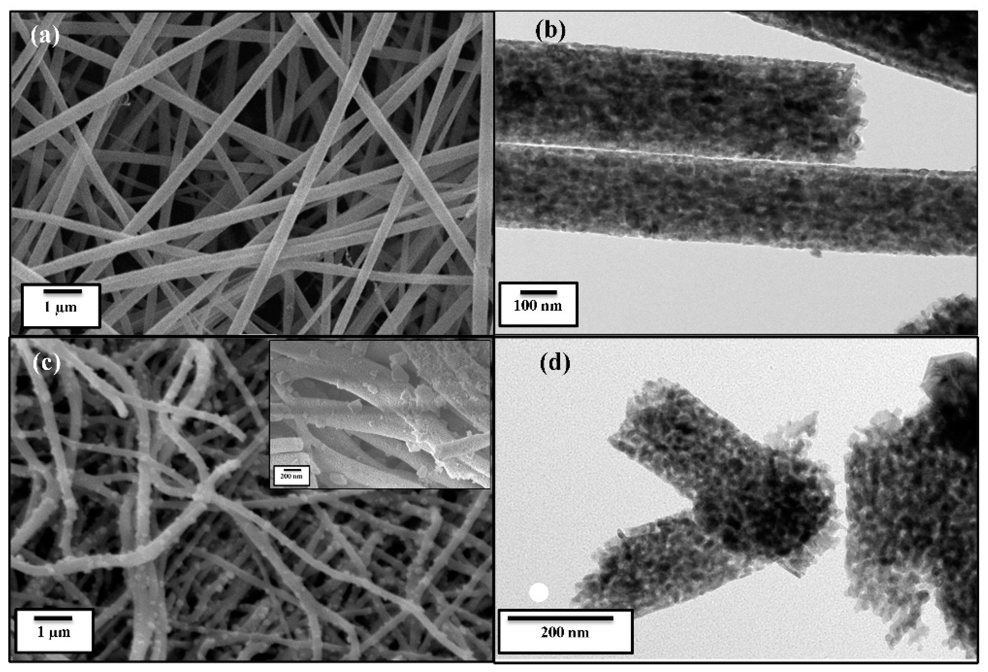
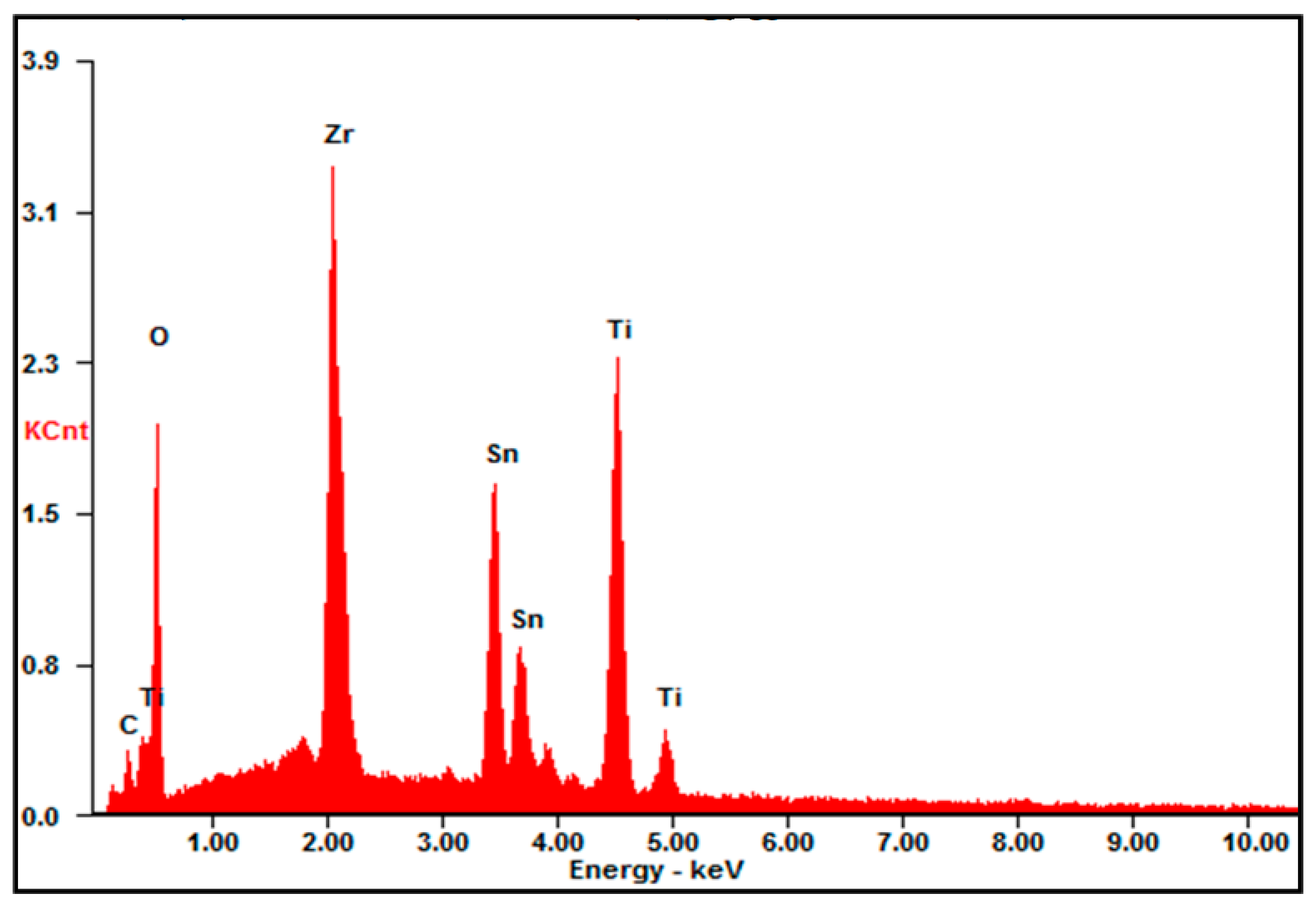
Figure 2 shows FESEM and TEM micrographs of the pure TiO2 and ZSTS composite nanofibers through different magnifications. Figure 2a shows that the surface of the pure TiO2 nanofibers is quite smooth and has a diameter of around 200 nm, which is also confirmed by the TEM image (Figure 2b). The low- (Figure 2c) and high- (inset Figure 2c) magnification FESEM micrograph shows that the ZSTS composite is mainly composed of perforated nanofibers with an average diameter of about 150 nm, authenticated by the TEM image as well (Figure 2d). It possesses rough and fibrous surfaces, which are built up of nanoparticles on the surface of the nanofibers. Figure 3 gives a typical EDS spectrum of ZSTS nanofibers, signifying that the nanocomposite comprises Ti, Sn, Zr and O.
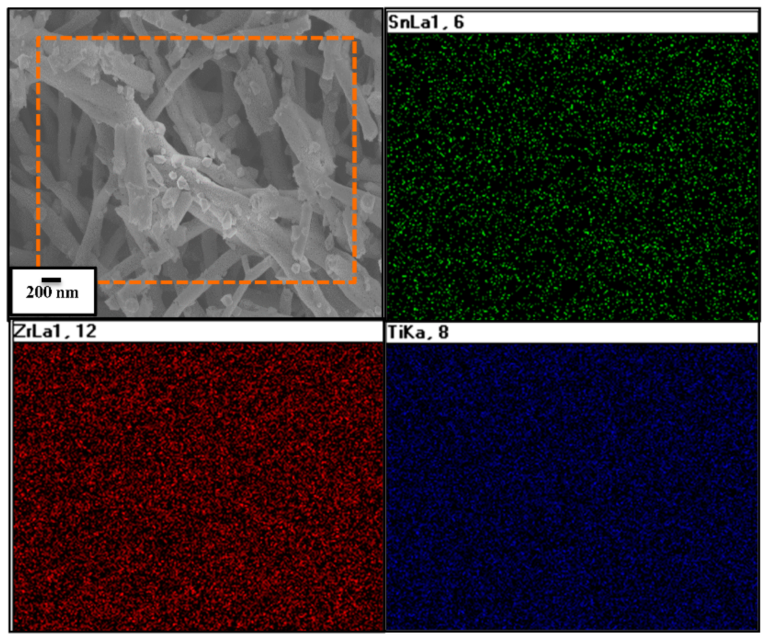
Furthermore, the elemental mapping shown in Figure 4 confirms that the electrospun nanofibers have been formed of Zr0.5Sn0.5TiO3 and SnO2 materials (Figure 4). The electron probe microanalysis (EPMA) profile shows that the composite possesses Zr, Sn and Ti elements. The elemental mapping picture evidently demonstrates that Zr, Sn and Ti are consistently dispersed all through the surface of the ZSTS composite nanofibers.
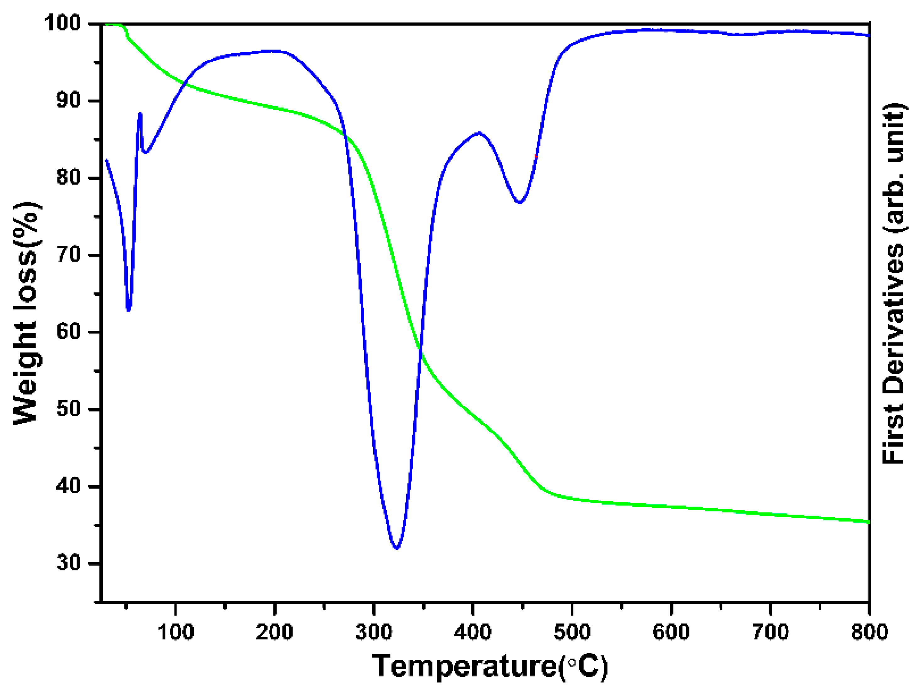
Figure 5 depicts the TGA thermogram of the composite nanofiber precursors from room temperature to 800 °C. The TGA illustrates that the ZSTS composite sample has a total weight reduction of ~64%. The evaporation of water and solvent at temperatures between 25 and 200 °C was the primary cause of the first weight loss (~11%). After 200 °C, the weight loss increased to more than 40% because of the PVAc polymer degrading up to 400 °C, with a significant exothermic peak occurring at 325 °C. The final weight loss (~13%) at temperatures between 400 and 500 °C was caused by the complete disintegration of PVAc, the degradation of the metal salt in the composite and the precipitation of the metal oxide.
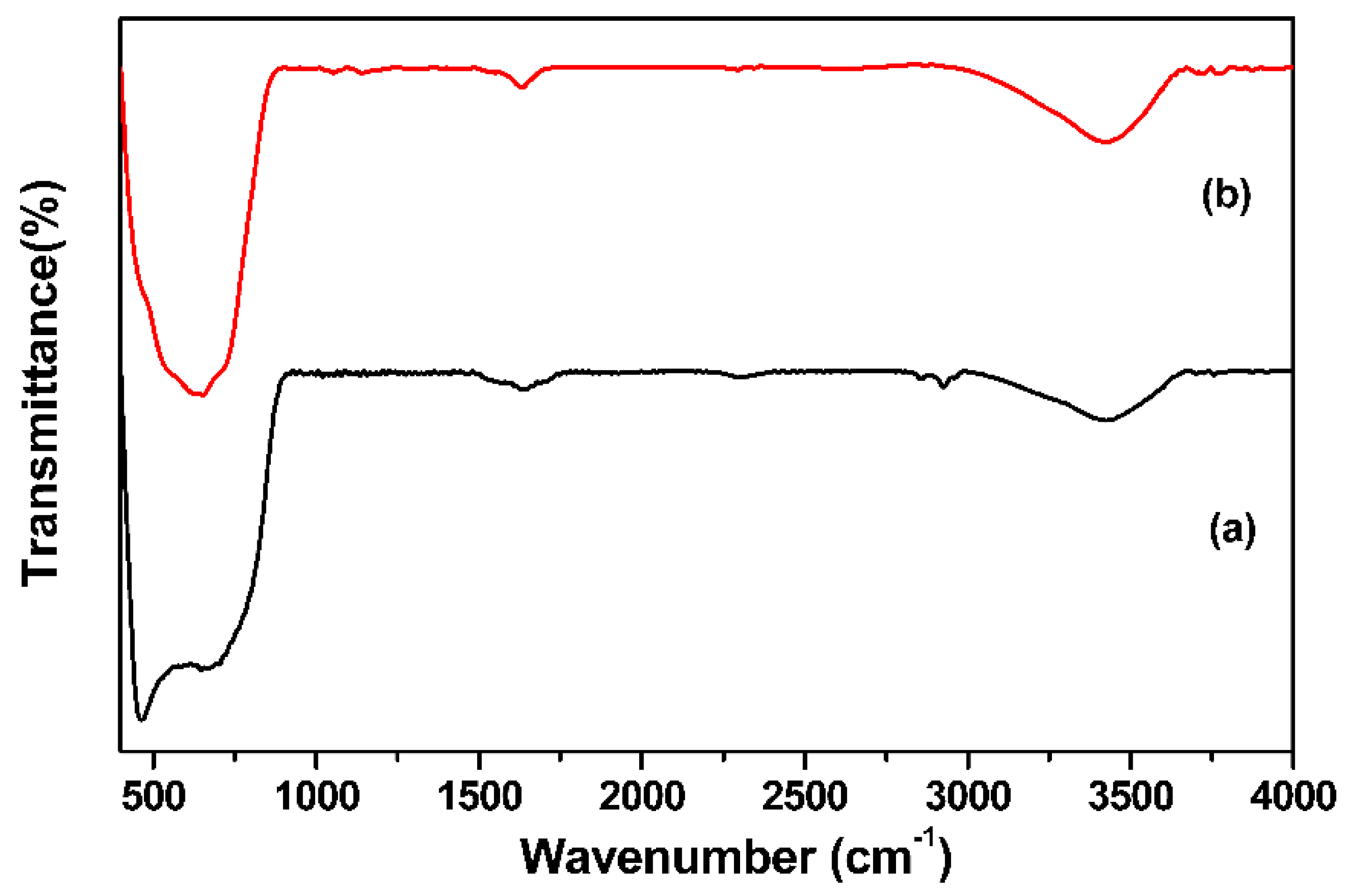
In the IR spectra of TiO2, a broad band at 3403 cm−1, which is associated with the stretching mode of the OH group, and a minor band at 1631 cm−1, which denotes the bending mode of molecular water, were visible (Figure 6a). Characteristic peaks at 480 and 616 cm−1 indicate the bending and stretching mode of the Ti–O bond [37]. Figure 6b displays the FT–IR spectrum of the heterostructured ZSTS. It shows that the observed wide vibrational band (centered around ~634 cm−1) could be defined as a metal–oxygen (M–O) stretching vibration [38].
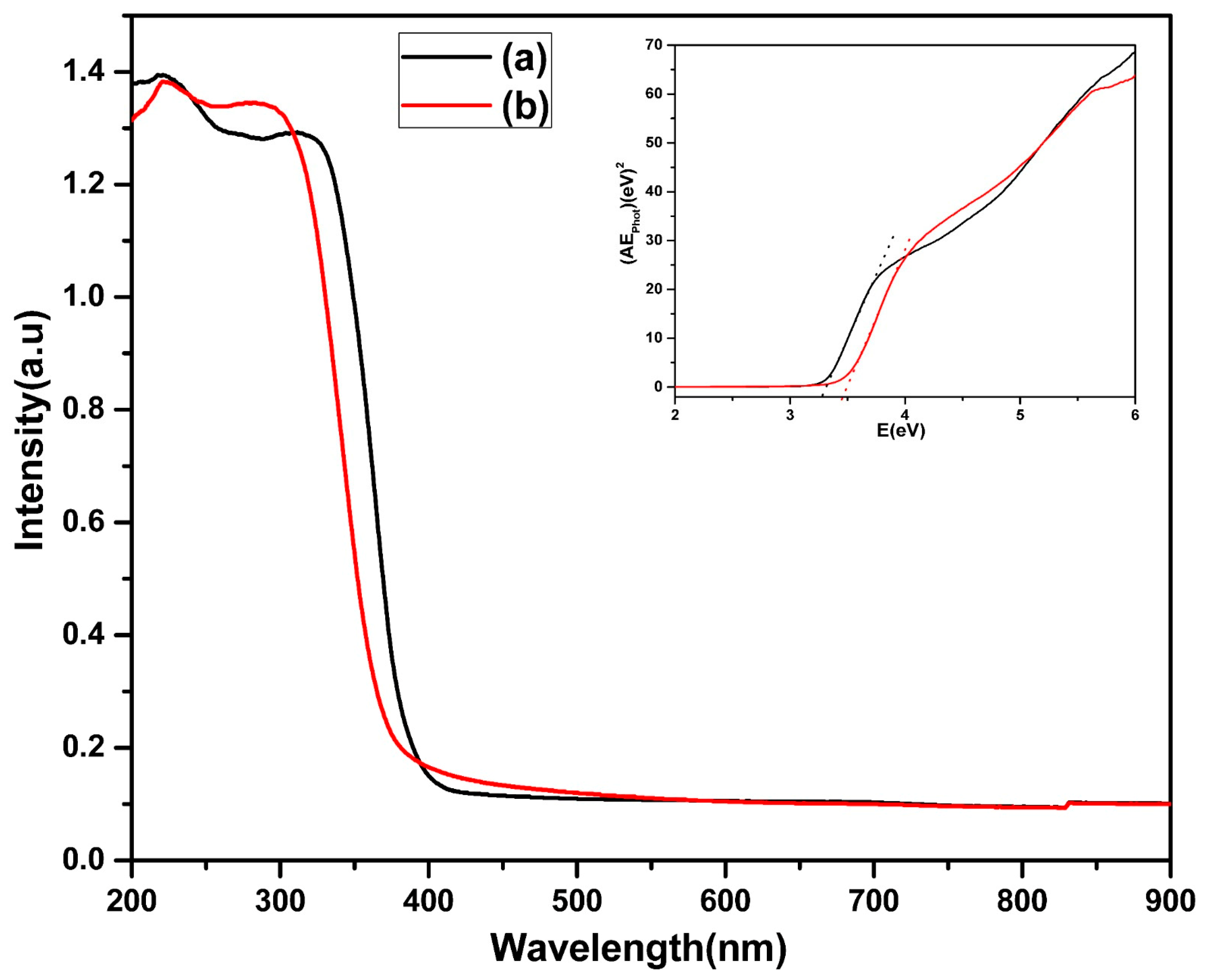
The absorption of light obtained from the diffuse reflectance UV is displayed in Figure 7. The absorption edge for both the samples falls in the UV region (λ < 400 nm). A blue shift in the spectra of the ZSTS nanocomposite is observed compared to the TiO2 photocatalyst. The presence of zirconium and tin oxide, which have a wider band gap than titania, may be responsible for the shift in the intensity of the light absorption in the composite sample. To estimate the bandgap energy (Eg) of the samples, Eg was calculated from the plot of (αhν)1/2 versus the energy of the stimulating light (hν) using equation αhν = A(hν − Eg)n/2 [39]. As a result, the band gaps of the TiO2 and ZSTS nanofibers were determined to be 3.28 and 3.45 eV, respectively (inset Figure 7).
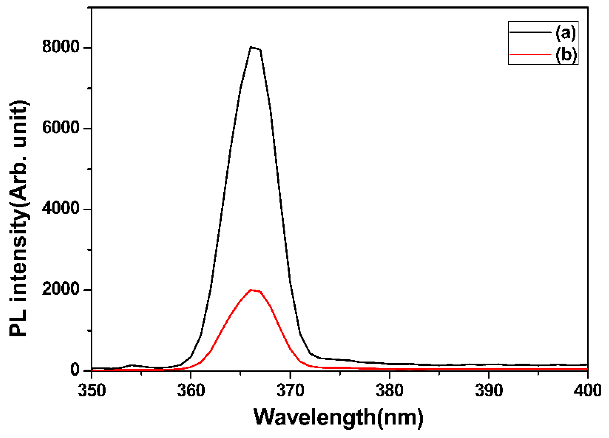
Figure 8 shows the prepared ZSTS nanofibers’ PL spectra. Each sample has an emission peak with a central wavelength of roughly 366 nm. The ZSTS composites exhibit considerable quenching of the PL when compared to TiO2. The strength of the PL peaks, which are connected to the recombination of the electron–hole pairs within the semiconductor, further suggests that there is an effective charge transfer within the ZSTS nanofibers. The photogenerated electron–hole pairs can readily transfer at the heterostructured interface in the case of the ZSTS composite, leading to greater photocatalytic activity when exposed to UV light.
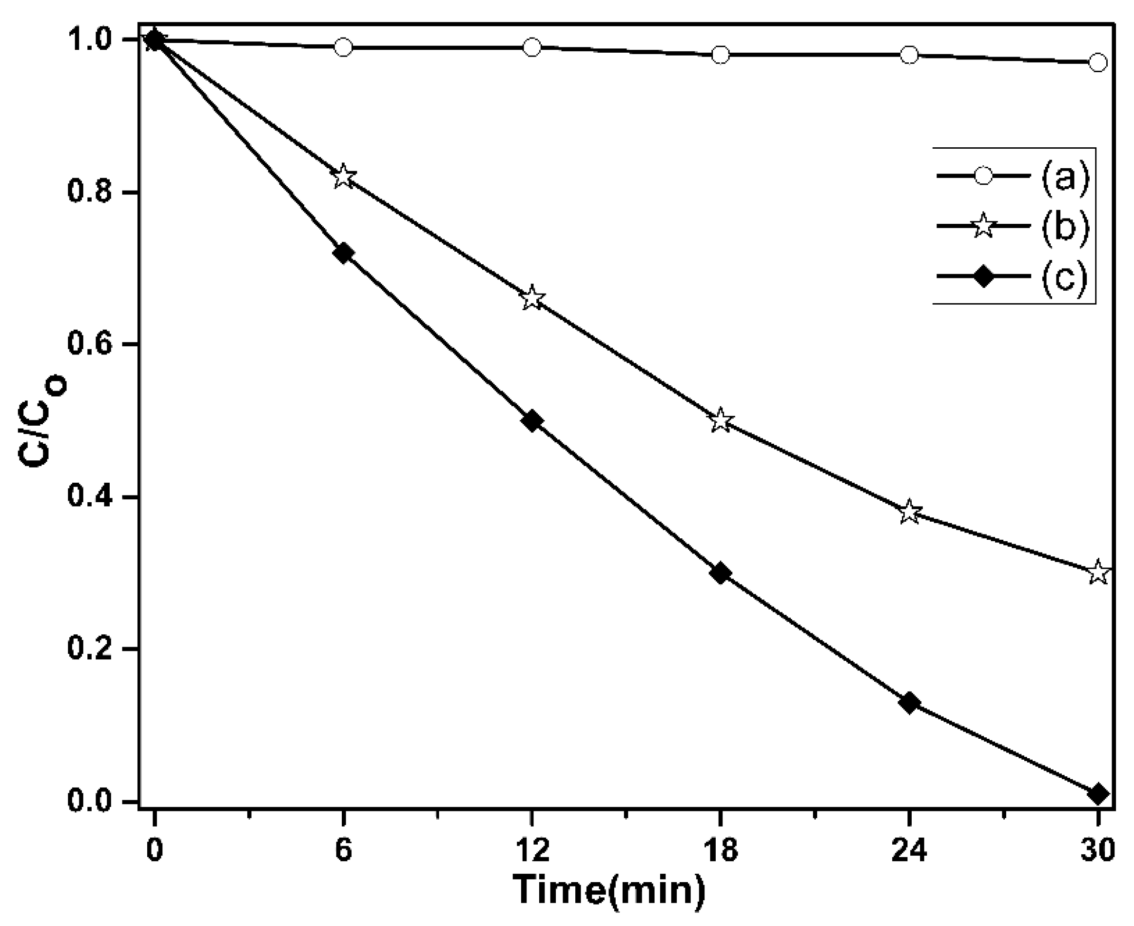
The photocatalytic activity of the pure TiO2 and ZSTS composite was assessed using the photodegradation of RhB dye under UV light irradiation, as shown in Figure 9. For comparison, RhB degradation without the photocatalyst was also carried out. The outcomes showed that RhB degradation was extremely slow in the absence of the photocatalyst. The initial concentration of the RhB dye solution used was 20 ppm. Pure TiO2 decolorizes RhB by 70% in 30 min. The ZSTS composite nanofibers, however, show a noticeably improved photocatalytic performance. The composite sample demonstrates that all of the dye can be broken down within 30 min. The experimental findings showed that ZSTS composite photocatalysts have greater photocatalytic activity than pure TiO2 photocatalysts. This improvement in photocatalytic efficiency could be due to the heterostructure that forms when Zr0.5Sn0.5TiO3 and SnO2 come together, since it is crucial for the separation of electron–hole pairs. Therefore, it seems more likely that the superior photocatalytic activity of ZSTS is attributable to a more effective separation of the electron–hole pairs or to a significantly reduced propensity for electron and hole recombination.
Numerous studies have also documented how combining TiO2 with SnO2 increases their photocatalytic activity. Owing to the charge separation that takes place at the interface between two semiconductors, this improvement is attributed to a drop in the rate of electron–hole recombination [40]. In addition, it has been discovered that the inclusion of Sn4+ ions in the TiO2 lattice increases the parent oxide’s exterior acidity [41], which may boost the photocatalytic activity. Also, a superior interaction with surface water tales place in the case of the TiO2/ZrO2 photocatalyst, which results in the detection of more O2H radicals. However, in the case of TiZr, the heteroatom takes part in the electron trapping sites that result in the establishment of Zr4+–O2•– species, and this possibly has an impact on the inherent photoactivity of matter [42].
To evaluate the reusability of the composites from the standpoint of practical applications, the ZSTS nanofibers were selected. The catalysts currently in use are easily separated from the solution and effectively dispersed when stirred. Centrifugation makes it easy to separate the catalyst from the cleansed solution. The dye did not adhere to the catalyst permanently; rather, photodegradation is what causes the cleaning. The ZSTS composite showed good catalytic stability, keeping a similar level of reactivity even after five cycles (97%) (Figure 10). The unavoidable catalyst loss during recycling should be the cause of the minor reduction in efficiency.
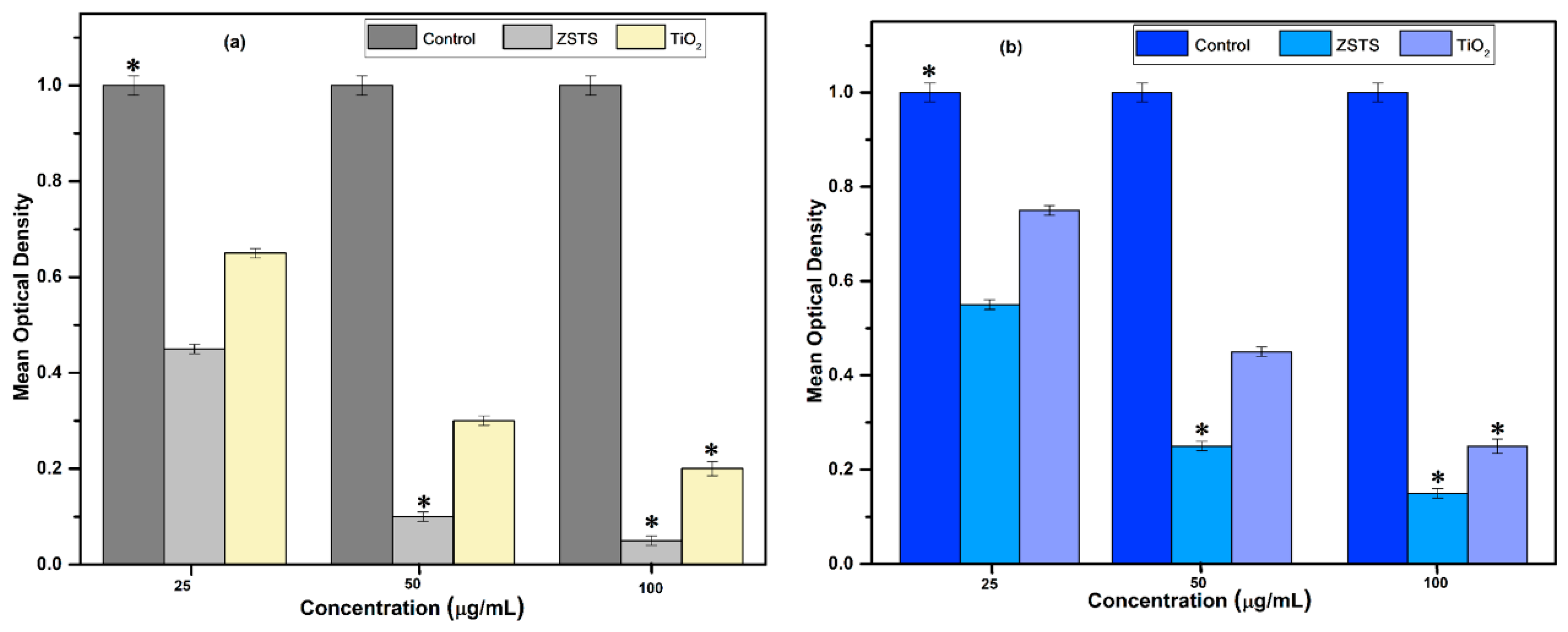
Antibiotic resistance has incurred staggering health care expenditures in the community over the last decade, necessitating immediate action. This demands a thorough examination of the innovative possibilities. With the ability to autonomously control and manipulate both atoms and molecules, nanotechnology is a multidisciplinary field of study that has enabled significant advancements in numerous scientific fields [43,44]. In the present study, the antibacterial efficacy of TiO2 and ZSTS composite nanofibers against two typical bacteria was demonstrated and is shown in Figure 11a. E. coli, which frequently causes severe diarrhea, and S. aureus, an opportunistic pathogen that frequently causes respiratory infections and food poisoning, are the bacteria that were studied. By treating them using ZSTS nanofibers and the pure TiO2, bacterial growth as an indicator of turbidity was observed and computed in order to assess their antibacterial capabilities. The outcomes attained point out that the ability of TiO2 nanofibers to inhibit the growth of bacteria is exclusively associated with its selected concentration and UV irradiation, comparable to previous outcomes [33]. In UV/TiO2 organization, investigators have proposed diverse elimination mechanisms, including negative effects on deoxyribonucleic acid, membrane impairment that causes the cell contents to leak out, and observed reductions in respiratory operations owing to the loss of precursor enzymes [33]. Nevertheless, there is still a dearth of concrete proof regarding photocatalytic death. In particular, aqueous suspensions produced reactive oxygen species (ROS) when the TiO2 solution was exposed to UV radiation. A hole (h+) is produced in the valence band via charge separation, while an electron (e) is produced in the conduction band. Hydroxyl radicals (OH) are produced at the surface of the excited particle by elusive electrons in the valence band holes of water and/or hydroxyl ions. The superoxide anion (O2) is created when electrons reduce oxygen. The ROS produced by UV-irradiated TiO2 nanofibers may have initially interacted with the cell wall, produced free radicals that can enter into the interior of the cell, disrupt the internal contents and cause the cells to deform, leading to disorganization and leakage [33]. In the current investigation, it was found that, in comparison to the pure TiO2 nanofibers, the ZSTS nanocomposites demonstrated action against bacterial growth that was more severe (Figure 11b). The current findings suggest that ZSTS nanocomposites may kill certain bacteria and interact with them robustly at a high concentration after 12 h of incubation, regardless of the mechanism involved. However, when the concentration of ZSTS nanocomposites in the suspension was reduced to 25 µg/mL, no palpable antibacterial activity was detected. The shape, increase in surface area [45] and combined outcome are attributed to the amplified activity of the ZSTS nanocomposites. The ZSTS nanofibers in this instance showed significant antibacterial activity against E. coli. Additionally, it was discovered that ZSTS nanocomposites and TiO2 nanofibers make E. coli more sensitive. The two strains’ different cell structures have been shown to be the cause of increased sensitivity [45,46]. The shape of microorganisms is primarily responsible for variances in their sensitivity. Gram-negative bacteria’s cell walls are made up of an exterior lipid membrane and a thin layer of peptidoglycan. ROS, which cause the cells of microorganisms to die, are produced as a result of the effectiveness of the metal oxide. Thus, oxidation and cell death happen due to ROS targeting several distinct locations and biomolecules in the respective microorganisms. However, S. aureus, a Gram-positive bacterium, has a dense covering of peptidoglycan that prevents nanoparticles from easily entering S. aureus. It could be interpreted that in the case of Gram-negative bacteria, the desired outcome may be achieved via the electrostatic attraction between negatively charged bacteria and positively charged metal ions, which causes bacteria to rupture and die. A similar interpretation of ZrO2 nanoparticles has been reported elsewhere [47].
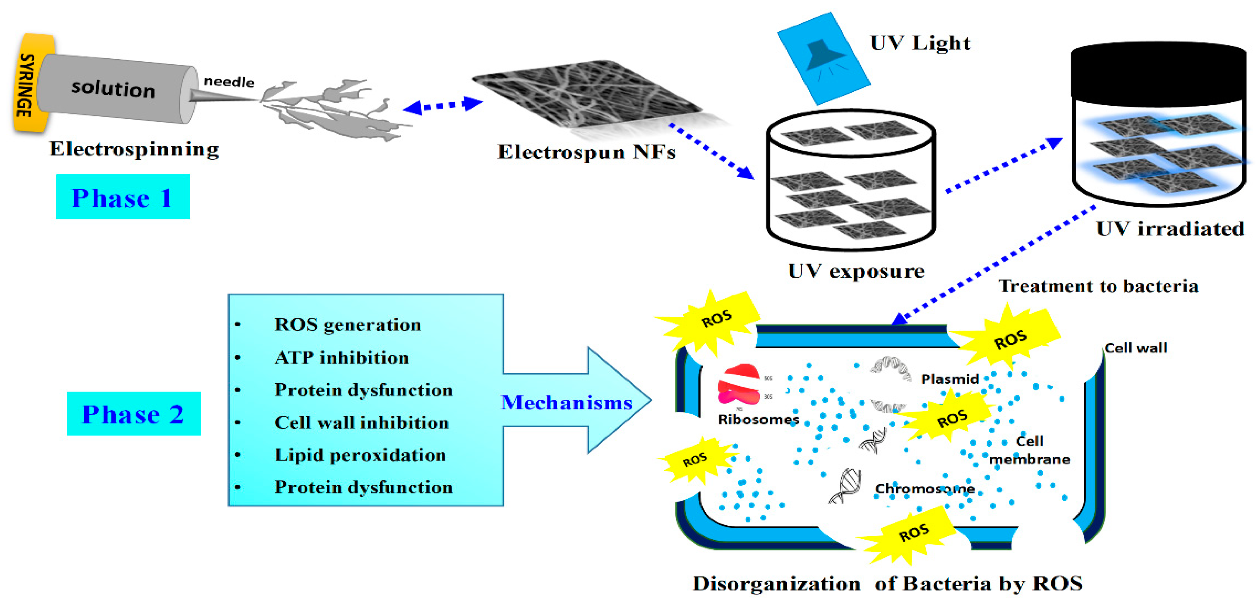
Previous researchers have documented the bactericidal effects of cotton treated with ZrO2 and ZrO2 nanoparticles against infections [48]. In yet another investigation, the antibacterial uses of ZrO2 NPs in tooth decay assertions were identified [7]. According to previous studies, ZSTS nanocomposites may exaggeratedly increase production of ROS, boost ATPase activity, and cause the expulsion of proteins and electrolytes, leading to the death of bacteria [49]. As such, ZSTS nanocomposites are a unique material that may be utilized for the production of antimicrobial composites. ZSTS nanocomposites have a strong ability to kill bacteria, making them a suitable material for the fabrication of novel bactericides and expanding their range of possible uses. The likely mechanism is shown in Figure 12. Although ZSTS nanocomposites have demonstrated effective antibacterial activity, more research is required to determine their other physicochemical characteristics before they can be employed with confidence. In the end, according to the preliminary information gathered herein, ZSTS nanofibers might be employed topically to regulate the spread of bacterial toxicities and in order to manage the purification of industrial and sewage wastewater.

4. Conclusions

In summary, TiO2 and ZSTS nanofiber photocatalysts were prepared via the electrospinning method. The experimentation evaluating the photodegradation of RhB and PL investigation revealed that ZSTS nanofibers have a noteworthy effect on the separation of photogenerated charge carriers and UV light photocatalytic action. Furthermore, in 30 min, the composite completely bleached the colorant. The produced photocatalysts also exhibited stability when exposed to UV light. The synergetic effects between Zr0.5Sn0.5TiO3 and SnO2 were mostly responsible for the increased photocatalytic activity of the ZSTS composites. The above findings contribute a very effective and robust photocatalytic material for eliminating biological and chemical pollutants, in addition to offering updated knowledge on how to develop some effective ceramics/metal oxide nanofibrous photocatalysts. Interestingly, the ZSTS demonstrated efficient antimicrobial potential for both Gram-negative and Gram-positive bacteria; however, due to their negatively charged cell surface and scavenging activity, these photocatalysts were more vigorous against Gram-negative bacteria. Despite the substantial advancements, there are still a number of problems associated with antibacterial nanofibers that need to be resolved. First, it is necessary to attempt the manufacturing of composite antibacterial nanofibers on a large scale, as it is currently primarily restricted to the laboratory, and to investigate the biocompatibility of these high-aspect-ratio nanofibers with human cell lines. Therefore, the ZSTS could act as new material for environmental applications.

Author Contributions

Conceptualization, J.S.A., M.S.H. and T.A.; methodology, J.S.A. and M.S.H.; software, M.A.M.A.; validation, A.F.S. and L.S.A.; formal analysis, B.H.A.A.-D.; investigation, T.A.; resources, A.F.S.; data curation, A.A.A. and L.S.A.; writing—original draft preparation, M.S.H., T.A. and J.S.A.; writing—review and editing, T.A. and A.A.A. All authors have read and agreed to the published version of the manuscript.

Funding

This research received funding from Deputy for Research and Innovation Ministry of Education, Kingdom of Saudi Arabia for this research through a grant (NU/IFC/2/SERC/-/1) under the Institutional Funding Committee at Najran University, Kingdom of Saudi Arabia.

Institutional Review Board Statement

Not applicable.

Informed Consent Statement

Not applicable.

Data Availability Statement

The data presented in this study are available in article.

Acknowledgments

The authors would like to acknowledge the support of the Deputy for Research and Innovation Ministry of Education, Kingdom of Saudi Arabia, for this research through a grant (NU/IFC/2/SERC/-/1) under the Institutional Funding Committee at Najran University, Kingdom of Saudi Arabia.

Conflicts of Interest

The authors declare no conflict of interest.

References

  1. Badgley, B.D.; Ferguson, J.; Heuvel, A.V.; Kleinheinz, G.T.; McDermott, C.M.; Sandrin, T.R.; Kinzelman, J.; Junion, E.A.; Byappanahalli, M.N.; Whitman, R.L. Multi-scale temporal and spatial variation in genotypic composition of Cladophora-borne Escherichia coli populations in Lake Michigan. Water Res. 2011, 45, 721–731. [Google Scholar] [CrossRef] [PubMed]
  2. DeFlorio-Barker, S.; Wing, C.; Jones, R.M.; Dorevitch, S. Estimate of incidence and cost of recreational waterborne illness on United States surface waters. Environ. Health 2018, 17, 1–10. [Google Scholar] [CrossRef] [Green Version]
  3. Korajkic, A.; McMinn, B.R.; Harwood, V.J. Relationships between microbial indicators and pathogens in recreational water settings. Int. J. Environ. Res. Public Health 2018, 15, 2842. [Google Scholar] [CrossRef] [PubMed] [Green Version]
  4. Shannon, M.A.; Bohn, P.W.; Elimelech, M.; Georgiadis, J.G.; Marinas, B.J.; Mayes, A.M. Science and technology for water purification in the coming decades. Nature 2008, 452, 301–310. [Google Scholar] [CrossRef] [PubMed]
  5. Phukan, A.; Bhattacharjee, R.P.; Dutta, D.K. Stabilization of SnO2 nanoparticles into the nanopores of modified Montmorillonite and their antibacterial activity. Adv. Powder Technol. 2017, 28, 139–145. [Google Scholar] [CrossRef]
  6. Otero-González, L.; García-Saucedo, C.; Field, J.A.; Sierra-Álvarez, R. Toxicity of TiO2, ZrO2, Fe0, Fe2O3, and Mn2O3 nanoparticles to the yeast, Saccharomyces cerevisiae. Chemosphere 2013, 93, 1201–1206. [Google Scholar] [CrossRef]
  7. Fathima, J.B.; Pugazhendhi, A.; Venis, R. Synthesis and characterization of ZrO2 nanoparticles-antimicrobial activity and their prospective role in dental care. Microb. Pathog. 2017, 110, 245–251. [Google Scholar] [CrossRef]
  8. Jeevanandam, J.; Barhoum, A.; Chan, Y.S.; Dufresne, A.; Danquah, M.K. Review on nanoparticles and nanostructured materials: History, sources, toxicity and regulations. Beilstein J. Nanotechnol. 2018, 9, 1050–1074. [Google Scholar] [CrossRef] [Green Version]
  9. Patra, J.K.; Das, G.; Fraceto, L.F.; Campos, E.V.R.; del Pilar Rodriguez-Torres, M.; Acosta-Torres, L.S.; Diaz-Torres, L.A.; Grillo, R.; Swamy, M.K.; Sharma, S. Nano based drug delivery systems: Recent developments and future prospects. J. Nanobiotechnology 2018, 16, 1–33. [Google Scholar] [CrossRef] [Green Version]
  10. Jeyaraj, M.; Gurunathan, S.; Qasim, M.; Kang, M.-H.; Kim, J.-H. A comprehensive review on the synthesis, characterization, and biomedical application of platinum nanoparticles. Nanomaterials 2019, 9, 1719. [Google Scholar] [CrossRef] [Green Version]
  11. Palit, S.; Hussain, C.M. Nanodevices applications and recent advancements in nanotechnology and the global pharmaceutical industry. In Nanomaterials in Diagnostic Tools and Devices; Elsevier: Amsterdam, The Netherlands, 2020; pp. 395–415. [Google Scholar]
  12. Lin, J.; Jimmy, C.Y.; Lo, D.; Lam, S. Photocatalytic Activity of Rutile Ti1− xSnxO2Solid Solutions. J. Catal. 1999, 183, 368–372. [Google Scholar] [CrossRef]
  13. Wang, J.; Jing, L.; Xue, L.; Qu, Y.; Fu, H. Enhanced activity of bismuth-compounded TiO2 nanoparticles for photocatalytically degrading rhodamine B solution. J. Hazard. Mater. 2008, 160, 208–212. [Google Scholar] [CrossRef]
  14. Reddy, B.M.; Sreekanth, P.M.; Reddy, E.P.; Yamada, Y.; Xu, Q.; Sakurai, H.; Kobayashi, T. Surface Characterization of La2O3− TiO2 and V2O5/La2O3− TiO2 Catalysts. J. Phys. Chem. B 2002, 106, 5695–5700. [Google Scholar] [CrossRef]
  15. Hong, S.-S.; Lee, M.S.; Park, S.S.; Lee, G.-D. Synthesis of nanosized TiO2/SiO2 particles in the microemulsion and their photocatalytic activity on the decomposition of p-nitrophenol. Catal. Today 2003, 87, 99–105. [Google Scholar] [CrossRef]
  16. Asahi, R.; Morikawa, T.; Ohwaki, T.; Aoki, K.; Taga, Y. Visible-light photocatalysis in nitrogen-doped titanium oxides. Science 2001, 293, 269–271. [Google Scholar] [CrossRef]
  17. Tahir, M.N.; Eberhardt, M.; Therese, H.A.; Kolb, U.; Theato, P.; Müller, W.E.; Schröder, H.C.; Tremel, W. From Single Molecules to Nanoscopically Structured Functional Materials: Au Nanocrystal Growth on TiO2 Nanowires Controlled by Surface-Bound Silicatein. Angew. Chem. Int. Ed. 2006, 45, 4803–4809. [Google Scholar] [CrossRef] [PubMed]
  18. Stafford, U.; Gray, K.A.; Kamat, P.V. Photocatalytic degradation of organic contaminants: Halophenols and related model compounds. Heterog. Chem. Rev. 1996, 3, 77–104. [Google Scholar] [CrossRef]
  19. Vassiliev, S.Y.; Yusipovich, A.; Rogynskaya, Y.E.; Chibirova, F.K.; Skundin, A.; Kulova, T. Nanostructured SnO2-TiO2 films as related to lithium intercalation. J. Solid State Electrochem. 2005, 9, 698–705. [Google Scholar] [CrossRef]
  20. Wu, C.; Shen, L.; Yu, H.; Huang, Q.; Zhang, Y.C. Synthesis of Sn-doped ZnO nanorods and their photocatalytic properties. Mater. Res. Bull. 2011, 46, 1107–1112. [Google Scholar] [CrossRef]
  21. Jing, L.; Xin, B.; Yuan, F.; Xue, L.; Wang, B.; Fu, H. Effects of surface oxygen vacancies on photophysical and photochemical processes of Zn-doped TiO2 nanoparticles and their relationships. J. Phys. Chem. B 2006, 110, 17860–17865. [Google Scholar] [CrossRef]
  22. Hou, Y.; Wang, X.; Wu, L.; Chen, X.; Ding, Z.; Wang, X.; Fu, X. N-doped SiO2/TiO2 mesoporous nanoparticles with enhanced photocatalytic activity under visible-light irradiation. Chemosphere 2008, 72, 414–421. [Google Scholar] [CrossRef] [PubMed]
  23. Krous, E.; Patel, D.; Langston, P.; Menoni, C.; Markosyan, A.; Route, R.; Fejer, M.; Nguyen, D.; Emmert, L.; Rudolph, W. Scandium Oxide Thin Films Deposited by Dual Ion Beam Sputtering for High-Power Laser Applications. In Optical Interference Coatings; P FA10; Optica Publishing Group: Washington, DC, USA, 2010. [Google Scholar]
  24. Stehlin, F.; Bourgin, Y.; Spangenberg, A.; Jourlin, Y.; Parriaux, O.; Reynaud, S.; Wieder, F.; Soppera, O. Direct nanopatterning of 100 nm metal oxide periodic structures by Deep-UV immersion lithography. Opt. Lett. 2012, 37, 4651–4653. [Google Scholar] [CrossRef] [PubMed]
  25. Park, K.H.; Hong, C.K. Morphology and photoelectrochemical properties of TiO2 electrodes prepared using functionalized plant oil binders. Electrochem. Commun. 2008, 10, 1187–1190. [Google Scholar] [CrossRef]
  26. Ishizuka, S.; Yamada, A.; Matsubara, K.; Fons, P.; Sakurai, K.; Niki, S. Alkali incorporation control in Cu (In, Ga) Se 2 thin films using silicate thin layers and applications in enhancing flexible solar cell efficiency. Appl. Phys. Lett. 2008, 93, 124105. [Google Scholar] [CrossRef]
  27. Gusmano, G.; Bianco, A.; Viticoli, M.; Kaciulis, S.; Mattogno, G.; Pandolfi, L. Study of Zr1− xSnxTiO4 thin films prepared by a polymeric precursor route. Surf. Interface Anal. Int. J. Devoted Dev. Appl. Tech. Anal. Surf. Interfaces Thin Film. 2002, 34, 690–693. [Google Scholar] [CrossRef]
  28. Hu, C.; Sun, J.; Long, C.; Wu, L.; Zhou, C.; Zhang, X. Synthesis of nano zirconium oxide and its application in dentistry. Nanotechnol. Rev. 2019, 8, 396–404. [Google Scholar] [CrossRef]
  29. Gupta, V.; Singh, S.; Rawat, K.; Bohidar, H.; Solanki, P.R. Cytotoxicity and antimicrobial activity of transition metal oxide nanoparticles. Adv. Sci. Lett. 2014, 20, 1650–1653. [Google Scholar] [CrossRef]
  30. Imran, M.; Riaz, S.; Sanaullah, I.; Khan, U.; Sabri, A.N.; Naseem, S. Microwave assisted synthesis and antimicrobial activity of Fe3O4-doped ZrO2 nanoparticles. Ceram. Int. 2019, 45, 10106–10113. [Google Scholar] [CrossRef]
  31. Jangra, S.L.; Stalin, K.; Dilbaghi, N.; Kumar, S.; Tawale, J.; Singh, S.P.; Pasricha, R. Antimicrobial activity of zirconia (ZrO2) nanoparticles and zirconium complexes. J. Nanosci. Nanotechnol. 2012, 12, 7105–7112. [Google Scholar] [CrossRef]
  32. Thirumagal, K.; Rajeshkumar, S.; ROY, A. In vitro Cytotoxic Effect of Zirconium Oxide Nanoparticle and Its Antibacterial Activity Against Oral Pathogens. Plant Cell Biotechnol. Mol. Biol. 2020, 21, 28–36. [Google Scholar]
  33. Hassan, M.S.; Amna, T.; Mishra, A.; Yun, S.-I.; Kim, H.-C.; Kim, H.-Y.; Khil, M.-S. Fabrication, characterization and antibacterial effect of novel electrospun TiO2 nanorods on a panel of pathogenic bacteria. J. Biomed. Nanotechnol. 2012, 8, 394–404. [Google Scholar] [CrossRef] [PubMed]
  34. Amna, T.; Hassan, M.S.; Sheikh, F.A.; Seo, H.C.; Kim, H.-C.; Alotaibi, N.; Alshahrani, T.; Khil, M.-S. Natural mulberry biomass fibers doped with silver as an antimicrobial textile: A new generation fabric. Text. Res. J. 2021, 91, 00405175211013422. [Google Scholar] [CrossRef]
  35. Gao, Y.; Bach Truong, Y.; Zhu, Y.; Louis Kyratzis, I. Electrospun antibacterial nanofibers: Production, activity, and in vivo applications. J. Appl. Polym. Sci. 2014, 131. [Google Scholar] [CrossRef]
  36. Ramanathan, P.T.; Abdullah, M.S.; Amalraj, L. Preparation and Characterization of Tin Dioxide Thin Film by Nebulized Spray Pyrolysis Technique. J. Bloomers Res. 2013, 5, 651–655. [Google Scholar]
  37. He, Z.; Que, W.; Chen, J.; He, Y.; Wang, G. Surface chemical analysis on the carbon-doped mesoporous TiO2 photocatalysts after post-thermal treatment: XPS and FTIR characterization. J. Phys. Chem. Solids 2013, 74, 924–928. [Google Scholar] [CrossRef]
  38. Shaposhnik, D.; Pavelko, R.; Llobet, E.; Gispert-Guirado, F.; Vilanova, X. Hydrogen sensors on the basis of SnO2-TiO2 systems. Procedia Eng. 2011, 25, 1133–1136. [Google Scholar] [CrossRef] [Green Version]
  39. Huang, G.; Zhu, Y. Synthesis and photoactivity enhancement of ZnWO 4 photocatalysts doped with chlorine. CrystEngComm 2012, 14, 8076–8082. [Google Scholar] [CrossRef]
  40. Shang, J.; Yao, W.; Zhu, Y.; Wu, N. Structure and photocatalytic performances of glass/SnO2/TiO2 interface composite film. Appl. Catal. A Gen. 2004, 257, 25–32. [Google Scholar] [CrossRef]
  41. Shibata, K.; Kiyoura, T.; Kitagawa, J.; Sumiyoshi, T.; Tanabe, K. Acidic properties of binary metal oxides. Bull. Chem. Soc. Jpn. 1973, 46, 2985–2988. [Google Scholar] [CrossRef] [Green Version]
  42. Fresno, F.; Hernández-Alonso, M.D.; Tudela, D.; Coronado, J.M.; Soria, J. Photocatalytic degradation of toluene over doped and coupled (Ti,M)O2 (M = Sn or Zr) nanocrystalline oxides: Influence of the heteroatom distribution on deactivation. Appl. Catal. B Environ. 2008, 84, 598–606. [Google Scholar] [CrossRef]
  43. Bayda, S.; Adeel, M.; Tuccinardi, T.; Cordani, M.; Rizzolio, F. The history of nanoscience and nanotechnology: From chemical–physical applications to nanomedicine. Molecules 2019, 25, 112. [Google Scholar] [CrossRef] [PubMed] [Green Version]
  44. Li, P.; Su, M.; Wang, X.; Zou, X.; Sun, X.; Shi, J.; Zhang, H. Environmental fate and behavior of silver nanoparticles in natural estuarine systems. J. Environ. Sci. 2020, 88, 248–259. [Google Scholar] [CrossRef]
  45. Ann, L.C.; Mahmud, S.; Bakhori, S.K.M.; Sirelkhatim, A.; Mohamad, D.; Hasan, H.; Seeni, A.; Rahman, R.A. Antibacterial responses of zinc oxide structures against Staphylococcus aureus, Pseudomonas aeruginosa and Streptococcus pyogenes. Ceram. Int. 2014, 40, 2993–3001. [Google Scholar] [CrossRef]
  46. Azam, A.; Ahmed, A.S.; Oves, M.; Khan, M.S.; Habib, S.S.; Memic, A. Antimicrobial activity of metal oxide nanoparticles against Gram-positive and Gram-negative bacteria: A comparative study. Int. J. Nanomed. 2012, 7, 6003. [Google Scholar] [CrossRef] [PubMed] [Green Version]
  47. Tabassum, N.; Kumar, D.; Verma, D.; Bohara, R.A.; Singh, M. Zirconium oxide (ZrO2) nanoparticles from antibacterial activity to cytotoxicity: A next-generation of multifunctional nanoparticles. Mater. Today Commun. 2021, 26, 102156. [Google Scholar] [CrossRef]
  48. Gowri, S.; Gandhi, R.R.; Sundrarajan, M. Structural, optical, antibacterial and antifungal properties of zirconia nanoparticles by biobased protocol. J. Mater. Sci. Technol. 2014, 30, 782–790. [Google Scholar] [CrossRef]
  49. Du, T.; Chen, S.; Zhang, J.; Li, T.; Li, P.; Liu, J.; Du, X.; Wang, S. Antibacterial activity of manganese dioxide nanosheets by ros-mediated pathways and destroying membrane integrity. Nanomaterials 2020, 10, 1545. [Google Scholar] [CrossRef] [PubMed]
Figure 1. XRD spectra of (a) TiO2, and (b) ZSTS nanofibers (“●” represents Zr0.5Sn0.5TiO3, whereas “◆” represents SnO2).
Materials 16 05148 g001
Figure 2. FESEM and TEM images of (a,b) TiO2 and (c,d) ZSTS nanofibers (inset shows FESEM of ZSTS at high magnification).
Materials 16 05148 g002
Figure 3. EDX spectrum of ZSTS composite nanofibers.
Materials 16 05148 g003
Figure 4. EPMA pattern of ZSTS nanofibers.
Materials 16 05148 g004
Figure 5. TGA spectra and first derivatives of the as-synthesized ZSTS nanofibers (Green color line shows the weight loss whereas, the blue color shows the first derivatives).
Materials 16 05148 g005
Figure 6. FT−IR spectra of (a) TiO2, and (b) ZSTS nanofibers.
Materials 16 05148 g006
Figure 7. UV–Vis diffuse reflectance spectra of (a) TiO2, and (b) ZSTS nanofibers (inset shows plot of Kubelka–Munk function vs. photon energy).
Materials 16 05148 g007
Figure 8. PL spectra of (a) TiO2, and (b) Zr0.5Sn0.5TiO3/SnO2 nanofibers.
Materials 16 05148 g008
Figure 9. Photocatalytic degradation of RhB dye solution (a) without using TiO2 and ZSTS, (b) with TiO2, and (c) ZSTS nanofibers.
Materials 16 05148 g009
Figure 10. Photocatalytic degradation of RhB dye by ZSTS nanofibers in number of cycles.
Materials 16 05148 g010
Figure 11. Antimicrobial susceptibility test of TiO2 and ZSTS nanofibers (0–100 µg/mL) against (a) E. coli and (b) S. aureus. Data represent the mean values ± standard deviation of three replicates. * p < 0.005 vs. control.
Materials 16 05148 g011
Figure 12. Schematic illustrations of fabrication of ZSTS nanofibers via electrospinning and the bactericidal mechanism thereof.
Materials 16 05148 g012
Disclaimer/Publisher’s Note: The statements, opinions and data contained in all publications are solely those of the individual author(s) and contributor(s) and not of MDPI and/or the editor(s). MDPI and/or the editor(s) disclaim responsibility for any injury to people or property resulting from any ideas, methods, instructions or products referred to in the content.