Article

A Robust Strontium Coordination Polymer with Selective and Sensitive Fluorescence Sensing Ability for Fe3+ Ions

1
State Key Laboratory of Structural Chemistry, Fujian Institute of Research on the Structure of Matter, The Chinese Academy of Sciences, Fuzhou 350002, China
2
College of Chemistry and Materials Science, Fujian Normal University, Fuzhou 350007, China
3
Fujian Science & Technology Innovation Laboratory for Optoelectronic Information of China, Fuzhou 350108, China
*
Authors to whom correspondence should be addressed.
Materials 2023, 16(2), 577; https://doi.org/10.3390/ma16020577
Submission received: 14 December 2022 / Revised: 2 January 2023 / Accepted: 4 January 2023 / Published: 6 January 2023

Abstract

:
Exploration of sensitive and selective fluorescence sensors towards toxic metal species is of great importance to solve metal pollution issues. In this work, a three-dimensional (3D) strontium coordination polymer of Sr2(tcbpe) (H4tcbpe = 1,1,2,2-tetrakis(4-(4-carboxy-phenyl)phenyl)ethene) has been synthesized and developed as a fluorescent sensor to Fe3+ ions. Sr2(tcbpe) shows a mechanochromic fluorescence with emission shifting from blue of the pristine to green after being ground. Notably, based on a fluorescence quenching mechanism, Sr2(tcbpe) displays a sensitive and selective fluorescent sensing behavior to Fe3+ ions with a detection limit of 0.14 mM. Moreover, Sr2(tcbpe) exhibits high tolerance to water in a wide pH range (pH = 3–13), demonstrating that Sr2(tcbpe) is a potential fluorescent sensor of Fe3+ in water.

1. Introduction

In the past two decades, great progress has been made in fluorescent coordination polymers (FL-CPs), which have been applied in bioimaging, light emitting, chemical sensing, and so forth [1,2,3,4,5]. Many fluorescence (FL) mechanisms have been developed in FL-CPs, exemplified by linker- or metal-centered emission energy transfer between metals and ligands (LMCT or MLCT) [6,7,8]. Based on the desired features of CPs, therefore, their fluorescence can also be custom-made by careful choice of inorganic metal cation and organic linker. The most important representatives are rare earth (RE)-based CPs, whose fluorescence is derived from RE metal centers. RE-based CPs have received intense attention due to their bright and narrow characteristic emission bands and long emission lifetime [9,10,11,12]. However, the scarcity of RE sources to some extent restricts their future applications. Alkaline earth (AE) metals such as Ca2+ and Sr2+, like RE cations, usually exhibit abundant and flexible coordination modes, making them good candidates as alternative metals to construct FL-CPs [13,14,15]. Because of their d0 electron configuration, AE2+ ions are very suitable to build FL-CPs with ligand-centered luminescence by using emissive organic linkers with a unique chromophore. AE metals are also good candidates to build FL-CPs because of their economic and environmental advantages. However, thus far, FL-CPs constructed from AE metals are comparatively rare [13,14,15].
Metal ions play essential roles in biological metabolism. However, metal species usually exist at a trace concentration level in biological systems, while an excess or deficiency of metals would bring great harm to the biological environment and even threaten life. For instance, excess Fe3+ can cause Alzheimer’s disease, while a lack of Fe3+ can result in hemochromatosis [16,17]. Therefore, many efforts have been devoted to monitoring the Fe3+ ions. A variety of FL-CP sensors towards Fe3+ ions have been developed in the past decade, which are mostly constructed from RE and transition metal (TM) ions [18,19,20]. However, as far as we know, AE-based FL-CPs with sensitive and selective Fe3+ sensing performance are comparatively rare [13,15].
Bearing this in mind, herein, we report a Sr-based FL-CP of Sr2(tcbpe) (H4tcbpe = 1,1,2,2-tetrakis(4-(4-carboxy-phenyl)phenyl)ethene). Sr2(tcbpe) exhibits an interestingly mechanochromic FL inherited from the tetraphenyl ethylene emitting center in H4tcbpe with aggregation-induced emission (AIE) characteristics. Sr2(tcbpe) shows FL sensing performance towards Fe3+ with good selectivity and sensitivity, representing the first FL Sr-CP sensor to Fe3+ ions. It also possesses an excellent tolerance to solvents and water even in acid/base conditions, indicating that Sr2(tcbpe) is a promising FL sensor to probe Fe3+ ions in water.

2. Materials and Methods

Reagents. SrCl2 (≥99%, Adamas-beta, Shanghai Titan Chemical Co., Ltd., Shanghai, China); DMF (N,N-dimethyl-Formamide, AR, Greagent, Shanghai Titan Chemical Co., Ltd.); formic acid (≥98%, Adamas-beta, Shanghai Titan Chemical Co., Ltd.). H4tcbpe has been synthesized using a previously reported method [21].
Preparation of Sr2(tcbpe). A mixture of strontium chloride (50 mg) and H4tcbpe (20 mg) in 5 mL DMF, 2 mL H2O, and 500 μL formic acid was sealed into a 20 mL glass vial and heated at 120 °C for 2 days. Light-yellow block-like crystals were obtained by filtration after the vial was cooled to room temperature (50 mg, 30% yield based on strontium, Figures S1 and S2). Elemental analyses calculated for Sr2(tcbpe): C 61.76%, H 3.26%. Found: C61.20%, H 3.83%.
Physical measurements. Powder X-ray diffraction (PXRD) patterns were recorded on a Rigaku MiniFlex II diffractometer using CuKα radiation (λ = 1.54178 Å). A graphite monochromator was used and the generator power settings were set at 44 kV and 40 mA (Figure S3). Data were collected in a 2θ range of 3 and 35° with a scanning speed of 1.0°/min. Thermogravimetric (TG) data were collected on a NETZSCH STA449C thermogravimetric analyzer with a temperature ramping rate of 10 °C/min from 30 to 700 °C under nitrogen gas flow (Figure S4). Elemental analyses for C, H, and O were performed on a German Elementary Vario EL III instrument. Single crystal X-ray diffraction data were collected with graphite-monochromated MoKα (λ = 0.71073 Å) using an XtaLAB Synergy R, HyPix diffractometer at 298(2) K.
FL measurements. The as-made crystalline samples of Sr2(tcbpe) were manually ground to obtain a fine powder. A 2 mg powdered sample was dispersed in 2 mL of the given organic solvents or 10−2 M metal ion solution by ultrasonication to obtain stable FL suspensions. The FL emulsion was then placed in a 1 cm width quartz cell to record the FL spectra using a PerkinElmer LS55 FL spectrometer. The FL detection experiments were carried out by adding varied amounts of 0.5 × 10⁻² M Fe3+ ions into the prepared suspensions with a pipette. For all FL measurements, the excitation wavelength was monitored at 380 nm and the corresponding emission wavelengths were monitored from 400 nm to 700 nm.
Stability measurements. A 15 mg as-made crystalline sample was immersed in 2 mL of organic solvents or water with a range of pH values over 24 h. Then the immersed samples were collected by filtration and used for further PXRD measurements.
X-ray crystallography. A single crystal suitable for single-crystal X-ray diffraction (SCXRD) was selected under an optical microscope and glued to a thin glass fiber. The structure was solved by direct methods and refined with full-matrix least-squares techniques using the SHELX2018 package [22]. The CCDC number for Sr2(tcbpe) is 2224915. The detailed crystallographic data and structure-refinement parameters are summarized in Table 1.

3. Results and Discussion

3.1. Crystal Structure Analysis

Single-crystal analysis indicates that Sr2(tcbpe) displays a three-dimensional (3D) structure. As seen in Figure 1a, there are two crystallographically independent Sr2+ cations. Sr1 is eight-coordinated by four water molecules (of which two as bridging linkers and two as terminal molecules) and four monodentate coordinating carboxylic groups from four different tabpe4− ligands (Figure S5a). Sr2 is nine-coordinated exhibiting similar coordination to RE ions. The Sr2 is connected with three carboxylic groups from three different tcbpe4− ligands in chelating coordination, one carboxylic group in a monodentate coordinating mode, and two bridging water molecules (Figure S5b). The stretching vibration peak at ~3550 cm−1 for the –OH of the free carboxylic group in H4tcbpe has disappeared, which further demonstrated the coordination between the Sr2+ and tcbpe4− ligands (Figure S2). The Sr1 and Sr2 are interconnected by one bridging water and one carboxylic group to form a 1D zigzag chain along the b direction as the secondary building unit (SBU, Figure S5c,d). Such 1D chain-like SBUs are bridged by tcbpe4− ligand with only one coordination mode to connect the six neighboring 1D chains to generate a 3D nonporous structure (Figure 1b,c).

3.2. FL Studies

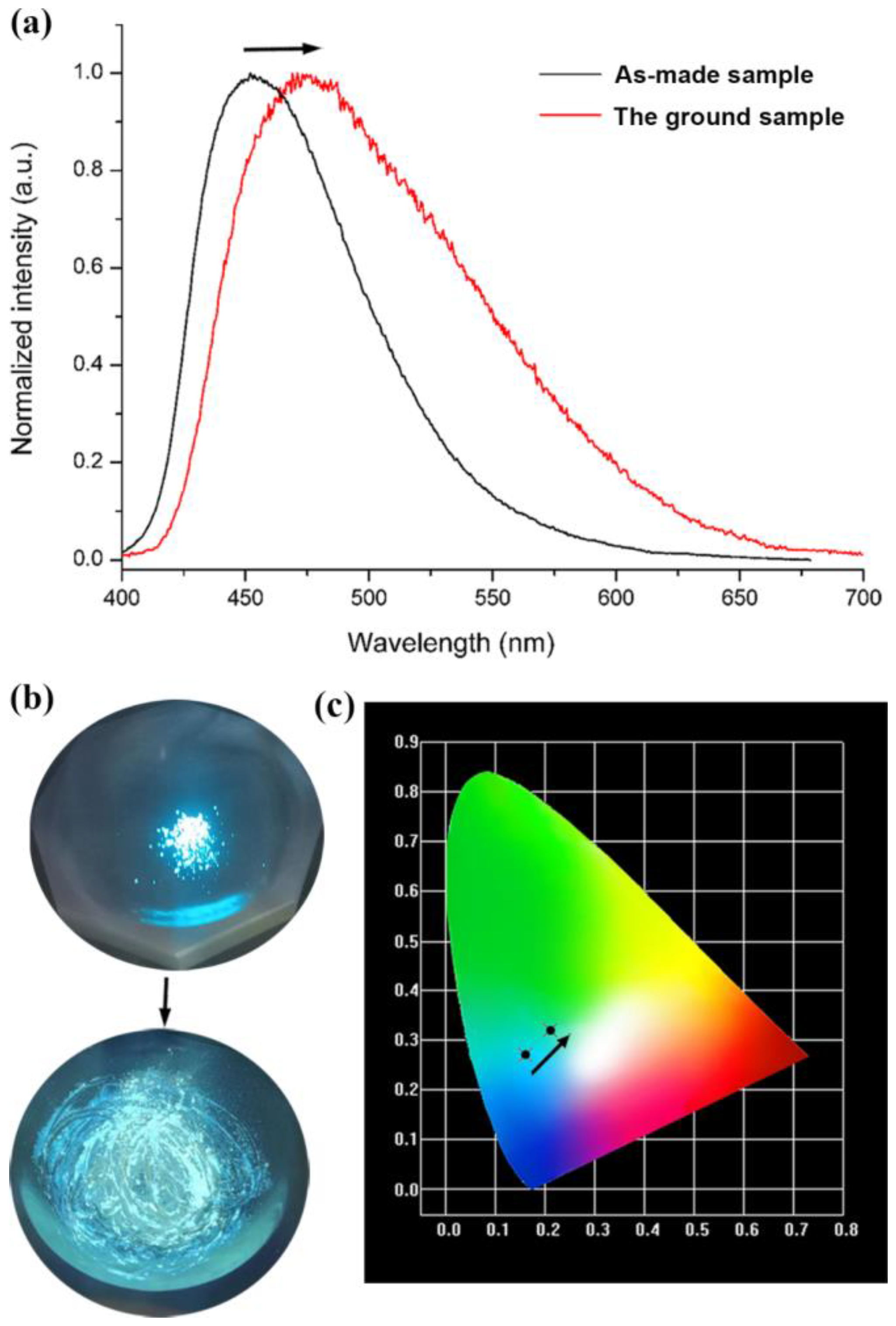
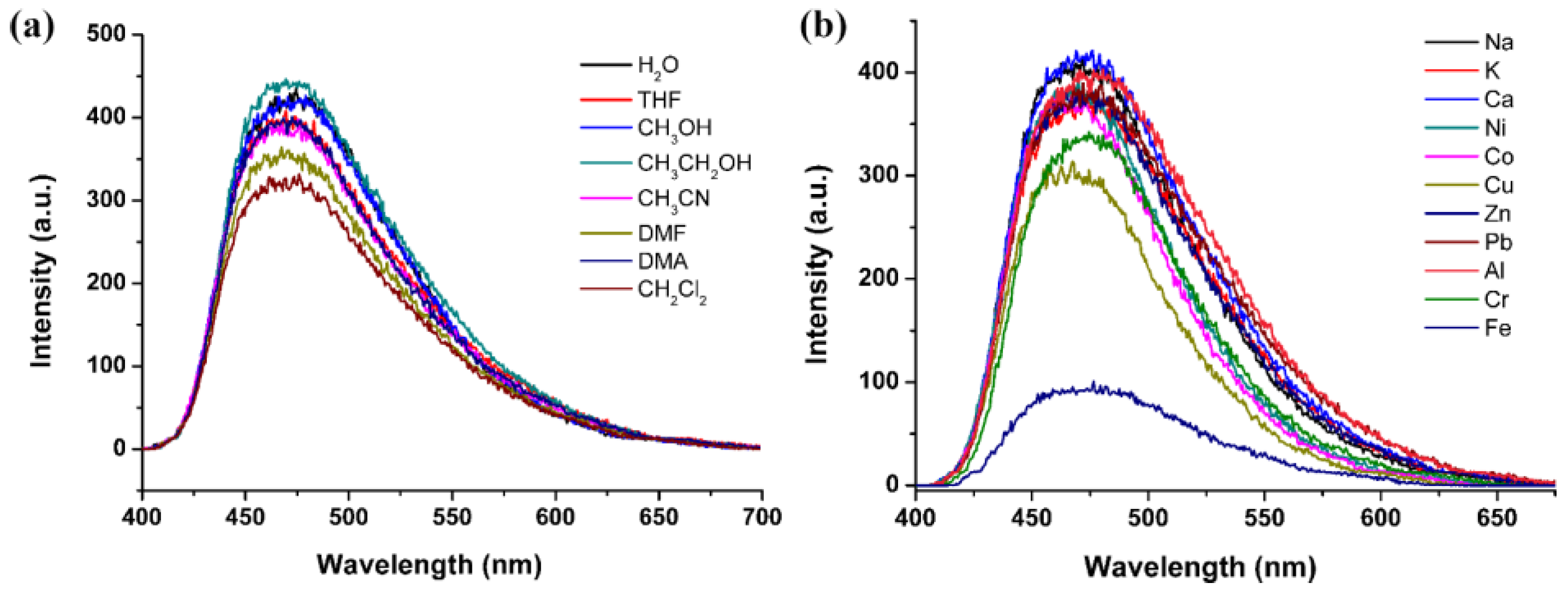
H4tcbpe is a bright emissive ligand with aggregation-induced emission (AIE) [23,24]. Therefore, ligands bearing this AIE center of tetraphenyl ethylene usually show sensitive FL to external stimuli [23,24,25]. Herein, the AIE character is inherited by Sr2(tcbpe), which exhibits interestingly mechanochromic FL. As depicted in Figure 2a, the solid-state FL spectrum of the as-made Sr2(tcbpe) shows a blue emission maximized at 470 nm when excited by 410 nm light (Figures S6 and S7). A mechanoresponsive bathochromic shift in FL emission after grinding is observed—that is, a shift from the blue emission centered at 470 nm to a green emission maximized at 485 nm (inset in Figure 2a, Figures S7 and S8, ESI). Consequently, the chromaticity coordinate for the as-made sample is (0.16, 0.27), while that for the ground sample is (0.21, 0.32) (Figure 2b). When dispersed in various lab solvents (e.g., N,N-dimethylformamide (DMF), N,N-dimethylacetamide (DMA), acetone, and ethanol), the powdered Sr2(tcbpe) exhibits no solvent-dependent FL (Figure S9), which showed, to some extent, a quenching of FL intensity (Figure 3a). The powdered samples of Sr2(tcbpe) were dispersed in various 0.5󠆼 × 10−2 M M(AC)n solutions (n = 1–3, M = Na+, K+, Ca2+, Zn2+, Cu2+, Co2+, Ni2+, Pb2+, Fe3+, Al3+, and Cr3+) to test their FL-sensing selectivity to metal ions. As can be seen in Figure 2b, although different metal ions exhibit mild FL-quenching effects to Sr2(tcbpe), only the Fe3+ ion almost entirely quenches its FL, demonstrating that the material is a selective FL sensor to Fe3+ ions.
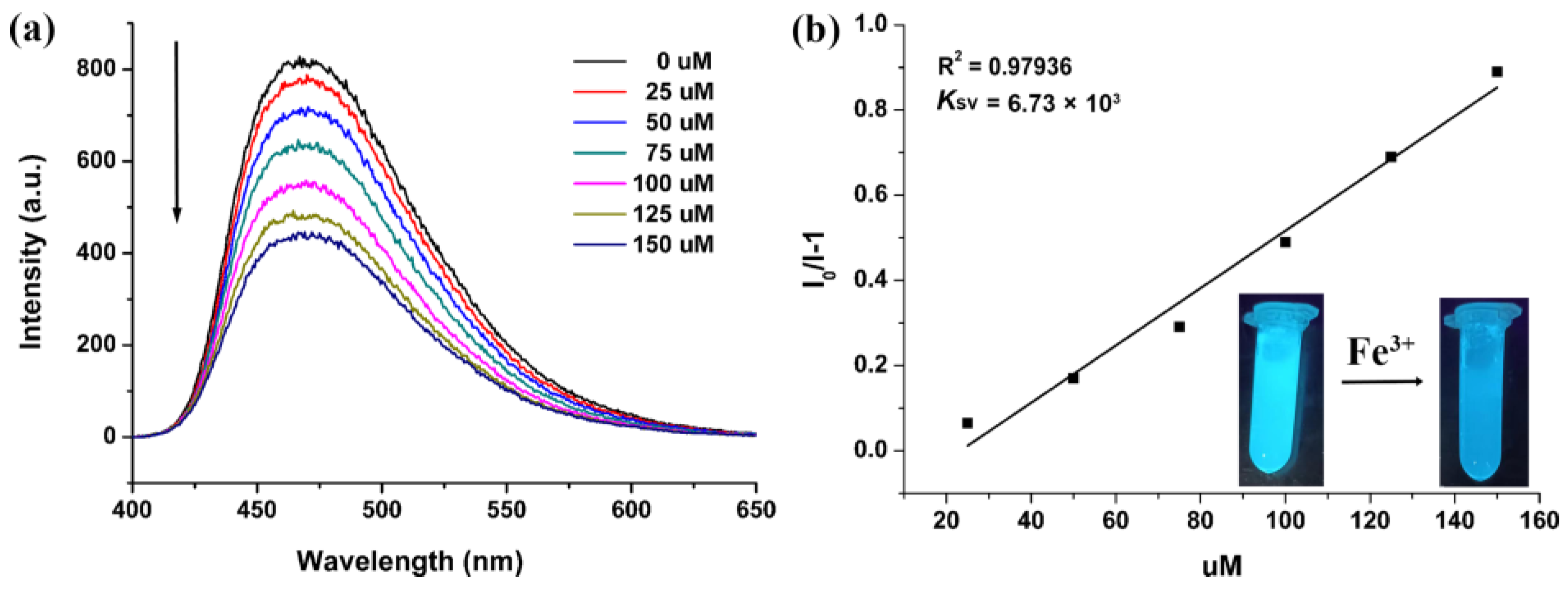
The FL-quenching percentage was quantitatively monitored by the addition of different amounts of 0.5󠆼 × 10−2 M Fe3+ ions into the FL emulsion (2 mg Sr2(tcbpe) dispersed in 2 mL H2O). The FL intensity of Sr2(tcbpe) is gradually quenched with increasing Fe3+ ion content, and the FL was quenched by almost 50% at a concentration of 0.1 mM Fe3+ ions (Figure 4a). As shown in Figure 4b, the Stern–Volmer equation (I0/I = 1 + Ksv[M], in which I0 and I are the FL intensity of Sr2(tcbpe) without and with the addition of Fe3+, and [M] is the molarity of Fe3+ and Ksv is the quenching constant), exhibits good linear behavior. The value of Ksv was found to be 6.73 × 10³ M−1. The limit of detection was obtained as 0.14 mM from the ratio of 3δ/slope. The detection sensitivity to Fe3+ ions is even comparable to that of porous FL-CP sensors [26,27,28,29,30,31,32,33,34,35,36,37,38,39], Table S1.
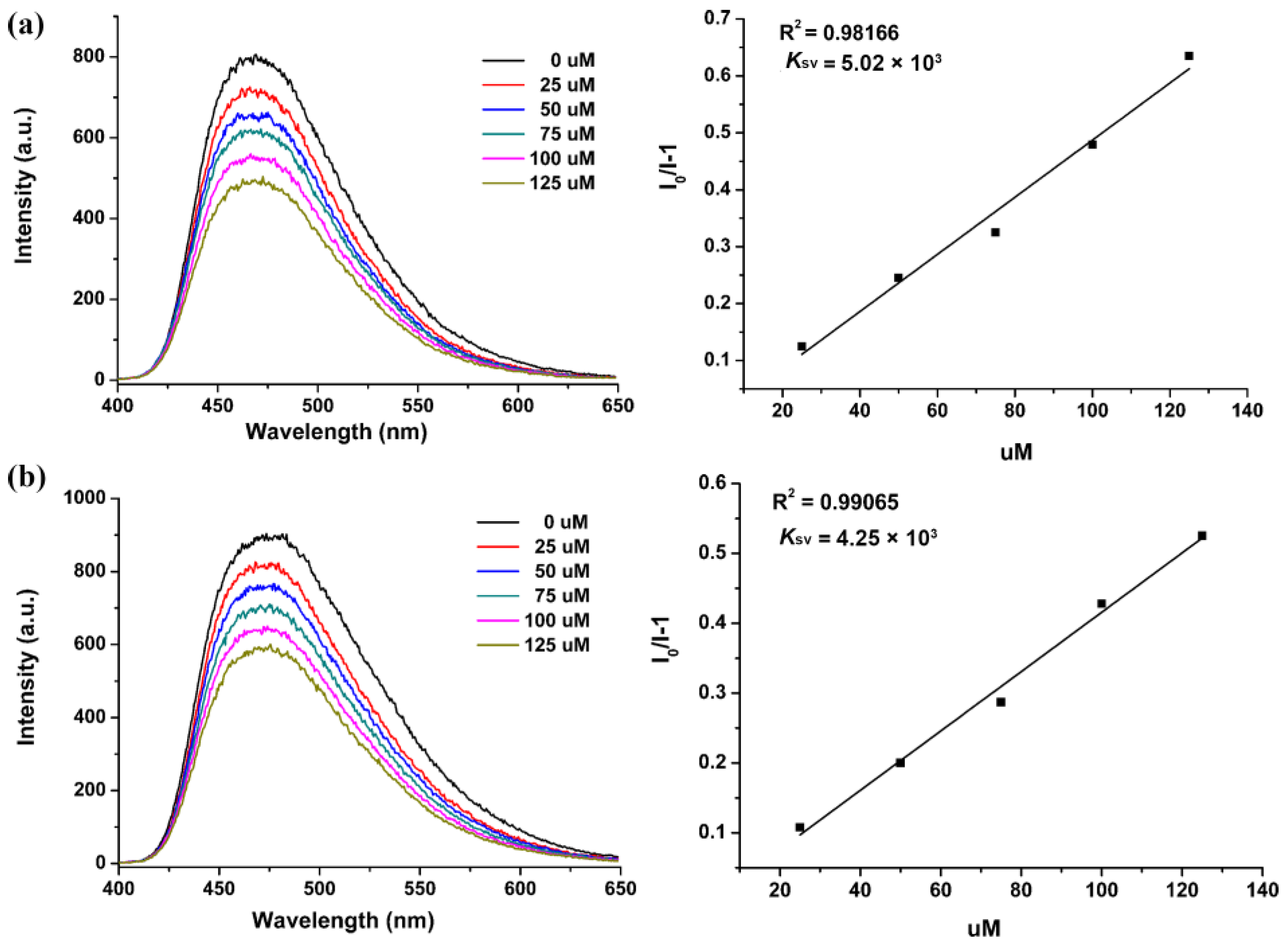
Selectivity is an important parameter for FL sensors. Na+, Ca2+, etc. are usually coexisting ions in water in nature. Therefore, the powdered sample of Sr2(tcbpe) was dispersed in separate 0.5󠆼 × 10−2 M Na+ and Ca2+ aqueous solutions to check the sensing selectivity towards Fe3+. As depicted in Figure 5, the FL intensity of Sr2(tcbpe) in these interferential metal ions showed a similar quenching response to Fe3+ as that of the FL emulsion dispersed in water. The decrease in FL intensity also exhibits a good linear relationship with Fe3+ concentration. The Ksv values are 5.02 × 10³ and 4.25 × 10³ M−1 for Fe3+ ions (Insets of Figure 5), respectively, which are comparable to that in water. The results demonstrate that Sr2(tcbpe) possesses a good sensing selectivity toward Fe3+ even in water systems with various interferential metal ions.
The as-made Sr2(tcbpe) also exhibits excellent solvent- and water-tolerances even in acid/base conditions. The prepared compound has exhibited good anti-solvent stability, as demonstrated by a comparative study of the PXRD patterns for the samples before and after immersion in common lab solvents (Figure S10). As shown in Figure S11, the PXRD patterns of the samples immersed in water with pH values of between 3 to 13 remain the same as the simulated pattern, indicating that the skeleton of Sr2(tcbpe) is still maintained in acid or base water environments. The good selectivity and chemical stability make Sr2(tcbpe) a promising FL sensor for Fe3+ in water. Fe3+ ions due to their d5 configuration possess a strong electron-withdrawing ability. The UV light-excited electrons of Sr2(tcbpe) transfer to the Fe3+ ions, thus resulting in a decrease in the FL intensities of the Sr2(tcbpe) [28,29,30,31].

4. Conclusions

An AIE-emitting ligand of H4tcbpe has been coordinated with Sr2+ to assemble an FL Sr-CP. The obtained compound, Sr2(tcbpe), exhibits a mechanoresponsive FL shifting from blue to green emission. Remarkably, the water-stable Sr2(tcbpe) represents the first FL Sr-CP sensor for Fe3+ ions with good sensitivity and selectivity. More AE-based FL-CPs sensors designed to detect toxic species will be created in our lab in the future.

Supplementary Materials

The following supporting information can be downloaded at: https://www.mdpi.com/article/10.3390/ma16020577/s1. Figure S1: Photograph of the as-made sample of Sr2(tcbpe); Figure S2: IR spectra for the H4tcbpe ligand and the as-made sample for Sr2(tcbpe); Figure S3: Experimental PXRD pattern of Sr2(tcbpe) compared with the simulated pattern; Figure S4: TG curve of the as-made Sr2(tcbpe); Figure S5: Extra structural details for Sr2(tcbpe); Figure S6: UV-Vis absorption spectrum for the as-made Sr2(tcbpe); Figures S7 and S8: FL spectra for Sr2(tcbpe) before and after grinding; Figure S9: Photographs of the powdered Sr2(tcbpe) dispersed in solvents excited at 365 nm UV light; Figures S10 and S11: PXRD patterns of the sample immersed in different lab solvents and water in a wide range of pH (3–13); Table S1: Comparison of Ksv for the reported CPs FL sensors for Fe3+ ion.

Author Contributions

Data collection and curation, Z.-W.L. and B.T.; writing—review and editing, Z.-F.W.; supervision and project administration, X.-Y.H. All authors have read and agreed to the published version of the manuscript.

Funding

This research was funded by the Fujian Science & Technology Innovation Laboratory for Opto-electronic Information of China (2021ZR130), the Natural Science Foundation of Fujian Province (2021J01513), and the National Natural Science Foundation of China (No. 22175178).

Institutional Review Board Statement

Not applicable.

Informed Consent Statement

Not applicable.

Data Availability Statement

Not applicable.

Conflicts of Interest

The authors declare no conflict of interest.

References

  1. Chowdhury, M.A. Metal-organic-frameworks for biomedical applications in drug delivery, and as MRI contrast agents. J. Biomed. Mater. Res. Part A 2017, 105, 1184–1194. [Google Scholar] [CrossRef] [PubMed]
  2. Bastug, E.; Kursunlu, A.N.; Güler, E. A fluorescent clever macrocycle: Deca-bodipy bearing a pillar [5]arene and its selective binding of asparagine in half-aqueous medium. J. Lumin. 2020, 225, 117343. [Google Scholar] [CrossRef]
  3. Taner, B.; Kursunlu, A.N.; Güler, E. The example of calix[4]pyrrole derivative containing Bodipy unit: Fluorometric and colorimetric sensor for F ion. Acta A Mol. Biomol. Spectrosc. 2014, 118, 903–907. [Google Scholar] [CrossRef] [PubMed]
  4. Lustig, W.P.; Mukherjee, S.; Rudd, N.D.; Desai, A.V.; Li, J.; Ghosh, S.K. Metal–organic frameworks: Functional luminescent and photonic materials for sensing applications. Chem. Soc. Rev. 2017, 46, 3242–3285. [Google Scholar] [CrossRef]
  5. Mendes, R.F.; Figueira, F.; Leite, J.P.; Gales, L.; Almeida Paz, F.A. Metal–organic frameworks: A future toolbox for biomedicine? Chem. Soc. Rev. 2020, 49, 9121–9153. [Google Scholar] [CrossRef]
  6. Allendorf, M.D.; Bauer, C.A.; Bhakta, R.K.; Houk, R.J.T. Luminescent metal–organic frameworks. Chem. Soc. Rev. 2009, 38, 1330–1352. [Google Scholar] [CrossRef]
  7. Lustig, W.P.; Li, J. Luminescent metal–organic frameworks and coordination polymers as alternative phosphors for energy efficient lighting devices. Coord. Chem. Rev. 2018, 373, 116–147. [Google Scholar] [CrossRef]
  8. Zhao, Y.; Li, D. Lanthanide-functionalized metal–organic frameworks as ratiometric luminescent sensors. J. Mater. Chem. C 2020, 8, 12739–12754. [Google Scholar] [CrossRef]
  9. Cui, Y.; Chen, B.; Qian, G. Lanthanide metal-organic frameworks for luminescent sensing and light-emitting applications. Coord. Chem. Rev. 2014, 273–274, 76–86. [Google Scholar] [CrossRef]
  10. Hasegawa, Y.; Nakanishi, T. Luminescent lanthanide coordination polymers for photonic applications. RSC Adv. 2015, 5, 338–353. [Google Scholar] [CrossRef]
  11. Rocha, J.; Carlos, L.D.; Paz, F.A.A.; Ananias, D. Luminescent multifunctional lanthanides-based metal–organic frameworks. Chem. Soc. Rev. 2011, 40, 926–940. [Google Scholar] [CrossRef] [PubMed]
  12. Yan, B. Luminescence response mode and chemical sensing mechanism for lanthanide-functionalized metal–organic framework hybrids. Inorg. Chem. Front. 2021, 8, 201–233. [Google Scholar] [CrossRef]
  13. Wu, Z.-F.; Tan, B.; Lustig, W.P.; Velasco, E.; Wang, H.; Huang, X.-Y.; Li, J. Magnesium based coordination polymers: Syntheses, structures, properties and applications. Coord. Chem. Rev. 2019, 399, 213025. [Google Scholar] [CrossRef]
  14. Ye, G.; Chen, C.; Lin, J.; Peng, X.; Kumar, A.; Liu, D.; Liu, J. Alkali /alkaline earth-based metal–organic frameworks for biomedical applications. Dalton Trans. 2021, 50, 17438–17454. [Google Scholar] [CrossRef] [PubMed]
  15. Zang, Y.; Li, L.-K.; Zang, S.-Q. Recent development on the alkaline earth MOFs (AEMOFs). Coord. Chem. Rev. 2021, 440, 213955. [Google Scholar] [CrossRef]
  16. Hureau, C. Coordination of redox active metal ions to the amyloid precursor protein and to amyloid-β peptides involved in Alzheimer disease. Part 1: An overview. Coord. Chem. Rev. 2012, 256, 2164–2174. [Google Scholar] [CrossRef]
  17. Kozlowski, H.; Luczkowski, M.; Remelli, M.; Valensin, D. Copper, zinc and iron in neurodegenerative diseases (Alzheimer’s, Parkinson’s and prion diseases). Coord. Chem. Rev. 2012, 256, 2129–2141. [Google Scholar] [CrossRef]
  18. Cui, Y.; Yue, Y.; Qian, G.; Chen, B. Luminescent Functional Metal–Organic Frameworks. Chem. Rev. 2012, 112, 1126–1162. [Google Scholar] [CrossRef]
  19. Hu, Z.; Deibert, B.J.; Li, J. Luminescent metal–organic frameworks for chemical sensing and explosive detection. Chem. Soc. Rev. 2014, 43, 5815–5840. [Google Scholar] [CrossRef] [Green Version]
  20. Wang, H.; Lustig, W.P.; Li, J. Sensing and capture of toxic and hazardous gases and vapors by metal–organic frameworks. Chem. Soc. Rev. 2018, 47, 4729–4756. [Google Scholar] [CrossRef]
  21. Lustig, W.P.; Shen, Z.; Teat, S.J.; Javed, N.; Velasco, E.; O’Carroll, D.M.; Li, J. Rational design of a high-efficiency, multivariate metal–organic framework phosphor for white LED bulbs. Chem. Sci. 2020, 11, 1814–1824. [Google Scholar] [CrossRef] [PubMed] [Green Version]
  22. Sheldrick, G.M. Crystal Structure Refinement with SHELXL. Acta Crystallogr. Sect. C Struct. Chem. 2015, 71, 3–8. [Google Scholar] [CrossRef] [PubMed] [Green Version]
  23. Wu, X.-H.; Luo, P.; Wei, Z.; Li, Y.-Y.; Huang, R.-W.; Dong, X.-Y.; Li, K.; Zang, S.-Q.; Tang, B.Z. Guest-triggered aggregation-induced emission in silver chalcogenolate cluster metal–organic frameworks. Adv. Sci. 2019, 6, 1801304. [Google Scholar] [CrossRef] [Green Version]
  24. Wu, Z.-F.; Velasco, E.; Shan, C.; Tan, K.; Zhang, Z.-Z.; Hu, Q.-Q.; Xing, K.; Huang, X.-Y.; Li, J. Robust fluorescent calcium coordination polymers as Cu2+ sensors with high sensitivity and fast response. J. Mater. Chem. C 2020, 8, 6820–6825. [Google Scholar] [CrossRef]
  25. Zhu, Z.-H.; Bi, C.; Zou, H.-H.; Feng, G.; Xu, S.; Tang, B.Z. Smart tetraphenylethene-based luminescent metal–organic frameworks with amide-assisted thermofluorochromics and piezofluorochromics. Adv. Sci. 2022, 9, 2200850. [Google Scholar] [CrossRef] [PubMed]
  26. Chen, C.-H.; Wang, X.-S.; Li, L.; Huang, Y.-B.; Cao, R. Highly selective sensing of Fe3+ by an anionic metal–organic framework containing uncoordinated nitrogen and carboxylate oxygen sites. Dalton Trans. 2018, 47, 3452–3458. [Google Scholar] [CrossRef]
  27. Luo, Y.-H.; Xie, A.D.; Chen, W.-C.; Shen, D.; Zhang, D.-E.; Tong, Z.-W.; Lee, C.-S. Multifunctional anionic indium–organic frameworks for organic dye separation, white-light emission and dual-emitting Fe3+ sensing. J. Mater. Chem. C 2019, 7, 14897–14903. [Google Scholar] [CrossRef]
  28. Sun, W.; Wang, J.; Zhang, G.; Liu, Z. A luminescent terbium MOF containing uncoordinated carboxyl groups exhibits highly selective sensing for Fe3+ ions. RSC Adv. 2014, 4, 55252–55255. [Google Scholar] [CrossRef]
  29. Wu, Z.-F.; Huang, X.-Y. A series of Mg–Zn heterometallic coordination polymers: Synthesis, characterization, and fluorescence sensing for Fe3+, CS2, and nitroaromatic compounds. Dalton Trans. 2017, 46, 12597–12604. [Google Scholar] [CrossRef]
  30. Yao, J.; Liu, Y.-E.; Yang, L.-B.; Dou, A.-N.; Hou, C.-F.; Xu, Q.-Q.; Huang, B.; Zhu, A.-X. Novel alkaline earth metal–organic frameworks with thiophene groups for selective detection of Fe3+. CrystEngComm 2020, 22, 5970–5979. [Google Scholar] [CrossRef]
  31. Zhang, J.; Zhao, L.; Liu, Y.; Li, M.; Li, G.; Meng, X. Two luminescent transition-metal–organic frameworks with a predesigned ligand as highly sensitive and selective iron(iii) sensors. New J. Chem. 2018, 42, 6839–6847. [Google Scholar] [CrossRef]
  32. Zhang, X.; Zhuang, X.; Zhang, N.; Ge, C.; Luo, X.; Li, J.; Wu, J.; Yang, Q.; Liu, R. A luminescent sensor based on a Zn (II) coordination polymer for selective and sensitive detection of NACs and Fe3+ ion. CrystEngComm 2019, 21, 1948. [Google Scholar] [CrossRef]
  33. Das, A.; Biswas, S. A multi.-responsive carbazole-functionalized Zr (IV)-based metal-organic framework for selective sensing of Fe (III), cyanide and p-nitrophenol. Sens. Actuator B-Chem. 2017, 250, 121. [Google Scholar] [CrossRef]
  34. Das, A.; Biswas, S.; Trivedi, V.; Biswas, S. Extraordinary sensitivity for H2S and Fe (III) sensing in aqueous medium by Al-MIL-53-N3 metal–organic framework: In vitro and in vivo applications of H2S sensing. Dalton Trans. 2018, 47, 2690. [Google Scholar] [CrossRef] [PubMed]
  35. Rath, B.B.; Vittal, J.J. Water stable Zn (II) metal–organic framework as a selective and sensitive luminescent probe for Fe (III) and chromate ions. Inorg. Chem., 2020, 59, 8818. [Google Scholar] [CrossRef]
  36. Xu, T.-Y.; Nie, H.-J.; Li, J.-M.; Shi, Z.-F. Luminescent Zn (II)/Cd (II) coordination polymers based on 1-(tetrazol-5-H)-3, 5-bis (1-triazole) benzene for sensing Fe3+, Cr2O72−, and CrO42− in water. J. Solid State Chem. 2020, 287, 121342. [Google Scholar] [CrossRef]
  37. Zhu, H.; Fu, L.; Liu, D.; Li, Y.-H.; Dong, G.-Y. Three water-stable luminescent Zn (II) coordination polymers for highly sensitive and selective sensing of acetylacetone and Fe3+ ions. J. Solid State Chem. 2020, 286, 121265. [Google Scholar] [CrossRef]
  38. Chen, Z.; Mi, X.; Lu, J.; Wang, S.; Li, Y.; Dou, J.; Li, D. From 2D→ 3D interpenetration to packing: N coligand-driven structural assembly and tuning of luminescent sensing activities towards Fe3+ and Cr2O72− ions. Dalton Trans. 2018, 47, 6240. [Google Scholar] [CrossRef]
  39. Yang, Y.-J.; Wang, M.-J.; Zhang, K.-L. A novel photoluminescent Cd (II)–organic framework exhibiting rapid and efficient multi-responsive fluorescence sensing for trace amounts of Fe3+ ions and some NACs, especially for 4-nitroaniline and 2-methyl-4-nitroaniline. J. Mater. Chem. C 2016, 4, 11404. [Google Scholar] [CrossRef]
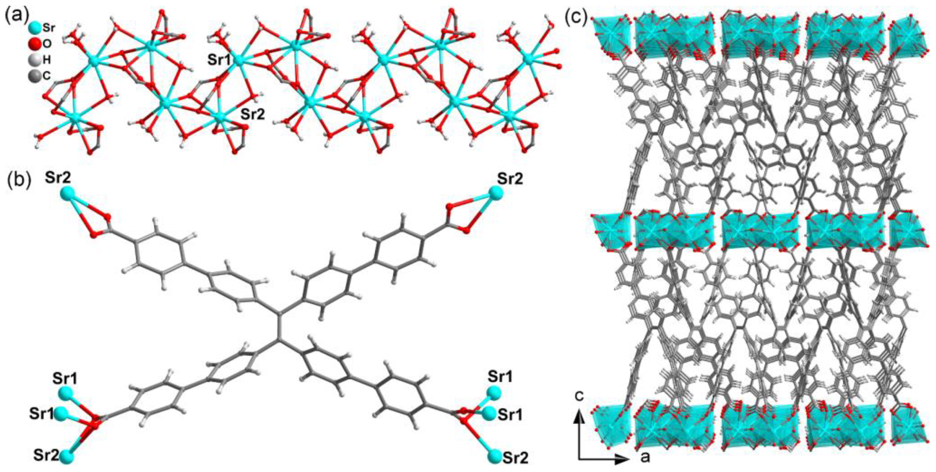
Figure 1. (a) The coordination environments of Sr2+ ions in the 1D chain-like SBU of structure Sr2(tcbpe). (b) The coordination mode of tcbpe4− in Sr2(tcbpe). (c) The 3D skeleton of Sr2(tcbpe) seen from the b-axis.
Materials 16 00577 g001
Figure 2. (a) The FL spectra of the as-made and ground samples of Sr2(tcbpe) measured at room temperature. (b) Photographic image of the luminescent changes of Sr2(tcbpe) under 365 nm light before and after grinding. (c) Photograph of the CIE chromaticity diagram for Sr2(tcbpe) before and after grinding.
Materials 16 00577 g002
Figure 3. The FL of the powdered Sr2(tcbpe) dispersed in different solvents (a) and 0.5󠆼 × 10−2 M aqueous solution containing different metal ions (b).
Materials 16 00577 g003
Figure 4. (a) FL of Sr2(tcbpe) with the addition of 0.5󠆼 × 10−2 M Fe3+ ions in an increasing amount. (b) The corresponding Ksv curve. Insets show the FL photographs of Sr2(tcbpe) before and after the addition of Fe3+ ions.
Materials 16 00577 g004
Figure 5. (a) FL spectra of Sr2(tcbpe) dispersed in 0.5󠆼 × 10−2 M Na+ aqueous solutions by adding Fe3+ ions at various concentrations and the corresponding Ksv curve. (b) FL spectra of Sr2(tcbpe) dispersed in 0.5 × 10⁻² M Ca2+ solutions with the addition of Fe3+ ions and the corresponding Ksv curve.
Materials 16 00577 g005
Table 1. Crystallographic data and structural refinement details for Sr2(tcbpe).
Empirical formulaSr2C54H40O12
Formula weight1056.10
Crystal systemMonoclinic
Space groupP21
T/K298(2)
λ0.71073
a10.0613(2)
b9.8710(2)
c22.7537(3)
β90.0016(11)
V32259.79(7)
Z2
Dc/Mg·m−31.552
μ/mm−12.428
F(000)1072
Measured refls.50,879
Independent refls.11,257
Rint0.0504
No. of parameters638
GOF1.038
Flack parameter0.401(10)
aR1, bwR2 [I > 2σ(I)]0.0490, 0.1226
aR1, bwR2 (all data)0.0516, 0.1238
aR1 = ∑║Fo│ − │Fc║/∑│Fo│. b wR2 = {∑w[(Fo)2 − (Fc2)]2/∑w(Fo2)2}1/2.
Disclaimer/Publisher’s Note: The statements, opinions and data contained in all publications are solely those of the individual author(s) and contributor(s) and not of MDPI and/or the editor(s). MDPI and/or the editor(s) disclaim responsibility for any injury to people or property resulting from any ideas, methods, instructions or products referred to in the content.