Article

The Influence of High-Energy Milling on the Phase Formation, Structural, and Photoluminescent Properties of CaWO4 Nanoparticles

1
Institute of General and Inorganic Chemistry, Bulgarian Academy of Sciences, Acad. G. Bonchev, Str. Bld. 11, 1113 Sofia, Bulgaria
2
Institute of Optical Materials and Technologies “Acad. Jordan Malinowski”, Bulgarian Academy of Sciences, 1113 Sofia, Bulgaria
*
Author to whom correspondence should be addressed.
Materials 2024, 17(15), 3724; https://doi.org/10.3390/ma17153724 (registering DOI)
Submission received: 19 June 2024 / Revised: 15 July 2024 / Accepted: 19 July 2024 / Published: 27 July 2024

Abstract

:
CaWO4 nanoparticles were obtained by facile mechanochemical synthesis at room temperature, applying two different milling speeds. Additionally, a solid-state reaction was employed to assess the phase composition, structural, and optical characteristics of CaWO4. The samples were analyzed by X-ray diffraction (XRD), transition electron microscopy (TEM), and Raman, infrared (IR), ultraviolet–visible (UV–Vis) reflectance, and photoluminescence (PL) spectroscopies. The phase formation of CaWO4 was achieved after 1 and 5 h of applied milling speeds of 850 and 500 rpm, respectively. CaWO4 was also obtained after heat treatment at 900 °C for 12 h. TEM and X-ray analyses were used to calculate the average crystallite and grain size. The Raman and infrared spectroscopies revealed the main vibrations of the WO4 groups and indicated that more distorted structural units were formed when the compound was synthesized by the solid-state method. The calculated value of the optical band gap of CaWO4 significantly increased from 2.67 eV to 4.53 eV at lower and higher milling speeds, respectively. The determined optical band gap of CaWO4, prepared by a solid-state reaction, was 5.36 eV. Blue emission at 425 (422) nm was observed for all samples under an excitation wavelength of 230 nm. CaWO4 synthesized by the solid-state method had the highest emission intensity. It was established that the intensity of the PL peak depended on two factors: the morphology of the particles and the crystallite sizes. The calculated color coordinates of the CaWO4 samples were located in the blue region of the CIE diagram. This work demonstrates that materials with optical properties can be obtained simply and affordably using the mechanochemical method.

1. Introduction

Divalent metal tungstates with the general formula AWO4 (where A = Ca, Sr, Ba, Pb, Zn) have important scientific and industrial applications in various fields, such as scintillators in high-energy physics [1,2], laser and stimulated Raman-scattering active media [3], medical devices and drug delivery [4], catalysts [5,6,7], and luminescent materials [8,9,10,11]. Among the metal tungstates, calcium tungstate is a very attractive material due to its excellent thermal and chemical stability, as well as its good photovoltaic [12], electrical [13], magnetic [14], catalytic [5,15,16], and optical properties [7,8,9,17]. CaWO4 has a scheelite-type structure with the space group I41/a and unit-cell parameters a = 5.243 Å and c = 11.373 Å [ICDD PDF#00-041-1431]. In this crystal structure, the alkaline ions are coordinated with eight oxygen atoms and form CaO8 polyhedra, and the tungstaten atoms are coordinated by four oxygen atoms in tetrahedral symmetry. The WO4 polyhedra are connected to CaO8 through common corners [18]. This compound is a semiconductor material with a wide band gap from 3.3 eV to 5.8 eV, which relates to different samples: bulk, nanoparticles, or film [5,9,12,13,19,20,21,22,23,24,25,26].
According to a number of studies, CaWO4 exhibits blue [4,10,11,24,25,26], blue-green [9,22], and green luminescence at room temperature [5,8,23,27]. Blue emission is commonly attributed to electronic transitions in the WO4 groups, while green originates from WO3 defect centers associated with oxygen vacancies. It is interesting to note that the particle shape of CaWO4 influences the kind and position of the emission of this compound. The CaWO4 with nano-rods and spherical morphology shows broad emission with maximum at 415–417 nm with different intensity [28]. This same crystal phase, with dumbbell, coral, rod, and dendrite particle morphologies, displays emission at 416 nm with various intensities and profiles [29]. The microspheres of CaWO4 exhibit emission at 420 nm, and the absolute PL intensity decreases with increasing temperature [30]. CaWO4 with a round morphology and an average size below 70 nm shows asymmetric blue-green emission at room temperature [31]. The aggregates of CaWO4 nano- and microcrystals possess intense luminescence emissions at 484 and 491 nm, respectively [22]. A broad emission peak at 521 nm was observed for CaWO4 with nearly spherical or elongated grains obtained by the molten salt method [7]. Emissions at 305 and 460 nm of CaWO4 with irregular particle morphology were described by Y. Wang et al., obtained by solvothermal synthesis [8].
Different physicochemical routes have been applied for the preparation of CaWO4, such as solid-state reactions [12,13,22], the Czochralski technique [2], hydrothermal synthesis [4,10,16,23], the sonochemical method [5,6], the molten salt method [8], solvothermal synthesis [9,28,31], co-precipitation [10,25], the double decomposition flux reaction method [32], electrospinning preparation [33], and mechanochemical synthesis [15,19,20,34].
Mechanochemical synthesis is one important and modern method for preparing nanomaterials in various fields from inorganic to metal–organic or organic materials [35,36,37,38]. Mechanical activation, as a combination of heat and pressure, causes different effects: defects and fresh surface areas that accelerate the synthesis of final products. The chemical reaction during the ball milling process depends on several factors, such as the nature of the initial reagents, the type of mills (horizontal rotary ball mill, planetary ball mill, vibration, shakier mill, etc.), milling speeds, milling time, the ball-to-powder weight ratio (BPR), and the milling environment (dry or wet). The milling speed has a decisive role for the chemical reaction at a constant BPR, the number and diameter of the balls, and the volume of the milling chamber [39,40,41,42]. Recently, mechanochemical treatment has been used for the preparation of optical materials due to the following advantages: an enhanced diffusion process, a short reaction time, and improved luminescent properties [43,44].
The literature data indicate that a lower milling speed has primarily been used for mechanochemical activation for the preparation of CaWO4. L. Cheng et al. used a lower speed (300/450 rpm) to prepare AWO4 (A = Ca, Ba, Sr) ceramics with good microwave dielectric properties [34]. P. Jena et al. obtained CaWO4 with a lower optical band gap after 12 h of mechanochemical treatment at 300 rpm and additional heat treatment at 1100 °C for 4 h [19]. S. Balamurugan et al. reported a thorough investigation of the phase formation of CaWO4 at a 300 rpm milling speed using various amounts of reaction mixtures with varying numbers and diameters of WC balls, under dry and wet conditions [20]. The authors obtained the pure phase of CaWO4 after 10 h of milling time in a dry atmosphere. Keeping in mind the above-mentioned literature data, as well as our previous results for the direct mechanochemical synthesis of similar inorganic compounds, we chose 2.5 g of reaction mixture (CaCO3 + WO3) and applied two grinding speeds, 500 and 850 rpm, to produce CaWO4.
The aim of the present work is to verify the possibility of direct synthesis in a short reaction time at a higher grinding rate. To the best of our knowledge, the influence of high-energy ball milling on the photoluminescent properties of CaWO4 has not been reported. The structural and luminescent properties obtained are compared with those of CaWO4 obtained by solid-state reaction.

2. Materials and Methods

2.1. Direct Mechanochemical Synthesis

The reagents used in the mechanochemical treatment were CaCO3 (Merck KGaA, Amsterdam, The Netherlands, 99.9% purity) and WO3 (Merck KGaA, Amsterdam, The Netherlands, 99.9% purity). The stoichiometric ratio of the initial materials was 1:1, which corresponds to the crystal CaWO4 phase. The high-energy ball milling of the initial mixture was carried out in a planetary ball mill (Fritsch premium line, Pulverisette No 7, FRITSCH GmbH, Idar-Oberstein, Germany), where two different milling speeds were applied: 500 and 850 rpm. The activation was performed in air atmosphere, and the ball-to-powder weight ratio was 10:1. To minimize the temperature during milling, the process was carried out in periods of 15 min, with rest periods of 5 min according to our previous studies [15,39,40,41]. The labels of the milled samples were as follows: CaWO4-I for the milling speed of 500 rpm; CaWO4-II for the milling speed of 850 rpm.

2.2. Solid-State Reaction

The initial reagents for the solid-state reaction were identical to those used in the mechanochemical activation i.e., CaCO3 (Merck KGaA, Amsterdam, The Netherlands, 99.99%) and WO3 (Merck KGaA, Amsterdam, The Netherlands, 99.99%). The stoichiometric ratio was the same (1:1). The initial mixture was homogenized in an agate mortar. Subsequently, the mixture was transferred to an aluminum crucible and heated at 900 °C for 12 h in an electrical furnace. The heat treatment was carried out according to the data in reference [13]. The prepared sample was labeled CaWO4-III.

2.3. Characterization

The XRD powder patterns were collected using the Bruker D8 Advance X-ray powder diffractometer, Karlsruhe, Germany, equipped with a CuKa radiation source (1.542 A) and the LynxEye PSD detector. The measurement range was from 5.5° to 120.0° 2θ, with a step of 0.02° 2θ and a counting time of 1.0 s/strip (for a total of 175.0 s/step, according to the PSD detector). The qualitative phase analysis was performed using the DIFFRAC.EVA v.4 software program [45] and the ICDD PDF-2 (2021) reference database. The unit cell parameters and crystallites size were calculated using the whole profile of the powder pattern and Topas v.4.2 software [46]. TEM observation was performed by a JEOL JEM-2100 microscope (Akishima, Japan) at an accelerating voltage of 200 kV. The preparation procedure of the specimens consisted of dispersing them in ethanol by ultrasonic treatment and then dripping them onto standard Cu grids. The Raman spectra were recorded using the Via Qontor Raman Confocal Microscope (Renishaw plc, Wotton-under-Edge, UK) with a laser wavelength of 532 nm (Nd:YAG-Laser). The laser power on the sample was maintained at 1% of the nominal power, so no heating effects on the powder sample could be observed. The excitation light was focused and collected using a ×50 LWD objective lens. Infrared spectra were registered in the range of 1200–400 cm−1 on a Nicolet-320 FTIR spectrometer (Madison, WI, USA) using the KBr pellet technique with a spectral resolution of 2 nm. The diffuse-reflectance spectra were recorded with a Thermo Evolution 300 UV-Vis Spec-trophotometer equipped with a Praying Mantis device (Madison, WI, USA) with Spectralon reflectance standard. For recording the background, Spectralon was used. The PL emission spectra were measured on a Horiba Fluorolog 3-22 TCS spectrophotometer (Longjumeau, France) equipped with a 450 W Xenon Lamp as the excitation source. The automated modular system, with the highest sensitivity among those available on the market, was used, allowing for the measurement of light emission of practically any type of sample. Double-grating monochromators were used for emissions in the range of 200–950 nm. All spectra were measured at room temperature.

3. Results and Discussion

3.1. Phase Formation and Morphology

3.1.1. X-ray Powder Diffraction Analysis

A comparative analysis of the phase formation, morphology, and symmetry of the structural units and optical properties of the mechanochemically and solid-state-obtained CaWO4 powders was carried out. The influence of the milling speed on the reaction time, crystallite size, and defects in the CaWO4 was studied by X-ray diffraction analysis (Figure 1A,B). The XRD pattern of the initial mixture shows the principal peaks of the monoclinic WO3 (PDF# 01-072-0677) and the orthorhombic CaCO3 (PDF# 00-041-1475). The milling speed of 500 rpm led to a decrease in the intensity and the broadening of all diffraction lines after 1 h of milling time (Figure 1A). This was a result of a decrease in the particle size, destruction of the long-range order, and the partial amorphization of both reagents. Increasing the milling time to 3 h caused the appearance of reflections characteristic for tetragonal CaWO4 (PDF#00-041-1431). This is an indication of the beginning of the chemical reaction between the activated reagents. The reaction finished after 5 h of milling time (Figure 1A). The possibility of phase formation after a short time was checked using a higher milling speed of 850 rpm. In this case, the principal peaks of CaWO4 were observed after 15 min of milling time. But the diffraction lines typical of the initial WO3 at 2θ = 23.20 and 23.95° were detected (Figure 1B). The pure CaWO4 was synthesized after 30 min of milling time, which is shorter compared to the synthesis at the 500 rpm milling speed. The increase in the milling time to 60 min did not affect the phase composition. In both cases, the diffraction lines were broad, which was attributed to the smaller size of the mechanochemically synthesized CaWO4 particles. The TEM images of these samples also show the formation of particles with lower dimensions (Figure 2A,B). Compared to the research reported in Ref. [20], we found that the amount of 2.5 g of the reaction mixture, higher milling speeds of 500 and 850 rpm, and dry media were more suitable conditions for the rapid preparation of CaWO4. Figure 1C exhibits the XRD pattern of CaWO4 after heat treatment at 900 °C for 12 h. A remarkable narrowing of the diffraction lines was observed due to the higher crystallinity of the CaWO4 in comparison to the mechanochemically obtained CaWO4 powders. No additional diffraction lines were found, meaning that the obtained sample was pure single phase. The lattice parameters (a, c, unit cell), lattice strain, and crystallite size of the CaWO4 obtained from the XRD refinement are shown in Table 1.
Table 1 presents the calculated unit cell parameters, crystallite sizes, and isotropic microstrains of the three synthesized samples. These were compared with data published in the literature [18]. It is clear from the values that there was no significant change in the parameters or volumes of the unit cells of the CaWO4 regardless of the conditions and method of preparation. Quite expectedly, as the mechanochemical activation energy increased, the crystallite size of the treated sample decreased. This was particularly clearly observed in the obtained values for the crystal lattice microstrains, which almost doubled when the milling speeds increased from 500 rpm to 850 rpm. The lattice strain decreased with an increasing crystallite size due to the prolongation of the heat treatment at a high temperature (CaWO4-III).

3.1.2. TEM Analysis

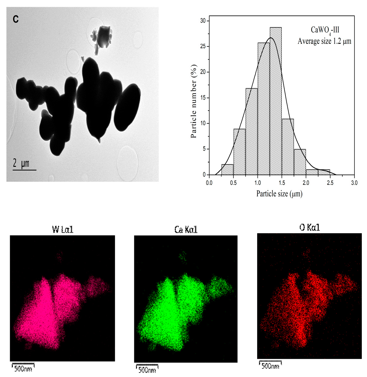
The size and morphology of the particles are important factors for the optical application of CaWO4. Figure 2A–C presents the TEM images, particle size distribution, and EDS mapping of the investigated samples. The particles of CaWO4-I and II obtained by mechanochemical treatment (milling speeds of 500 and 850 rpm) are well separated from each other, with a quasihexagonal form (Figure 2A,B). The size histograms illustrate that both CaWO4 nanoparticles exhibited good dispersion and a narrower size distribution. The particle size distribution of the CaWO4-I obtained at the lower milling speed of 500 rpm was between 5 and 30 nm, with most particles in the range between 10 and 15 nm (Figure 2A). At the higher milling speed, the particles had an average size of 20 nm (Figure 2B). Agglomeration of the particles was not observed in either of the mechanochemically synthesized samples. As might be expected, the particles obtained by solid-state synthesis were much larger, with an oval shape and average particle size of 1200 nm (Figure 2C). The particle distribution was between 500 and 2500 nm, with most particles tending to be between 1000 and 1500 nm in size. In this case, the agglomeration of the particles was visible. We can infer from the TEM results that significantly smaller particles were produced by high-energy ball milling. The EDS mapping demonstrates that the elements Ca, W, and O were disseminated on the entire surface of the samples prepared by mechanochemical activation and solid-state reaction.

3.2. Raman and Infrared Spectroscopy

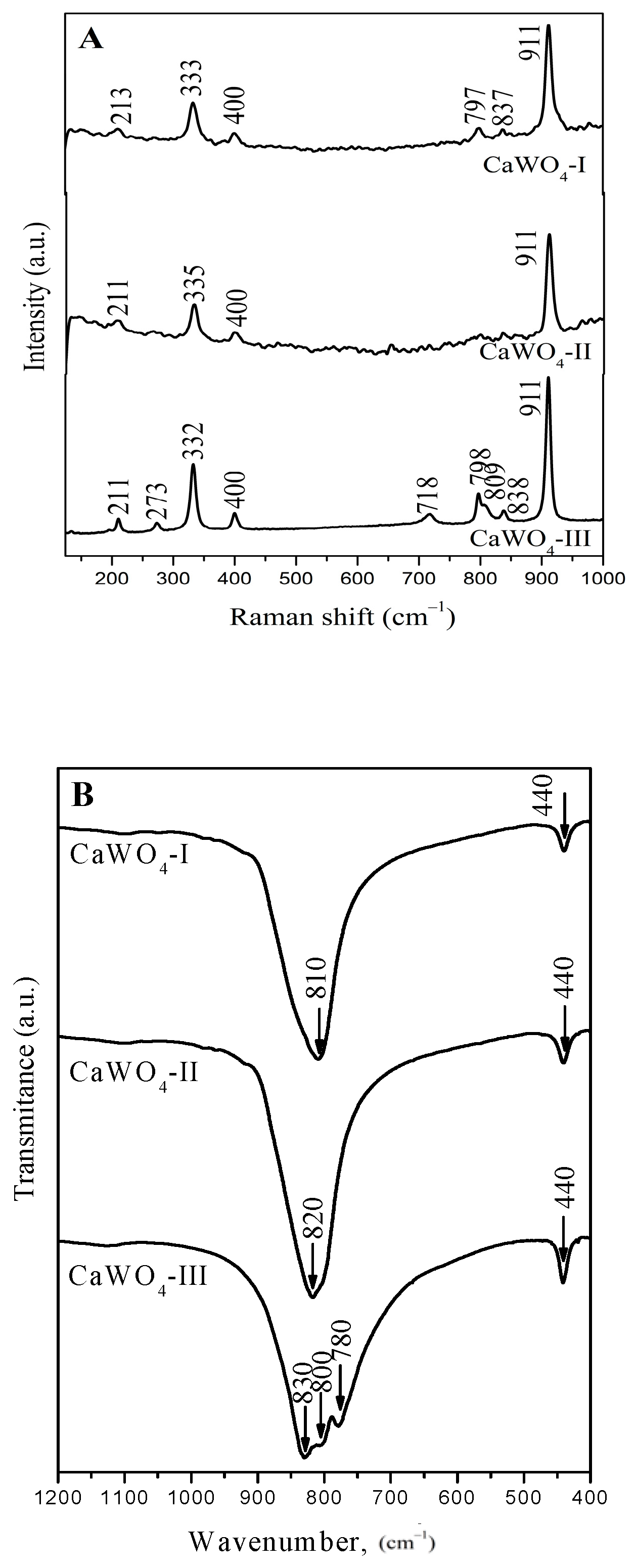
Both types of spectroscopic spectra of the obtained products evidence the formation of CaWO4 with a scheelite type structure with different degrees of WO4 destruction. Figure 3A shows the Raman spectra of the CaWO4 phase excited at 532 nm (Nd:YAG-Laser). The Raman bands can be divided into two groups—internal and external modes—due to the weak coupling between the WO4 and CaO8 groups and keeping in mind the literature data [5,18,23,47]. The internal modes are attributed to the oscillations inside the [WO4]2− units with an immovable mass center. The external or lattice phonons correspond to the motion of the Ca2+cations and WO4 units [47,48].
The spectrum of CaWO4-III produced by the solid-state reaction was used to assign the Raman peaks: the strong band at 911 cm−1 was due to the symmetric stretching ν1 of W–O bond in the WO4 tetrahedra [5,18,23,47,48]. The appearance of several absorption bands in the range of 838–718 cm−1 can be attributed to the elimination of the degeneracy of the ν3 vibration of different crystallographically nonequivalent WO4 tetrahedra with different local symmetry (Td, C3, C2v) [49]. The observed band at 332 cm−1 and the weak band at 400 cm−1 are the result of the ν2 vibrations of the W-O bond [47,48]. The low frequency bands at 273 and 210 cm−1 could be assigned to the translational mode of the ν(Ca–O) and the bending vibration of the [WO4]2− group in the CaWO4, respectively [23,50]. The absence of the peaks in the range from 840 to 400 cm−1 in the CaWO4-II sample after 1 h of milling time at 850 rpm indicates the generated WO4 units were more symmetric compared to the other samples (CaWO4-I and CaWO4-III). These results were confirmed by the infrared (IR) spectroscopy (Figure 3B). The IR spectra of the CaWO4-I and CaWO4-II obtained by mechanochemical synthesis display one absorption band around 810 (820) cm−1 due to the ν3 vibration of the WO4 structural unit building the crystalline structure of the CaWO4 [15,17,25]. The low-intensity band at 440 cm−1 is attributed to the ν4 modes of the same structural groups [51]. In contrast, the IR spectrum of the CaWO4-III prepared by solid-state reaction exhibited more absorption bands between 900 and 700 cm−1. In this case, the appearance of the bands at 830, 800, and 780 cm−1 are assigned to the elimination of the ν3 vibration degeneracy of the WO4 tetrahedral with a lower local symmetry [52]. Similar results were established for BaMoO4 obtained by solid-state reaction [41]. The results above show that more symmetrical WO4 structural units were formed due to the high-energy ball milling (Figure 3B).

3.3. Optical Properties

3.3.1. UV–Vis Absorbance Spectroscopy

The optical behavior of the CaWO4 powders obtained by both techniques was investigated using UV-Vis (Figure 4A,B) and photoluminescence spectroscopies (Figure 5 and Figure 6). The UV–visible absorption spectra was transformed in the Kubelka–Minck function using the following formula [F(R)]: F(R∞) = (1 − R∞)2/(2R∞) = K/S. The R is the diffuse reflectance of the sample, K is the absorption coefficient, and S is the scattering coefficient. The reflectance of the sample depends on the ratio of K to S but not on the absolute values of K and S [53,54]. A strong absorption in the range of 210–250 nm was observed for all obtained samples (Figure 4A). The position of the main peak depends on the processing conditions i.e., the milling speeds (CaWO4-I and CaWO4-II) and the solid-state method (CaWO4-III). In the UV-Vis spectrum, the CaWO4-I sample exhibited a wide range from 210 to 370 nm with a maximum peak at 250 nm. The absorption band shifted up to 220 nm when the milling speed was increased to 850 rpm. CaWO4-III, obtained by solid-state reaction, also showed a tendency toward a blue shift, with a notable absorption peak recorded at 210 nm (Figure 4A). The narrowest absorption line was detected for this sample. This fact can be attributed to the higher crystallinity nature of the solid-state reaction-obtained powder with the highest crystalline size (D = 370 nm). The bands between 210 and 250 nm were attributed to the charge-transfer transitions within the WO42− complex in the scheelite type inorganic phases [5,7,17,24,25,30]. In addition, a secondary absorption band between 250 and 320 nm was also visible. Keeping in mind the reported data, this band corresponds to the creation of the excitonic state in A2+ ions (A2+ = Ba, Sr, Ca) [33,55]. A similar UV-Vis spectra was reported for CaWO4 obtained by different methods, including the sonochemical method [5], solvothermal method [9,23], solid-state reaction [12,13], and hydrothermal route [16].
The calculated Eg values of the CaWO4 powders obtained by mechanochemical activation at 500 and 850 rpm were 2.88 and 4.59 eV, respectively. The optical band gap value of CaWO4-I (5 h, 500 rpm) is similar to those of CaWO4 obtained by 12 h of milling time and heat-treated at 1100 °C [19]. The calculated optical band gap of CaWO4-III produced by the solid-state technique is 5.27 eV (Figure 4B). This increase in the band gap is a result of the structural improvements, according to the XRD analysis. The value is close to that of the CaWO4 thin film obtained by the chemical solution method [21] and the CaWO4 powders obtained by solid-state reaction [13], the polymeric precursor method, and the microwave-assisted hydrothermal method [56]. The changes in the crystallite size and microstructure of the CaWO4 obtained by both preparation methods influenced the electron structure, and this is reason for the difference in value of the optical band gaps and their optical features [57].

3.3.2. Luminescent Properties

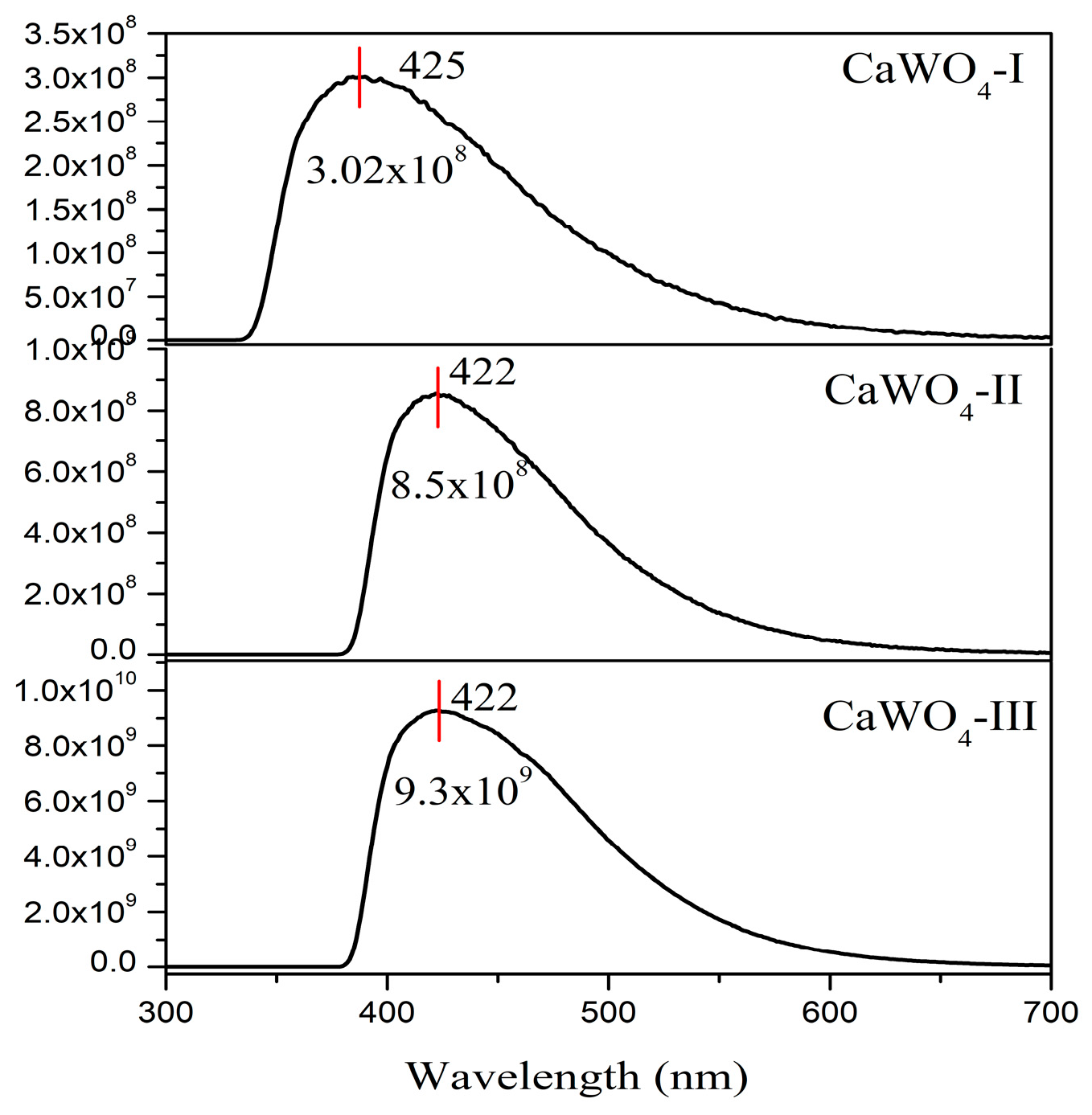
Figure 5 compares the emission spectra of the CaWO4 synthesized by both procedures: mechanochemical activation (500 and 850 rpm) and solid-state reaction (900 °C for 12 h). All samples show a broad blue band with a maximum at around 422–425 nm upon excitation at 230 nm (absorption of the WO4 groups). The emission lines show no variation in shape, but there is a noticeable difference in the emission intensity. The emission peaks of all prepared CaWO4 are in good agreement with the literature data for blue emission [58,59]. In our case, although the obtained crystalline phases possessed different particle size and morphologies, the emission spectrum profiles were the same. The blue emission at 425 nm, with a full width at the half maximum of 98 nm, was registered for CaWO4-I obtained using the lower milling speed (500 rpm), while the blue emission was changed at 422 nm, and a lower full width at the half maximum for CaWO4-II was obtained after the short milling time (1 h) at 850 rpm. The same blue maximum emission (422 nm) was observed with a full width at the half maximum of 100 nm for the solid-state-synthesized sample (CaWO4-III). This demonstrates that the as-prepared broader blue CaWO4 materials are promising candidates for WLEDs.
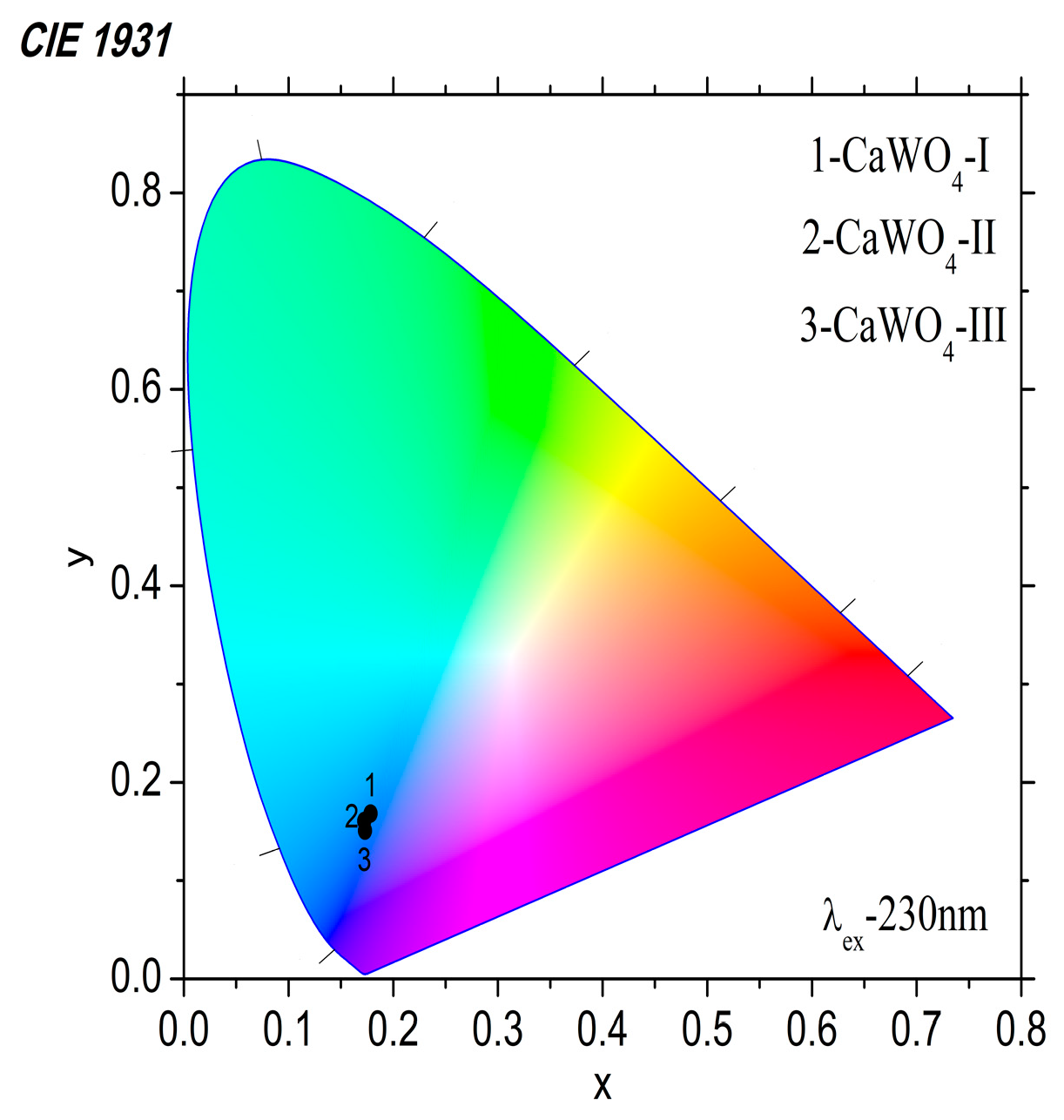
The PL intensities at 425–422 nm gradually increased with an increasing crystallite size. The value of the emission intensity was: ~3.02 × 10⁸ for the CaWO4-I prepared after 5 h of milling time and a milling speed of 500 rpm; ~8.5 × 10⁸ was recorded for the CaWO4-II obtained after a 1 h milling time and a milling speed of 850 rpm; ~9.3 × 10⁹ was recorded for the CaWO4-III obtained by the solid-state technique. The highest blue emission of CaWO4-III was probably due to its good crystallinity, oval-shaped particles, and highly asymmetric WO4 structural units. Similar results regarding the emission intensity were discussed by F. Lei et al. [60]. According to the literature data, the PL intensity and emission behavior of MWO4 with the scheelite and wolframite type structure depends on the surface chemistry, morphology, and particle size [61]. The lower crystallite size that results from mechanochemical treatment may be the cause of the lowest blue emissions for CaWO4-I and CaWO4-II [62]. The Commission International de I’Eclairage (CIE)’s chromaticity diagram of the CaWO4 samples excited at 230 nm is shown in Figure 6. As the main peak in the luminescence spectra is in the range between 400 and 600 nm and peaks at 425 (422) nm, it is expected that the emitted light will be blue. The calculated chromaticity coordinates are very close values and fall into the blue range on the CIE diagram (Figure 6 and Table 2). Blue light is in high demand because it can be mixed with yellow or participate in the tricolor (red, green, blue) to produce white light.

4. Conclusions

The variation in the milling speed promoted the rapid synthesis of the CaWO4 from 5 h at 500 rpm to 1 h at 850 rpm without additional heat treatment. We demonstrated that the mechanochemical treatment led to the formation of particles with a quasihexagonal form and a narrower size distribution. CaWO4 was also obtained by solid-state reaction at 900 °C for 12 h. Agglomeration of the oval particles was obtained using the solid-state technique. It was established that the method of preparation influences the crystallinity nature, macrostrains, and the symmetry of the WO4 entity and its optical properties. It was established that the optical band gap of CaWO4 strongly depends on the milling conditions, i.e., an Eg increase from 2.85 eV to 4.59 eV with an increase in the milling speed. The CaWO4-III obtained by the solid-state reaction possessed higher crystallinity, asymmetric WO4 units, and oval particles, which resulted in a higher blue emission intensity compared to the mechanochemically synthesized samples.

Author Contributions

Conceptualization, M.G.; methodology, M.G., R.I., and I.K.; software, P.T.; and P.I.; validation, M.G. and I.K.; formal analysis, M.G. and P.T.; investigation, M.G.; resources, R.I. and I.K.; data curation, M.G., R.I., I.K., and P.T.; writing—original draft preparation, M.G., P.T., and I.K.; writing—review and editing, R.I.; visualization, M.G. and I.K.; supervision, R.I. All authors have read and agreed to the published version of the manuscript.

Funding

This research received no external funding.

Institutional Review Board Statement

Not applicable.

Informed Consent Statement

Not applicable.

Data Availability Statement

The data are contained within the article.

Acknowledgments

Research equipment of the distributed research infrastructure INFRAMAT (part of the Bulgarian National roadmap for research infrastructures), supported by the Bulgarian Ministry of Education and Science under contract D01-322/30, November 2023, were used in this investigation. The Raman equipment and investigation were used within the framework of the “Center of excellence mechatronics and clean technologies”, with financial support from the European regional development fund within the program “Research, Innovation and Digitalisation for Smart Transformation, 2021–2027”.

Conflicts of Interest

The authors declare no conflicts of interest.

References

  1. Nitsch, K.; Nikl, M.; Ganschow, S.; Reiche, P.; Uecker, R. Growth of lead tungstate single crystal scintillators. J. Cryst. Growth 1996, 165, 163–165. [Google Scholar] [CrossRef]
  2. Nagirnyi, V.; Feldbach, E.; Onsson, L.J.; Kirm, M.; Lushchik, A.; Lushchik, C.; Nagornaya, L.L.; Ryzhikov, V.D.; Savikhiu, F.; Svensson, G.; et al. Excitonic and recombination processes in CaWO4 and CdWO4 scintillators under synchrotron irradiation. Radiat. Meas. 1998, 29, 247–250. [Google Scholar] [CrossRef]
  3. Kaminskii, A.A.; Eichler, H.J.; Ueda, K.; Klassen, N.V.; Redkin, B.S.; Li, L.E.; Findeisen, J.; Jaque, D.; Garcia-Sole, J.; Fernandez, J.; et al. Properties of Nd3+-doped and undoped tetragonal PbWO4, NaY(WO4)2, CaWO4, and undoped monoclinic ZnWO4 and CdWO4 as laser-active and stimulated Raman scattering-active crystals. Appl. Opt. 1999, 38, 4533–4547. [Google Scholar] [CrossRef] [PubMed]
  4. Tain, B.; Wang, M.; Liu, C.; Liu, S.; Jin, L.; Yan, X.; Li, C.; Wang, Z. Luminescent hollow CaWO4 microspheres: Template-free synthesis, characterization and application in drug delivery. RSC Adv. 2015, 5, 104172–104178. [Google Scholar]
  5. Andrade Neto, N.F.; Dias, B.P.; Tranquilin, R.L.; Longo, E.; Li, M.; Bomio, M.R.D.; Motta, F.V. Synthesis and characterization of Ag+ and Zn2+ co doped CaWO4 nanoparticles by a fast and facile sonochemical method. J. Alloys Compd. 2020, 823, 153617. [Google Scholar] [CrossRef]
  6. Sousa, P.B.; Gouveia, A.F.; Sczancoski, J.C.; Nogueira, I.C.; Longo, E.; San-Miguel, M.A.; Cavalcante, L.S. Electronic structure, optical and sonophotocatalytic properties of spindle-like CaWO4 microcrystals synthesized by the sonochemical method. J. Alloys Compd. 2021, 855, 157377–157386. [Google Scholar] [CrossRef]
  7. Shana, Z.; Wang, Y.; Ding, H.; Huang, F. Structure-dependent photocatalytic activities of MWO4 (M = Ca, Sr, Ba). J. Mol. Catal. A Chem. 2009, 302, 54–58. [Google Scholar] [CrossRef]
  8. Wang, Y.; Ma, J.; Tao, J.; Zhu, X.; Zhou, J.; Zhao, Z.; Xie, L.; Tian, H. Synthesis of CaWO4 nanoparticles by a molten salt method. Mater. Lett. 2006, 60, 291–293. [Google Scholar] [CrossRef]
  9. Wang, Y.; Wu, C.; Geng, L.; Chen, S. Unexpected formation of scheelite-structured Ca1−xCdxWO4 (0 ≤ x ≤ 1) continuous solid solutions with tunable photoluminescent and electronic properties. Phys. Chem. Chem. Phys. 2017, 19, 23204–23212. [Google Scholar] [CrossRef]
  10. Zhang, Y.; Abraha, A.; Zhang, R.; Shahbazyan, T.; Fadavi, M.; Heydari, E.; Dai, Q. Luminescence properties of CaWO4 and CaWO4:Eu3+ nanostructures prepared at low temperature. Opt. Mater. 2018, 84, 115–122. [Google Scholar] [CrossRef]
  11. Zhu, J.; Li, C.; Sun, Y.; Yang, C.; Zhao, Y.; Zhu, Z.; Wang, D.; Hu, Z.; Chou, S.; Li, L.; et al. Enhanced photoluminescence of hollow CaWO4 microspheres: The fast fabrication, structural manipulation, and exploration of the growth mechanism. Mater. Chem. Front. 2022, 6, 1046–1055. [Google Scholar] [CrossRef]
  12. Kim, D.W.; Cho, I.S.; Shina, S.S.; Lee, S.; Noh, T.H.; Kim, D.H.; Jung, H.S.; Hong, K.S. Electronic band structures and photovoltaic properties of MWO4 (M = Zn, Mg, Ca, Sr) compounds. J. Solid State Chem. 2011, 184, 2103–2107. [Google Scholar]
  13. Khobragade, N.; Sinha, E.; Rout, S.K.; Kar, M. Structural, optical and microwave dielectric properties of Sr1−xCaxWO4 ceramics prepared by the solid-state reaction route. Ceram. Int. 2013, 39, 9527–9635. [Google Scholar] [CrossRef]
  14. Sun, B.; Jia, X.J.; Wu, J.H.; Chen, P. Controlled synthesis and room-temperature ferromagnetism of CaWO4 nanostructures. J. Alloys Compd. 2015, 653, 95–99. [Google Scholar] [CrossRef]
  15. Georgieva, R.; Gancheva, M.; Ivanov, G.; Shipochka, M.; Markov, P.; Nihtianova, D.; Iordanova, R.; Naydenov, A. Synthesis, characterization and activity of Pd/CaWO4 catalyst in the complete oxidation of C1–C6 alkanes and toluene. React. Kinet. Mech. Catal. 2021, 132, 811–827. [Google Scholar] [CrossRef]
  16. Nobre, F.X.; Muniz, R.; Nascimento, E.R.; Amorim, R.S.; Silva, R.S.; Almeida, A.; Agostinho Moreira, J.; Tavares, P.B.; Brito, W.R.; Couceiro, P.R.C.; et al. Hydrothermal temperature dependence of CaWO4 nanoparticles: Structural, optical, morphology and photocatalytic activity. J. Mater. Sci. Mater. Electron. 2021, 32, 9776–9794. [Google Scholar] [CrossRef]
  17. Pereira, P.F.S.; Gouveia, A.F.; Assis, M.; Oliveira, R.C.; Pinatti, I.M.; Penha, M.; Gonçalves, R.F.; Gracia, L.; Andrés, J.; Longo, E. ZnWO4 nanocrystals: Synthesis, morphology, and photoluminescence and photocatalytic properties. Phys. Chem. Chem. Phys. 2018, 20, 1923–1937. [Google Scholar] [CrossRef] [PubMed]
  18. Burbank, R.D. Absolute integrated intensity measurement: Application to CaWO4 and comparison of several refinements. Acta Cryst. 1965, 18, 88–97. [Google Scholar] [CrossRef]
  19. Jena, P.; Kumar, D.; Verma, N.K.; Singh, A.K. Estimation of Lattice Strain and Optical Properties of Scheelite-Type AWO4 (A = Ca, Sr, Ba) Nanocrystalline Materials Synthesized by Mechanical Activation. AIP Conf. Proc. 2019, 2115, 030199–030204. [Google Scholar]
  20. Balamurugan, S.; Thomas, S.M.; Ashika, S.A.; Sana Fathima, T.K. Profound impact on different properties of calcium tungstate scheelite, CaWO4 phase stabilized via wider synthesis conditions. Inorg. Chem. Commun. 2023, 155, 111090–111099. [Google Scholar] [CrossRef]
  21. Maurera, M.A.M.A.; Souza, A.G.; Soledade, L.E.B.; Pontes, F.M.; Longo, E.; Leite, E.R.; Varela, J.A. Microstructural and optical characterization of CaWO4 and SrWO4 thin films prepared by a chemical solution method. Mater. Lett. 2004, 58, 727–732. [Google Scholar] [CrossRef]
  22. Paikaray, R.; Badapanda, T.; Mohapatra, H.; Richhariya, T. Investigation of Structural, Photoluminescence and Thermoluminescence Properties of Scheelite-Type CaWO4 Phosphor. Trans. Electr. Electron. Mater. 2023, 24, 73–81. [Google Scholar] [CrossRef]
  23. Cavalcante, L.S.; Longo, V.M.; Sczancoski, J.C.; Almeida, M.A.P.; Batista, A.A.; Varela, J.A.; Orlandi, M.O.; Longo, E.; Siu Li, M. Electronic structure, growth mechanism and photoluminescence of CaWO4 crystals. Cryst. Eng. Commun. 2012, 14, 853–868. [Google Scholar] [CrossRef]
  24. Kaur, P.; Khanna, K.; Kumar, R.; Chandra, E. Rare earth doped CaWO4 and CaMoO4 thin film for white light emission. J. Vac. Sci. Technol. B 2021, 39, 012205. [Google Scholar] [CrossRef]
  25. Wang, S.; Gao, H.; Sun, G.; Li, Y.; Wang, Y.; Liu, H. Structure characterization, optical and photoluminescence properties of scheelite-type CaWO4 nanophosphors: Effects of calcination temperature and carbon skeleton. Opt. Mater. 2020, 99, 109562–109573. [Google Scholar] [CrossRef]
  26. Compos, A.B.; Simoes, A.Z.; Longo, E.; Varela, J.A.; Lang, V.M. Mechanisms behind blue, green, and red photoluminescence emissions in CaWO4 and CaMoO4 powders. Appl. Phys. Lett. 2007, 91, 051923–051927. [Google Scholar] [CrossRef]
  27. Nikl, M. Wide Band Gap Scintillation Materials: Progress in the Technology and Material Understanding. Phys. Status Solidi A 2000, 178, 595–620. [Google Scholar] [CrossRef]
  28. Chen, S.J.; Li, J.; Chen, X.T.; Hong, J.M.; Xue, Z.; You, X.Z. Solvothermal synthesis and characterization of crystalline CaWO4 nanoparticles. J. Cryst. Growth 2003, 253, 361–365. [Google Scholar] [CrossRef]
  29. Chen, Z.; Gong, Q.; Zhu, J.; Yuan, Y.P.; Qian, L.W.; Qian, X.F. Controllable synthesis of hierarchical nanostructures of CaWO4 and SrWO4 via a facile low-temperature route. Mater. Res. Bull. 2009, 44, 45–50. [Google Scholar] [CrossRef]
  30. Zhang, Q.; Yao, W.T.; Chen, X.; Zhu, L.; Fu, Y.; Zhang, G.; Sheng, L.; Yu, S.H. Nearly Monodisperse Tungstate MWO4 Microspheres (M = Pb, Ca): Surfactant-Assisted Solution Synthesis and Optical Properties. Cryst. Growth Des. 2007, 8, 1423–1431. [Google Scholar] [CrossRef]
  31. Kaowphong, S.; Thongtem, T.; Thongtem, S. Solvothermal Preparation of nano-sized CaWO4 particles. Solid State Phenom. 2007, 124–126, 1265–1268. [Google Scholar] [CrossRef]
  32. Arora, S.K.; Chudasama, B. Crystallization and optical properties of CaWO4 and SrWO4. Cryst. Res. Technol. 2006, 41, 1089–1095. [Google Scholar] [CrossRef]
  33. Hou, Z.; Li, C.; Yang, J.; Lian, H.; Yang, P.; Chai, R.; Chenga, Z.; Lin, J. One-dimensional CaWO4 and CaWO4:Tb3+ nanowires and nanotubes: Electrospinning preparation and luminescent properties. Chem. Phys. Lett. 2015, 636, 22–25. [Google Scholar] [CrossRef]
  34. Cheng, L.; Liu, P.; Qu, S.X.; Zhang, H.W. Microwave dielectrical properties of AWO4 (A = Ca, Ba, Sr) ceremics synthesized via high energy ball milling. J. Alloys Compd. 2013, 581, 553–557. [Google Scholar] [CrossRef]
  35. Ryu, W.H.; Kim, D.H.; Kang, S.H.; Kwon, H.S. Electrochemical properties of nanosized Li-rich layered oxide as positive electrode materials for Li-Ion batteries. RSC Adv. 2013, 3, 8527–8534. [Google Scholar] [CrossRef]
  36. Gaur, A.; Chauhan, V.S.; Vaish, R. Planetary ball milling induced piezocatalysis for dye degradation using BaTiO3 ceramics. Environ. Sci. Adv. 2023, 2, 462–472. [Google Scholar] [CrossRef]
  37. Stolle, A.; Szuppa, T.; Leonhardt, S.E.S.; Ondruschka, B. Ball milling in organic synthesis: Solutions and challenges. Chem. Soc. Rev. 2011, 40, 2317–2329. [Google Scholar] [CrossRef] [PubMed]
  38. Dong, Y.; Zhang, S.; Du, X.; Hong, S.; Zhao, S.; Chen, Y.; Chen, X.; Sang, K. Boosting the electrical double-layer capacitance of graphene by self-doped defects through ball-milling. Adv. Funct. Mater. 2019, 29, 1901127–1901137. [Google Scholar] [CrossRef]
  39. Mancheva, M.; Iordanova, R.; Dimitriev, Y. Mechanochemical synthesis of nanocrystalline ZnWO4 at room temperature. J. Alloys Compd. 2011, 509, 15–20. [Google Scholar] [CrossRef]
  40. Dimitriev, Y.; Gancheva, M.; Iordanova, R. Effects of the mechanical activation of zinc carbonate hydroxide on the formation and properties of zinc oxides. J. Alloys Compd. 2012, 519, 161–166. [Google Scholar] [CrossRef]
  41. Gancheva, M.; Koseva, I.; Iordanova, R.; Avdeev, G.; Burdina, G.; Ivanov, P. Synthesis and luminescent properties of barium molybdate nanoparticles BaMoO4. Materials 2023, 16, 7025–7036. [Google Scholar] [CrossRef]
  42. Şimsek, T.; Ceylan, A.; Askin, G.S.; Özcan, S. Band gap engineering of ZnO nanocrystallites prepared via ball-milling. J. Polytech. 2022, 25, 89–94. [Google Scholar]
  43. Havasi, V.; Tátrai, D.; Szabó, G.; Varga, E.; Erdőhelyi, A.; Sipos, G.; Kónya, Z.; Kukovecz, A. On the effects of milling and thermal regeneration on the luminescence properties of Eu2+ and Dy3+ doped strontium aluminate phosphors. J. Lumin. 2020, 219, 116917–116939. [Google Scholar] [CrossRef]
  44. Martín-Rodríguez, R.; Valiente, R.; Pesquera, C.; González, F.; Blanco, S.; Potin, V.; Marco de Lucas, M.C. Optical properties of nanocrystalline-coated Y2O3:Er3+, Yb3+ obtained by mechano-chemical and combustion synthesis. J. Lumin. 2009, 129, 1109–1114. [Google Scholar] [CrossRef]
  45. DIFFRAC. EVA V.4, User’s Manual; Bruker AXS: Karlsruhe, Germany, 2014.
  46. TOPAS V4.2: General Profile and Structure Analysis Software for Powder Diffraction Data, User’s Manual; Bruker AXS: Karlsruhe, Germany, 2009.
  47. Porto, S.P.S.; Scoot, J.K. Raman spectra of CaWO4, SrWO4, CaMoO4 and SrMoO4. Phys. Rev. 1967, 157, 716–719. [Google Scholar] [CrossRef]
  48. Basiev, T.T.; Sobol, A.A.; Voronko, Y.K.; Zverev, P.C. Spontaneous Raman spectroscopy of tungstate and molybdate crystals for Raman lasers. Opt. Mater. 2000, 15, 205–216. [Google Scholar] [CrossRef]
  49. Nakamoto, K. Inarered and Raman Spectra of Inorganic and Coordination Compounds, 3rd ed.; John Wiley & Sons: New York, NY, USA; Chichester, UK; Brisbane, Australia; Toronto, ON, Canada, 1978; pp. 145–148. [Google Scholar]
  50. García-Lopez, K.B.; Martínez-Martínez, R.; Meza-Rocha, A.N.; Vargas-García, V.; Lozada-Morales, R.; Agustín-Serrano, R.; Rubio-Rosas, E.; Cedillo del Rosario, G.; Carmona-Tellez, S. Commercial 3D resin blended with TTA functionalized CaWO4:Eu (III), synthesis route and a luminescence study. Opt. Mater. 2023, 142, 114008–114018. [Google Scholar] [CrossRef]
  51. Scott, J.F. Lattice Perturbations in CaWO4 and CaMoO4. J. Chem. Phys. 1968, 48, 874–876. [Google Scholar] [CrossRef]
  52. Iordanova, R.S.; Milanova, M.K.; Kostov, K.L. Glass formation in the MoO3–CuO system. Phys. Chem. Glasses Eur. J. Glass Sci. Technol. B 2006, 47, 631–637. [Google Scholar]
  53. Kubelka, P. New contributions to the optics of intensely light-scattering materials. J. Opt. Soc. Am. 1948, 38, 448–457. [Google Scholar] [CrossRef]
  54. Balakrishna, A.; Kumar, V.; Kumar, A.; Ntwaeaborwa, O.M. Structural and photoluminescence features of Pr3+-activated different alkaline sodium-phosphate-phosphors. J. Alloys Compd. 2016, 686, 533–539. [Google Scholar] [CrossRef]
  55. Spassky, D.A.; Ivanov, S.N.; Kolobanov, V.N.; Mikhailin, V.V.; Zemskov, V.N.; Zadneprovski, B.I.; Potkin, L.I. Optical and luminescent properties of the lead and barium molybdates. Radiat. Meas. 2004, 38, 607–610. [Google Scholar] [CrossRef]
  56. Alencara, L.D.S.; Lima, N.A.; Mesquita, A.; Fernando, L.; Probst, D.; Batalhad, D.C.; Rosmaninho, M.G.; Fajardod, H.V.; Balzer, R.; Bernardia, M.I.B. Effect of different synthesis methods on the textural properties of calcium tungstate CaWO4 and Its Catalytic Properties in the Toluene Oxidation. Mater. Res. 2018, 21, 20170961–20170975. [Google Scholar] [CrossRef]
  57. Wang, W.S.; Zhen, L.; Xu, C.Y.; Yang, L.; Shao, W.Z.; Chen, Z.L. Aqueous solution synthesis and photoluminescence properties of two-dimensional dendritic PbWO4 nanostructures. Mater. Res. Bull. 2014, 56, 1–7. [Google Scholar] [CrossRef]
  58. Thongtem, T.; Kungwankunakorn, S.; Kuntalue, B.; Phuruangrat, A.; Thongtem, S. Limunescence and absorbance of highly crystalline CaMoO4, SrMoO4, CaWO4 and SrWO4 nanoparticleas synthesized by co-precipitation method at room temperature. J. Alloys Compd. 2010, 506, 475–481. [Google Scholar] [CrossRef]
  59. Boyle, T.J.; Yang, P.; Hattar, K.; Hernandez-Sanchez, B.A.; Neville, M.L.; Hoppe, S. Synthesis and characterization of solvothermal processed calcium tungstate nanomaterials from alkoxide precursors. Chem. Mater. 2014, 26, 965–975. [Google Scholar] [CrossRef]
  60. Lei, F.; Chen, H.H.; Zhang, Q.; Zhao, J.T. Surfactant-assisted hydrothermal synthesis, physical characterization, and photoluminescence of PbWO4. Cryst. Growth Des. 2009, 9, 3730–3736. [Google Scholar] [CrossRef]
  61. Hernandez-Sanchez, B.A.; Boyle, T.J.; Pratt, H.D., III; Rodriguez, M.A.; Brewer, L.N.; Dunphy, D.R. Morphological and phase controlled tungsten based nanoparticles: Synthesis and characterization of scheelite, wolframite, and oxide nanomaterials. Chem. Mater. 2008, 20, 6643–6656. [Google Scholar] [CrossRef]
  62. Gokul, B.; Matheswaran, P.; Sathyamoorthy, R. Influence of annealing on physical properties of CdO thin films prepared by SILAR method. J. Mater. Sci. Technol. 2013, 29, 17–21. [Google Scholar] [CrossRef]
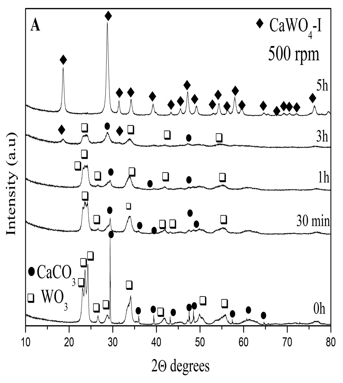
Figure 1. XRD patterns of the initial mixture and mechanochemically activated sample at 500 rpm (A), mechanochemically activated sample at 850 rpm (B), and CaWO4 obtained after solid-state reaction (C).
Materials 17 03724 g001aMaterials 17 03724 g001b
Figure 2. (A) TEM image, particle size distribution, and EDS mapping of CaWO4-I obtained after 5 h of milling time at 500 rpm. (B) TEM image, particle size distribution, and EDS mapping of CaWO4-II obtained after 1 h of milling time at 850 rpm. (C) TEM image, particle size distribution, and EDS mapping of CaWO4-III obtained by solid-state reaction.
Materials 17 03724 g002aMaterials 17 03724 g002b
Figure 3. (A) Raman spectra of the samples synthesized by mechanochemical treatment (CaWO4-I, 5 h at 500 rpm; CaWO4-II, 1 h at 850 rpm) and solid-state reaction (CaWO4-III). (B) Infrared spectra of the samples synthesized by mechanochemical treatment (CaWO4-I, 5 h at 500 rpm; CaWO4-II, 1 h at 850 rpm) and solid-state reaction (CaWO4-III).
Materials 17 03724 g003
Figure 4. (A) UV-Vis spectra of the samples obtained by mechanochemical synthesis treatment (CaWO4-I, 5 h at 500 rpm; CaWO4-II, 1 h at 850 rpm) and solid-state reaction (CaWO4-III). (B) Tauc’s plot of the samples obtained by mechanochemical synthesis treatment (CaWO4-I, 5 h at 500 rpm; CaWO4-II, 1 h at 850 rpm) and solid-state reaction (CaWO4-III).
Materials 17 03724 g004
Figure 5. Emission spectra of samples obtained by mechanochemical treatment (CaWO4-I, 5 h at 500 rpm; CaWO4-II, 1 h at 850 rpm) and solid state reaction (CaWO4-III).
Materials 17 03724 g005
Figure 6. CIE chromaticity diagram of samples obtained by mechanochemical treatment (CaWO4-I, 5 h at 500 rpm; CaWO4-II, 1 h at 850 rpm) and solid-state reaction (CaWO4-III).
Materials 17 03724 g006
Table 1. Unit cell parameters, crystallite size, and lattice microstrain values of the obtained samples.
CaWO4-I
5 h, 500 rpm
CaWO4-II
1 h, 850 rpm
CaWO4-III
900 °C-12 h
CaWO4
(PDF#00-041-1431)
Unit cell parameters
a/c (Å)
5.244(1)
11.388(2)
5.242(1)
11.375(2)
5.238(1)
11.377(2)
5.243
11.373
Unit cell volume (Å3)313.19(1)312.598(3)312.22(1)312.63
Crystallites size (nm)31.5(1)24.4(7)369(5)n.a.
Lattice strain ε0 × 10−46.92(10)12.55(8)2.067(15)n.a.
Table 2. Emission peaks, FWHM, and CIE coordinates (x, y) of the synthesized samples.
SamplesEmission Peak,
nm
FWHM,
nm
xy
CaWO4-I,
5 h at 500 rpm
425980.1780.168
CaWO4-II
1 h at 850 rpm
422920.1730.150
CaWO4-III
900 °C for 12 h
4221000.1720.161
Disclaimer/Publisher’s Note: The statements, opinions and data contained in all publications are solely those of the individual author(s) and contributor(s) and not of MDPI and/or the editor(s). MDPI and/or the editor(s) disclaim responsibility for any injury to people or property resulting from any ideas, methods, instructions or products referred to in the content.