


Flexural Strength and Morphological Study of Different Multilayer Zirconia Dental Materials


Abstract
1. Introduction
2. Materials and Methods
3. Results
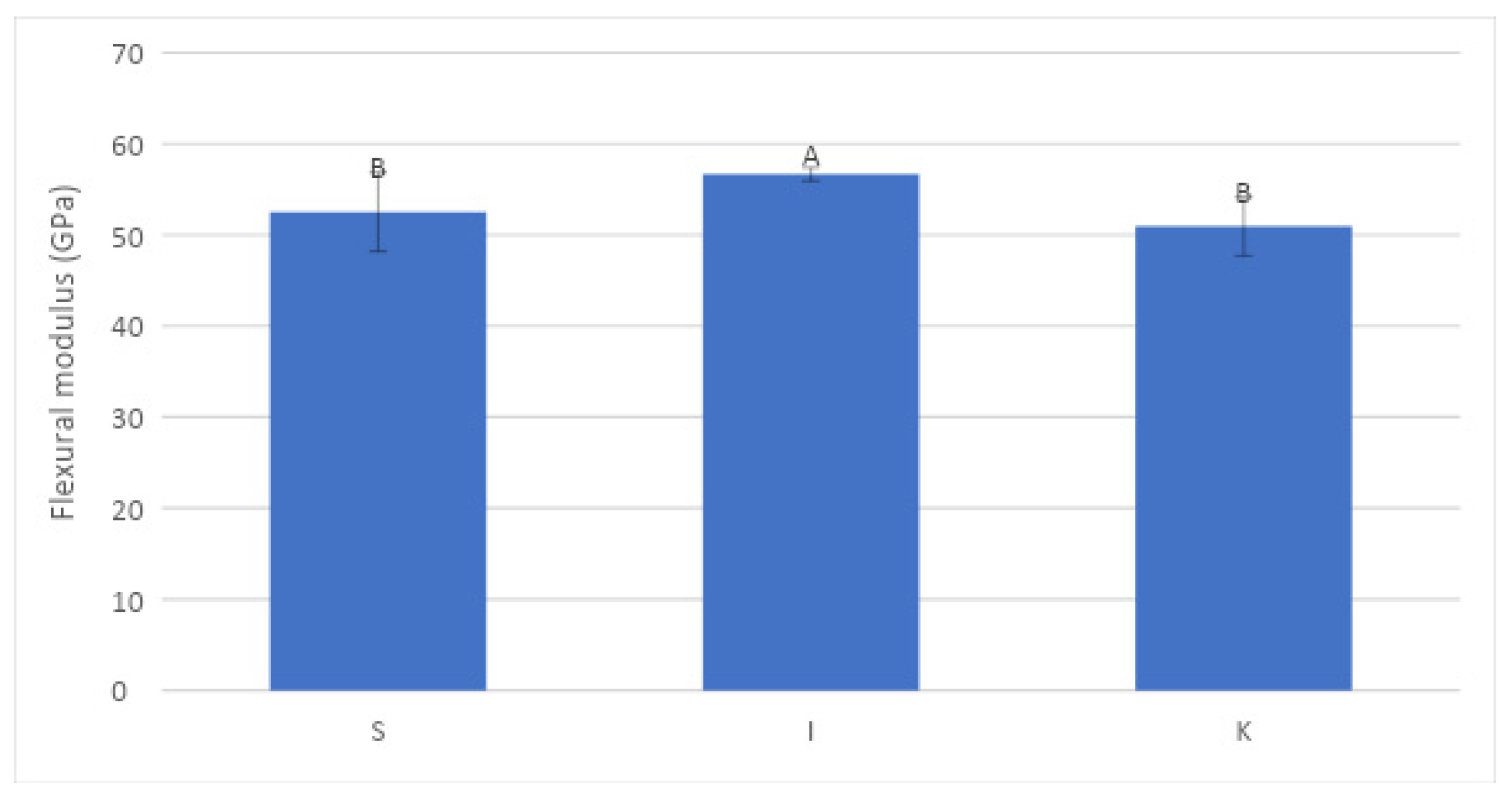
3.1. Mechanical Properties
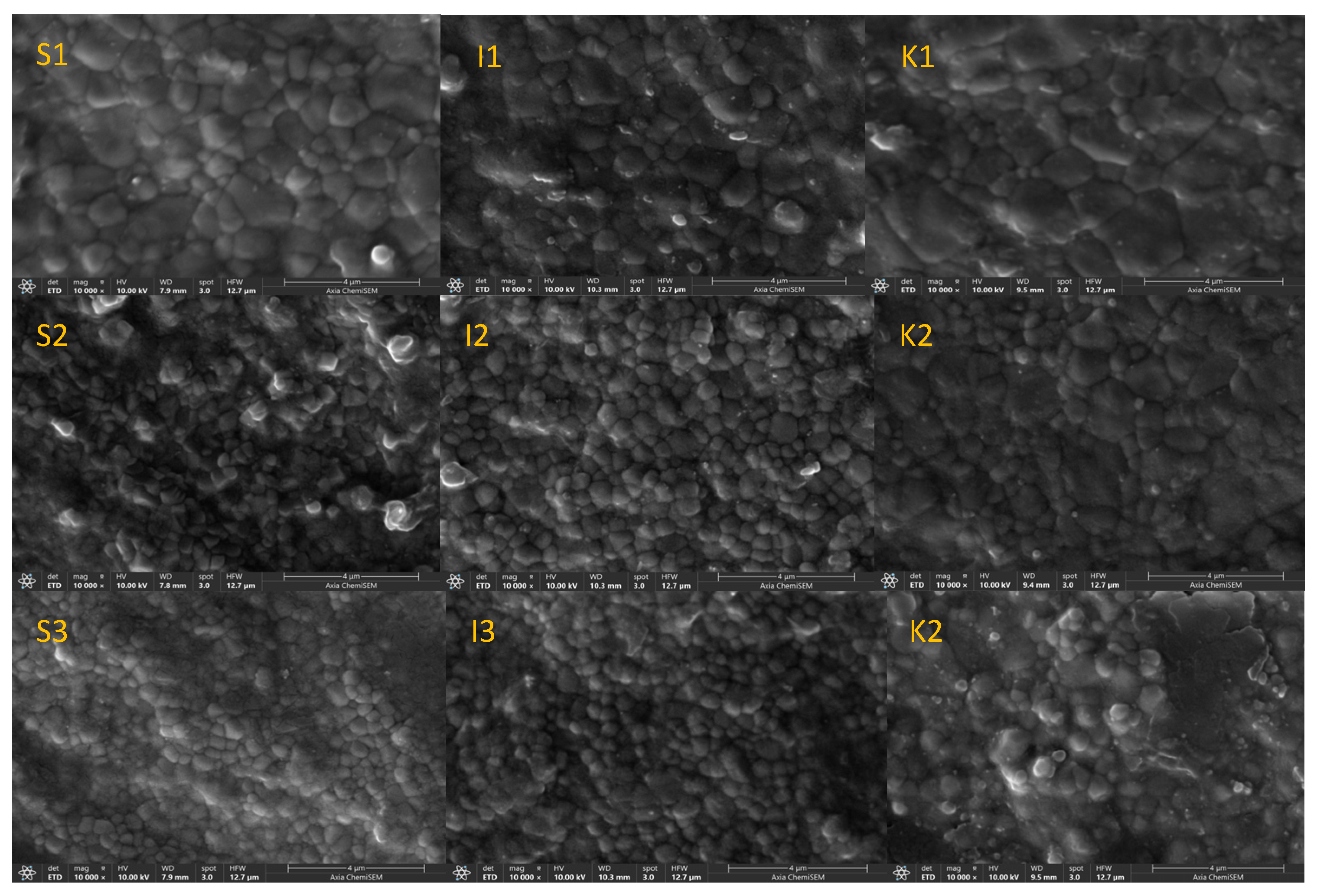
3.2. SEM Microstructural Analysis
3.3. PXRD Quantitative Phase Analysis
4. Discussion
5. Conclusions
Author Contributions
Funding
Institutional Review Board Statement
Informed Consent Statement
Data Availability Statement
Conflicts of Interest
References
- Jakovac, M.; Klaser, T.; Radatović, B.; Bafti, A.; Skoko, Ž.; Pavić, L.; Žic, M. Impact of Sandblasting on Morphology, Structure and Conductivity of Zirconia Dental Ceramics Material. Materials 2021, 14, 2834. [Google Scholar] [CrossRef] [PubMed]
- Denry, I.; Kelly, J.R. State of the Art of Zirconia for Dental Applications. Dent. Mater. 2008, 24, 299–307. [Google Scholar] [CrossRef]
- Manicone, P.F.; Rossi Iommetti, P.; Raffaelli, L. An Overview of Zirconia Ceramics: Basic Properties and Clinical Applications. J. Dent. 2007, 35, 819–826. [Google Scholar] [CrossRef] [PubMed]
- Piconi, C.; Maccauro, G. Zirconia as a Ceramic Biomaterial. Biomaterials 1999, 20, 1–25. [Google Scholar] [CrossRef] [PubMed]
- Işeri, U.; Özkurt, Z.; Yalniz, A.; Kazazoglu, E. Comparison of Different Grinding Procedures on the Flexural Strength of Zirconia. J. Prosthet. Dent. 2012, 107, 309–315. [Google Scholar] [CrossRef] [PubMed]
- Kosmač, T.; Oblak, C.; Jevnikar, P.; Funduk, N.; Marion, L. The Effect of Surface Grinding and Sandblasting on Flexural Strength and Reliability of Y-TZP Zirconia Ceramic. Dent. Mater. 1999, 15, 426–433. [Google Scholar] [CrossRef]
- Kelly, J.R.; Denry, I. Stabilized Zirconia as a Structural Ceramic: An Overview. Dent. Mater. 2008, 24, 289–298. [Google Scholar] [CrossRef]
- Lysaght, M.; Webster, T.J. Biomaterials for Artificial Organs; Elsevier Science: Amsterdam, The Netherlands, 2010; ISBN 9781845696535. [Google Scholar]
- Conrad, H.J.; Seong, W.-J.; Pesun, I.J. Clinical Implications Current Ceramic Materials and Systems with Clinical Recommendations: A Systematic Review. J. Prosthet. Dent. 2007, 98, 389–404. [Google Scholar] [CrossRef]
- Michailova, M.; Elsayed, A.; Fabel, G.; Edelhoff, D.; Zylla, I.M.; Stawarczyk, B. Comparison between Novel Strength-Gradient and Color-Gradient Multilayered Zirconia Using Conventional and High-Speed Sintering. J. Mech. Behav. Biomed. Mater. 2020, 111, 103977. [Google Scholar] [CrossRef]
- Almohammed, S.N.; Alshorman, B.; Abu-Naba’a, L.A. Optical Properties of Five Esthetic Ceramic Materials Used for Monolithic Restorations: A Comparative In Vitro Study. Ceramics 2022, 5, 961–980. [Google Scholar] [CrossRef]
- Zhang, Y. Making Yttria-Stabilized Tetragonal Zirconia Translucent. Dent. Mater. 2014, 30, 1195–1203. [Google Scholar] [CrossRef]
- Reyes, A.R.; Dennison, J.B.; Powers, J.M.; Sierraalta, M.; Yaman, P. Translucency and Flexural Strength of Translucent Zirconia Ceramics. J. Prosthet. Dent. 2023, 129, 644–649. [Google Scholar] [CrossRef]
- Vichi, A.; Zhao, Z.; Paolone, G.; Scotti, N.; Mutahar, M.; Goracci, C.; Louca, C. Factory Crystallized Silicates for Monolithic Metal-Free Restorations: A Flexural Strength and Translucency Comparison Test. Materials 2022, 15, 7834. [Google Scholar] [CrossRef]
- Kawajiri, Y.; Ikeda, H.; Nagamatsu, Y.; Masaki, C.; Hosokawa, R.; Shimizu, H. PICN Nanocomposite as Dental CAD/CAM Block Comparable to Human Tooth in Terms of Hardness and Flexural Modulus. Materials 2021, 14, 1182. [Google Scholar] [CrossRef] [PubMed]
- Jakovac, M.; Klaser, T.; Bafti, A.; Skoko, Ž.; Pavić, L.; Žic, M. The Effect of Y3+ Addition on Morphology, Structure, and Electrical Properties of Yttria-Stabilized Tetragonal Zirconia Dental Materials. Materials 2022, 15, 1800. [Google Scholar] [CrossRef] [PubMed]
- Machry, R.V.; Borges, A.L.S.; Pereira, G.K.R.; Kleverlaan, C.J.; Venturini, A.B.; Valandro, L.F. Influence of the Foundation Substrate on the Fatigue Behavior of Bonded Glass, Zirconia Polycrystals, and Polymer Infiltrated Ceramic Simplified CAD-CAM Restorations. J. Mech. Behav. Biomed. Mater. 2021, 117, 104391. [Google Scholar] [CrossRef] [PubMed]
- Ilie, N. Frequency-Related Viscoelastic Properties in High Translucent CAD-CAM Resin-Based Composites. J. Mech. Behav. Biomed. Mater. 2021, 118, 104427. [Google Scholar] [CrossRef]
- Albelasy, E.; Hamama, H.H.; Tsoi, J.K.H.; Mahmoud, S.H. Influence of Material Type, Thickness and Storage on Fracture Resistance of CAD/CAM Occlusal Veneers. J. Mech. Behav. Biomed. Mater. 2021, 119, 104485. [Google Scholar] [CrossRef] [PubMed]
- Van Noort, R. The Future of Dental Devices Is Digital. Dent. Mater. 2012, 28, 3–12. [Google Scholar] [CrossRef] [PubMed]
- Jakovac, M.; Klaser, T.; Radatović, B.; Skoko, Ž.; Pavić, L.; Žic, M. Surface Characterization and Conductivity of Two Types of Lithium-Based Glass Ceramics after Accelerating Ageing. Materials 2020, 13, 5632. [Google Scholar] [CrossRef]
- Ban, S.; Okuda, Y.; Noda, M.; Tsuruki, J.; Kawai, T.; Kono, H. Contamination of Dental Zirconia before Final Firing: Effects on Mechanical Properties. Dent. Mater. J. 2013, 32, 1011–1019. [Google Scholar] [CrossRef]
- Işerı, U.; Ozkurt, Z.; Kazazoğlu, E.; Küçükoğlu, D. Influence of Grinding Procedures on the Flexural Strength of Zirconia Ceramics. Braz. Dent. J. 2010, 21, 528–532. [Google Scholar] [CrossRef] [PubMed]
- Chevalier, J.; Gremillard, L.; Virkar, A.V.; Clarke, D.R. The Tetragonal-Monoclinic Transformation in Zirconia: Lessons Learned and Future Trends. J. Am. Ceram. Soc. 2009, 92, 1901–1920. [Google Scholar] [CrossRef]
- Yi, Y.A.; Ahn, J.S.; Park, Y.J.; Jun, S.H.; Lee, I.B.; Cho, B.H.; Son, H.H.; Seo, D.G. The Effect of Sandblasting and Different Primers on Shear Bond Strength between Yttria-Tetragonal Zirconia Polycrystal Ceramic and a Self-Adhesive Resin Cement. Oper. Dent. 2015, 40, 63–71. [Google Scholar] [CrossRef]
- Eriksson, C.; Masaki, N.; Yao, I.; Hayasaka, T.; Setou, M. MALDI Imaging Mass Spectrometry—A Mini Review of Methods and Recent Developments. Mass Spectrom. 2013, 2, S0022. [Google Scholar] [CrossRef]
- Inokoshi, M.; Zhang, F.; De Munck, J.; Minakuchi, S.; Naert, I.; Vleugels, J.; Van Meerbeek, B.; Vanmeensel, K. Influence of Sintering Conditions on Low-Temperature Degradation of Dental Zirconia. Dent. Mater. 2014, 30, 669–678. [Google Scholar] [CrossRef] [PubMed]
- Okada, M.; Taketa, H.; Torii, Y.; Irie, M.; Matsumoto, T. Optimal Sandblasting Conditions for Conventional-Type Yttria-Stabilized Tetragonal Zirconia Polycrystals. Dent. Mater. 2019, 35, 169–175. [Google Scholar] [CrossRef] [PubMed]
- Strasser, T.; Preis, V.; Behr, M.; Rosentritt, M. Roughness, Surface Energy, and Superficial Damages of CAD/CAM Materials after Surface Treatment. Clin. Oral Investig. 2018, 22, 2787–2797. [Google Scholar] [CrossRef]
- Craciunescu, E.; Sinescu, C.; Negrutiu, M.L.; Pop, D.M.; Lauer, H.C.; Rominu, M.; Hutiu, G.; Bunoiu, M.; Duma, V.F.; Antoniac, I. Shear Bond Strength Tests of Zirconia Veneering Ceramics after Chipping Repair. J. Adhes. Sci. Technol. 2016, 30, 666–676. [Google Scholar] [CrossRef]
- Špehar, D.; Jakovac, M. Nove Spoznaje o Cirkonij-Oksidnoj Keramici Kao Gradivnom Materijalu u Fiksnoj Protetici. Acta Stomatol. Croat. 2015, 49, 137–144. [Google Scholar] [CrossRef]
- ISO 6872:2015; Dentistry: Ceramic Materials. ISO: Geneva, Switzerland, 2015.
- Wibisono, G.; Nikraz, H.R. Resilient Modulus Values of Western Australia Asphalt Pavement. IOP Conf. Ser. Mater. Sci. Eng. 2019, 615, 012129. [Google Scholar] [CrossRef]
- Carrabba, M.; Keeling, A.J.; Aziz, A.; Vichi, A.; Fabian Fonzar, R.; Wood, D.; Ferrari, M. Translucent Zirconia in the Ceramic Scenario for Monolithic Restorations: A Flexural Strength and Translucency Comparison Test. J. Dent. 2017, 60, 70–76. [Google Scholar] [CrossRef] [PubMed]
- Kwon, S.J.; Lawson, N.C.; McLaren, E.E.; Nejat, A.H.; Burgess, J.O. Comparison of the Mechanical Properties of Translucent Zirconia and Lithium Disilicate. J. Prosthet. Dent. 2018, 120, 132–137. [Google Scholar] [CrossRef] [PubMed]
- Camposilvan, E.; Leone, R.; Gremillard, L.; Sorrentino, R.; Zarone, F.; Ferrari, M.; Chevalier, J. Aging Resistance, Mechanical Properties and Translucency of Different Yttria-Stabilized Zirconia Ceramics for Monolithic Dental Crown Applications. Dent. Mater. 2018, 34, 879–890. [Google Scholar] [CrossRef] [PubMed]
- De Souza, G.M.; Zykus, A.; Ghahnavyeh, R.R.; Lawrence, S.K.; Bahr, D.F. Effect of Accelerated Aging on Dental Zirconia-Based Materials. J. Mech. Behav. Biomed. Mater. 2017, 65, 256–263. [Google Scholar] [CrossRef] [PubMed]
- Siarampi, E.; Kontonasaki, E.; Andrikopoulos, K.S.; Kantiranis, N.; Voyiatzis, G.A.; Zorba, T.; Paraskevopoulos, K.M.; Koidis, P. Effect of in Vitro Aging on the Flexural Strength and Probability to Fracture of Y-TZP Zirconia Ceramics for All-Ceramic Restorations. Dent. Mater. 2014, 30, e306–e316. [Google Scholar] [CrossRef]
- Stawarczyk, B.; Frevert, K.; Ender, A.; Roos, M.; Sener, B.; Wimmer, T. Comparison of Four Monolithic Zirconia Materials with Conventional Ones: Contrast Ratio, Grain Size, Four-Point Flexural Strength and Two-Body Wear. J. Mech. Behav. Biomed. Mater. 2016, 59, 128–138. [Google Scholar] [CrossRef]
- Xu, Y.; Han, J.; Lin, H.; An, L. Comparative Study of Flexural Strength Test Methods on CAD/CAM Y-TZP Dental Ceramics. Regen. Biomater. 2015, 2, 239–244. [Google Scholar] [CrossRef]
- Wendler, M.; Belli, R.; Petschelt, A.; Mevec, D.; Harrer, W.; Lube, T.; Danzer, R.; Lohbauer, U. Chairside CAD/CAM Materials. Part 2: Flexural Strength Testing. Dent. Mater. 2017, 33, 99–109. [Google Scholar] [CrossRef]
- Wang, H.; Aboushelib, M.N.; Feilzer, A.J. Strength Influencing Variables on CAD/CAM Zirconia Frameworks. Dent. Mater. 2008, 24, 633–638. [Google Scholar] [CrossRef] [PubMed]
- Inokoshi, M.; Shimizubata, M.; Nozaki, K.; Takagaki, T.; Yoshihara, K.; Minakuchi, S.; Vleugels, J.; Van Meerbeek, B.; Zhang, F. Impact of Sandblasting on the Flexural Strength of Highly Translucent Zirconia. J. Mech. Behav. Biomed. Mater. 2021, 115, 104268. [Google Scholar] [CrossRef] [PubMed]
- Pereira, G.K.R.; Guilardi, L.F.; Dapieve, K.S.; Kleverlaan, C.J.; Rippe, M.P.; Valandro, L.F. Mechanical Reliability, Fatigue Strength and Survival Analysis of New Polycrystalline Translucent Zirconia Ceramics for Monolithic Restorations. J. Mech. Behav. Biomed. Mater. 2018, 85, 57–65. [Google Scholar] [CrossRef] [PubMed]
- Matsui, K.; Yoshida, H.; Ikuhara, Y. Grain-Boundary Structure and Microstructure Development Mechanism in 2–8 Mol% Yttria-Stabilized Zirconia Polycrystals. Acta Mater. 2008, 56, 1315–1325. [Google Scholar] [CrossRef]
- Strasser, T.; Wertz, M.; Koenig, A.; Koetzsch, T.; Rosentritt, M. Microstructure, Composition, and Flexural Strength of Different Layers within Zirconia Materials with Strength Gradient. Dent. Mater. 2023, 39, 463–468. [Google Scholar] [CrossRef] [PubMed]
- Kolakarnprasert, N.; Kaizer, M.R.; Kim, D.K.; Zhang, Y. New Multi-Layered Zirconias: Composition, Microstructure and Translucency. Dent. Mater. 2019, 35, 797–806. [Google Scholar] [CrossRef] [PubMed]
- Inokoshi, M.; Liu, H.; Yoshihara, K.; Yamamoto, M.; Tonprasong, W.; Benino, Y.; Minakuchi, S.; Vleugels, J.; Van Meerbeek, B.; Zhang, F. Layer Characteristics in Strength-Gradient Multilayered Yttria-Stabilized Zirconia. Dent. Mater. 2023, 39, 430–441. [Google Scholar] [CrossRef]
- Inokoshi, M.; Shimizu, H.; Nozaki, K.; Takagaki, T.; Yoshihara, K.; Nagaoka, N.; Zhang, F.; Vleugels, J.; Van Meerbeek, B.; Minakuchi, S. Crystallographic and Morphological Analysis of Sandblasted Highly Translucent Dental Zirconia. Dent. Mater. 2018, 34, 508–518. [Google Scholar] [CrossRef]
- Cokic, S.M.; Vleugels, J.; Van Meerbeek, B.; Camargo, B.; Willems, E.; Li, M.; Zhang, F. Mechanical Properties, Aging Stability and Translucency of Speed-Sintered Zirconia for Chairside Restorations. Dent. Mater. 2020, 36, 959–972. [Google Scholar] [CrossRef]
- Lughi, V.; Sergo, V. Low Temperature Degradation -Aging- of Zirconia: A Critical Review of the Relevant Aspects in Dentistry. Dent. Mater. 2010, 26, 807–820. [Google Scholar] [CrossRef]
- Lazar, D.R.R.; Bottino, M.C.; Özcan, M.; Valandro, L.F.; Amaral, R.; Ussui, V.; Bressiani, A.H.A. Y-TZP Ceramic Processing from Coprecipitated Powders: A Comparative Study with Three Commercial Dental Ceramics. Dent. Mater. 2008, 24, 1676–1685. [Google Scholar] [CrossRef]
- Juntavee, N.; Attashu, S. Effect of Different Sintering Process on Flexural Strength of Translucency Monolithic Zirconia. J. Clin. Exp. Dent. 2018, 10, e821–e830. [Google Scholar] [CrossRef] [PubMed]


| Material Name | Manufacturer | Sample Name | Flexural Strength | LOT Number |
|---|---|---|---|---|
| ZirCAD Prime | Ivoclar Vivadent (Schaan, Liechtenstein) | I | 1200 MPa | Z05RMR |
| Cercon ht ML | Dentsply Sirona (Charlotte, NC, USA) | S | 1200 MPa | 0018041712-1057 |
| Katana ZIRCONIA YML | Kuraray Noritake (Tokyo, Japan) | K | 1100 MPa | EJHNA |
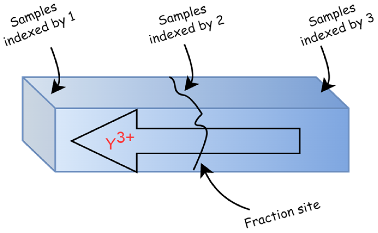
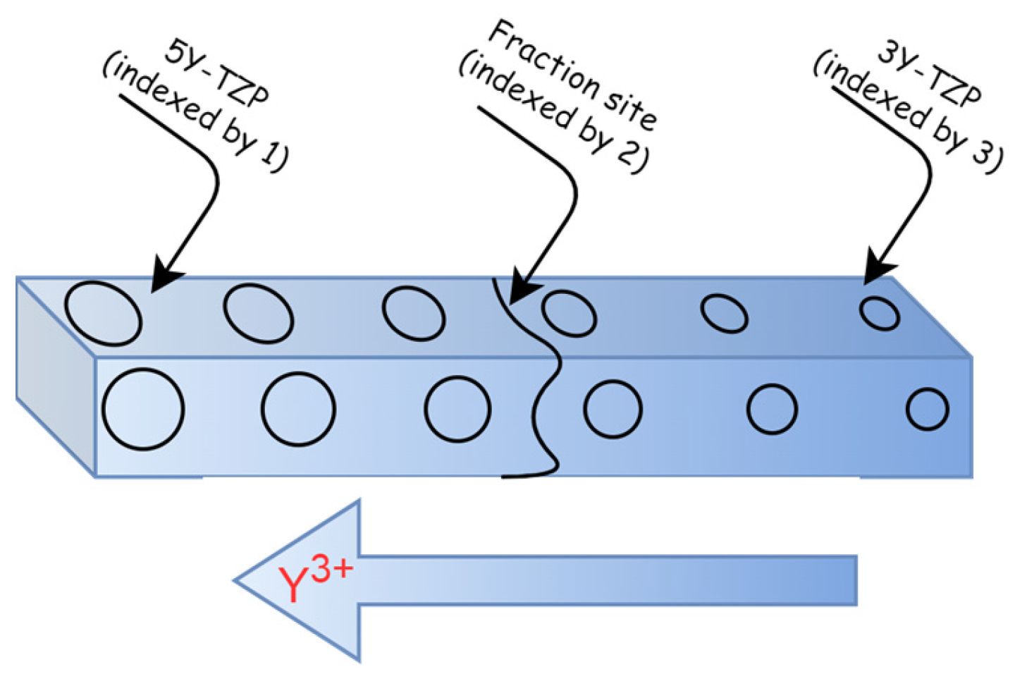
| Sample | 5Y-TZP | Fracture Site | 3Y-TZP |
|---|---|---|---|
 | |||
| S (Sirona) | S1 | S2 | S3 |
| I (Ivoclar) | I1 | I2 | I3 |
| K (Katana) | K1 | K2 | K3 |
| Sample | Phase | Sample Index | ||
| 5Y-TZP | Fracture Site | 3Y-TZP | ||
| 1 | 2 | 3 | ||
| ||||
| S | c-ZrO2 (wt %) | 56.4 | 22.8 | 22.5 |
| t-ZrO2 (wt %) | 43.6 | 77.2 | 77.5 | |
| I | c-ZrO2 (wt %) | 50.1 | 13.5 | 13.9 |
| t-ZrO2 (wt %) | 49.9 | 86.5 | 86.1 | |
| K | c-ZrO2 (wt %) | 60.8 | 47.3 | 31.6 |
| t-ZrO2 (wt %) | 39.2 | 52.7 | 68.4 | |
Disclaimer/Publisher’s Note: The statements, opinions and data contained in all publications are solely those of the individual author(s) and contributor(s) and not of MDPI and/or the editor(s). MDPI and/or the editor(s) disclaim responsibility for any injury to people or property resulting from any ideas, methods, instructions or products referred to in the content. |
© 2024 by the authors. Licensee MDPI, Basel, Switzerland. This article is an open access article distributed under the terms and conditions of the Creative Commons Attribution (CC BY) license (https://creativecommons.org/licenses/by/4.0/).
Share and Cite
Labetić, A.; Klaser, T.; Skoko, Ž.; Jakovac, M.; Žic, M. Flexural Strength and Morphological Study of Different Multilayer Zirconia Dental Materials. Materials 2024, 17, 1143. https://doi.org/10.3390/ma17051143
Labetić A, Klaser T, Skoko Ž, Jakovac M, Žic M. Flexural Strength and Morphological Study of Different Multilayer Zirconia Dental Materials. Materials. 2024; 17(5):1143. https://doi.org/10.3390/ma17051143
Chicago/Turabian StyleLabetić, Andrea, Teodoro Klaser, Željko Skoko, Marko Jakovac, and Mark Žic. 2024. "Flexural Strength and Morphological Study of Different Multilayer Zirconia Dental Materials" Materials 17, no. 5: 1143. https://doi.org/10.3390/ma17051143
APA StyleLabetić, A., Klaser, T., Skoko, Ž., Jakovac, M., & Žic, M. (2024). Flexural Strength and Morphological Study of Different Multilayer Zirconia Dental Materials. Materials, 17(5), 1143. https://doi.org/10.3390/ma17051143