Article

Phytophthora mediterranea sp. nov., a New Species Closely Related to Phytophthora cinnamomi from Nursery Plants of Myrtus communis in Italy

1
Dipartimento Territorio e Sistemi Agro-Forestali, Università degli Studi di Padova, Viale dell’Università, 16, 35020 Legnaro, Italy
2
Dipartimento di Agraria, Università degli Studi di Sassari, Viale Italia, 39, 07100 Sassari, Italy
3
Agenzia Regionale Fo.Re.S.T.A.S., viale Luigi Merello, 86, 09123 Cagliari, Italy
*
Author to whom correspondence should be addressed.
Forests 2021, 12(6), 682; https://doi.org/10.3390/f12060682
Submission received: 4 May 2021 / Revised: 18 May 2021 / Accepted: 25 May 2021 / Published: 27 May 2021
(This article belongs to the Special Issue Emerging Pathogens in Forest Ecosystems)

Abstract

:
Monitoring surveys of Phytophthora related diseases in four forest nurseries in Italy revealed the occurrence of fourteen Phytophthora species to be associated with collar and root rot on fourteen plants typical of Mediterranean and alpine regions. In addition, a multilocus phylogeny analysis based on nuclear ITS and ß-tubulin and mitochondrial cox1 sequences, as well as micromorphological features, supported the description of a new species belonging to the phylogenetic clade 7c, Phytophthora mediterranea sp. nov. Phytophthora mediterranea was shown to be associated with collar and root rot symptoms on myrtle seedlings. Phylogenetically, P. mediterranea is closely related to P. cinnamomi but the two species differ in 87 nucleotides in the three studied DNA regions. Morphologically P. mediterranea can be easily distinguished from P. cinnamomi on the basis of its smaller sporangia, colony growth pattern and higher optimum and maximum temperature values. Data from the pathogenicity test showed that P. mediterranea has the potential to threaten the native Mediterranean maquis vegetation. Finally, the discovery of P. cinnamomi in alpine nurseries, confirms the progressive expansion of this species towards cold environments, probably driven by climate change.

1. Introduction

Over the last few years, the horticultural industry has had a progressive expansion worldwide, which resulted in an increase in the trade of plants and commodities among continents [1,2]. In Italy, the horticulture industry is an important component of the agricultural sector; its production covers over 30,000 hectares with 100,000 employees and has a sellable production of about 2.5 billion euros per year [3].
Over the last few decades, the exponential increase in plant materials and commodities traded, together with limited and often ineffective diagnostic measures to detect pests and pathogens at the borders have contributed to a rapid diffusion of exotic and invasive species worldwide [4,5]. Among the most destructive pathogens that are introduced annually through trade and transport of plant material, many belong to the genus Phytophthora [6,7].
The genus Phytophthora encompasses a range of morphologically and ecologically diverse taxa grouped into 12 well-defined phylogenetic clades [8,9,10]. Some species have a saprophytic or opportunistic lifestyle, whereas others are aggressive pathogens that cause root and collar rot, bleeding canker and leaf blight symptoms in a huge number of plant species [11,12]. In particular, Phytophthora spp. are responsible for severe outbreaks with reduced vitality and quality of nursery plants [13]. In addition to the direct economic losses caused to the horticulture industry, Phytophthora spp. pose a threat to biodiversity of natural ecosystems as they are often introduced through the seedlings used for reforestation and restoration programmes [14,15,16]. In this regard, the P. ramorum and P. lateralis outbreaks in North America and P. cinnamomi in Australia are emblematic, having caused the devastation of extensive ecosystems [17,18,19,20].
Due to intensive cultivation techniques and the occurrence of different plant species in an often limited space, nurseries are an ideal place for many Phytophthora species [13]. Moreover, the proximity of many plant species and possible encounters between genetically related species can easily give rise to new host–pathogen associations as well as hybridization phenomena [21]. High-humidity conditions and recurrent water irrigation can also favour the reproduction and dissemination of these pathogens [22,23,24,25]. Many Phytophthora species, in fact, need water to complete their biological cycle [13,26]. Furthermore, many agronomic practices such as recycling of soil and pots could be pervasive to increase Phytophthora inoculum inside nurseries [13,27,28,29]. At the same time, the intensive use of chemicals can promote the formation of fungicide-resistance in pathogen populations [30,31,32].
Phytophthora occurrence in North American, Australian and European nurseries is well documented [33,34,35,36,37,38,39]. The increased attention paid to Phytophthora species in the last decade has allowed the knowledge about biology and ecology of many species to be expanded, as well as the discovery of about 30 new species in nurseries [10]. Some of these new taxa are related to the aquatic environment and characterized by a saprophytic lifestyle [40,41,42,43,44]. In contrast, others such as P. niederhauserii and P. kernoviae are polyphagous and potentially invasive [45,46].
With the expansion of ornamental horticulture, Phytophthora related diseases are also becoming a serious problem in Italy. Recent studies have revealed a very high diversity of Phytophthora species in ornamental nurseries [47,48]. This is linked both to the widespread presence of invasive and ubiquitous species such as P. nicotianae and P. palmivora, but also of new species, such as P. parvispora, a cryptic species closely related to P. cinnamomi [48,49]. At the same time, the discovery of rare species on nursery plants, such as Phytophthora pistaciae, characterized by a limited geographic distribution points out the risks posed by nursery material to the conservation and integrity of natural ecosystems in the new areas [50].
Therefore, given the constant discovery of new or rare Phytophthora species in Italian nurseries, a thorough study was conducted to isolate, identify and characterize the main Phytophthora species associated with symptomatic plants in four nurseries spanning from the Mediterranean to the alpine climate region.

2. Materials and Methods

2.1. Surveys and Sampling Procedure

Field surveys were conducted from spring 2019 to autumn 2020 in four Italian forest nurseries located in Sardinia (N1 and N2), Veneto (N3) and Friuli Venezia Giulia (N4) regions (Table 1). In each nursery, all potting plants between 6 months and 5 years were visually checked for the presence of Phytophthora related disease symptoms such as chlorosis, defoliation, shoot blight, sudden death, as well as collar and root rot. Among the monitored plants, 76 were randomly selected for diagnostic analyses. The sampled species included Abies alba, Arbutus unedo, Alnus incana, Castanea sativa, Fagus sylvatica, Helichrysum italicum, Ilex aquifolium, Juglans regia, Laurus nobilis, Lavandula officinalis, Myrtus communis, Phyllirea latifolia, Pistacia lentiscus and Quercus ilex (Table 1). The plant samples were sealed in plastic bags, labelled and used for Phytophthora isolations within 24–48 h.

2.2. Isolation of Phytophthora Species

In the laboratory, plant samples were initially checked for collar and root symptoms and then used for Phytophthora isolation. From each sample about 300 g of rhizosphere soil and roots were positioned inside plastic containers with 2 L of distilled water. After 24 h the water surface was cleaned and young Quercus suber and Sambucus nigra leaves placed on the surface as bait [51]. Containers were kept at 20 °C under natural daylight for 3–7 days. Leaves showing necrotic spots were cut in small pieces (5 mm2) and placed on petri dishes containing potato dextrose agar (PDA 39 g/L, Oxoid Ltd., Basingstoke, UK) supplemented with 100 mL/L of carrot juice, 0.013 g/L of pimaricin and 0.05 g/L of hymexazol (PDA+) [50]. Hyphal tips typical of Phytophthora from the emerging colonies were sub-cultured on PDA and carrot agar (CA) [11] and incubated at 20 °C in the dark.
Isolations of Phytophthora species were also performed from collar and root lesions. After removing the outer bark, inner bark fragments were aseptically cut from the margin of necrotic lesions with a sterile scalpel and placed onto 90 mm Petri dishes containing PDA+. The dishes were incubated in the dark at 20 °C and examined every 6–12 h. Pure colonies were obtained as described above.

2.3. Morphological Identification and Characterization of Isolates

All Phytophthora isolates were grouped into morphotypes on the basis of colony growth patterns including surface and reverse colony appearance observed after 7 days of incubation on PDA and CA at 20 °C in the dark and morpho-biometric data of oogonia and sporangia. To enhance sporangia production, CA plugs (5 mm diameter) of each isolate, taken from 4-day-old colonies, were positioned inside petri dishes containing unsterile pond water and three cork oak fine roots (1 cm long). Petri dishes were kept at 20 °C in the dark and sporangia production was assessed every 12 h for 4 days.
Colony growth patterns of the new species were evaluated on 7-day-old cultures incubated at 25 °C in the dark on CA, PDA and malt extract agar (MEA, 20 g/L, Oxoid Ltd., Basingstoke, UK). In addition, size and shape of fifty chlamydospores, hyphal swellings and sporangia were recorded. Cardinal temperatures for growth were evaluated on 90 mm CA plates incubated at 5, 10, 15, 20, 25, 30, 32, 35, 37 and 40 °C (±0.5 °C) in the dark for 96 h. Five replicate plates for each isolate were made. An isolate of both P. parvispora (CB86) and P. cinnamomi (CB21) obtained in the study were also included in the growth bioassay for comparison.
Measurements and photos of the main morphological structures were taken at 400× and 600× magnification and recorded using the software Motic Images Plus 3.0 paired with a Moticam 10+ camera connected to a Motic BA410E microscope. Sporangia dimensions are presented as mean values ± standard deviation.
Representative isolates of each species were stored on PDA and CA slants under oil in the culture collection of the Dipartimento Territorio e Sistemi Agro-Forestali, Università degli Studi di Padova. Ex-type culture of the new species was deposited at the Westerdijk Fungal Biodiversity Institute, Utrecht, The Netherlands, and nomenclatural data in MycoBank (www.MycoBank.org, accessed on 4 May 2021). The holotype was lodged with the herbarium of Westerdijk Fungal Biodiversity Institute as a dried culture on CA.

2.4. DNA Extraction, Polymerase Chain Reaction (PCR) Amplification and Sequencing

For all isolates genomic DNA was extracted from mycelium of 5-day-old colonies grown on PDA at 20 °C using Instagene Matrix (BioRad Laboratories, Hercules, CA, USA). The entire internal transcribed spacer (ITS) region of the ribosomal DNA, including the 5.8S rRNA gene, was amplified and sequenced using primers ITS1 and ITS4 [52]. ITS sequences were used to confirm the identification at species level. For four isolates belonging to the clade 7 [8] including those of the new species, other two DNA regions namely ß-tubulin (Btub) and cytochrome c oxidase subunit I (cox1), were amplified and sequenced using the primer-pairs TUBUF2/TUBUR1 and FM84/FM83 [53,54], respectively. Polymerase chain reactions (PCR) were performed as described in Bregant et al. [51].
PCR products were purified using a EUROGOLD gel extraction kit according to the manufacturer’s instructions (EuroClone S.p.A., Pero, Italy). Both strands were sequenced by BMR Genomics DNA sequencing service (www.bmr-genomics.it, access on 4 May 2021). Sequences were edited with FinchTV v1.4.0 (Geospiza, Inc., http://www.geospiza.com/finchtv, accessed on 4 May 2021) and compared with sequences of ex-type culture available in GenBank using the BLASTn algorithm (http://blast.ncbi.nlm.nih.gov, access on 4 May 2021). New sequences were deposited in GenBank (Table 2 and Table 3). Alignments and trees are available in TreeBase (study ID 28180).

2.5. Phylogenetic Analysis

ITS, Btub and cox1 sequences of four isolates obtained in this survey were compiled in a dataset together with 78 sequences from 26 Phytophthora species representative of described species in the clade 7 (including ex-type culture) for which molecular data are available in GenBank (Table 2).
Sequence alignments were performed with ClustalX v. 1.83 [55], using the parameters reported in Bregant et al. [51]. Alignments were checked and edited with BioEdit Alignment Editor v. 7.2.5 [56]. Phylogenetic analyses were completed with MEGA-X 10.1.8 [57]. All gaps were included in the analyses. The best model of DNA sequence evolution was determined automatically by the software. Maximum likelihood (ML) analysis was performed with a neighbour-joining (NJ) starting tree generated by the software. A bootstrap analysis (1000 replicates) was used to estimate the robustness of nodes.

2.6. Pathogenicity Test

Pathogenicity of the new Phytophthora species was tested on 3-year-old myrtle (Myrtus communis) and lentisk (Pistacia lentiscus) seedlings grown in plastic pots (20 cm diameter, 2 L volume). Eight seedlings of each plant species were inoculated with the isolate CB84 (ex-type culture). In addition, the same number of each plant species were inoculated with the isolate CB21 of P. cinnamomi for comparison. Finally, the same number of seedlings were used as control. Inoculation was performed at the collar. The inoculated point was surface-disinfected with 70% ethanol and a small piece of outer bark (5 mm diameter) was removed with a flame-sterilised cork borer and replaced with an agar-mycelium plug of the same size, taken from the margin of an actively growing colony on CA. The inoculation point was covered with cotton wool soaked in sterile water and wrapped with an aluminium foil to retain moisture. Controls were inoculated with a sterile CA plug.
All inoculated seedlings were kept in a climatic chamber at 25 °C and watered every two days until the end of the experimental period. After 20 days, all plants were checked for the presence of disease symptoms and the length of necrotic lesion surrounding the inoculation site was measured after removing the outer bark with a sterile scalpel.
Re-isolation was performed by transferring 10 pieces of inner bark taken around the margin of the necrotic lesions onto PDA+. Growing colonies were sub-cultured onto CA and PDA, incubated in the dark at 20 °C and identified by morphological and molecular analysis (ITS region) to confirm Koch’s postulates.

2.7. Data Analysis

Data from the pathogenicity assay were first checked for normality and then subjected to analysis of variance (ANOVA). Significant differences among mean values were determined using Fisher’s least significant differences multiple range test (p = 0.05) after one-way ANOVA using XLSTAT 2008 software (Addinsoft).

3. Results

3.1. Symptomatology

Phytophthora related diseases were observed in all investigated nurseries. Regardless of the species, potted plants showed wilting foliage, chlorosis, stunted growth and sudden death symptoms (Figure 1). Sudden death was very common in plants of the alpine nursery (N3) and, in particular, of silver fir and beech seedlings.
All canopy symptoms described above were associated with collar and root rot symptoms. In most cases, the root system was completely compromised and fine roots absent (Figure 1). The mortality rates, especially in one-year-old seedlings, were very high with losses close to 60–70%.

3.2. Phytophthora Species Associated with Nursery Plants

Isolations performed on 76 symptomatic plants yielded a total of 66 Phytophthora isolates. The largest number of isolates (36) was obtained from site N1, followed by site N2 (22), whereas a total of 8 isolates were obtained from sites N3 and N4.
On the basis of micromorphological features and ITS sequence data, fourteen Phytophthora species belonging to seven phylogenetic clades were identified: P. cinnamomi (26 isolates), P. nicotianae (9), P. palmivora (5), P. pseudocryptogea (4), P. megasperma (3), P. plurivora (3), P. parvispora (3), P. pseudosyringae (3), P. pistaciae (2), P. bilorbang (2), P. acerina (1), P. cactorum (1), P. citrophthora (1) and P. psychrophila (1). For each species BLAST searches against GenBank showed 100% identity to reference sequences of representative strains including those of ex-type culture (Table 3). Two isolates obtained from declining myrtle seedlings could not be assigned on the basis of morphological features and ITS sequence data to any formally described species or informally designated Phytophthora and were therefore considered a new taxon unknown to science.
Among the detected species, P. cinnamomi was the dominant one, being isolated from five different hosts in three nurseries (Table 3). Phytophthora nicotianae was the most abundant species in the nursery N2. The other species were isolated only from a single host. Interestingly, some of the monitored plant species were positive for two or three Phytophthora species.

3.3. Phylogenetic Analysis

Phylogenetic relationships among the Phytophthora species belonging to clade 7 and four representative isolates obtained in this study were elucidated using a multilocus analysis based on the sequences of ITS, Btub and cox1 regions.
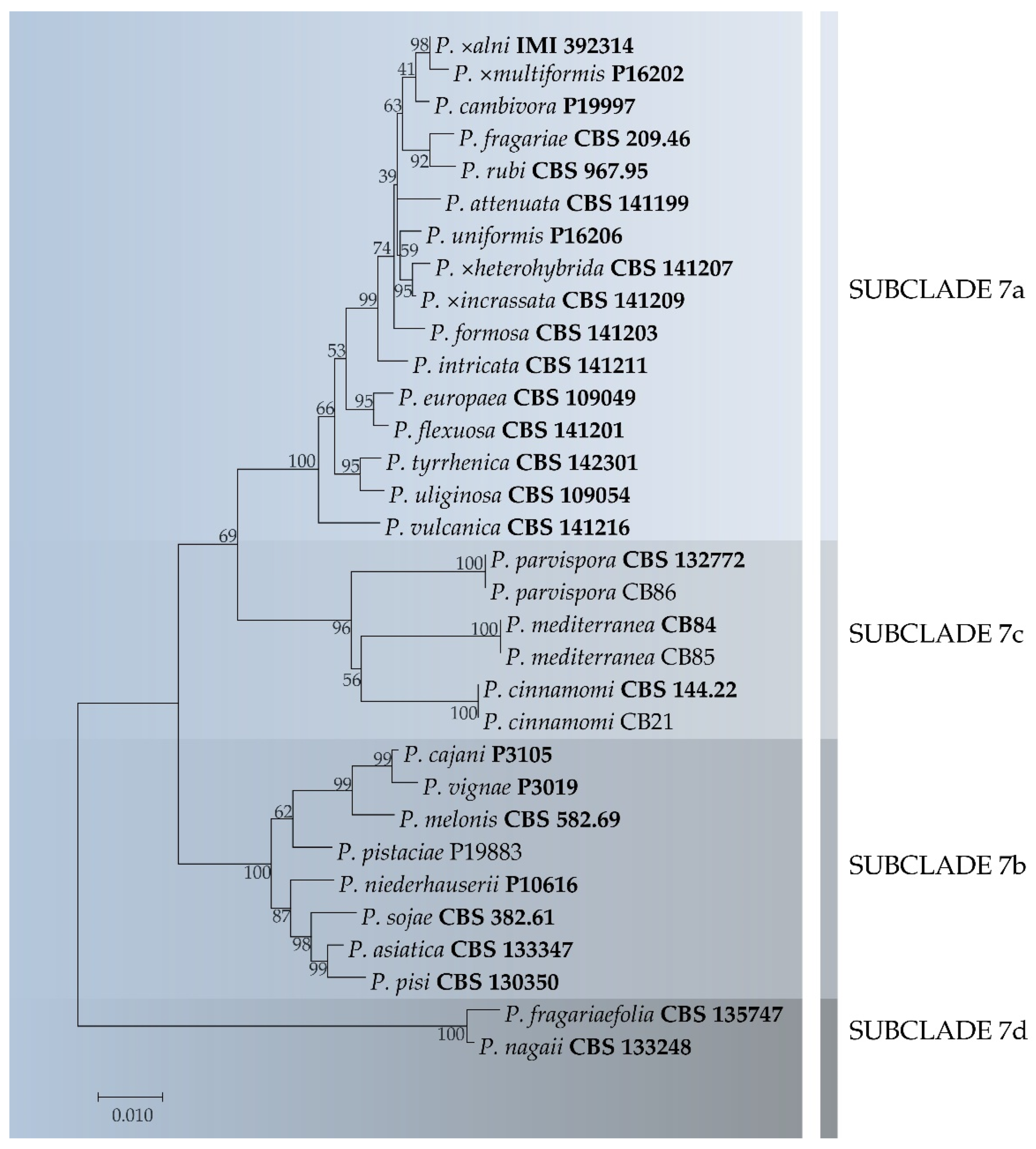
Fragments of approximately 800, 920 and 1020 bp were obtained for ITS, Btub and cox1 regions, respectively. Individual gene phylogenies revealed similar tree topologies, indicating that the three loci could be combined (data not shown). The ML evolutionary reconstruction allowed for the differentiation of 27 distinct lineages within clade 7, corresponding to 27 species and hybrids (Figure 2). The four isolates obtained in this study were distributed into three sub-clades in clade 7c (Figure 2). In particular, isolate (CB21) clustered with ex-culture type of P. cinnamomi and isolate (CB86) with ex-culture type of P. parvispora. The remaining two isolates clustered together in a well-supported terminal clade (ML bootstrap = 100%) and were considered to represent a new species described here as Phytophthora mediterranea sp. nov. (Figure 2).
Phylogenetically, the new Phytophthora species is closely related to P. cinnamomi, from which it can be distinguished on the basis of 15, 23 and 49 bp in ITS, Btub and cox1 loci, respectively. The other evolutionarily closest species, P. parvispora differs by a total of 105 nucleotides in the investigated DNA regions.

3.4. Taxonomy

Phytophthora mediterranea Bregant, Mulas and Linaldeddu sp. nov. (Figure 3).
MycoBank: MB839612.
Etymology: the epithet refers to the Mediterranean region, where the species was originally discovered.
Holotype: CBS H-24768.
Host/distribution: potted Myrtus communis plants with root rot symptoms in Italy.
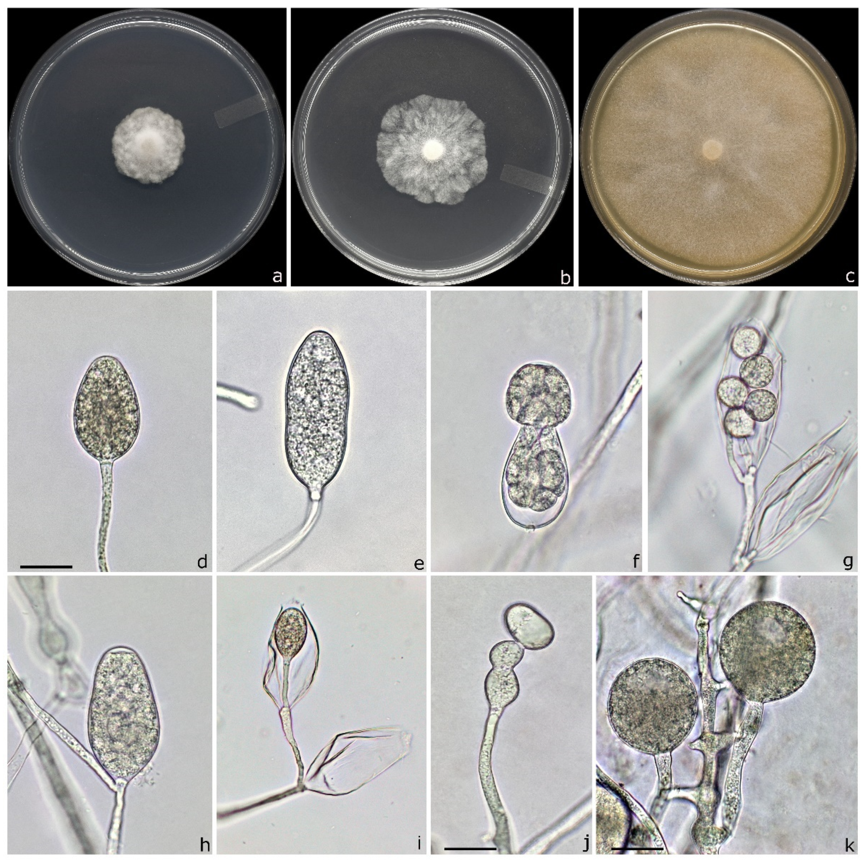
Description: sporangia were abundantly produced in unsterile pond water after 36–48 h of incubation at 20 °C. They were non-papillate, rarely semi-papillate, persistent, from ovoid to ellipsoid and less frequently elongated or distorted (Figure 3d,e,h). Typically, sporangia were borne terminally on unbranched sporangiophores and only occasionally on compound sporangiophores. Sporangial proliferation was usually external and very rarely internal (nested and extended) (Figure 3g–i).
Sporangia size ranged from 27.2 to 64.9 µm in length (av. 42.9 ± 9.3 µm) and from 20.1 to 46.8 µm in breadth (av. 29.8 ± 5.3 µm) with a length/breadth ratio of 1.4 ± 0.1 (n = 50). Zoospores were abundantly produced in liquid cultures after 36–48 h (Figure 3f,g). Globose to sub-globose, irregular and catenulate hyphal swellings were abundantly produced on CA (Figure 3j). Spherical chlamydospores were mostly terminal and only occasionally lateral and intercalary, and were abundantly produced on CA and water (Figure 3k). Chlamydospores ranged from 14.9 to 37.5 µm in diameter (av. 24.5 ± 5.0 µm). No gametangia were observed on pure cultures on CA suggesting a heterothallic behaviour.
Cultural characteristics: colonies were stellate on PDA and radiate on MEA whereas on CA showed a regular margin with a cottony and aerial mycelium without a distinct pattern. On PDA and MEA colony growth was slow, whereas on CA colony reached 80 mm diameter in 7 d at 25 °C.
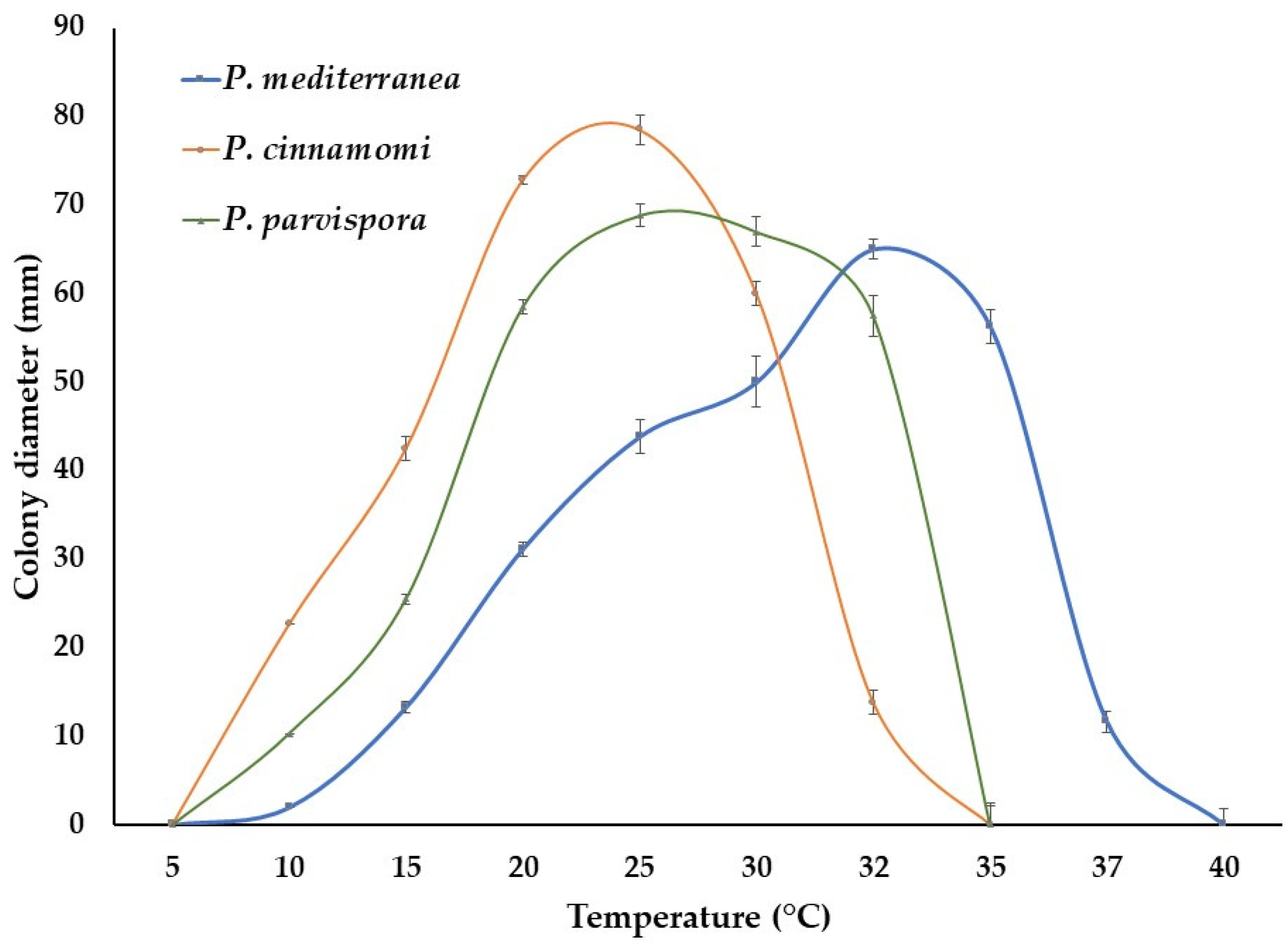
Cardinal temperatures: minimum <10 °C, maximum >37 °C and optimum 32 °C. Both isolates failed to grow at 40 °C and mycelium did not resume growth when plates were moved to 20 °C.
Material examined: ITALY: Oristano, isolated from roots of a potted plant of Myrtus communis, 18 April 2019, isolated by Antonio Mulas, HOLOTYPE CBS H-24768, a dried culture on CA, culture ex-holotype CB84 = CBS 147720. ITALY: Oristano, isolated from rhizosphere and fine roots of a potted plant of Myrtus communis, 12 November 2020, isolated by Carlo Bregant (culture CB85).
Notes: Phytophthora mediterranea differs from the closely related species P. cinnamomi and P. parvispora through a combination of unique morphological characters and molecular data such as sporangia shape and sizes, growth pattern on different culture media, a higher maximum and optimum temperature value for growth (Figure 4) as well as a total of 87 (P. cinnamomi) and 105 (P. parvispora) fixed differences in the ITS, Btub and cox1 sequences.

3.5. Pathogenicity Test

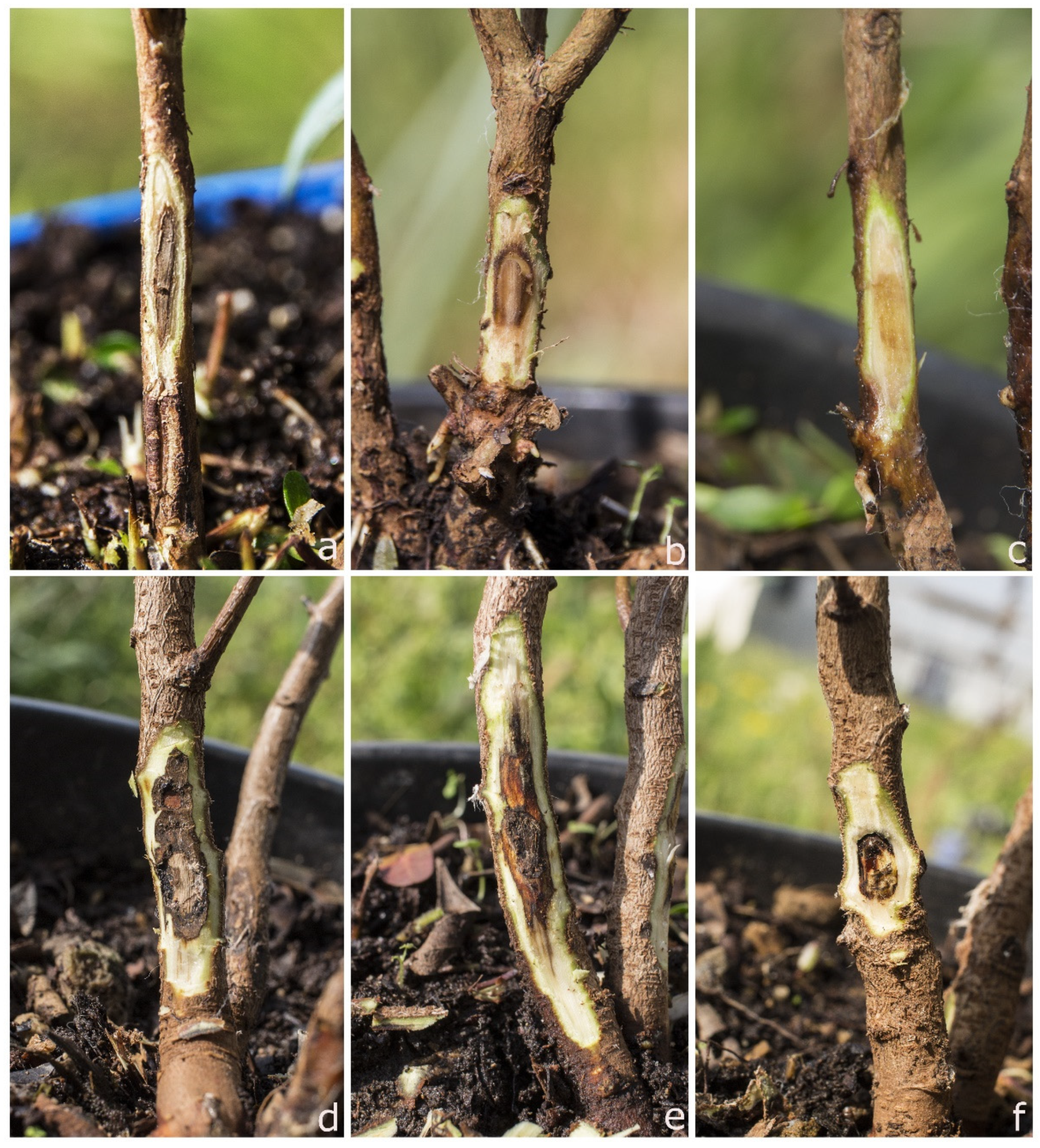
In artificial inoculation trials, P. mediterranea was shown to be pathogenic on myrtle and lentisk seedlings. After 20 days from inoculation, all plants inoculated with P. mediterranea displayed necrotic inner bark lesions that spread up and down from the inoculation site (Figure 5). On myrtle, the necrotic lesions caused by the isolate of P. mediterranea were significantly larger than those caused by P. cinnamomi; in contrast on lentisk, P. cinnamomi showed itself to be more aggressive than P. mediterranea (Table 4).
Control plants inoculated with sterile CA plugs remained symptomless, only a light brown discoloration limited to the inoculation point was observed on lentisk seedlings (Figure 5). Both Phytophthora species were successfully re-isolated from all inoculated plants, fulfilling Koch’s postulates. No Phytophthora isolates were re-isolated from control plants.

4. Discussion

The results obtained allowed us to expand knowledge on the diversity of Phytophthora species in Italian forest nurseries. The investigated nurseries are located in sites with very different climatic conditions, ranging from the Mediterranean (N2) to alpine ones (N3), including an intermediate climate typical of temperate hilly and low mountain areas (N1 and N4). These differences were also reflected in the plant species cultivated, which were typical of the different geographic regions.
Over the past two decades, diseases caused by Phytophthora species have been reported in a wide range of economically important ornamental plants worldwide and are considered yield-limiting factors in nursery production [13,29].
Our findings demonstrated that all the investigated nurseries were severally impacted by Phytophthora diseases. The N1 in particular was the most affected by the problem, and basically all cultivated plant species typical of the Mediterranean region were affected by Phytophthora infections.
In total, 15 Phytophthora species were isolated in pure culture from 14 hosts and identified on the basis of morphological features and DNA sequence analysis. Nine Phytophthora species were isolated in N1, five in N2, two in N4 and one in N3. The species obtained are representative of seven phylogenetic clades and included 14 previously known species: P. acerina, P. bilorbang, P. cactorum, P. cinnamomi, P. citrophthora, P. megasperma, P. nicotianae, P. palmivora, P. parvispora, P. pistaciae, P. plurivora, P. pseudocryptogea, P. pseudosyringae and P. psychrophila. In addition, two isolates could not be assigned to any known species and are therefore described here as P. mediterranea sp. nov.
Phytophthora mediterranea was shown to be associated with collar and root rot symptoms of myrtle seedlings. Phylogenetically, P. mediterranea is closely related to P. cinnamomi but the two species differ in 87 nucleotides in the three DNA regions studied. Morphologically P. mediterranea can be easily distinguished from P. cinnamomi on the basis of its smaller sporangia, colony growth pattern and higher optimum and maximum temperature values. Data from the pathogenicity tests showed that P. mediterranea has the potential to threaten the native Mediterranean maquis vegetation. A study is currently in progress to evaluate the susceptibility of the main Mediterranean maquis species to this new pathogen (Linaldeddu, unpublished data). The high optimum temperature value for growth of 32 °C and the high production of long-term survival propagules (chlamydospores) suggest that P. mediterranea is well adapted to survive in Mediterranean environments.
Phytophthora mediterranea belongs to clade 7c together with P. cinnamomi and P. parvispora, which were also obtained in this study. Phytophthora cinnamomi was the dominant species as it has been detected on five different hosts confirming its well-known polyphagous nature [11,58], and in three of the four monitored nurseries. The three nurseries are characterized by different environmental conditions ranging from the Mediterranean climate of site 2 located in Sardinia to the cold habitat of site 3 in the pre-Alps. This aspect emphasizes the potential of P. cinnamomi to survive in very different environments including low temperature habitats, as confirmed by a recent study in which this pathogen was detected in an outdoor blueberry stand in Germany [59]. Cold regions such as alpine and sub-alpine regions were long considered P. cinnamomi free, due to the inactivity of this species in soil at temperatures below 10 °C [60]. Analysis on current distribution data, global change and a forecast model, have allowed a global map to be created, identifying the cold areas of the planet as those at greatest risk of introduction and spread of P. cinnamomi in the future [61]. The discovery of P. cinnamomi in mountain nurseries in Italy confirms this trend and poses the risk of diffusion of this pathogen on new susceptible hosts among the alpine species.
Phytophthora nicotianae was the second dominant species. It has been isolated from four host species confirming the results of previously studies on the role of the nursery trade as one of the main ways of spreading for this pathogen [48,62]. The other twelve species have been isolated from a single plant host and only one nursery. Differences in species diversity among nurseries are difficult to interpret. It is plausible that different agronomical practices including sanitation, water management, origin and treatment of seeds as well as host plant diversity could have a role in the differences observed in terms of Phytophthora assemblages. Some species such as P. parvispora and P. pistaciae have confirmed a specificity towards some plant hosts already reported in other studies [49,50]. Phytophthora parvispora, long considered a variety of P. cinnamomi, is closely linked to Arbutus unedo plants in both nursery and natural areas of the Mediterranean Basin [49]. It has also recently been reported in North American nurseries associated with seedlings destined for reforestation programmes and in pomegranate orchards in Turkey [63,64]. Phytophthora pistaciae is a species characterized by a very limited geographic distribution and host range; it was originally described in Iran associated with Pistacia vera gummosis [65] and more recently in Italy on potted Pistacia lentiscus seedlings [50].
The other species, except P. acerina, have already been reported in different nurseries in Europe, Australia and North America on several plant hosts [34,39,48,63]. Nonetheless, seven new Phytophthora–host associations were detected in this study: P. psychrophila/Ilex aquifolium, P. pseudosyringae/I. aquifolium, P. pseudocryptogea/Laurus nobilis, P. megasperma/L. nobilis, P. citrophthora/L. nobilis, P. bilorbang/Phyllirea latifolia and P. palmivora/P. latifolia.
Phytophthora acerina was originally described in Northern Italy from declining maple trees [66] and more recently on declining olives with sudden death symptoms [67]. Its reports have increased in the last years in both natural and agricultural environments in Italy and its role in the onset of sudden death and dieback of kiwi, walnut and pomegranate orchards as well as oaks, chestnut and ash forests has been ascertained (Linaldeddu, unpublished data). In a nursery, P. acerina appears to be a very rare species, this is the first report on nursery plants in Europe, but it was recently reported in nursery plants in California [68]. The discovery of P. acerina and P. plurivora on seedlings potentially destined to reforestation programmes highlights the risk of further spread of these species, directly associated with the decline of grey alder on the Alps [51].

5. Conclusions

The findings obtained in this study highlight the occurrence of multiple Phytophthora species in Italian forest nurseries. Many of the species isolated are common in Italian nurseries, while others are rare or have never been reported. Phytophthora pistaciae is an example of a rare and possibly exotic species with a high risk of spread to the Mediterranean maquis. In addition, a new species closely related to P. cinnamomi and P. parvispora was isolated from potted myrtle plants. The myrtle is a typical shrub of the Mediterranean Basin, and Sardinia is recognized as one of its main local centres of diversity [69]. Its fruit and leaves exhibit antioxidant, antibacterial and antifungal properties, as well as being used for their content of essential oils and most commonly as an ingredient in a locally made liquor [70]. Nursery plants are often used for the creation of new orchards [71]. The use of infected plants could represent an important pathway for P. mediterranea.
Finally, the discovery of P. cinnamomi in alpine nurseries confirms the progressive expansion of this species towards the coldest areas of the globe, probably driven by climate change. A survey is currently in progress to map the distribution of Phytophthora species in alpine nurseries in Italy, with the purpose of evaluating the risk of diffusion of these pathogens through restoration and reforestation programmes.

Author Contributions

Conceptualization, B.T.L.; field survey, sample collection and assay, B.T.L., C.B., A.A.M., G.R. and G.P.; data analysis, B.T.L., C.B., G.R., A.D. and L.M.; funding acquisition B.T.L. and L.M.; draft writing B.T.L., C.B. and G.R.; review and manuscript editing, B.T.L., G.P., L.M. All authors have read and agreed to the published version of the manuscript.

Funding

This research was funded by grant number DOR2103497/21 “Impatto delle specie invasive di Phytophthora sulla biodiversità delle formazioni forestali in Italia” and by the “Fondo di Ateneo per la ricerca 2020”. Internal funding was provided by the University of Sassari.

Data Availability Statement

Data sharing not applicable.

Conflicts of Interest

The authors declare no conflict of interest.

References

  1. Hulme, P.E. Trade, transport and trouble: Managing invasive species pathways in an era of globalization. J. Appl. Ecol. 2009, 46, 10–18. [Google Scholar] [CrossRef]
  2. Bradley, B.A.; Blumenthal, D.M.; Regan, E.; Grosholz, E.D.; Lawler, J.J.; Miller, L.P.; Sorte, C.J.B.; D’Antonio, C.M.; Diez, J.M.; Dukes, J.S.; et al. Global Change, Global Trade, and the Next Wave of Plant Invasions. Front. Ecol. Environ. 2012, 10, 20–28. [Google Scholar] [CrossRef] [Green Version]
  3. ISTAT. Stima Preliminare dei Conti Economici Dell’agricoltura; Archivio, id252819; ISTAT: Rome, Italy, 2021. [Google Scholar]
  4. Brasier, C. The biosecurity threat to the UK and global environment from international trade in plants. Plant Pathol. 2008, 57, 792–808. [Google Scholar] [CrossRef]
  5. Liebhold, A.M.; Brockehoff, E.G.; Garret, L.J.; Parke, J.L.; Britton, K.O. Live plant imports: The major pathway for forest insect and pathogen invasions of the US. Front. Ecol. Environ. 2012, 10, 135–143. [Google Scholar] [CrossRef]
  6. Liebhold, A.M.; Brockerhoff, E.G.; Kalisz, S.; Nuñez, M.A.; Wardle, D.A.; Wingfield, M.J. Biological invasions in forest ecosystems. Biol. Invasions 2017, 19, 3437–3458. [Google Scholar] [CrossRef]
  7. Ghelardini, L.; Luchi, N.; Pecori, F.; Pepori, A.L.; Danti, R.; Rocca, G.D.; Capretti, P.; Tsopelas, P.; Santini, A. Ecology of invasive forest pathogens. Biol. Invasions 2017, 19, 3183–3200. [Google Scholar] [CrossRef]
  8. Yang, X.; Tyler, B.M.; Hong, C. An expanded phylogeny for the genus Phytophthora. IMA Fungus 2017, 8, 355–384. [Google Scholar] [CrossRef] [PubMed] [Green Version]
  9. Jung, T.; Horta Jung, M.; Cacciola, S.O.; Cech, T.; Bakonyi, J.; Seress, D.; Mosca, S.; Schena, L.; Seddaiu, S.; Pane, A.; et al. Multiple new cryptic pathogenic Phytophthora species from Fagaceae forests in Austria, Italy, and Portugal. IMA Fungus 2017, 8, 219–244. [Google Scholar] [CrossRef] [PubMed] [Green Version]
  10. Scott, P.; Bader, M.K.F.; Burgess, T.I.; Hardy, G.; Williams, N. Global biogeography and invasion risk of the plant pathogen genus Phytophthora. Environ. Sci. Policy 2019, 101, 175–182. [Google Scholar] [CrossRef]
  11. Erwin, C.D.; Ribeiro, O.K. Phytophthora Diseases Worldwide; APS Press: Saint Paul, MN, USA, 1996. [Google Scholar]
  12. Benson, D.M.; Jones, J.K. Diseases of Woody Ornamentals and Trees in Nurseries; APS Press: St. Paul, MN, USA, 2001. [Google Scholar]
  13. Parke, J.L.; Redekar, N.R.; Eberhart, J.L.; Funahashi, F. Hazard analysis for Phytophthora species in container nurseries: Three case studies. HortTechnology 2019, 29, 745–755. [Google Scholar] [CrossRef] [Green Version]
  14. Webber, J.F.; Mullett, M.; Brasier, C.M. Dieback and mortality of plantation Japanese larch (Larix kaempferi) associated with infection by Phytophthora ramorum. New Dis. Rep. 2010, 22, 19. [Google Scholar] [CrossRef] [Green Version]
  15. Prospero, S.; Vercauteren, A.; Heungens, K.; Belbahri, L.; Rigling, D. Phytophthora diversity and population structure of Phytophthora ramorum in Swiss ornamental nurseries. Plant Pathol. 2013, 62, 1063–1071. [Google Scholar] [CrossRef]
  16. Simamora, A.V.; Paap, T.; Howard, K.; Stukely, M.J.; Hardy, G.E.S.J.; Burgess, T.I. Phytophthora contamination in a nursery and its potential dispersal into the natural environment. Plant Dis. 2017, 102, 132–139. [Google Scholar] [CrossRef] [PubMed] [Green Version]
  17. Zentmyer, G.A. Origin and Distribution of Phytophthora cinnamomi; California Avocado Society: San Juan Capistrano, CA, USA, 1985; Volume 69, pp. 89–96. Available online: http://www.avocadosource.com/cas_yearbooks/cas_69_1985/cas_1985_pg_89-96.pdf (accessed on 4 May 2021).
  18. Hansen, E.M.; Streito, J.C.; Delatour, C. First confirmation of Phytophthora lateralis in Europe. Plant Dis. 1999, 83, 587. [Google Scholar] [CrossRef] [PubMed]
  19. Rizzo, D.M.; Garbelotto, M.; Davidson, J.M.; Slaughter, G.W.; Koike, S.T. Phytophthora ramorum as the cause of extensive mortality of Quercus spp and Lithocarpus densiflorus in California. Plant Dis. 2002, 86, 205–214. [Google Scholar] [CrossRef] [Green Version]
  20. Cobb, R.C.; Haas, S.E.; Kruskamp, N.; Dillon, W.W.; Swiecki, T.J.; Rizzo, D.M.; Frankel, S.J.; Meentemeyer, R.K. The magnitude of regional-scale tree mortality caused by the invasive pathogen Phytophthora ramorum. Earth’s Future 2020, 8, e2020EF001500. [Google Scholar] [CrossRef]
  21. Man in’t Veld, W.A.; De Cock, A.W.A.M.; Summerbell, R.C. Natural hybrids of residents and introduced Phytophthora species proliferating on multiple new hosts. J. Plant Pathol. 2007, 117, 25–33. [Google Scholar] [CrossRef]
  22. McDonald, J.D.; Alishtayeh, M.S.; Kabashima, J.; Stites, J. Occurrence of Phytophthora species in recirculated nursery irrigation effluents. Plant Dis. 1994, 78, 607–611. [Google Scholar] [CrossRef]
  23. Themann, K.; Werres, S.; Lüttmann, R.; Diener, H.A. Observations of Phytophthora spp. in water recirculation systems in commercial hardy ornamental nursery stock. Eur. J. Plant Pathol. 2002, 108, 337–343. [Google Scholar] [CrossRef]
  24. Hong, C.X.; Moorman, G.W. Plant pathogens in irrigation water: Challenges and opportunities. Crit. Rev. Plant Sci. 2005, 24, 189–208. [Google Scholar] [CrossRef]
  25. Redekar, N.R.; Eberhart, J.L.; Parke, J.L. Diversity of Phytophthora, Pythium and Phytopythium species in recycled irrigation water in a container nursery. Phytobiomes 2019, 3, 31–45. [Google Scholar] [CrossRef] [Green Version]
  26. Ivors, K.L.; Moorman, G.W. Oomycete plant pathogens in irrigation water. In Biology, Detection, and Management of Plant pathogens in Irrigation Water; Hong, C., Moorman, G.W., Wohanka, W., Büttner, C., Eds.; APS Press: St. Paul, MN, USA, 2017; pp. 57–64. [Google Scholar]
  27. Shishkoff, N. Persistence of Phytophthora ramorum in soil mix and roots of nursery ornamentals. Plant Dis. 2007, 91, 1245–1249. [Google Scholar] [CrossRef] [PubMed] [Green Version]
  28. Crone, M.; McComb, J.A.; O’Brien, P.A.; Hardy, G.E.S.J. Survival of Phytophthora cinnamomi as oospores, stromata, and thick-walled chlamydospores in roots of symptomatic and asymptomatic annual and herbaceous perennial plant species. Fungal Biol. 2013, 117, 112–123. [Google Scholar] [CrossRef] [PubMed] [Green Version]
  29. Bienapfl, J.C.; Balci, Y. Movement of Phytophthora spp. in Maryland’s nursery trade. Plant Dis. 2014, 98, 134–144. [Google Scholar] [CrossRef] [PubMed] [Green Version]
  30. Bruin, G.C.A.; Edgington, L.V. Adaptive resistance in Peronosporales to metalaxyl. Can. J. Plant Pathol. 1981, 3, 201–206. [Google Scholar] [CrossRef]
  31. Dobrowolski, M.; Shearer, B.; Colquhoun, I.; O’brien, P.; Hardy, G.E.S.J. Selection for decreased sensitivity to phosphite in Phytophthora cinnamomi with prolonged use of fungicide. Plant Pathol. 2008, 57, 928–936. [Google Scholar] [CrossRef]
  32. González-Tobón, J.; Childers, R.A.; Regnier, M.; Rodriguez, A.; Olave, C.; Fry, W.E.; Restrepo, S.; Danies, G. Is the phenomenon of mefenoxam-acquired resistance in Phytophthora infestans universal? Plant Dis. 2019, 104, 211–221. [Google Scholar] [CrossRef]
  33. Hardy, G.E.S.J.; Sivasithamparam, K. Phytophthora spp. associated with container-grown plants in nurseries in Western Australia. Plant Dis. 1988, 72, 435–437. [Google Scholar] [CrossRef] [Green Version]
  34. Davison, E.M.; Drenth, A.; Kumar, S.; Mack, S.; Mackie, A.E.; McKirdy, S. Pathogens associated with nursery plants imported into Western Australia. Australas. Plant Pathol. 2006, 35, 473–475. [Google Scholar]
  35. Moralejo, E.; Pérez-Sierra, A.M.; Alvarez, L.A.; Belbahri, L.; Lefort, F.; Descals, E. Multiple alien Phytophthora taxa discovered on diseased ornamental plants in Spain. Plant Pathol. 2009, 58, 100–110. [Google Scholar] [CrossRef]
  36. Yakabe, L.E.; Blomquist, C.L.; Thomas, S.L.; MacDonald, J.D. Identification and frequency of Phytophthora species associated with foliar diseases in California ornamental nurseries. Plant Dis. 2009, 93, 883–890. [Google Scholar] [CrossRef] [PubMed] [Green Version]
  37. Pérez-Sierra, A.; Jung, T. Phytophthora in woody ornamental nurseries. In Phytophthora: A Global Perspective; Lamour, K., Ed.; CABI: Wallingford, UK, 2013; pp. 166–177. [Google Scholar]
  38. Parke, J.L.; Knaus, B.J.; Fieland, V.J.; Lewis, C.; Grünwald, N.J. Phytophthora community structure analyses in Oregon nurseries inform systems approaches to disease management. Phytopathology 2014, 104, 1052–1062. [Google Scholar] [CrossRef] [PubMed] [Green Version]
  39. Molnar, C.; Nikolaeva, E.; Kim, S.; Olson, T.; Bily, D.; Kim, J.-E.; Kang, S. Phytophthora diversity in Pennsylvania nurseries and greenhouses inferred from clinical samples collected over four decades. Microorganisms 2020, 8, 1056. [Google Scholar] [CrossRef] [PubMed]
  40. Scott, P.; Taylor, P.; Gardner, J.; Puértolas, A.; Panda, P.; Addison, S.; Hood, I.; Burgess, T.; Horner, I.; Williams, N.; et al. Phytophthora aleatoria sp. nov., associated with root and collar damage on Pinus radiata from nurseries and plantations. Australas. Plant Pathol. 2019, 48, 313–321. [Google Scholar] [CrossRef]
  41. Hong, C.X.; Gallegly, M.E.; Richardson, P.A.; Kong, P.; Moorman, G.W.; Lea-Cox, J.D.; Ross, D.S. Phytophthora hydropathica, a new pathogen identified from irrigation water, Rhododendron catawbiense and Kalmia latifolia. Plant Pathol. 2010, 59, 913–921. [Google Scholar] [CrossRef]
  42. Yang, X.; Copes, W.E.; Hong, C.X. Phytophthora mississippiae sp. nov., a new species recovered from irrigation reservoirs at a plant nursery in Mississippi. J. Plant Pathol. Microb. 2013, 4, 1–7. [Google Scholar] [CrossRef] [Green Version]
  43. Bertier, L.; Brouwer, H.; de Cock, A.W.A.M.; Cooke, D.E.L.; Olsson, C.H.B.; Höfte, M. The expansion of Phytophthora clade 8b: Three new species associated with winter grown vegetable crops. Persoonia 2013, 31, 63–76. [Google Scholar] [CrossRef] [Green Version]
  44. Yang, X.; Gallegly, M.E.; Hong, C. A high-temperature tolerant species in Clade 9 of the genus Phytophthora: P. hydrogena sp. nov. Mycologia 2014, 106, 57–65. [Google Scholar] [CrossRef] [Green Version]
  45. Brasier, C.M.; Beales, P.A.; Kirk, S.A.; Denman, S.; Rose, J. Phytophthora kernoviae sp. nov., an invasive pathogen causing bleeding stem lesions on forest trees and foliar necrosis of ornamentals in the UK. Mycol. Res. 2005, 109, 853–859. [Google Scholar] [CrossRef] [Green Version]
  46. Abad, Z.G.; Abad, J.A.; Cacciola, S.O.; Pane, A.; Faedda, R.; Moralejo, E.; Pérez-Sierra, A.; Abad-Campos, P.; Alvarez-Bernaola, L.A.; Bakonyi, J.; et al. Phytophthora niederhauserii sp. nov., a polyphagous species associated with ornamentals, fruit trees and native plants in 13 countries. Mycologia 2014, 106, 431–447. [Google Scholar] [CrossRef]
  47. Migliorini, D.; Ghelardini, L.; Tondini, E.; Luchi, N.; Santini, A. The potential of symptomless potted plants for carrying invasive soilborne plant pathogens. Divers. Distrib. 2015, 21, 1218–1229. [Google Scholar] [CrossRef]
  48. Prigigallo, M.I.; Mosca, S.; Cacciola, S.O.; Cooke, D.E.L.; Schena, L. Molecular analysis of Phytophthora diversity in nursery-grown ornamental and fruit plants. Plant Pathol. 2015, 64, 1308–1319. [Google Scholar] [CrossRef] [Green Version]
  49. Scanu, B.; Hunter, G.C.; Linaldeddu, B.T.; Franceschini, A.; Maddau, L.; Jung, T.; Denman, S. A taxonomic re-evaluation reveals that Phytophthora cinnamomi and P. cinnamomi var. parvispora are separate species. For. Pathol. 2014, 44, 1–20. [Google Scholar] [CrossRef]
  50. Linaldeddu, B.T.; Mulas, A.A.; Bregant, C.; Piras, G.; Montecchio, L. First Report of Phytophthora pistaciae causing root and collar rot on nursery plants of Pistacia lentiscus in Italy. Plant Dis. 2020, 104, 1564. [Google Scholar] [CrossRef]
  51. Bregant, C.; Sanna, G.P.; Bottos, A.; Maddau, L.; Montecchio, L.; Linaldeddu, B.T. Diversity and pathogenicity of Phytophthora species associated with declining alder trees in Italy and description of Phytophthora alpina sp. nov. Forests 2020, 11, 848. [Google Scholar] [CrossRef]
  52. White, T.J.; Bruns, T.; Lee, S.; Taylor, J. Amplification and direct sequencing of fungal ribosomal RNA genes for phylogenetics. In PCR Protocols, a Guide to Methods and Applications; Innis, M.A., Gelfand, D.H., Sninsky, J.J., White, T.J., Eds.; Academic Press: San Diego, CA, USA, 1990; pp. 315–322. [Google Scholar]
  53. Martin, F.N.; Tooley, P.W. Phylogenetic relationships among Phytophthora species inferred from sequence analysis of mitochondrially encoded cytochrome oxidase I and II genes. Mycologia 2003, 95, 269–284. [Google Scholar] [CrossRef]
  54. Kroon, L.P.N.M.; Bakker, F.T.; Van Den Bosch, G.B.M.; Bonants, P.J.M.; Flier, W.G. Phylogenetic analysis of Phytophthora species based on mitochondrial and nuclear DNA sequences. Fungal Genet. Biol. 2004, 41, 766–782. [Google Scholar] [CrossRef]
  55. Thompson, J.D.; Gibson, T.J.; Plewniak, F.; Jeanmougin, F.; Higgins, D.G. The CLUSTAL_X windows interface: Flexible strategies for multiple sequence alignment aided by quality analysis tools. Nucleic Acids Res. 1997, 25, 4876–4882. [Google Scholar] [CrossRef] [Green Version]
  56. Hall, T.A. BioEdit: A user-friendly biological sequence alignment editor and analysis program for Windows 95/98/NT. In Nucleic Acids Symposium Series; Oxfrod University Press: Oxfrod, UK, 1999; Volume 41, pp. 95–98. [Google Scholar]
  57. Kumar, S.; Stecher, G.; Li, M.; Knyaz, C.; Tamura, K. MEGA X: Molecular Evolutionary Genetics Analysis across computing platforms. Mol. Biol. Evol. 2018, 35, 1547–1549. [Google Scholar] [CrossRef]
  58. Hardham, A.R.; Blackman, L.M. Phytophthora cinnamomi. Mol. Plant Pathol. 2018, 19, 260–285. [Google Scholar] [CrossRef] [PubMed] [Green Version]
  59. Nechwatal, J.; Jung, T. A study of Phytophthora spp. in declining highbush blueberry in Germany reveals that P. cinnamomi can thrive under Central European outdoor conditions. J. Plant. Dis. Prot. 2021. [Google Scholar] [CrossRef]
  60. Weste, G.; Marks, G. The biology of Phytophthora cinnamomi in Australasian forests. Annu. Rev. Phytopathol. 2020, 25, 207–229. [Google Scholar] [CrossRef]
  61. Burgess, T.I.; Scott, J.K.; McDougall, K.L.; Stukely, M.J.; Crane, C.; Dunstan, W.A.; Brigg, F.; Andjic, V.; White, D.; Rudman, T.; et al. Current and projected global distribution of Phytophthora cinnamomi, one of the world’s worst plant pathogens. Glob. Chang. Biol. 2017, 23, 1661–1674. [Google Scholar] [CrossRef] [Green Version]
  62. Mammella, M.A.; Martin, F.N.; Cacciola, S.O.; Coffey, M.D.; Faedda, R.; Schena, L. Analyses of the population structure in a global collection of Phytophthora nicotianae isolates inferred from mitochondrial and nuclear DNA sequences. Phytopathology 2013, 103, 610–622. [Google Scholar] [CrossRef] [Green Version]
  63. Rooney-Latham, S.; Blomquist, K.L.; Kosta, K.L.; Gou, Y.Y.; Woods, P.W. Phytophthora species are common on nursery stock grown for restoration and revegetation purposes in California. Plant Dis. 2019, 103, 448–455. [Google Scholar] [CrossRef] [Green Version]
  64. Kurbetli, İ.; Karaca, G.; Aydoğdu, M. Phytophthora species causing root and collar rot of pomegranate in Turkey. Eur. J. Plant. Pathol. 2020, 157, 485–496. [Google Scholar] [CrossRef]
  65. Mirabolfathy, M.; Cooke, D.E.L.; Duncan, J.M.; Williams, N.A.; Ershad, D.; Alizadeh, A. Phytophthora pistaciae sp. nov. and P. melonis: The principal causes of pistachio gummosis in Iran. Mycol. Res. 2001, 105, 1166–1175. [Google Scholar] [CrossRef]
  66. Ginetti, B.; Moricca, S.; Squires, J.N.; Cooke, D.E.L.; Ragazzi, A.; Jung, T. Phytophthora acerina sp. nov., a new species causing bleeding cankers and dieback of Acer pseudoplatanus trees in planted forests in northern Italy. Plant Pathol. 2014, 63, 858–876. [Google Scholar] [CrossRef] [Green Version]
  67. Linaldeddu, B.T.; Bregant, C.; Montecchio, L.; Favaron, F.; Sella, L. First report of Phytophthora acerina, P. pini and P. plurivora causing root rot and sudden death on olive trees in Italy. Plant Dis. 2020, 104, 996. [Google Scholar] [CrossRef]
  68. Frankel, S.J.; Conforti, C.; Hillman, J.; Ingolia, M.; Shor, A.; Benner, D.; Alexander, J.M.; Bernhardt, E.; Swiecki, T.J. Phytophthora introductions in restoration areas: Responding to protect California native flora from human-assisted pathogen spread. Forests 2020, 11, 1291. [Google Scholar] [CrossRef]
  69. Medail, F.; Quezel, P. Biodiversity hotspots in the Mediterranean basin: Setting global conservation priorities. Conserv. Biol. 1999, 13, 1510–1513. [Google Scholar] [CrossRef]
  70. Melito, S.; Chessa, I.; Erre, P.; Podani, J.; Mulas, M. The genetic diversity of Sardinian myrtle (Myrtus communis L.) populations. Electron. J. Biotechnol. 2013, 16, 7. [Google Scholar] [CrossRef]
  71. Mulas, M. Problematiche legate alla coltivazione del mirto. Italus Hortus 2004, 11, 308–312. [Google Scholar]
Figure 1. Overview of the forest nursery N1 (a), symptoms of wilting, sudden death and collar and root rot observed on: Abies alba (b), Alnus incana (c,d), Arbutus unedo (e), Castanea sativa (f,g), Fagus sylvatica (h), Helichrysum italicum (i), Ilex aquifolium (j), Juglans regia (k), Laurus nobilis (l), Lavandula officinalis (m), Myrtus communis (n), Phyllirea latifolia (o), Pistacia lentiscus (p) and Quercus ilex (q) seedlings. On the right from the top, colony morphology of: Phytophthora acerina, P. bilorbang, P. cactorum, P. cinnamomi, P. citrophthora, P. mediterranea, P. megasperma, P. nicotianae, P. palmivora, P. parvispora, P. pistaciae, P. plurivora, P. pseudocryptogea, P. pseudosyringae and P. psychrophila after 7 days growth at 25 °C on CA in the dark.
Forests 12 00682 g001
Figure 2. Maximum likelihood tree obtained from combined ITS, Btub and cox1 sequences of Phytophthora species belonging to clade 7. Data are based on the general time reversible model. A discrete gamma distribution was used to model evolutionary rate differences among sites. The tree is drawn to scale, with branch lengths measured in the number of substitutions per site. Bootstrap support values in percentage (1000 replicates) are given at the nodes. Ex-type cultures are reported in bold.
Forests 12 00682 g002
Figure 3. Colony morphology of Phytophthora mediterranea (CB84) after 7 days growth at 25 °C on potato dextrose agar (PDA) (a), malt extract agar (MEA) (b) and carrot agar (CA) (c). Sporangia observed in pond water: non-papillate, persistent and ovoid (d), elongated (e), releasing zoospores (f,g). External (h) and internal proliferations (i), hyphal swellings (j) and terminal chlamydospores (k). Scale bars = 20 µm.
Forests 12 00682 g003
Figure 4. Mean colony diameter (± standard deviation) of P. cinnamomi (CB21), P. mediterranea (CB84) and P. parvispora (CB86) after 96 h on CA in the dark at different temperatures.
Forests 12 00682 g004
Figure 5. Symptoms observed on myrtle and lentisk seedlings 20 days after inoculation with Phytophthora mediterranea (a,d) and P. cinnamomi (b,e). Control seedlings (c,f).
Forests 12 00682 g005
Table 1. Nurseries information and plant species monitored for Phytophthora.
NurseryElevation
(m. a.s.l.)
Geographic
Coordinates
Plant Species *
N186040°25′54′′ N8°58′43′′ ECs (12), Ia (6), Jr (12), Ln (6), Phl (6)
N21639°57′43′′ N8°36′02′′ EAu (4), Hi (4), Lo (2), Mc (3), Pl (7), Qi (6)
N3107746°06′54′′ N12°26′05′′ EAa (2), Fs (2)
N419146°11′51′′ N13°14′13′′ EAi (4)
* In brackets the number of plants collected: Abies alba (Aa), Arbutus unedo (Au), Alnus incana (Ai), Castanea sativa (Cs), Fagus sylvatica (Fs), Helichrysum italicum (Hi), Ilex aquifolium (Ia), Juglans regia (Jr), Laurus nobilis (Ln), Lavandula officinalis (Lo), Myrtus communis (Mc), Phyllirea latifolia (Phl), Pistacia lentiscus (Pl) and Quercus ilex (Qi).
Table 2. Phytophthora isolates used in the phylogenetic analysis. Ex-type cultures are reported in bold and newly generated sequences are indicated in italics.
SpeciesCodeHostGenBank Accession Number
ITSBtubCox1
P. asiaticaCBS 133347Pueraria lobataAB688422AB539560AB740169
P. attenuataCBS 141199Castanopsis carlesiiKU517154KU899277LC595899
P. cajaniP3105Cajanus cajanMG783386MH493912MH136859
P. cinnamomiCBS 144.22Cinnamomum sp.MG865473MH493920MH136869
P. cinnamomiCB21Abies albaMW892397MW900442MW900446
P. cambivoraP19997Castanea sativaMG783387MH493913MH136860
P. europaeaCBS 109049Quercus roburMG865488MH493935MH136884
P. flexuosaCBS 141201Fagus hayataeKU517152KU899302LC595910
P. formosaCBS 141203Araucaria sp.KU517153KU899270LC595912
P. fragariaeCBS 209.46Fragaria ×ananassaMG865494MH493938MH136890
P. fragariaefoliaCBS 135747Fragaria ×ananassaMG865495MH493939MH136891
P. intricataCBS 141211Quercus tarokoensisKU517155KU899284LC595921
P. niederhauseriiP10616Hedera helixMG865552MH493988MH136944
P. melonisCBS 582.69Cucumis sativusMG865536MH493974MH136931
P. mediterraneaCB84Myrtus communisMW892398MW900443MW900447
P. mediterraneaCB85M. communisMW892399MW900444MW900448
P. nagaiiCBS 133248Rosa sp.MG865547MN207274MH136940
P. parvisporaCBS 132772Arbutus unedoKC478667KC609402KC609413
P. parvisporaCB86A. unedoMW892401MW900445MW900449
P. pisiCBS 130350Pisum sativusMG865567MH494000MH477754
P. pistaciaeP19883Pistacia veraKT183043KX251749LC595934
P. rubiCBS 967.95Rubus idaeusMG865584MH494011MH136976
P. sojaeCBS 382.61Glycine maxMG865587MN207279MH136979
P. tyrrhenicaCBS 142301Quercus ilexKU899188KU899265LC595950
P. uliginosaCBS 109054Q. roburMG865597MH494023MH136988
P. uniformisP16206Alnus glutinosaMK496514MH493905MH136992
P. vignaeP3019Vigna sinensisMG865598MH494024MH136989
P. vulcanicaCBS 141216Fagus sylvaticaMF036209MF036235LC595951
P. ×alniIMI 392314A. glutinosaMK496513MH493903MH136991
P. ×heterohybridaCBS 141207WaterKU517151KU899290LC595953
P. ×incrassataCBS 141209WaterKU517156KU899286LC595954
P. ×multiformisP16202A. glutinosaMG783372MH493904MK493472
Table 3. Number of isolates of Phytophthora species obtained from monitored plants in the investigated nurseries.
SpeciesAccession
Number
ITS CladePlant Species *Nursery
P. acerinaMW8923952Ai (1)N4
P. bilorbangMW9599116Phl (2)N1
P. cactorumMW8923961Cs (1)N1
P. cinnamomiMW8923977Cs (8), Fs (2), Aa (2), Jr (8), Qi (6)N1-2-3
P. citrophthoraMW9599162Ln (1)N1
P. mediterraneaMW8923987Mc (2)N2
P. megaspermaMW9599136Ln (3)N1
P. nicotianaeMW8924001Lo (1), Pl (2), Hi (4), Mc (2)N2
P. palmivoraMW9599174Phl (5)N1
P. parvisporaMW8924017Au (3)N2
P. pistaciaeMW8924027Pl (2)N2
P. plurivoraMW8924032Ai (3)N4
P. pseudocryptogeaMW9599128Ln (4)N1
P. pseudosyringaeMW9599143Ia (3)N1
P. psychrophilaMW9599153Ia (1)N1
* In brackets the number of Phytophthora isolates on: Abies alba (Aa), Arbutus unedo (Au), Alnus incana (Ai), Castanea sativa (Cs), Fagus sylvatica (Fs), Helichrysum italicum (Hi), Ilex aquifolium (Ia), Juglans regia (Jr), Laurus nobilis (Ln), Lavandula officinalis (Lo), Myrtus communis (Mc), Phyllirea latifolia (Phl), Pistacia lentiscus (Pl) and Quercus ilex (Qi).
Table 4. Mean lesion length (cm) (± standard deviation) caused by Phytophthora mediterranea (CB84) and P. cinnamomi (CB21) on myrtle (M) and lentisk (L) seedlings and percentage of positive re-isolations.
SpeciesMyrtle *Lentisk *Re-Isolation
Frequency (%)
Phytophthora mediterranea1.01 ± 0.4a1.9 ± 0.6b100 (M) and 100 (L)
Phytophthora cinnamomi0.68 ± 0.3b2.3 ± 0.8a100 (M) and 100 (L)
Control-0.5 ± 0.2c-
* Values in column with the same letter do not differ significantly at p = 0.05, according to LSD multiple range test.
Publisher’s Note: MDPI stays neutral with regard to jurisdictional claims in published maps and institutional affiliations.