Article

Effects of Bacillus cereus NJSZ-13 on Fatty Acid Metabolism of Bursaphelenchus xylophilus

Co-Innovation Center for Sustainable Forestry in Southern China, College of Forestry and Grassland, Nanjing Forestry University, Nanjing 210037, China
*
Author to whom correspondence should be addressed.
These authors contributed equally to this work.
Forests 2023, 14(10), 2065; https://doi.org/10.3390/f14102065
Submission received: 12 September 2023 / Revised: 5 October 2023 / Accepted: 11 October 2023 / Published: 16 October 2023
(This article belongs to the Special Issue Advance in Pine Wilt Disease)

Abstract

:
In order to explore the changes in the fatty acid metabolism of Bursaphelenchus xylophilus under the treatment of Bacillus cereus NJSZ-13, the surface changes in lipid droplets were observed under a Zeiss microscope after the B. xylophilus was stained with modified Oil Red O. The triglyceride (TG) content in B. xylophilus was determined according to the TG kit instructions. The type and content of fatty acids in B. xylophilus were detected by gas chromatography–mass spectrometry (GC-MS); the stearyl coenzyme A of B. xylophilus was analyzed by real-time fluorescence quantitative PCR. The change in Bx-SCD (gene regulating stearyl CoA desaturase in B. xylophilus) expression was observed. The results showed that the lipid droplets of B. xylophilus treated with NJSZ-13 were broken to varying degrees, and the TGs in B. xylophilus decreased continuously. The total fatty acid content in the bodies of treated B. xylophilus decreased: the difference between the fermentation broth treatment and the control was extremely significant (p < 0.01); that between the fermentation filtrate and the control was significant (p < 0.05); and that between the bacterial suspension and the control was not significant (p > 0.05). Saturated fatty acids decreased in all treatments, but not significantly. Compared with the control group, the unsaturated fatty acid content in fermentation broth and fermentation filtrate treatments was extremely significantly reduced, and the unsaturated fatty acid content of the bacterial suspension group was significantly decreased, which indicated that NJSZ-13 mainly caused a decrease in the unsaturated fatty acids in B. xylophilus. The trend in changes in monounsaturated fatty acids and unsaturated fatty acids was the same, but for polyunsaturated fatty acids, the fermentation broth and fermentation filtrate treatments caused a significant decrease in content, but the bacterial suspension resulted in no significant change. The results showed that NJSZ-13 mainly caused a decrease in monounsaturated fatty acid content in B. xylophilus. In addition, the contents of C16:1, C18:1, and C18:2 fatty acids were significantly decreased after treatment with strain NJSZ-13 for 48 h, and the contents of C16:1, C18:1, C18:2, and C20:4 were extremely significantly decreased after the fermentation broth and filtrate treatments. The expression of Bx-SCD in B. xylophilus was significantly lower than that of the control (p < 0.0001). This study analyzed the changes in the content of related substances and relative gene expression in fatty acid metabolism of B. xylophilus treated with strain NJSZ-13, and preliminarily reveals the nematicidal mechanism of strain NJSZ-13 against B. xylophilus. This provides a theoretical basis for further exploration of the key cause of death induced by this strain in B. xylophilus.

1. Introduction

Pine wilt disease can cause the rapid wilting and death of pine [1]. The disease caused by the pine wood nematode (Bursaphelenchus xylophilus (Steiner & Buhrer, 1934) Nickle 1970 (Nematoda: Aphelenchoididae)) originated in North America, but has become prevalent in Asia in recent years [2]. China is the most seriously affected country in Asia. According to Announcement No. 5 issued by the State Forestry and Grassland Administration of China and various reports, 728 county-level administrative regions in 19 provinces (autonomous regions and municipalities) are affected by pine wilt disease as of 2021, covering an area of more than 1.8 million hectares, killing hundreds of millions of pine trees, and posing a serious threat to China’s forests and ecological security [3]. Over the years, people have attempted to control pine wilt disease using different approaches. These control methods can be roughly divided into physical control, chemical control, and biological control. Among them, chemical control is rapid and effective, but it causes environmental pollution and has poor ecological benefits. Therefore, microorganisms used to control B. xylophilus as an environmentally friendly biocontrol strategy have received much attention [4]. Bacillus cereus NJSZ-13 is an endophytic bacterium with high antagonistic activity against B. xylophilus [5,6]. It has reproductive toxicity, developmental toxicity, neurotoxicity, and other biological toxicity effects on B. xylophilus, but is safe for other organisms [7]. It is an ideal microorganism for controlling B. xylophilus.
Fatty acids are a type of aliphatic carboxylic acid containing 2–30 carbon atoms. There are three kinds of animal fatty acids: saturated fatty acids, monounsaturated fatty acids, and polyunsaturated fatty acids [8]. Saturated fatty acids refer to fatty acids that do not contain double bonds, such as stearic acid (18:0); monounsaturated fatty acids refer to fatty acids that only contain one double bond, such as oleic acid (18:1); and polyunsaturated fatty acids refer to fatty acids that contain multiple double bonds, such as octadecatrienoic acid (C18:4n3). Stearoyl CoA desaturase (SCD), also known as Δ-9 desaturase, is a key enzyme in fat synthesis [9]. SCD converts saturated fatty acids into monounsaturated fatty acids by introducing double bonds at the Δ-9 position of saturated fatty acids, such as palmitic acid (16:0) and stearic acid (18:0), which are obtained from food or synthesized endogenously into palmitoleic acid (16:1n7) and oleic acid (18:1n9) [10]. Monounsaturated fatty acids are not only important substrates for the synthesis of triglycerides (TGs), membrane phospholipids, cholesterol esters, and wax esters, but also substrates for the synthesis of polyunsaturated fatty acids (PUFAs) [11]. In addition, the normal ratio of unsaturated fatty acids to saturated fatty acids plays an important role in membrane fluidity [12], signal transmission [13], energy balance [10], and cell apoptosis [14]. SCD in the human body is extremely unstable, and is influenced and regulated by diet, hormones, and many transcription factors [15]. When the expression of the scd gene is too high, fat accumulates; and when the expression of the scd gene is too low, free saturated fatty acids accumulate, leading to endoplasmic reticulum stress responses and apoptosis [16]. In the study of model organism Caenorhabditis elegans, it was found that C. elegans has three genes (fat-5, fat-6, and fat-7), which jointly encode the scd gene. The scd gene of C. elegans has a direct impact on its stress resistance, growth and development, and fat metabolism [17]. Wang et al. [18] found that B. xylophilus has an scd gene homologous to that in C. elegans, named Bx-SCD. Their results showed that the expression of the Bx-SCD gene was positively correlated with changes in fatty acids. As an important component of B. xylophilus, fatty acids play an important role in its growth and development.
Does NJZS-13 affect the fatty acid metabolism of B. xylophilus? What is the impact? Is this effect related to SCD? Currently, none of these issues are clear. In a preliminary study, it was found that after 48 h of treatment with strain NJSZ-13 fermentation broth and fermentation filtrate, the mortality rate of B. xylophilus reached 100%, and that strain NJSZ-13 had an impact on various aspects such as egg production, egg hatching rate, and reproductive capacity of nematodes [7]. At the same time, strain NJSZ-13 affected several physiological and biochemical indicators of nematodes [19]. In order to further explore the nematicidal mechanism of strain NJSZ-13 against B. xylophilus, this study analyzed the changes in the content of related substances and the relative expression of genes involved in fatty acid metabolism of B. xylophilus after treatment with strain NJSZ-13.

2. Materials and Methods

2.1. Tested Nematodes and Strains

The AMA3 isolate of B. xylophilus was extracted from Pinus thunbergii in Ma’anshan, Anhui Province. AMA3 was provided by Nanjing Forestry University and extracted using the Baermann funnel method [20]. It is now stored in the resource bank of pine Nematoda isolates of Nanjing Forestry University.
Bacillus cereus NJSZ-13 is a kind of endophytic bacteria with nematicidal activity isolated from Pinus elliottii. All of the tested strains were obtained from the forest pathology laboratory of Nanjing Forestry University. NJSZ-13 has been deposited in the Chinese typical culture preservation center, with the accession number of CCTCC M 2016660.

2.2. Obtaining of the Bacterial Suspension and Fermentation Filtrate

The stored B. cereus NJSZ-13 was activated on an NA plate, transferred into an NB medium triangular flask, and placed on 200 R/min shaking table at 28 °C for 24 h. The mixture was then centrifuged at 10,000 R/min at 4 °C for 15 min and the supernatant was discarded. The suspension of bacteria in sterile water of the same volume as the discarded supernatant is the test bacterial suspension. Transfer the activated NJSZ-13 into an NB medium triangular flask and place it on a shaking table at 200 R/min and 28 °C for 4 days; the obtained shaking culture solution was the fermentation broth. The supernatant obtained by centrifugation was the fermentation filtrate [21].

2.3. NJSZ-13 Treatment of B. xylophilus

B. xylophilus (about 100,000) was mixed with the suspension, fermentation broth, and fermentation filtrate of NJSZ-13 at a concentration of 3 × 10⁸ in a 1.5 mL centrifuge tube, and then placed in a 25 °C incubator for 48 h. Sterile water and NB medium were used as a control. B. xylophilus treated for 48 h was centrifuged at 3500 R/min for 10 min and cleaned with sterile water three times before use.

2.4. Observation of Lipid Droplets of B. xylophilus Treated with Strain NJSZ-13

After NJSZ-13 treatment, M9 buffer solution was added to the B. xylophilus solution, which was kept at 4 °C for 10 min. The supernatant was discarded and B. xylophilus was cleaned with M9 buffer twice. The B. xylophilus fat was fixed with 4% paraformaldehyde at 4 °C for 30 min, quickly frozen with liquid nitrogen, and placed into a −80 °C refrigerator for 15 min. Then, it was thawed in a 43 °C water bath. The thawed samples were centrifuged at 3500 R/min for 1 min and the supernatant was removed and then washed three times with 1 × phosphate buffer (1 × PBS, pH = 7.2–7.4). After centrifugation, the supernatant was discarded and the nematode samples were soaked in 2:3 Triton X-100 and isopropanol Oil Red O saturated solution for 30 min. After being washed with 1 × PBS three times, observations were made and images were taken with a Carl Zeiss microscope.

2.5. Determination of Fatty Acid Content in B. xylophilus by Strain NJSZ-13

Methanol (1 mL) and 2% sulfuric acid (1 mL) were added to the solution after NJSZ-13 treatment, and it was then chilled at −80 °C for 1 h. After cooling, 1 mL n-hexane was added and shaken for several minutes, and the supernatant was centrifuged. The supernatant was sent to Nanjing Int Biotechnology Co., Ltd. (Nanjing, China) for the determination of fatty acid components.

2.6. qRT-PCR Detection of Genes Related to Fatty Acid Synthesis Pathways in Nematodes

2.6.1. Extraction and Detection of Total RNA from B. xylophilus

The B. xylophilus treated with NJSZ-13 were transferred into a 1.5 mL RNA-free centrifuge tube and centrifuged at 3500 R/min for 2 min, after which the supernatant was discarded, and washed with PBS three times. The sample centrifuge tube was placed directly into liquid nitrogen for quick freezing, and then it in an ultra-low temperature refrigerator at −80 °C for 20 min. After grinding with a tissue grinder, Trizol (1 mL) was added and the sample was allowed to stand at room temperature for 10 min. It was then centrifuged at 4 °C 12,000 R/min for 5 min. The supernatant was transferred to a new 1.5 mL RNA-free EP tube, 1/5 Trizol volume of chloroform was added, and it was shaken vigorously by hand for 15 s to make ensure it was fully dissolved. The supernatant was allowed to stand on ice for 5 min, and then centrifuged at 4 °C and 12,000 R/min for 15 min. The centrifuge tube was divided into three layers: the upper layer was a colorless RNA layer, the middle layer was a white protein layer, and the lower layer was a red Trizol layer. Approximately 400–500 μL of the upper layer was placed into a new 1.5 mL RNA-free centrifuge tube, and an equal volume of isopropanol was added. The centrifuge tube was manually turned upside down so that it would mix well and allowed to stand for 10 min, after which it was centrifuged at 4 °C and 12,000 R/min for 10 min. The supernatant was discarded, and 1 mL of 75% ethanol prepared with DEPC water was added. The centrifuge tube was gently turned upside down several times and centrifuged at 4 °C and 12,000 R for 15 min, after which the supernatant was discarded and the procedure was repeated once. The product was placed at room temperature for 2–5 min. Then, 10–20 μL Rnase-free water was added to dissolve the precipitate and heated at 50 °C for 2–5 min. A total of 1.5 μL RNA was aspirated from each tube, and its concentration and purity were determined using a Nanodrop spectrophotometer. The purity of RNA was determined by the A260/A280 ratio. If the ratio was between 1.8 and 2.0, the purity of RNA was good; if the ratio was less than 1.8, there might have been protein residue in the RNA sample; if the ratio was greater than 2.0, the RNA was broken.

2.6.2. Reverse Transcription of RNA

In this experiment, a 20 μL system was used for reverse transcription in a common PCR apparatus. The reaction system was a two-step process. In the first step, 1 μL RNA, 4 μL 4× g DNA Wiper Mix, and 11 μL Rnase-free ddH2O were added. After mixing, the reaction system was incubated at 42 °C for 2 min, and then taken out and placed in a cooler. In the second step, 4 μL 5 × HiScript II qRT SuperMix was added to the reaction solution obtained in the first step, and thoroughly mixed with a pipette gun. The solution was placed into an reverse transcription instrument (Eppendorf AGPCR) for reverse transcription.

2.6.3. Real Time Fluorescent Quantitative PCR

Using Q-Bx-SCD-f (5′-CGC CTT CCA GAA TGC TA-3′) and Q-Bx-SCD-R (5′-CCG AGA GGT CTA ATT TGG CT-3′) as primers, RT-qPCR was performed according to ChamQ SYBR qPCR Master Mix. The reaction system was 20 μL, with 0.4 μL upstream primer, 0.4 μL downstream primer, 2 μL cDNA template, 10 μL chamq SYBR qPCR Master Mix, and 7.2 μL ddH2O. Actin-1 was used as an internal reference gene. RT-qPCR was carried out following a two-step method. The reaction procedure comprised pre-denaturation at 95 °C for 30 s, and 40 cycles of denaturation at 95 °C for 10 s, annealing at 60 °C for 34 s, and extension at 95 °C for 15 s. RT-PCR data analysis was calculated by the Δ CT method of the reference gene; that is, the expression level of the target gene was m = log2 (2a + 1), where a = CT (actin) − CT (target gene). Prism software (Version 9.5.0, San Diego, CA, USA)was used to perform a group t-test to analyze the significance of the differences between the treatments.

3. Results

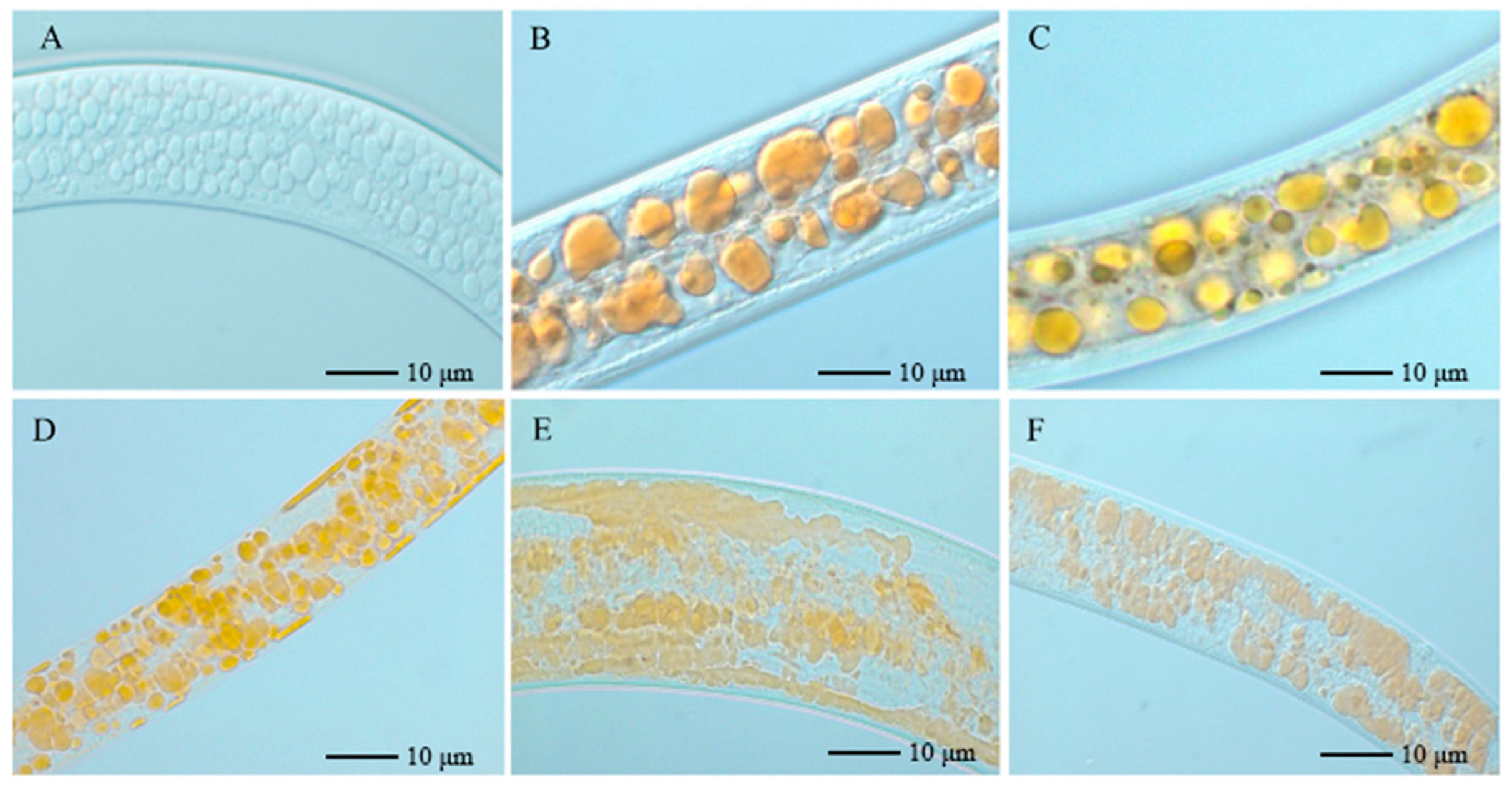
3.1. Effect of Strain NJSZ-13 on Lipid Droplets of B. xylophilus

B. xylophilus was treated with the suspension, fermentation liquid, and fermentation filtrate of NJSZ-13, and then stained with Oil Red O. It can be seen from Figure 1 that although a small amount of lipid droplets were broken, most lipid droplets were intact. After treatment with the fermentation broth and fermentation filtrate, the lipid droplets of the nematode were almost broken, and the contents were overflowing.

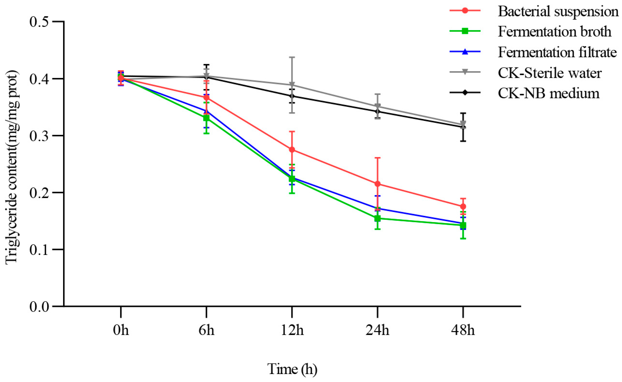
3.2. Effect of NJSZ-13 on Triglyceride Content in B. xylophilus

Triglycerides (TGs) are an important component of the body fat in B. xylophilus, and are widely present in the intestinal and subcutaneous tissue cells of B. xylophilus in the form of lipid droplets [18]. The TG content in B. xylophilus can directly reveal the effect of NJSZ-13 on the fatty acid metabolism of B. xylophilus. The effect of strain NJSZ-13 on the TG content of B. xylophilus is shown in Figure 2. It can be seen from the figure that the TGs in B. xylophilus treated with NJSZ-13 decreased significantly, and the decrease following the fermentation broth and fermentation filtrate was more obvious than that following the bacterial suspension. The fasted decrease occurred at 0–6 h and the largest decrease occurred over 6–48 h.

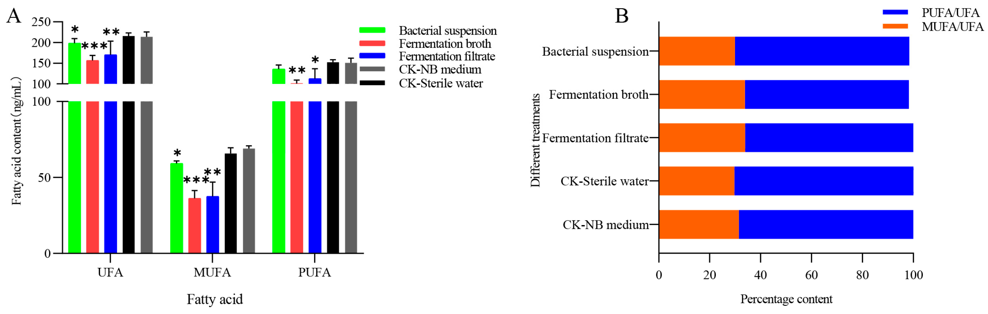
3.3. Effect of NJSZ-13 on Fatty Acid Content in B. xylophilus

The total fatty acid content in B. xylophilus treated with NJSZ-13 was significantly lower than that of NB medium (p < 0.01) (Figure 3), the content of fermentation filtrate was significantly different (p < 0.05), and the content of the bacterial suspension was not significantly different. Saturated fatty acids were decreased by all treatments, but not significantly. The results showed that there was a significant difference in the amount of unsaturated fatty acids between the bacterial suspension treatment and the control sterile water, while the amount of unsaturated fatty acids in the fermentation broth and fermentation filtrate-treated group were significantly lower than those in the control group (p < 0.01), indicating that NJSZ-13 mainly caused the decrease of unsaturated and fatty acid content in B. xylophilus. The monounsaturated fatty acids in B. xylophilus treated with the bacterial suspension were significantly different from those treated with control sterile water (p < 0.05), while the fermentation broth and fermentation filtrate were significantly different from those treated with the control NB medium (p < 0.01). However, with regard to the polyunsaturated fatty acids, there was no significant change in B. xylophilus treated with the bacterial suspension, but they were significantly decreased after fermentation broth treatment (p < 0.01), and significantly decreased after fermentation filtrate treatment (p < 0.05) (Figure 4). The results showed that NJSZ-13 mainly caused the decrease in monounsaturated fatty acid content in B. xylophilus, which may also be due to the low monounsaturated fatty acid content in B. xylophilus. In addition, the contents of C16:1, C18:1, and C18:2 fatty acids decreased significantly (p < 0.05) after the treatment with strain NJSZ-13 for 48 h. The contents of C16:1, C18:1, C18:2, and C20:4 fatty acids decreased significantly after the treatment with fermentation broth and fermentation filtrate compared with the control NB medium, the content was significantly decreased (p < 0.01) (Figure 5).

3.4. Effect of Strain NJSZ-13 on Bx-SCD Expression in B. xylophilus

The results (Table 1) showed that the expression of Bx-SCD in the suspension treated group was significantly lower than that in the control group (p < 0.0001), and the expression of Bx-SCD in the fermentation broth and fermentation filtrate treated groups was also significantly lower than that in the control group (p < 0.0001).

4. Discussion

Adipose tissue is an important component of the bodies of B. xylophilus. The fat content in the body often changes due to changes in gene expression in the metabolic pathway caused by changes in food intake, temperature, or drugs. Under normal conditions, the contents of phospholipids, TGs, wax esters, cholesterol esters, and cholesterol are maintained at normal levels [22]. In a high-glucose state, scd gene expression is increased, resulting in the transformation of 16:0/18:0 to 16:1/18:1 fatty acids, increasing the synthesis of phospholipids, TGs, wax esters, and cholesterol esters I [23]. With the increase in scd gene expression level, the content of cholesterol decreases because cholesterol is the substrate of cholesterol ester synthesis, and the decrease in cholesterol reduces the expression of the scd gene. The scd has three coding genes, namely, fat-5, fat-6, and fat-7. The fat-5 gene mainly catalyzes the transformation from C16:0 to C16:1 (N-7), while fat-6 and fat-7 mainly catalyze the transformation from C18:0 to C16:1 (N-9) [24]. Previous studies have shown that nematicides can cause disordered lipid metabolism in nematodes, resulting in a series of biological effects. For example, Hongmei et al. [25] showed that C. elegans was infected by Pseudomonas aeruginosa and stained with Oil Red O. The body of C. elegans was black, the body fat was significantly reduced, and the remaining fat was mainly distributed at both ends of the body.
Lipid droplets are a kind of monolayer membrane organelle with many lipid droplet proteins on their surface. TGs and esterified cholesterol form the intermediate lipid core [26]. Lipid droplets store fat, which can buffer the toxic effect of a large amount of fat in the body [27]. There are a large number of functional proteins on the surface of lipid droplets, which suggests that lipid droplets may be involved in the processes of intracellular material metabolism, transport, and cell signal transduction. It has been proven that TG storage and decomposition, arachidonic acid metabolism, and prostaglandin synthesis are related to lipid droplets, which indicates that lipid droplets participate in lipid anabolism [28].
In this study, the lipid droplets of B. xylophilus treated with NJSZ-13 were ruptured to varying degrees, the substances in the lipid droplets leaked out, and the triglyceride content continued to decrease with the increase in treatment time. Zhang Jingjing et al. [29] screened the downstream target genes of sbp-1 with RNA interference and detected the changes in fatty acid content by gas chromatography. The results showed that the zinc ion level affected the accumulation of triglyceride in nematodes by affecting the activity of SCDs in B. xylophilus, changing the ratio of monounsaturated fatty acids (C16:1n7 and C18:1n9) to saturated fatty acids (C16:0 and C18:0). Yihong [30] fixed and stained C. elegans treated with different concentrations of Pu’er tea water extract with Nile red. It was found that after treatment with Pu’er tea water extract, the lipid droplets of C. elegans became smaller, the density decreased, and the fat content decreased. The results showed that compared with the control group, the ratio of TAG/Total lipid decreased significantly (5%). The stearic acid (18:0) content increased and the oleic acid (18:1n9) content decreased, which confirmed that Pu’er tea could significantly reduce the expression of SCD and SREBP at the protein and mRNA levels. In this study, 26 kinds of fatty acids were detected by GC-MS after NJSZ-13 treatment. The total fatty acid content showed a downward trend, and the unsaturated fatty acids decreased significantly, among which C16:1, C18:1, C18:2, and C20:4 decreased significantly compared with the control, whereas other fatty acids decreased but not significantly. To further explore the molecular mechanism by which NJSZ-13 induced changes in the fatty acid content in nematodes, the relative expression of SCD, a key enzyme in fatty acid synthesis, was determined. The results showed that the expression of scd in B. xylophilus treated with NJSZ-13 was significantly lower than that in the control, which was consistent with the decrease in C16:1 and C18:1. In addition, the sharp decrease in triglycerides and the rupture of lipid droplets at storage sites in this study may also lead to the decrease in unsaturated fatty acids, because fatty acids are decomposed from stored triglycerides by a variety of lipases, and the decomposed fatty acids are synthesized through complex biochemical reactions. Unsaturated fatty acids are stored in the form of triglycerides, which is a conversion cycle process.
This study explored the changes in fatty acid metabolism of B. xylophilus under the treatment of B.cereus NJSZ-13. Treatment with strain NJSZ-13 caused damage to lipid droplets and a decrease in triglycerides and total fatty acid content in the body of B. xylophilus, leading to a decrease in their ability to adapt to adverse conditions such as nutrient deficiency and cold, thereby affecting their survival. After treatment with strain NJSZ-13, the expression level of the Bx-SCD gene in B. xylophilus decreased, resulting in the accumulation of free saturated fatty acids in the nematode, leading to stress response and cell apoptosis in the endoplasmic reticulum of B. xylophilus. This may be an important reason for the increased mortality rate of B. xylophilus. Through experimental analysis, it can be concluded that the effect of NJSZ-13 on the fatty acids of B. xylophilus is multifaceted. This analysis of metabolism and molecular level of fatty acids in nematodes has provided a preliminary outline of the nematicidal mechanism of strain NJSZ-13 in B. xylophilus, providing theoretical support for subsequent research.

5. Conclusions

To investigate the changes in the fatty acid metabolism of B. xylophilus under strain NJSZ-13 treatment, this study observed the surface changes in lipid droplets in B. xylophilus treated with strain NJSZ-13, measured the changes in triglyceride content in the body of B. xylophilus, and detected the changes in type and content of fatty acids in the body of B. xylophilus. The expression level of stearyl CoA desaturase in B. xylophilus was also analyzed. The treatment of strain NJSZ-13 causes damage to lipid droplets and a decrease in triglycerides and total fatty acid content in the body of B. xylophilus, leading to a decrease in their ability to adapt to adverse conditions, such as nutrient deficiency and cold, thereby affecting their survival. After treatment with strain NJSZ-13, the expression level of the Bx-SCD gene in B. xylophilus decreased, resulting in the accumulation of free saturated fatty acids in the nematode, leading to stress responses and cell apoptosis in the endoplasmic reticulum of B. xylophilus. This may be an important reason for the increased mortality rate of B. xylophilus.

Author Contributions

Conceptualization, M.P. and J.X.; methodology, M.P. and J.X.; formal analysis, M.P. and J.X.; investigation, M.P., J.X. and S.H.; resources, S.H. and Y.S.; data curation, M.P. and J.X.; writing—original draft preparation, M.P. and J.X.; writing—review and editing, M.P. and J.X.; visualization, M.P. and J.X.; supervision J.T.; project administration, J.T.; funding acquisition, J.T. All authors have read and agreed to the published version of the manuscript.

Funding

This study was funded by the China National key Research and Development Program (2021YFD1400900).

Data Availability Statement

The data presented in this study are available on request from the corresponding author.

Conflicts of Interest

The authors declare no conflict of interest.

References

  1. Kim, N.; Jeon, H.W.; Mannaa, M. Induction of resistance against pine wilt disease caused by Bursaphelenchus xylophilus using selected pine endophytic bacteria. Plant Pathol. 2019, 68, 434–444. [Google Scholar] [CrossRef]
  2. Wang, L.C.; Su, S.R.; Chen, F.M.; Dong, X.Y.; Tian, C.L.; Wang, Y. Preliminary investigations of vector beetle and nematode species in Pinus massoniana forest in Huangshan City. J. Nanjing For. Univ. (Nat. Sci. Ed.) 2022, 46, 29–35. [Google Scholar]
  3. Ding, X.L.; Zhang, Y.; Lin, S.X.; Ye, J.R. An overview of high-throughput sequencing techniques applied on Bursaphelenchus xylophilus. J. Nanjing For. Univ. (Nat. Sci. Ed.) 2022, 46, 1–7. [Google Scholar]
  4. Chen, J. Study on the Control of Bursaphelenchus xylophilus. J. Jilin For. Sci. Technol. 2019, 48, 41–48. [Google Scholar]
  5. Yin, Y.N.; Tan, J.J.; Li, M.W.; Xu, J.L.; Hao, D.J. A study on the biocontrol of pine wilt disease by Bacillus cereus NJSZ-13. J. Nanjing For. Univ. (Nat. Sci. Ed.) 2021, 45, 152–158. [Google Scholar]
  6. Wu, J.W.; Yin, Y.N.; Tan, J.J.; Hao, D.J. A preliminary study on resistance of Pinus massoniana induced by Bacillus cereus NJSZ-13 strain to Bursaphelenchus xylophilus. J. Nanjing For. Univ. (Nat. Sci. Ed.) 2022, 46, 53–58. [Google Scholar]
  7. Xu, J.L.; Tan, J.J.; Hao, D.J. Effect of Bacillus cereus NJSZ-13 strain on oviposition and reproduction of Bursaphelenchus xylophilus. J. Nanjing For. Univ. (Nat. Sci. Ed.) 2021, 45, 209–214. [Google Scholar]
  8. Nakamura, M.T.; Yudell, B.E.; Loor, J.J. Regulation of energy metabolism by long-chain fatty acids. Prog. Lipid Res. 2014, 53, 124–144. [Google Scholar] [CrossRef]
  9. Byberg, L.; Kilander, L.; Lemming, E.W.; Michalsson, K.; Vessby, B. Cancer death is related to high palmitoleic acid in serum and to polymorphisms in the SCD-1 gene in healthy Swedish men. Am. J Clin. Nutr. 2014, 99, 551–558. [Google Scholar] [CrossRef]
  10. Paton, C.M.; Ntambi, J.M. Biochemical and physiological function of stearoyl-CoA desaturase. Am. J. Physiol. Endocrinol. Metab. 2009, 297, E28–E37. [Google Scholar] [CrossRef]
  11. Strittmatter, P.; Spatz, L.; Corcoran, D.; Rogers, M.J.; Setlow, B.; Redline, R. Purification and properties of rat liver microsomal stearyl coenzyme A desaturase. Proc. Natl. Acad. Sci. USA 1974, 71, 4565–4569. [Google Scholar] [CrossRef] [PubMed]
  12. Brock, T.J.; Browse, J.; Watts, J.L. Genetic regulation of unsaturated fatty acid composition in C. elegans. PLoS Genet. 2006, 2, e108. [Google Scholar] [CrossRef] [PubMed]
  13. Kim, J.H.; Kim, Y.; Lee, S.D.; Lopez, I.; Arnold, R.S.; Lambeth, J.D.; Suh, P.G.; Ryu, S.H. Selective activation of phospholipase D2 by oleic acid. FEBS Lett. 1999, 454, 42–46. [Google Scholar] [CrossRef] [PubMed]
  14. Elizabeth, K.; Cameron, S.; Liling, Z.; Tracy, T.; Mariella, R.; Allen, V. Chronic palmitate but not oleate exposure induces endoplasmic reticulum stress, which may contribute to INS-1 pancreatic beta-cell apoptosis. Endocrinology 2006, 147, 3398–3407. [Google Scholar]
  15. Karaskov, E.; Scott, C.; Zhang, L.; Teodoro, T.; Ravazzola, M.; Volchuk, A. Diet, cancer and the lipidome. Endocrinology 2006, 147, 3398–3407. [Google Scholar] [CrossRef]
  16. Wang, L.; Folsom, A.R.; Eckfeldt, J.H. Plasma fatty acid composition and incidence of coronary heart disease in middle-aged adults: The Atherosclerosis Risk in Communities (ARIC) Study. Nutr. Metab. Cardiovasc. Dis. 2003, 13, 256–266. [Google Scholar] [CrossRef] [PubMed]
  17. Napier, J.A.; Hey, S.J.; Lacey, D.J.; Shewry, P.R. Identification of a Caenorhabditis elegans Δ6-fatty-acid-desaturase by heterologous expression in Saccharomyces cerevisiae. Biochem. J. 1998, 330, 611–614. [Google Scholar] [CrossRef] [PubMed]
  18. Wang, B.W. Molecular Mechanism of Low Temperature Response of Pine Wood Nematode. Doctor’s Thesis, Northeast Forestry University, Harbin, China, 2007. [Google Scholar]
  19. Pan, M.; Xu, J.L.; Tan, J.J.; Sun, Y.F.; Ye, J.R. Effects of Bacillus cereus NUSZ-13 on physiology and biochemical indexesof Bursaphelenchus xylophilus. J. Northeast For. Univ. 2022, 51, 134–138. [Google Scholar]
  20. Shen, L.; Lin, X.; Liu, F.; Huang, Y.; Ye, J.; Tan, J. Early Diagnosis of Pine Wood Nematode Disease Based on Chlorophyll Fluorescence Parameters and Organic Acids. Forests 2023, 14, 1582. [Google Scholar] [CrossRef]
  21. Pan, M.; Wang, Y.R.; Tan, J.J.; Liu, F.; Hu, J.F. Optimization of fermentation conditions for Bacillus pumilus LYMC-3 to antagonize Sphaeropsis sapinea. Fermentation 2023, 9, 482. [Google Scholar] [CrossRef]
  22. Ashrafi, K. Obesity and the regulation of fat metabolism. WormBook 2007, 9, 1–20. [Google Scholar] [CrossRef] [PubMed]
  23. Peng, G.; Du, Y.L.; Xu, S.M.; Liu, P.S. Stearoyl-CoA desaturase-1 and lipid metabolism. Chin. Bull. Life Sci. 2011, 23, 1101–1105. [Google Scholar]
  24. Watts, J.L.; Browse, J. A palmitoyl-CoA-specific Δ9 fatty acid desaturase from Caenorhabditis elegans. Biochem. Biophys. Res. Commun. 2000, 272, 263–269. [Google Scholar] [CrossRef]
  25. Liang, H.M. Study on Pseudomonas aeruginosa ATCC27853 Strain Toxicity in Caenorhaditis elegans. Master’s Thesis, Lanzhou University, Lanzhou, China, 2015. [Google Scholar]
  26. Martin, S.; Parton, R.G. Lipid droplets: A unified view of a dynamic organelle. Nat. Rev. Mol. Cell Biol. 2006, 7, 373–378. [Google Scholar] [CrossRef]
  27. Friedman, J.M. Obesity: Causes and control of excess body fat. Nature 2009, 459, 340–342. [Google Scholar] [CrossRef] [PubMed]
  28. Zhang, S.Y.; Yalan, D.U.; Wang, Y. Lipid droplet—A cellular organelle for lipid metabolism. Acta Biophys. Sin. 2010, 26, 97–105. [Google Scholar]
  29. Zhang, J.J. Study on the Molecular Mechanism of Zinc Regulation of Fat Metabolism in Caenorhabditis elegans. Doctor’s Thesis, University of Chinese Academy of Sciences, Beijing, China, 2016. [Google Scholar]
  30. Ding, H.Y. Study on the Inhibition of SREBP and SCD by Pu’er Tea in Reducing Fat Storage of Caenorhabditis elegans. Master’s Theis, University of Chinese Academy of Sciences, Beijing, China, 2014. [Google Scholar]
Figure 1. Effect of strain NJSZ-13 on lipid droplets of B. xylophilus. (A) Unstained. (B) Sterile water treatment was used as a control. (C) NB medium treatment as a control. (D) Treatment with bacterial suspension. (E) Treatment with fermentation broth. (F) Treatment with fermentation filtrate.
Forests 14 02065 g001
Figure 2. Effect of strain NJSZ-13 on triglyceride content in B. xylophilus.
Forests 14 02065 g002
Figure 3. Effect of NJSZ-13 on fatty acid content in B. xylophilus. (A) Changes in FA, SFA, and UFA content (FA: total fatty acids; SFA: saturated fatty acids; UFA: unsaturated fatty acids). (B) Ratio of saturated fatty acids and unsaturated fatty acids. Data are expressed as mean ±SEM; * p < 0.05, ** p < 0.01 and *** p < 0.001 compared with the control.
Forests 14 02065 g003
Figure 4. Effect of NJSZ-13 on unsaturated fatty acid content in B. xylophilus. (A) Changes in UFA, MUFA, and PUFA content (MUFA: monosaturated fatty acids; PUFA: polyunsaturated fatty acids). (B) Ratio of mono- and poly-unsaturated fatty acids to unsaturated fatty acids. Data are expressed as mean ± SEM; * p < 0.05, ** p < 0.01 and *** p < 0.001 compared with the control.
Forests 14 02065 g004
Figure 5. Effect of NJSZ-13 on the fatty acid content in B. xylophilus. C16:0: palmitic acid; C16:1: methyl palmitoleate; C18:0: methyl stearate; C18:1: methyl oleate; C18:2: methyl trans oleate; C18:3: methyl linolenate; C20:0: arachidonic acid methyl ester; C20:1: cis-11-eicosenoic acid methyl ester; C20:2:11,14-eicosadienoic acid methyl ester; C20:3: eicosatrienoic acid methyl ester; C20:4: methyl arachidonate. Data are expressed as mean ±SEM; * p < 0.05, ** p < 0.01, and *** p < 0.001 compared with the control.
Forests 14 02065 g005
Table 1. Effect of strain NJSZ-13 on the Bx-SCD expression of B. xylophilus.
Treatment48 h Bx-SCD Gene Expression
Bacterial suspension0.9525 B
Fermentation broth0.5737 C
Fermentation filtrate0.5165 C
CK-Sterile water1 A
CK-NB medium1 A
Capital letters A, B, and C indicate that there are extremely significant differences among different treatment groups.
Disclaimer/Publisher’s Note: The statements, opinions and data contained in all publications are solely those of the individual author(s) and contributor(s) and not of MDPI and/or the editor(s). MDPI and/or the editor(s) disclaim responsibility for any injury to people or property resulting from any ideas, methods, instructions or products referred to in the content.

