Article

Antifungal and Plant-Growth Promotion Effects of Bacillus velezensis When Applied to Coastal to Pine (Pinus thunbergii Parl.) Seedlings

1
Department of Forest Resources, College of Agriculture and Life Sciences, Chonnam National University, Gwangju 61186, Republic of Korea
2
National Institute of Forest Science, Forest Technology and Managenent Research Center, 498 Gwangneungsumogwon-ro, Soheul-eup, Pocheon-si 11186, Gyeonggi-do, Republic of Korea
3
Department of Urban and Regional Development, Hanyang University, 222 Wangsimni-ro, Seongdong-gu, Seoul 04763, Republic of Korea
*
Authors to whom correspondence should be addressed.
These authors contributed equally to this work.
Forests 2024, 15(1), 62; https://doi.org/10.3390/f15010062
Submission received: 21 November 2023 / Revised: 22 December 2023 / Accepted: 27 December 2023 / Published: 28 December 2023
(This article belongs to the Section Forest Health)

Abstract

:
Fungal diseases such as root rot and leaf blight cause substantial losses in coastal pine (Pinus thunbergii Parl.) seedling production, which hinders afforestation/forest restoration programs. We isolated and identified Fusarium oxysporum and Alternaria alternata as the causal agents of root rot and needle blight diseases and investigated the biocontrol efficacy against the fungal pathogens and growth promotion of coastal pine seedlings using Bacillus velezensis CE 100. The bacterium produced the hydrolytic enzymes chitinase, β-1,3-glucanase, and protease enzymes, and the crude enzyme fraction of the biocontrol strain caused the deformation of the fungal cell wall and antagonized F. oxysporum and A. alternata, causing respective inhibition of spore germination by 91.0% and 85.9% and mycelial growth by 58.3% and 54.3%, at a concentration of 1000 µL/mL. Consequently, the bacterial treatment improved the survival rate of seedlings 1.9 times relative to the control group. The bacterium secreted indole-acetic acid (IAA) phytohormone and enhanced root growth and absorption of nutrients, which notably enhanced the biomass production of coastal pine seedlings. Therefore, these results provide evidence that B. velezensis CE 100 is an effective antifungal and growth-promoting bacterium that can facilitate the production of high-quality coastal pine seedlings for the restoration and establishment of coastal forests.

1. Introduction

Coastal forests (separating human settlement from the sea) play a vital role in protecting and stabilizing the environment by serving as windbreaks and obstructing drift sands [1,2]. They are also a notable component of cultural landscapes, providing ecological habitats for animals and other forms of life, giving beautiful scenery for recreation, suitable microclimate, and economic and social-cultural forest service [3]. Coastal pine (Pinus thunbergii Parl.) is an evergreen coniferous tree belonging to the Pinaceae family, native to Korea, Japan, and China, with strong adaptability in dry and highly saline soils and is thus suitable for coastal land reforestation [2]. Specifically, coastal pine forests are effective windbreaks in the coastal areas, where they provide shelter and protection from natural disasters and conserve the surrounding natural environments such as the highly fragile coastal dunes [1,4,5]. Besides the protective and conservation effects, costal pine forests are also thought to help in the sequestration of carbon, which is necessary in climate change mitigation [6,7]. Moreover, coastal pine is a highly valuable tree species in urban forest ecosystems, especially in coastal cities where they contribute to essential ecosystem services that substantially improve the health of human communities, such as improving air quality, psychological satisfaction via interactions with nature, and improving the microclimate, especially in forests parks [8,9].
However, the production of healthy coastal pine seedlings in the nursery is hampered by fungal diseases such as root rot and dumping-off caused by Fusarium oxysporum and needle blight by Alternaria alternata [10,11,12]. These fungal diseases cause seedling mortality, lowering the survival rate and the quality and growth vigor of seedlings, which could potentially affect field performance after transplanting [10,11,12]. F. oxysporum is a soil-borne fungal pathogen that infects seeds and seedlings mainly through the roots, where it penetrates the epidermal cells in the roots and first exhibits a biotrophic lifestyle before causing cell decomposition [13,14]. As the infection progresses, it damages the vascular system (phloem and xylem tissue), leading to growth retardation with various symptoms such as peeling and root rot symptoms, damping-off, and withering of needles, which ultimately results in seedling mortality under severe conditions [10,12,13,14]. Similarly, seedling blight disease caused by A. alternata causes severe losses in nursery seedlings and mature pine trees, with symptoms of sporadic leaf discoloration from off-white to light brown and finally to dark brown as the disease progresses [11]. Alternaria alternata is a necrotrophic fungus that causes tremendous losses in pine production, especially at early growing stages, since pine seedlings lack strong lignification and suberization of cell walls, making them highly vulnerable [11,15]. Under severe conditions, late blight symptoms spread to numerous needles, reducing the photosynthetic surface, suppressing plant growth/vigor, and reducing the rate of seedling survival in the nursery [11]. Thus, needle blight (late blight of pine) caused by A. alternata poses a great ecological threat, especially in Pinus sp., including their effects on coastal pine nursery production (Figure 1), which could negatively impact the coastal and urban forest ecosystems.
The most common control interventions have relied on the application of chemical fungicides such as azoxystrobin, pyridine-carboxamides, dithiocarbamates (mainly mancozeb), organosilicons (like flusilazole), dicarboximides (like iprodione, dimethachlon), demethylation inhibitors (DMI) (like difenoconazole) benzimidazoles (mainly carbendazim/thiophanate-methyl (TM)), Dithane M-45, and difenoconazole, either applied by seed dressing or direct spraying on plants to suppress fungal diseases, caused by F. oxysporum and A. alternata especially in nursery seedling and fruit production [16,17,18,19]. However, several studies have demonstrated that the continuous application of chemical fungicides is not only harmful to human health but also negatively affects biodiversity since they can cause toxicity to various non-target species, including beneficial microbes that would otherwise promote plant health [20,21,22]. Besides the health concerns caused by toxic fungicide residues, there is an alarming trend of fungicide resistance in various phytopathogens of economic importance in nursery seedling production, including A. alternata, which, among others, exhibited resistance against difenoconazole, a common demethylation inhibitor (DMI) fungicide and a potential for cross-resistance [17,18,23]. Some phytopathogenic strains of Fusarium oxysporum have demonstrated resistance against various classes of commonly used fungicides, such as benzimidazoles [24,25].
Due to the increasing concerns about the impact of fungicides on the ecosystem (especially reducing the biodiversity of non-target and beneficial microbes), human health, and fungicide resistance, researchers are exploring new eco-friendly alternative strategies in the management of fungal disease in forestry and agriculture [26,27,28,29,30,31]. Among the various alternatives, the application of plant growth-promoting bacteria (PGPB), including some Bacillus sp., has demonstrated a reliable biological control efficacy against different fungal phytopathogens via various mechanisms, such as the secretion of antibiotics, volatile compounds, and hydrolytic enzymes [26,32,33,34]. For instance, most PGPB secretes hydrolytic enzymes, including chitinase, β-glucanase, and protease, which can degrade the fungal cell wall components [26,27,28]. The fungal cell wall is mainly composed of chitin fibrils, which are interlinked with β-glucans and glycoproteins polymers and are vital for the growth, sporulation, and infection cycle of phytopathogens in host plants [35,36]. The degradation of the fungal cell wall via the activity of hydrolytic enzymes from PGPB inhibits the germination of fungal spores and reduces mycelial growth [26,27,35,36,37]. This consequently lowers the proliferation of phytopathogens and reduces the rate of plant infection and disease severity in various plants [27,35,36,37]. In addition, the inoculation of PGPB promotes the growth and production of various crops, seedlings, and forest trees via the production of phytohormones such as indole acetic acid (IAA), which promotes root formation and growth [38,39,40]. In terms of soil nutrient management, PGPB increases the concentration of essential plant nutrients in the rhizosphere, such as nitrogen via nitrogen fixation and ammonia-N production, and phosphorus via phosphate solubilization and their subsequent uptake by the roots [26,41,42]. Nursery seedlings and forest trees treated with PGPB exhibit better root development, which increases the absorption of mineral nutrients and water [28,40,43,44]. This has been associated with the increase in photosynthetic activity, which eventually promotes seedling/tree growth [28,42,45]. The production of healthy seedlings with a well-developed root system increases the growth vigor and facilitates successful competition (with weeds) for water, nutrients, and space, reducing transplantation stress, which consequently improves the survival rate and establishment in the field [44,46]. However, most PGPB agents are effective enough to substitute the need for chemical fungicides and fertilizers. Therefore, in this study, a PGPB strain, Bacillus velezensis CE 100, with strong chitinolytic, glycolytic, and proteolytic activity, was investigated for its efficacy in controlling fungal diseases caused by F. oxysporum and A. alternata in coastal pine seedlings, while simultaneously enhancing seedling growth via the secretion of IAA to produce healthy seedlings for coastal forest management services.

2. Materials and Methods

2.1. Biocontrol Fusariym Oxysporum and Alternaria Alternata

2.1.1. Preparation of Bacterial Strain and Cell Growth Pattern

The bacterial strain used in this study was previously isolated from potted soil of tomato plants and stored in 50% glycerol at −70 °C until used for further experiments [47]. In this study, a single colony from a 24 h plate culture grown at 30 °C was used to prepare the pre-inoculation culture in tryptone soy broth (TSB). After incubation at 30 °C for 48 h, the culture was used as the inoculum in all the experiments conducted in this study (to prevent variations due to dissociation).
The bacterial growth pattern was investigated by inoculating 100 µL of the pre-inoculum into Pink-brown (PB) medium, which is composed of a mixture of essential nutrient compounds such as 3 g/L NPK (20-20-20) and 0.2 g KH2PO4, energy and protein sources such as 3 g/L sucrose, 0.5 g/L chitin powder and 0.6 g/L yeast extract, as well as secondary minerals such as 0.2 g/L MgSO4 and 0.1 g/L NaCl [48]. The bacterial culture was incubated for 10 days in an incubator (H1012 Incu-Shaker, Benchmark Scientific, Inc., Edison, NJ, USA) at a temperature of 30 °C and a speed of 120 rpm. During the 10-day incubation period, a 1 mL sample of the bacterial culture was collected daily, serially diluted, and used to enumerate the number of viable cells as colony-forming unit (CFU)/mL using plate count agar method on tryptone soy agar (TSA) for 24 h at 30 °C. The experiment was conducted in three replications.

2.1.2. Quantitative Analysis of Lytic Enzyme Production from the Bacterial Strain

To investigate chitinase, β-1,3 glucanase, and the protease-producing activity of the biocontrol strain during the incubation period, a 1 mL sample of bacterial broth cultures was collected daily from each replicate during the 10-day incubation. After centrifugation at 13,500 rpm for 10 min, the supernatants were used to analyze the hydrolytic enzyme activity.
Chitinase activity was determined, as described by Lingappa [49], based on the hydrolysis of chitin to N-acetyl-glucosamine (GlcNAc). Each reaction mixture was prepared in an Eppendorf tube containing 50 µL of the supernatant, 450 µL of sodium acetate buffer (50 mM, pH 5.0), and 500 µL of colloidal chitin (0.5%) solution. After incubating for 1 h at 37 °C, 200 µL of 1 N NaOH was added to end the reaction, and the contents were then centrifuged for 5 min at 35,000 rpm, 4 °C to obtain the supernatant. The reaction mixture was composed of 750 µL supernatant, 1 mL of Schales’ reagent, and 250 µL of distilled water. After boiling the mixture for 15 min at 100 °C, the color development for spectrophotometric analysis was achieved. A UV spectrophotometer (UV-1650PC, Shimadzu, Kyoto, Japan) was used to measure the absorbance of the reducing sugars at a wavelength of 420 nm. Chitinase activity was analyzed from the GlcNAc standard curve, with a unit of the enzyme being equal to the reducing activity that releases 1 µmol GlcNAc per hour at 37 °C.
The activity of β-glucanases enzyme was determined based on a method previously described by Liang et al. [50]. The reactants were 50 µL supernatant, 50 µL of laminarin (10 mg/mL), and 400 µL buffer (50 mM sodium acetate, pH 5.0). The reactants were incubated for 1 h at 37 °C before adding 1.5 mL of 3,5-dinitrosalicylic acid reagent to stop the reaction. The contents were boiled in a water bath for 5 min and the concentration of reducing sugars determined from the absorbance at 550 nm. β-glucanases activity was analyzed from the laminarin standard curve, with a unit of enzyme defined as the catalytic activity that released 1 µmol of glucose/h at 37 °C.
The activity of protease was determined using a method previously described by Ghorbel-Frikha et al. (2005) [51]. The reactant was composed of 50 µL supernatant, and 950 µL tris buffer (100 mM tris containing CaCl2 (2 mM) and casein (1%), adjusted to pH 8.0), and the contents were incubated for 15 min at 60 °C. After incubation, 500 µL trichloroacetic acid (TCA) was added to stop the reaction, and the contents were centrifuged for 10 min at 13,500 rpm, and the absorbance of the acid-soluble proteins was measured on a UV spectrophotometer at a wavelength of 280 nm. Protease activity was analyzed from the tyrosine standard curve, with a unit of enzyme defined as the catalytic activity that released 1 µg of tyrosine per min at 60 °C.

2.1.3. Preparation of Crude Enzyme Fraction from Bacterial Strain

The crude enzyme from the bacterial broth culture was obtained using a method previously described by Moon et al. [52]. Briefly, the bacterial culture was grown in PB medium for 7 days at 30 °C and 130 rpm. The bacterial culture broth was centrifuged for 30 min at 4 °C and 6000 rpm, and the supernatant was filtered through a double layer of Whatman No.6 filter paper (Whatman International Ltd., Maidstone, UK). The supernatant was precipitated by gradually (dropwise) adding a salt solution of ammonium sulfate with gentle stirring until 80% saturation and allowing the solution to settle for 24 h at 4 °C. The crude enzyme fraction (precipitate) was separated from the salt solution by centrifuging for 1 h at 4 °C and 6000 rpm. The crude enzyme fraction (pellet) was dissolved in a minimal amount of Tris-HCl buffer (20 mM, pH 8.2) and dialyzed extensively at 4 °C against the same buffer, and Tris-buffer was removed from the dialysis tubing using polyethylene glycol. The obtained crude enzyme fraction was then kept at −80 °C until its further use in the antifungal assay.

2.1.4. Preparation of Phytopathogenic Fungi from Coastal Pine Seedlings

During the growth of coastal pine seedlings, fungal disease symptoms such as root rot and needle blight were observed in the glasshouse experiment. The fungal pathogens were isolated from diseased roots and needles of infected seedlings under sterile conditions. Samples were sterilized for 5 min in 70% ethanol, cleaned in sterile distilled water (3 times), air dried under sterile conditions, and inoculated onto potato dextrose agar with streptomycin sulfate (0.05 g/L) at 25 °C. The hyphal tips from unique fungal colonies from the plant tissues were sub-cultured repeatedly until pure culture isolates were obtained. The pure colonies were identified based on the 18S rRNA gene sequence from Macrogen Inc. (Seoul, Republic of Korea). A pair of primers NS1 (5′-GTA GTC ATA TGC TTG TCT C-3′) and NS24 (5′-AAA CCT TGT TAC GAC TTT TA-3′) were used for PCR gene amplification. The gene sequences were compared with the sequences of other fungi from the NCBI database at a cut of 98% similarity using the basic local alignment and search tool (BLAST, http://www.ncbi.nlm.nih.gov/BLASI, accessed on 1 March 2021) program. A phylogenetic analysis based on the maximum likelihood method was performed using the MEGA X software system. The pathogenicity of the identified strains was confirmed based on Koch’s postulate. The isolates were inoculated on healthy seedlings, where they caused root rot and needle blight symptoms, and after re-isolation, the causal agents were confirmed as F. oxysporum and A. alternata. Then, F. oxysporum and A. alternata strains were then sub-cultured for 7 days at 25 °C on a PDA medium for further experiments.

2.1.5. The Effect of the Bacterial Strain on the Inhibition Spore Germination and Mycelium Growth

To examine the effect of the crude enzyme fraction on the inhibition of spore germination, the suspension of fungal spores was prepared from a 7-day-old culture of F. oxysporum and A. alternata grown on PDA at 25 °C. The fungal plates were flooded with sterile distilled water, gently scrubbed, and filtered through four layers of cheesecloth (four layers) under sterile conditions. The concentration of fungal spores was adjusted to 106/mL using a hemocytometer and used in the subsequent experiments. The crude enzyme fraction was prepared at concentrations of 100 µL/mL, 250 µL/mL, 500 µL/mL, and 1000 µL/mL in Tris-HCl buffer (20 mM, pH 8.2). The same concentration of fungal spore and an equal volume of the buffer solution without crude enzyme were used in the control group. Each experiment was performed in three replicates in Eppendorf tubes containing 500 µL of sterile 2 × PDB (48 g/L), 200 µL of spore suspension, and 300 µL of crude enzyme mixture for each respective concentration and then incubated for 24 h, at 25 °C. To count the number of germinated spores from 10 µL solution, 100 spores were examined from each tube using a light microscope (NSB-80T, Samwon, Seoul, Republic of Korea) at 200× magnification and a hemocytometer. The percentage of spore germination inhibition (SGI) was calculated as SGI (%) = (GSC − GST)/GSC × 100, where GSC and GST are the total number of germinated spores in the control and treatment groups, respectively.
To examine the mycelial growth inhibition effect against the phytopathogenic fungi, the different concentrations (100, 250, 500, and 1000) µL/mL of the crude enzyme fraction were used. Phytopathogenic fungal plugs of 5 mm were placed on one side of the PDA plate (90 mm diameter), and wells of (5 mm diameter) were made on the opposite side, 4 cm apart. Each well was inoculated with 25 µL of each concentration of crude enzyme. In control group contained only 25 µL of the buffer solution (20 mM Tris-HCl, pH 8.2) without the crude enzyme fraction, and all fungal plates were incubated for 7 days at 25 °C. The percentage of mycelial growth inhibition (MI) against F. oxysporum and A. alternata at each treatment concentration was calculated using the following formula: MI (%) = (MC − MT)/MC × 100, where MC and MT present radial mycelia growth in the control and treatment groups at each crude enzyme concentration.
The inhibition of spore germination and hyphal morphologies in the mycelial growth inhibition assay was observed on a light microscope at 200× magnification for each treatment group.

2.2. Growth Improvement in Coastal Pine Seedlings under Different Treatments

2.2.1. Analysis of Indole-Acetic Acid (IAA) Produced by the B. velezensis CE 100

The IAA production by the bacterial strain was determined as previously described by Rahman et al. [53]. The bacterial inoculum was grown in PB medium supplemented with 0.01% L-tryptophan at 30 °C and 120 rpm for 10 days. From 0 to 10 days, bacterial samples (2 mL) were prepared in three replications by centrifugation at 12,000 rpm at 15 min. After the centrifuge, 1 mL of bacterial supernatant, 2 mL of Salkowski reagent, and then 20 µL of phosphoric acid were mixed in glass tubes and incubated at room temperature under dark conditions for 30 min. Salkowski reagent was freshly prepared using 49 mL of 70% perchloride acid, 49 mL of deionized, and 2 mL of 0.5 M FeCl3. The control contained distilled water instead of the bacterial supernatant. The pinkish color developed during the reaction was an indicator of IAA accumulation, and color intensity was measured at 530 nm on a UV spectrometer and the concentration was calculated from the standard curve equation.

2.2.2. Greenhouse Experiment Conditions

Coastal pine seeds were bought from the National Forest Seed and Variety Center (Forest Services, Chungju-s-, Republic of Korea) and maintained at 4 °C and a relative humidity of 40% in a refrigerator. Prior to sowing, the seeds were soaked at a constant water flow for 7 days and then planted in a plastic tray with pots of approximately 170 cm3 at a depth of 1 cm in a medium of 20% vermiculite, 40% sand, 20% subsoil, and 20% topsoil. The seeds were sowed in February and began to germinate in March 2021 (under full sun and regular irrigation). The seedlings were managed using standard nursery management practices until used in the experiment.
The experiment was conducted using fully germinated coastal pine seedlings (7-week-old seedlings, with an average height of approximately 5 cm) in a greenhouse located about 35°17′ N, 126°90′ E, at Chonnam National University arboretum. Seedlings were irrigated using an automatic spray irrigation system, and the temperature was maintained between 20 and 25 °C all day; using a heating/cooling system and natural lighting (12–14 h of sunlight) provided the energy source to the seedlings. The fungal diseases observed in this study, root rot (caused by F. oxysporum) and needle blight (caused by A. alternata), emerged from natural infection, which was aided by irrigation water and air movement (from the fan), which are important for the dispersal of fungal spores in forest nurseries [54,55,56]. Two treatment groups (the bacterial treatment group (inoculation with the bacterial strain) and the control group (the bacterial culture was replaced with water) were set. Each treatment contained 30 seedlings (10 seedlings for each replicate), and a total of 60 seedlings (treatment and control) were used. For the bacterial treatment group, the bacterial broth culture was applied every 10 days at a rate of 50 mL/seedling (half of the treatment was applied by foliar spray and half by drenching the root zone). For the control group, the bacterial culture was replaced by an equal volume of water, and all treatments were applied at the same rate from March 2021 to September 2021.

2.2.3. Nutrient Content in Coastal Pine Seedlings

To estimate the total nitrogen (N) and phosphorus (P) content of coastal pine seedlings, 15 seedlings were randomly selected for each treatment. One half of the samples were combusted at 1200 °C, and total N content was analyzed using a Variomax CN elemental Analyzer (Elementar Analysensysteme GmbH, Langenselbold, Germany) connected with a thermal conductivity detector. The other samples were digested with nitric acid in a MARS Xpress microwave oven (CEM Corporation, Matthews, NC, USA), and total P was determined using Optima 8300 inductively coupled plasma optical emission spectroscopy (ICP-OES) (PerkinElmer, Waltham, MA, USA). The nutrient content of the seedlings was calculated using the formula previously described by Park et al. [44], i.e., Total N/P content (mg) = sample dry weight (g) × N/P concentration (%).

2.2.4. Antifungal and Seedling Growth Promotion Effect of the Bacterial Strain

To examine the antifungal effect of the biocontrol strain on coastal pine seedlings, the survival rates of coastal pine seedlings were investigated every month from March 2021 to September 2021. The experiment was conducted in a greenhouse, and consisted of the treatment and the control group, each having three replications assigned using a completely randomized design. Each replicate contained 10 coastal pine seedlings (30 seedlings for each group), and only seedlings with completely dried leaves/shoots were considered dead. The percentage of seedling survival was calculated as a ratio of the living seedlings at a given time to the total number of seedlings at the start of the experiment.
To examine growth-promoting effect of the bacterial strain on coastal pine seedlings, the shoot length, root length, and stem girth were investigated in September 2021. All the soil and debris were cleaned off in running tape water, and the shoots and roots were separated at the stem base and the coastal pine seedlings were dried at 45 °C to a constant weight in an oven and to obtain the dry weight.

2.3. Statistical Analysis

The statistical analyses were conducted using Statistical Package for the Social Sciences (SPSS), V. 25 (Armonk, NY, USA). The data for survival rate, nutrient content, growth and dry weight were subjected to t-test analysis with the significance level (p) set at 0.01. The data of cell growth, lytic enzyme production, IAA production, mycelial growth, and spore germination inhibition were analyzed using the ANOVA method, based on the Waller–Duncan test at p = 0.01. All the results are presented as mean ± standard deviations.

3. Results

3.1. Antagonistic Activity of B. velezensis CE 100

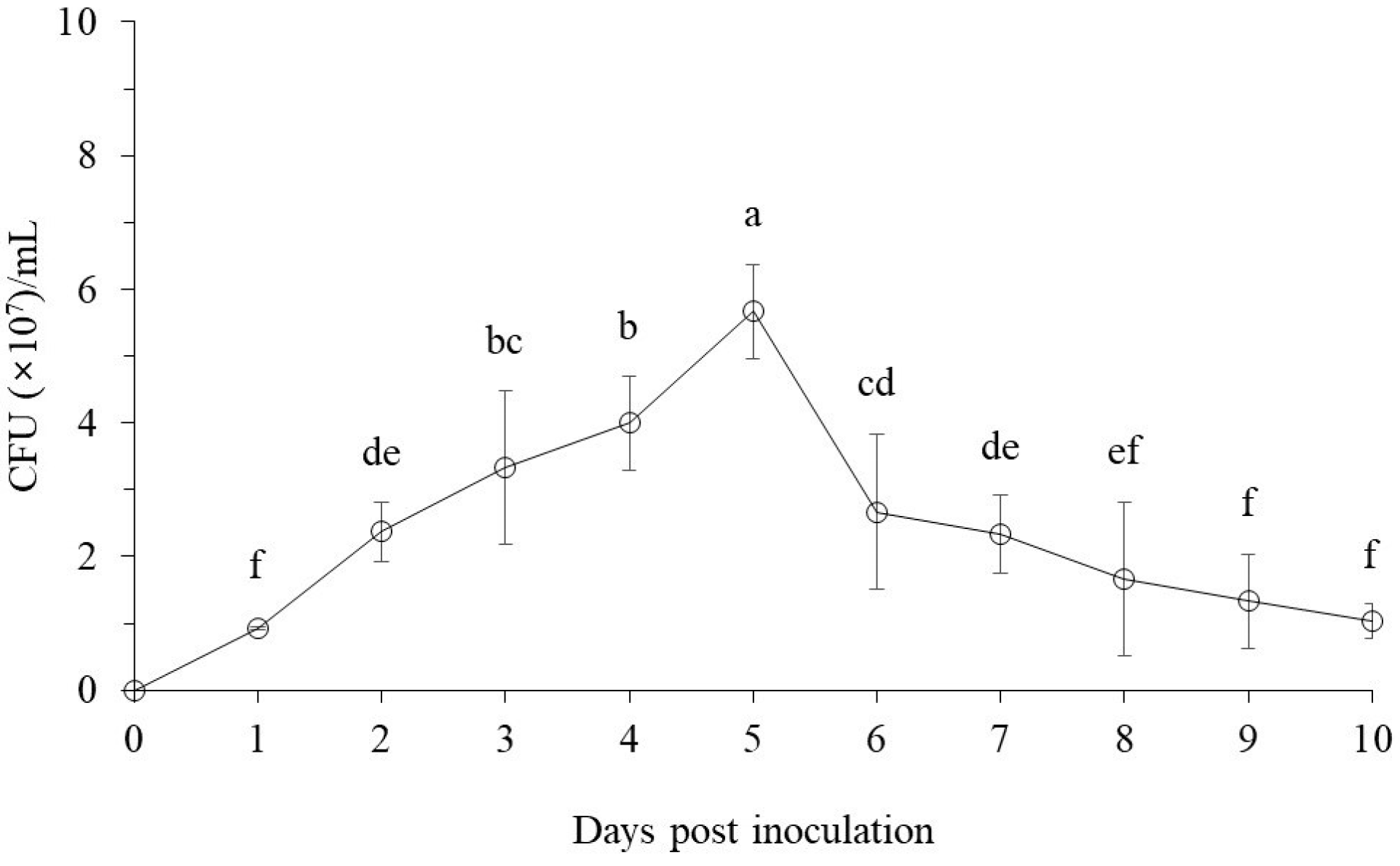
3.1.1. The Bacterial Cell Growth Pattern

The bacterial strain exhibited high cell growth in PB medium from 1 day to 10 days with over 107 CFU/mL units. The number of viable cells steadily increased for 5 days to peak at 5.7 × 10⁷ CFU/mL, and from then showed a sharp decline after 6 days to 2.8 × 10⁷ CFU/mL. After the peak level, the cell growth declined gradually to 1.3 × 10⁷ CFU/mL after 10 days (Figure 2).

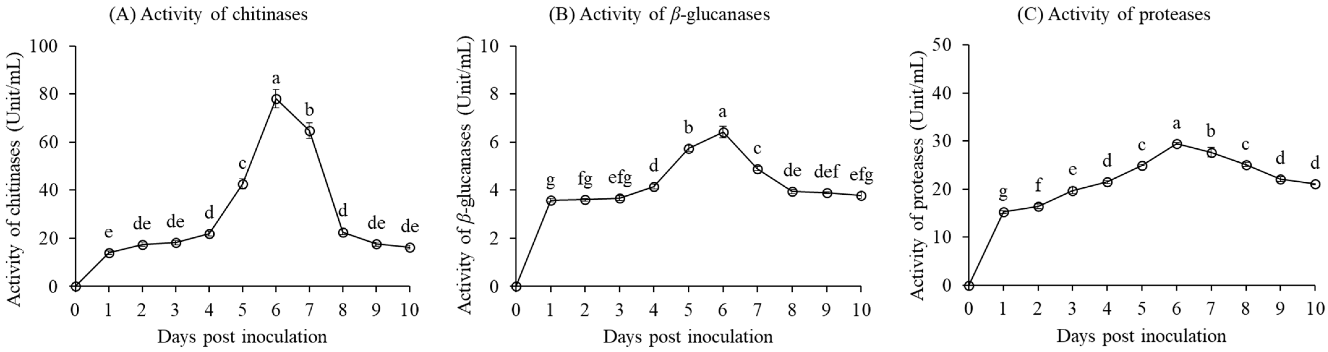
3.1.2. Lytic Enzyme Activity of the Bacterial Strain

The lytic activity of chitinases, β-glucanases, and proteases from the bacterial strain was relatively stable in the media and continued to increase after 5 days (Figure 3) when the cell growth was declining. Chitinase activity increased gradually from 14.1 units/mL after one day to 22.0 units/mL after 4 days of incubation (Figure 3A). Then, the activity of chitinases rapidly increased up to a peak activity of 78.2 units/mL after 6 days of incubation. After attaining the peak, the activity of chitinases rapidly declined to 22.6 units/mL after 8 days and then declined further at a gradual rate to 16.3 units/mL by the end of the experiment (after 10 days).
The activity of β-glucanases from the bacterial strain showed a rapid increase to 3.6 units/mL after 1 day of incubation and then increased very slowly to 3.7 units/mL after 3 days of incubation (Figure 3B). From 4 days to 6 days, the activity of β-glucanases gradually increased to a peak of 6.4 units/mL after 6 days of incubation. Then, the activity of β-glucanases gradually declined to 3.9 units/mL after 8 days and remained relatively stable until the end of the incubation period.
The activity of proteases produced by the bacterial strain showed a rapid increase to 15.2 units/mL after 1 day of incubation, followed by a gradual increase up to a peak of 29.4 units/mL after 6 days of incubation (Figure 3C). From 6 days to 10 days of incubation, the activity of proteases showed a gradual decline up to 21.0 units/mL on the final day of incubation.

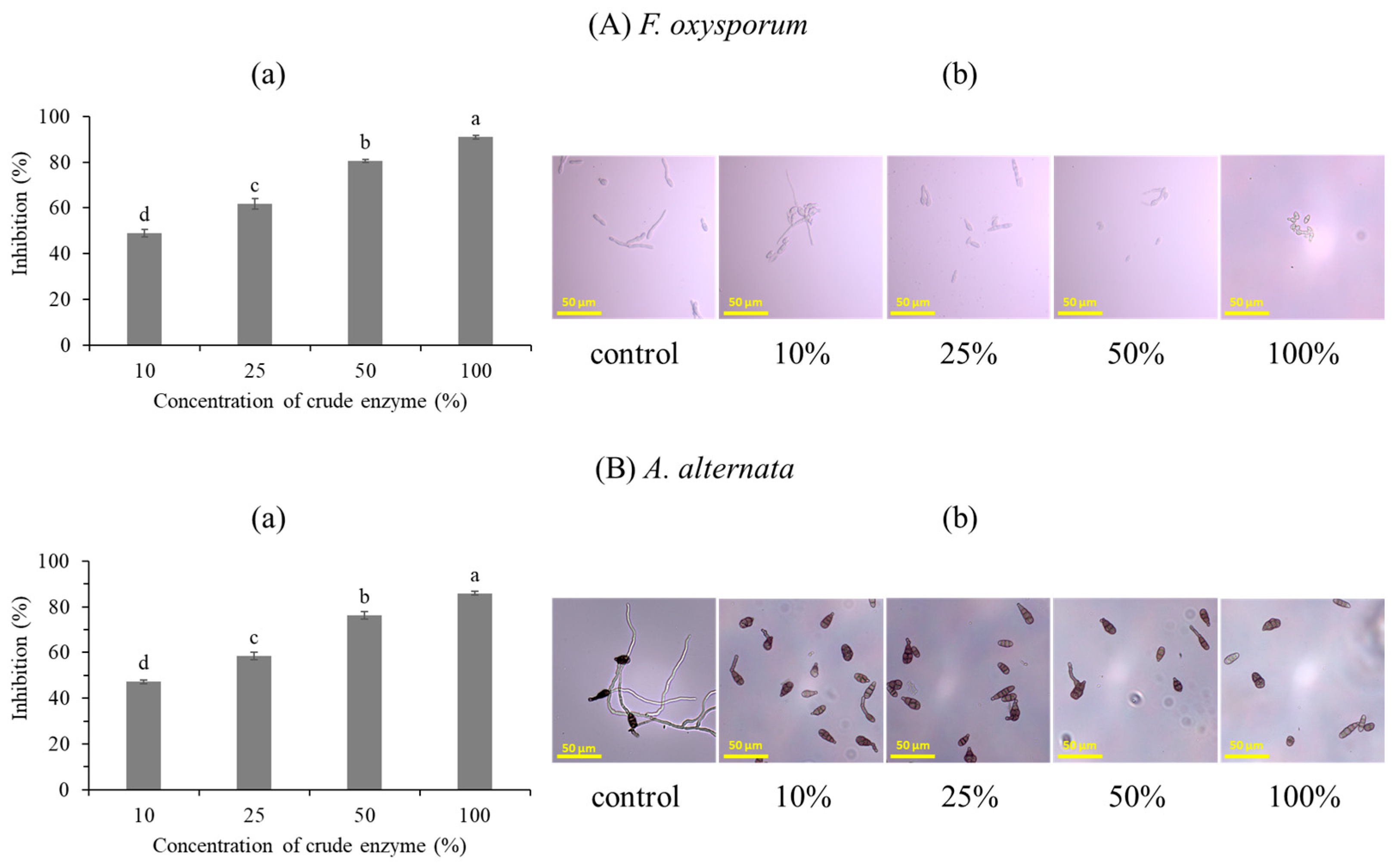
3.1.3. Antifungal Activity of the Bacterial Strain against F. oxysporum and A. alternata

The crude enzyme fraction of the bacterial strain inhibited spore germination of F. oxysporum and A. alternata at all treatment concentrations (100 µL/mL, 250 µL/mL, 500 µL/mL, and 1000 µL/mL) in a dose-dependent manner (Figure 4). The highest spore inhibition rate was recorded at a concentration of 1000 µL/mL, which corresponded to 91.0% and 85.9% against F. oxysporum and A. alternata, respectively. The lowest concentration of the crude enzyme fraction (100 µL/mL) inhibited spore germination by 49.0% and 47.2% against F. oxysporum and A. alternata, respectively. The microscopic images revealed that the crude enzyme fraction also suppressed germ tube elongation in germinated spores in a concentration-dependent manner against both phytopathogens.
The effect of the crude enzyme fraction based on paper disc assay also showed a concentration-dependent mycelial growth inhibition effect against phytopathogenic fungi (Figure 5). In the case of F. oxysporum, the highest inhibition rate was 58.3% at 1000 µL/mL, while only 40.3% inhibition was recorded at 100 µL/mL (Figure 5(Aa)). For A. alternata, the maximum inhibition rate was 54.3% at a concentration of 1000 µL/mL, while the lowest inhibition rate of 30.9% was recorded at a concentration of 100 µL/mL (Figure 5(Ba)).
In addition, the microscopic observations revealed that tall treatment levels caused severe morphological deformations in the hyphae of the two phytopathogenic fungi when relative to the control (Figure 5(Ab,Bb)). For F. oxysporum, high treatment concentration caused severe swelling of the hyphae with aggregation of cellular material, while the control group exhibited normal hyphae growth with an even distribution of cellular content. The same effect was exhibited with A. alternata, including the formation of dark bolus structures in the cells.

3.1.4. The Survival Rate of Coastal Pine Seedling in Greenhouse

The survival of coastal pine seedlings under greenhouse conditions showed a progressive decline from 100% in March to only 52.8% in September due to seedling mortality caused by phytopathogenic (F. oxysporum and A. alternata) infections in the control group (Table 1). The survival rate of coastal pine seedlings under the bacterial treatment group (the bacterial strain inoculation) remained uniform at 100.0% throughout the study period. There was no seedling mortality recorded under the bacterial treatment group, which increased the survival rate 1.9 times in relation to the control group (Table 1).

3.2. Effect of Bacterial Treatment on Coastal Pine Seedling Growth

3.2.1. The Production of Indole-3-Acetic Acid

The IAA phytohormone production rapidly increased to 2.7 mg/L on the first day of incubation and then continued to increase gradually up to a peak of 3.4 mg/L after 7 days of incubation. After attaining the peak, IAA concentration decreased slightly to 3.3 mg/L after 8 days and then remained relatively stable up to the end of the incubation period (Figure 6).

3.2.2. Effect of Treatment on the Nutrient Content in Coastal Pine Seedlings

The seedlings inoculated with the bacterial strain had significantly higher nutrient content relative to the control. For instance, inoculating seedlings with the bacterial culture increased the percentage of total N in coastal pine seedlings at least 2.7 times, from 0.5% in the control to 1.4% in the bacterial treatment during the experimental period. In addition, the bacterial treatment increased total P concentration 1.2 times from 0.13% in the control to 1.2% during the study period (Table 2).
The total N content (mg) in the seedlings treated with the bacterial culture broth was 43.55 mg, which is significantly higher than the control group with 0.65 mg. The total phosphorus content of the seedlings in the bacterial treatment group was 4.28 mg, which is also significantly higher than the control group with 0.15 mg (Table 2).

3.2.3. Effect Treatments on Coastal Pine Seedling Growth and Biomass Production

The average growth rate of the seedlings was notably higher in the bacterial treatment relative to the control based on all the measured parameters, including the girth (diameter at the root collar), the length of shoots and roots, and seedling dry weight (Table 3). Compared to the seedlings in the control group, the bacterial treatment improved the average stem girth 4.4 times, the shoot length 3.2 times, and the root length 1.4 times (Table 3).
The bacterial treatment also improved the biomass of coastal pine seedlings during the experimental period. The bacterial inoculation also increased the average shoot dry weight at least 31.6 times from 0.3 g in the control to 8.2 g in the bacterial treatment, and the root dry weight 11.1 times, from 0.2 g in the control to 2.2 g in the bacterial treatment (Table 3).

4. Discussion

4.1. Antifungal Effect of the Bacterial Strain against Phytopathogenic Fungi

This study demonstrates the biocontrol potential of B. velezensis CE 100 against fungal pathogens of economic importance in nursery seedling production under greenhouse conditions. The bacterium produced hydrolytic (cell wall-degrading) enzymes chitinases, β-glucanases, and proteases, with a peak production observed after 6 days of incubation (Figure 3). Since the cell growth of the bacterial strain peaked after 5 days, the high activity of hydrolytic enzymes beyond the active bacterial growth phase indicates the stability of the enzymes in the bacterial culture and potential for apoptosis, which correlates with the increase in the bioactivity [57]. The prolific production of cell wall degrading enzymes is an effective biocontrol strategy of PGPB against various plant phytopathogenic fungi [26,27,28]. In this study, different treatment levels of the crude enzyme fraction from the bacterial strain demonstrated effective inhibition of spore germination against F. oxysporum and A. alternata to a maximum rate of 91.0% and 85.9%, respectively, at 1000 µL/mL (Figure 4). The results of the microscopic examination revealed that the rate of spore germination inhibition and germ tube elongation increased with the increase in crude enzyme concentration against both phytopathogenic fungi (Figure 4). In addition, the crude enzyme fraction inhibited the rate of mycelium growth of F. oxysporum and A. alternata in a dose-dependent manner up to 58.3% and 40.3%, respectively, at 1000 µL/mL (Figure 5). The microscopic examination revealed that the severity of hyphal deformation increased with the increase in the concentration of the crude enzyme fraction. The crude enzyme treatments caused hyphal abnormalities, such as twisted and swollen hyphae with bolus structures that hindered normal mycelial growth (Figure 5). The inhibition effect of fungal spores and mycelial growth is based on cell wall degradation by the hydrolytic enzyme activity, such as chitinase, β-1,3 glucanase, and protease, which specifically target and hydrolyze the vital components of the fungal cell wall, such as chitin polymers, β-1,3 glucans, and glycoproteins protease [26,27,28].
The fungal cell wall is a vital structure for protecting and shielding the cellular contents against environmental pressure, provides the mechanical strength required to support fungal structure (shapes), and allows selective permeability for the in-flow of nutrients and release of membrane vesicles [58,59]. The phytopathogenic fungal cell wall is particularly a vital structure that provides the necessary force to facilitate turgor-driven penetration of plant cells during the infection phase, mainly by modifying the cell structures such as appressoria [60]. To support these vital functions, the fungal cell walls are generally composed of modifications of branched β-1,3-glucans linked to chitin polymers to form a central core structure with flexible, viscus, and elastic properties that are essential for mechanical strength (cell form/shape) and physiological functions of the fungal cell. However, the cell wall is also a suitable target for antifungal products, including chitinase and β-glucanases [26,27,28,58,61]. The hydrolysis of these vital structures results in cell deformation/ loss of shape and loss of turgor pressure, which is required for plant cell penetration and infection [26,27,28]. The outer layer of the fungal cell wall is largely composed of glycoprotein polymer which plays a vital role in preventing the entry of foreign material/toxins due to its hydrophobic properties. These glycoproteins are essential for environmental signaling and have adhesive properties that facilitate the attachment of fungal spores on the host surface to enable spore germination and infection [58,59,60]. The degradation of glycoprotein components by antifungal compounds such as proteases could potentially cause cell permeation, increase the osmotic pressure, and disrupt environmental signaling, which reduces the ecological fitness of the phytopathogenic, including loss of virulence [26,27,28,58,59,60]. Fungal spores are vital for the dispersion/spread of phytopathogenic fungi, including F. oxysporum and A. alternata, and the subsequent spreading of fungal diseases in forest nurseries and fields via irrigation water, rain splashes, and wind [54,55,56].
The degradation of phytopathogenic fungal cell walls and the subsequent deformation of fungal spores and hyphae renders the fungi less virulent and limits the spread of diseases. Thus, the inoculation with the bacterial strain in coastal pine nurseries notably increased the survival rate of seedlings relative to the control. Whereas the survival of seedlings under the control group dropped from 100.0% in March to only 52.8% in September, the survival rate in the bacterial treatment group remained at 100.0% throughout the study period (Table 1). The bacterial treatment improved the survival rate of coastal pine seedlings at least 1.9 times compared to the control. The improved survival rate of seedlings could be attributed to the deformation of the phytopathogenic fungal cells, which causes the suppression of spore germination and reduces the rate of mycelial growth. The reduced rate of spore and mycelial lowers the infection rate and thus improves the rate of seedling survival. These findings are consistent with other studies that demonstrated the effective role of biocontrol agents in suppressing fungal diseases in plants, including forest seedlings, via hydrolytic enzyme activity [28,40,61]. For instance, Paramanandham et al. (2017) demonstrated the effective inhibition of F. oxysporum and A. solani phytopathogens in Solanum lycopersicum by hydrolytic enzymes from other PGPB including Pseudomonas sp. [62]. Moon et al. [40] also demonstrated effective control of phytopathogenic infections in Chamaecyparis obtusa seedlings via the hydrolytic activity of β-1,3-glucanases and proteases from Bacillus velezensis CE 100. Similar findings were confirmed by Won et al. [28] demonstrate similar results against Pestalotiopsis maculans in Quercus acutissima seedlings.

4.2. Seedling Growth Promoting Effect of the Bacterial Strain

Besides the antifungal effect of biocontrol strain against phytopathogenic infections, the bacterium also demonstrated plant growth-promoting potential, which could be a result of IAA production [38,39,40]. IAA is an important phytohormone that stimulates root hair initiation and root development [38,39,40]. In this study, the bacterial strain produced IAA auxin up to 3.4 mg/L during the incubation period (Figure 6). The mechanism by which IAA promotes plant growth includes the enhancement of cell division and elongation in both the root and shoot systems, depending on plant-environmental signaling pathways, which especially results in an improved root architecture to maximize nutrient uptake [28,38,39,40]. The increase in IAA auxins in the rhizosphere stimulates root hair formation and lateral root morphogenesis in the region of root differentiation [38,39]. IAA also enhances the loosening of the cell wall and cell membrane along with the activation of the plasma membrane protonmotive force (H+ -ATPase proton pumps and a pH gradient), leading to osmotic water permeability [38,39,63]. This osmotic water intake builds the turgor pressure that facilitates plant cell expansion in both the root system to improve nutrient absorption and the elongation of the shoot system, which is then followed by cell differentiations and biomass depositions that leads to plant growth and development [38,39,63].
Besides the improved root architectural development due to IAA production, B. velezensis CE 100 has been previously demonstrated to enhance the availability of essential plant nutrients via ammonia-N production, N-fixation, and the solubilization of insoluble phosphates in the soil [26]. The improvement in root architectural development and nutrient availability in the rhizosphere consequently results in higher nutrient uptake by the seedlings under the bacterial treatment [28,44]. Thus, the higher nutrient content which was observed in coastal pine seedlings from the bacterial treatment compared to the control group (Table 2) could be a result of the enhanced root architectural development (Figure 1C) due to IAA and the improvement in nutrient availability [26,28,44]. The improvement in N and P content in the seedlings, along with the direct effect of IAA in the leaves, ultimately increases the photosynthetic rate and biomass production [28,44,64]. Hence, the treatment of coastal pine seedlings with the bacterial strain remarkedly improved seedling growth as indicated by an increase in stem girth, average shoot height, and average root length at least 4.4, 3.1, and 1.4 times relative to the control (Table 3). The bacterial treatment also improved biomass production in coastal pine seedlings, as indicated by an increase in the shoot dry weight (31.6 times) and the root dry weight (11.1 times) in relation to the seedlings in the control group. Won et al. (2021) also reported the production of IAAB and the improvement in nutrient uptake in oak seedlings by PGPB, which led to a higher photosynthetic rate and the subsequent enhancement in seedling biomass by more than 2 times in relation to the control [28]. Therefore, we demonstrated that the inoculation of PGPB can effectively promote the growth of coastal pine seedlings, which could potentially eliminate the need for applying chemical fertilizers that cause environmental pollution.

5. Conclusions

The hydrolytic enzymes chitinases, β-glucanases, and protease from the bacterial strain B. velezensis CE 100 have been demonstrated to effectively suppress the germination of phytopathogenic fungal spores and reduce the mycelial growth rate of F. oxysporum and A. alternata. These phytopathogens cause Fusarium wilt and leaf blight diseases in coastal pine seedlings in forest nurseries. Consequently, treatment of coastal pine seedlings with the bacterial culture notably increased the survival rate of coastal pine seedlings relative to the control during the experiment period. Moreover, the study demonstrated effective seedling growth promotion via the production of IAA. The seedlings treated with the bacterial strain showed a remarkedly higher rate of growth, as demonstrated by the improved root and shoot length and biomass production of the seedlings compared to those in the control group. Thus, the inoculation of this biocontrol strain can effectively serve as an alternative to synthetic fungicides and fertilizers to protect coastal pine seedlings from fungal infection (to enhance the survival rate) while simultaneously improving seedling growth to facilitate the production of high-quality seedlings for the restoration and establishment of urban forest ecosystems in coastal areas. Future studies should evaluate the field application of the bacterium in the management of fungal diseases and growth promotion of coastal pine trees.

Author Contributions

Conceptualization, P.S.-H.L. and Y.S.A.; investigation and methodology, J.-Y.Y., H.-S.K., J.-H.M., S.-J.W., V.C. and S.-I.C.; validation, P.S.-H.L. and Y.S.A.; formal analysis, J.-Y.Y., H.-S.K., J.-H.M. and H.B.A.; resources, P.S.-H.L. and Y.S.A.; writing—preparing the original draft, J.-Y.Y., H.-S.K., J.-H.M., H.B.A., P.S.-H.L. and Y.S.A.; writing—manuscript editing and review, H.B.A., P.S.-H.L. and Y.S.A.; supervision, administration of the project, and acquisition of funds, Y.S.A. All authors have read and agreed to the published version of the manuscript.

Funding

The study was supported by the “Regional Innovation Strategy (RIS)” via the NRF (National Research Foundation), which is funded by the Ministry of Education (MOE) of Korea (grant number 2021RIS-002).

Data Availability Statement

All data relevant to his manuscript can be obtained upon request from the corresponding authors.

Conflicts of Interest

The authors hereby declare that there is no conflict of interest regarding this article. The research funders did not play any role in deciding the study design, in the collection, analyses, and the interpretation of data, neither in the writing of the manuscript nor in the decision to publish the results.

References

  1. Lopes, A.I. Fighting drift sands with pine trees: Reforestation of coastal areas of NW Portugal at the end of eighteenth century. J. Coast. Conserv. 2023, 27, 42. [Google Scholar] [CrossRef]
  2. Zhu, J.; Gonda, Y.; Yu, L.; Li, F.; Yan, Q.; Sun, Y. Regeneration of a coastal pine (Pinus thunbergii Parl.) forest 11 years after thinning, Niigata, Japan. PLoS ONE 2012, 7, e47593. [Google Scholar] [CrossRef]
  3. Fujihara, M.; Ohnishi, M.; Miura, H.; Sawada, Y. Conservation and Management of the Coastal Pine Forest as a Cultural Landscape. In Landscape Ecology in Asian Cultures. Ecological Research Monographs; Hong, S., Kim, J.E., Wu, J., Nakagoshi, N., Eds.; Springer: Tokyo, Japan, 2011; pp. 235–248. [Google Scholar] [CrossRef]
  4. Zhu, J.; Matsuzaki, T.; Gonda, Y. Wind profiles in a coastal forest of Japanese black pine (Pinus thunbergii Parl.) with different thinning intensities. J. For. Res. 2001, 6, 287–296. [Google Scholar] [CrossRef]
  5. Sarmati, S.; Bonari, G.; Angiolini, C. Conservation status of Mediterranean coastal dune habitats: Anthropogenic disturbance may hamper habitat assignment. Rend. Lincei Sci. Fis. 2019, 30, 623–636. [Google Scholar] [CrossRef]
  6. Drius, M.; Carranza, M.L.; Stanisci, A.; Jones, L. The role of Italian coastal dunes as carbon sinks and diversity sources. A multi-service perspective. Appl. Geogr. 2016, 75, 127–136. [Google Scholar] [CrossRef]
  7. Rodrigues-Honda, K.C.D.S.; Junkes, C.F.D.O.; Lima, J.C.D.; Waldow, V.D.A.; Rocha, F.S.; Sausen, T.L.; Bayer, C.; Talamini, E.; Fett-Neto, A.G. Carbon sequestration in resin-tapped slash pine (Pinus elliottii Engelm.) subtropical plantations. Biology 2023, 12, 324. [Google Scholar] [CrossRef]
  8. Yoo, S.Y.; Kim, T.; Ham, S.; Choi, S.; Park, C.R. Importance of urban green at reduction of particulate matters in Sihwa Industrial Complex, Korea. Sustainability 2020, 12, 7647. [Google Scholar] [CrossRef]
  9. Shin, H.S.; Lee, S.C.; Choi, S.H.; Kang, H.M. Ecological characteristic and vegetation structure of Pinus thunbergii community in coastal forest of Busan metropolitan city, Korea. Korean J. Environ. Ecol. 2019, 33, 539–551. [Google Scholar] [CrossRef]
  10. Hocking, D.; Setliff, E.; Jaffer, A. Potential pathogenicities of fungi associated with damped-off pine seedlings in East African pine nurseries. Trans. Br. Mycol. Soc. 1968, 51, 227–232. [Google Scholar] [CrossRef]
  11. Zhang, M.J.; Zheng, X.R.; Li, H.; Chen, F.M. Alternaria alternata, the causal agent of a new needle blight disease on Pinus bungeana. J. Fungi 2023, 9, 71. [Google Scholar] [CrossRef] [PubMed]
  12. Luo, X.; Yu, C. First report of damping-off disease caused by Fusarium oxysporum in Pinus massoniana in China. J. Plant Dis Prot. 2020, 127, 401–409. [Google Scholar] [CrossRef]
  13. Dar, G.H.; Beig, M.A.; Ahanger, F.A.; Ganai, N.A.; Ahangar, M.A. Management of root rot caused by Rhizoctonia solani and Fusarium oxysporum in blue pine (Pinus wallichiana) through use of fungal antagonists. Asian J. Plant Pathol. 2011, 5, 62–67. [Google Scholar] [CrossRef]
  14. Stewart, J.E.; Kim, M.S.; James, R.L.; Dumroese, R.K.; Klopfenstein, N.B. Molecular characterization of Fusarium oxysporum and Fusarium commune isolates from a conifer nursery. Phytopathology 2006, 96, 1124–1133. [Google Scholar] [CrossRef]
  15. Hrunyk, N.; Gout, R.; Kovaleva, V. Regulation of gene expression for defensins and lipid transfer protein in Scots pine seedlings by necrotrophic pathogen Alternaria alternata (Fr.). Folia For. Pol. A. 2017, 59, 152–158. [Google Scholar] [CrossRef]
  16. Yanfei, H.; Hancheng, W.; Qingyuan, C.; Jin, W.; Changqing, Z.; Hongxue, L. Inhibitory activities of six fungicides against mycelial growth and conidial germination of Alternaria alternata. Chin. J. Pestic. Sci. 2016, 18, 263–267. [Google Scholar] [CrossRef]
  17. Avenot, H.; Morgan, D.; Michailides, T. Resistance to pyraclostrobin, boscalid and multiple resistance to Pristine® (pyraclostrobin+boscalid) fungicide in Alternaria alternata causing Alternaria late blight of pistachios in California. Plant Pathol. 2008, 57, 135–140. [Google Scholar] [CrossRef]
  18. He, M.H.; Wang, Y.P.; Wu, E.J.; Shen, L.L.; Yang, L.N.; Wang, T.; Shang, L.P.; Zhu, W.; Zhan, J. Constraining evolution of Alternaria alternata resistance to a demethylation inhibitor (DMI) fungicide difenoconazole. Front. Microbiol. 2019, 10, 1609. [Google Scholar] [CrossRef]
  19. Rajput, A.Q.; Arain, M.H.; Pathan, M.A.; Jiskani, M.M.; Lodhi, A.M. Efficacy of different fungicides against Fusarium wilt of cotton caused by Fusarium oxysporum f. sp. vasinfectum. Pak. J. Bot. 2006, 38, 875. [Google Scholar]
  20. Bozdogan, A.M. Assessment of total risk on non-target organisms in fungicide application for agricultural sustainability. Sustainability 2014, 6, 1046–1058. [Google Scholar] [CrossRef]
  21. Pirozzi, A.V.A.; Stellavato, A.; La Gatta, A.; Lamberti, M.; Schiraldi, C. Mancozeb, a fungicide routinely used in agriculture, worsens nonalcoholic fatty liver disease in the human HepG2 cell model. Toxicol. Lett. 2016, 249, 1–4. [Google Scholar] [CrossRef]
  22. Sharma, M.; Maheshwari, N.; Khan, F.H.; Mahmood, R. Carbendazim toxicity in different cell lines and mammalian tissues. J. Biochem. Mol. Toxicol. 2022, 36, e23194. [Google Scholar] [CrossRef]
  23. Yang, L.N.; He, M.H.; Ouyang, H.B.; Zhu, W.; Pan, Z.C.; Sui, Q.J.; Shang, L.P.; Zhan, J. Cross-resistance of the pathogenic fungus Alternaria alternata to fungicides with different modes of action. BMC Microbiol. 2019, 19, 205. [Google Scholar] [CrossRef]
  24. Chung, W.H.; Chung, W.C.; Ting, P.F.; Ru, C.C.; Huang, H.C.; Huang, J.W. Nature of resistance to methyl benzimidazole carbamate fungicides in Fusarium oxysporum f. sp. lilii and F. oxysporum f. sp. gladioli in Taiwan. J. Phytopathol. 2009, 157, 742–747. [Google Scholar] [CrossRef]
  25. González-Oviedo, N.A.; Iglesias-Andreu, L.G.; Flores-de la Rosa, F.R.; Rivera-Fernández, A.; Luna-Rodríguez, M. Genetic analysis of the fungicide resistance in Fusarium oxysporum associated to Vanilla planifolia. Rev. Mex. fitopatol. 2022, 40, 330–348. [Google Scholar] [CrossRef]
  26. Choub, V.; Ajuna, H.B.; Won, S.J.; Moon, J.H.; Choi, S.I.; Maung, C.E.H.; Kim, C.W.; Ahn, Y.S. Antifungal activity of Bacillus velezensis CE 100 against anthracnose disease (Colletotrichum gloeosporioides) and growth promotion of walnut (Juglans regia L.) trees. Int. J. Mol. Sci. 2021, 22, 10438. [Google Scholar] [CrossRef]
  27. Hong, S.; Kim, T.Y.; Won, S.-J.; Moon, J.-H.; Ajuna, H.B.; Kim, K.Y.; Ahn, Y.S. Control of fungal diseases and fruit yield improvement of strawberry using Bacillus velezensis CE 100. Microorganisms 2022, 10, 365. [Google Scholar] [CrossRef]
  28. Won, S.-J.; Moon, J.-H.; Ajuna, H.B.; Choi, S.-I.; Maung, C.E.H.; Lee, S.; Ahn, Y.S. Biological control of leaf blight disease caused by Pestalotiopsis maculans and growth promotion of Quercus acutissima Carruth container seedlings using Bacillus velezensis CE 100. Int. J. Mol. Sci. 2021, 22, 11296. [Google Scholar] [CrossRef]
  29. Ahmed, M.; El-Fiki, I. Effect of biological control of root rot diseases of strawberry using Trichoderma spp. Middle East J. Appl. Sci. 2017, 7, 482–492. [Google Scholar]
  30. Jochum, C.; Osborne, L.; Yuen, G. Fusarium head blight biological control with Lysobacter enzymogenes strain C3. Biol. Control 2006, 39, 336–344. [Google Scholar] [CrossRef]
  31. Yu, C.; Luo, X. Trichoderma koningiopsis controls Fusarium oxysporum causing damping-off in Pinus massoniana seedlings by regulating active oxygen metabolism, osmotic potential, and the rhizosphere microbiome. Biol. Control 2020, 150, 104352. [Google Scholar] [CrossRef]
  32. Balderas-Ruíz, K.A.; Bustos, P.; Santamaria, R.I.; González, V.; Cristiano-Fajardo, S.A.; Barrera-Ortíz, S.; Mezo-Villalobos, M.; Aranda-Ocampo, S.; Guevara-García, Á.A.; Galindo, E.; et al. Bacillus velezensis 83 a bacterial strain from mango phyllosphere, useful for biological control and plant growth promotion. AMB Express 2020, 10, 163. [Google Scholar] [CrossRef]
  33. Bouchard-Rochette, M.; Machrafi, Y.; Cossus, L.; Nguyen, T.T.A.; Antoun, H.; Droit, A.; Tweddell, R.J. Bacillus pumilus PTB180 and Bacillus subtilis PTB185: Production of lipopeptides, antifungal activity, and biocontrol ability against Botrytis cinerea. Biol. Control 2022, 170, 104925. [Google Scholar] [CrossRef]
  34. Choi, S.H.; Ahn, J.B.; Kozukue, N.; Levin, C.E.; Friedman, M. Distribution of free amino acids, flavonoids, total phenolics, and antioxidative activities of jujube (Ziziphus jujuba) fruits and seeds harvested from plants grown in Korea. J. Agric. Food Chem. 2011, 59, 6594–6604. [Google Scholar] [CrossRef]
  35. Plaza, V.; Silva-Moreno, E.; Castillo, L. Breakpoint: Cell wall and glycoproteins and their crucial role in the phytopathogenic fungi infection. Curr. Protein Pept. Sci. 2020, 21, 227–244. [Google Scholar] [CrossRef]
  36. Oliveira-Garcia, E.; Deising, H.B. Infection structure–specific expression of β-1, 3-glucan synthase is essential for pathogenicity of Colletotrichum graminicola and evasion of β-glucan–triggered immunity in maize. Plant Cell 2013, 25, 2356–2378. [Google Scholar] [CrossRef]
  37. Budi, S.W.; Van Tuinen, D.; Arnould, C.; Dumas-Gaudot, E.; Gianinazzi-Pearson, V.; Gianinazzi, S. Hydrolytic enzyme activity of Paenibacillus sp. strain B2 and effects of the antagonistic bacterium on cell integrity of two soil-borne pathogenic fungi. Appl. Soil Ecol. 2000, 15, 191–199. [Google Scholar] [CrossRef]
  38. Enders, T.A.; Strader, L.C. Auxin activity: Past, present, and future. Am. J. Bot. 2015, 102, 180–196. [Google Scholar] [CrossRef] [PubMed]
  39. Majda, M.; Robert, S. The role of auxin in cell wall expansion. Int. J. Mol. Sci. 2018, 19, 951. [Google Scholar] [CrossRef] [PubMed]
  40. Moon, J.-H.; Won, S.-J.; Maung, C.E.H.; Choi, J.-H.; Choi, S.-I.; Ajuna, H.B.; Ahn, Y.S. Bacillus velezensis CE 100 Inhibits root rot diseases (Phytophthora spp.) and promotes growth of Japanese cypress (Chamaecyparis obtusa Endlicher) seedlings. Microorganisms 2021, 9, 821. [Google Scholar] [CrossRef] [PubMed]
  41. Yu, X.; Liu, X.; Zhu, T.H.; Liu, G.H.; Mao, C. Isolation and characterization of phosphate-solubilizing bacteria from walnut and their effect on growth and phosphorus mobilization. Biol. Fertil. Soils 2011, 47, 437–446. [Google Scholar] [CrossRef]
  42. Richardson, A.E.; Barea, J.M.; McNeill, A.M.; Prigent-Combaret, C. Acquisition of phosphorus and nitrogen in the rhizosphere and plant growth promotion by microorganisms. Plant Soil 2009, 321, 305–339. [Google Scholar] [CrossRef]
  43. Wang, J.; Wen, X.; Zhang, X.; Li, S.; Zhang, D.Y. Co-regulation of photosynthetic capacity by nitrogen, phosphorus and magnesium in a subtropical Karst forest in China. Sci. Rep. 2018, 8, 7406. [Google Scholar] [CrossRef]
  44. Park, H.G.; Lee, Y.S.; Kim, K.Y.; Park, Y.S.; Park, K.H.; Han, H.O.; Park, C.M.; Ahn, Y.S. Inoculation with Bacillus licheniformis MH48 promotes nutrient uptake in seedlings of the ornamental plant Camellia japonica grown in Korean reclaimed coastal lands. Hortic. Sci. Technol. 2017, 35, 11–20. [Google Scholar] [CrossRef]
  45. Kuan, K.B.; Othman, R.; Abdul Rahim, K.; Shamsuddin, Z.H. Plant growth-promoting rhizobacteria inoculation to enhance vegetative growth, nitrogen fixation and nitrogen remobilisation of maize under greenhouse conditions. PLoS ONE 2016, 11, e0152478. [Google Scholar] [CrossRef]
  46. Davis, A.S.; Jacobs, D.F. Quantifying root system quality of nursery seedlings and relationship to outplanting performance. New For. 2005, 30, 295–311. [Google Scholar] [CrossRef]
  47. Choi, T.G.; Maung, C.E.H.; Lee, D.R.; Henry, A.B.; Lee, Y.S.; Kim, K.Y. Role of bacterial antagonists of fungal pathogens, Bacillus thuringiensis KYC and Bacillus velezensis CE 100 in control of root-knot nematode, Meloidogyne incognita and subsequent growth promotion of tomato. Biocontrol. Sci. Technol. 2020, 30, 685–700. [Google Scholar] [CrossRef]
  48. Ajuna, H.B.; Kim, I.; Han, Y.S.; Maung, C.E.H.; Kim, K.Y. Aphicidal activity of Bacillus thuringiensis strain AH-2 against cotton aphid (Aphis gossypii). Entomol. Res. 2021, 51, 151–160. [Google Scholar] [CrossRef]
  49. Lingappa, Y. Chitin media for selective isolation and culture of actinomycetes. Phytopathology 1962, 52, 317–323. [Google Scholar]
  50. Liang, Z.C.; Hseu, R.S.; Wang, H.H. Partial purification and characterization of a 1, 3-β-D-glucanase from Ganoderma tsugae. J. Ind. Microbiol. Biotechnol. 1995, 14, 5–9. [Google Scholar] [CrossRef]
  51. Ghorbel-Frikha, B.; Sellami-Kamoun, A.; Fakhfakh, N.; Haddar, A.; Manni, L.; Nasri, M. Production and purification of a calcium-dependent protease from Bacillus cereus BG1. J. Ind. Microbiol. Biotechnol. 2005, 32, 186–194. [Google Scholar] [CrossRef] [PubMed]
  52. Moon, J.-H.; Won, S.-J.; Maung, C.E.H.; Choi, J.-H.; Choi, S.-I.; Ajuna, H.B.; Ahn, Y.S.; Jo, Y.H. The Role of Lysobacter antibioticus HS124 on the control of fall webworm (Hyphantria cunea Drury) and growth promotion of Canadian poplar (Populus canadensis Moench) at Saemangeum reclaimed land in Korea. Microorganisms 2021, 9, 1580. [Google Scholar] [CrossRef] [PubMed]
  53. Rahman, A.; Sitepu, I.R.; Tang, S.Y.; Hashidoko, Y. Salkowski’s reagent test as a primary screening index for functionalities of rhizobacteria isolated from wild dipterocarp saplings growing naturally on medium-strongly acidic tropical peat soil. Biosci. Biotechnol. Biochem. 2010, 74, 2202–2208. [Google Scholar] [CrossRef] [PubMed]
  54. Widyastuti, S.M.; Tasik, S.; Harjono, H. Infection process of Fusarium oxysporum fungus: A cause of damping-off on Acacia mangium seedlings. Agrivita, J. Agric. Sci. 2013, 35, 110–118. [Google Scholar] [CrossRef]
  55. Hong, C.; Moorman, G. Plant pathogens in irrigation water: Challenges and opportunities. CRC Crit. Rev. Plant. Sci. 2005, 24, 189–208. [Google Scholar] [CrossRef]
  56. Bashan, Y.; Levanony, H.; Or, R. Wind dispersal of Alternaria alternata, a cause of leaf blight of cotton. J. Phytopathol. 1991, 133, 225–238. [Google Scholar] [CrossRef]
  57. Rice, K.C.; Bayles, K.W. Molecular control of bacterial death and lysis. Microbiol. Mol. Biol. 2008, 72, 85–109. [Google Scholar] [CrossRef]
  58. Gow, N.A.; Latge, J.P.; Munro, C.A. The fungal cell wall: Structure, biosynthesis, and function. Microbiol. Spectr. 2017, 5. [Google Scholar] [CrossRef]
  59. Beauvais, A.; Latgé, J.-P. Fungal cell wall. J. Fungi 2018, 4, 91. [Google Scholar] [CrossRef]
  60. Geoghegan, I.; Steinberg, G.; Gurr, S. The role of the fungal cell wall in the infection of plants. Trends Microbiol. 2017, 25, 957–967. [Google Scholar] [CrossRef]
  61. Latgé, J.P. The cell wall: A carbohydrate armour for the fungal cell. Mol. Microbiol. 2007, 66, 279–290. [Google Scholar] [CrossRef]
  62. Paramanandham, P.; Rajkumari, J.; Pattnaik, S.; Busi, S. Biocontrol potential against Fusarium oxysporum f. sp. lycopersici and Alternaria solani and tomato plant growth due to plant growth–promoting rhizobacteria. Int. J. Veg. Sci. 2017, 23, 294–303. [Google Scholar] [CrossRef]
  63. Haruta, M.; Gray, W.M.; Sussman, M.R. Regulation of the plasma membrane proton pump (H+-ATPase) by phosphorylation. Curr. Opin. Plant Biol. 2015, 28, 68–75. [Google Scholar] [CrossRef]
  64. Mir, A.R.; Siddiqui, H.; Alam, P.; Hayat, S. Foliar spray of Auxin/IAA modulates photosynthesis, elemental composition, ROS localization and antioxidant machinery to promote growth of Brassica juncea. Physiol. Mol. Biol. Plants. 2020, 26, 2503–2520. [Google Scholar] [CrossRef]
Figure 1. Experimental set-up in the nursery showing seedlings growth under control and treatment groups (A), symptoms of infections in the control group (B) and the healthy seedlings in Bacillus velezensis CE 100 treatment (C).
Forests 15 00062 g001
Figure 2. The cell growth pattern of bacterial strain over the 10-day period of incubation. Means with different superscript letters are significantly different (p < 0.01).
Forests 15 00062 g002
Figure 3. Lytic activity produced by the bacterial strain showing the concentration of Chitinases (A), β-glucanases (B), and proteases (C) over a 10-day period. Means with the same letter have statistically similar values (p < 0.01).
Forests 15 00062 g003
Figure 4. Spore germination inhibition rate (a) and light microscope image of spore germination inhibition effect (b) at different concentrations of the crude enzyme fraction against Fusarium oxysporum (A) and Alternaria alternata (B). Means with the same letter have statistically similar values (p < 0.01).
Forests 15 00062 g004
Figure 5. Mycelial growth inhibition rate (Aa), pictures of fungal colonies in the inhibition test (Ab), and light microscope image of mycelial growth inhibition effect (B) after treatment with different concentrations of the crude enzyme fraction against F. oxysporum (A) and A. alternata (B). Means with the same letter have statistically similar values (p < 0.01).
Forests 15 00062 g005
Figure 6. The concentration of IAA in the L-tryptophan enriched media secreted by the bacterial strain. Means with the same letter have statistically similar values (p < 0.01).
Forests 15 00062 g006
Table 1. The survival rate of coastal pine (Pinus thunbergii Parl.) seedlings during the experimental period.
TreatmentSurvival Rate (%)
MarchAprilMayJuneJulyAugustSeptember
Control100.0 ± 0.085.0 ± 5.6 *78.3 ± 5.8 *66.7 ± 7.6 *61.7 ± 7.6 *56.7 ± 3.5 *52.8 ± 1.8 *
Bacterial inoculation100.0 ± 0.0100.0 ± 0.0 *100.0 ± 0.0 *100.0 ± 0.0 *100.0 ± 0.0 *100.0 ± 0.0 *100.0 ± 0.0 *
An asterisk * indicates significantly different values between the treatments based on a t-test analysis (p < 0.01).
Table 2. Effect of bacterial treatment on the total nitrogen (N) and phosphorus (P) concentration and content of coastal pine (Pinus thunbergii Parl.) seedlings.
TreatmentConcentration (%)Content (mg)
Total NTotal PTotal NTotal P
Control0.54 ± 0.01 *0.13 ± 0.01 *0.65 ± 0.01 *0.15 ± 0.01 *
Bacterial inoculation1.43 ± 0.04 *0.15 ± 0.01 *43.55 ± 1.11 *4.28 ± 0.07 *
An asterisk * indicates significantly different values between the treatments based on a t-test analysis (p < 0.01).
Table 3. Growth and dry weight of coastal pine (Pinus thunbergii Parl.) seedlings by treatment group.
TreatmentStem Girth (mm)Length (cm)Dry Weight (cm)
Shoot Root Shoot Root
Control1.35 ± 0.49 *8.81 ± 2.54 *13.28 ± 7.56 *0.26 ± 0.21 *0.20 ± 0.15 *
Bacterial inoculation5.99 ± 1.06 *27.71 ± 5.23 *18.07 ± 4.55 *8.21 ± 2.91 *2.21 ± 0.60 *
An asterisk * indicates significantly different values between the treatments based on a t-test analysis (p < 0.01).
Disclaimer/Publisher’s Note: The statements, opinions and data contained in all publications are solely those of the individual author(s) and contributor(s) and not of MDPI and/or the editor(s). MDPI and/or the editor(s) disclaim responsibility for any injury to people or property resulting from any ideas, methods, instructions or products referred to in the content.

