Article

Exploring Functional Gene XsPDAT1’s Involvement in Xanthoceras sorbifolium Oil Synthesis and Its Acclimation to Cold Stress

1
College of Forestry, Shanxi Agricultural University, Jinzhong 030801, China
2
Engineering Research Center of Coal-Based Ecological Carbon Sequestration Technology of the Ministry of Education, Key Laboratory of National Forest and Grass Administration for the Application of Graphene in Forestry, Shanxi Datong University, Datong 037009, China
3
Agricultural College, Shihezi University, Shihezi 832003, China
*
Author to whom correspondence should be addressed.
Forests 2024, 15(10), 1822; https://doi.org/10.3390/f15101822
Submission received: 21 September 2024 / Revised: 15 October 2024 / Accepted: 16 October 2024 / Published: 18 October 2024
(This article belongs to the Special Issue Abiotic and Biotic Stress Responses in Trees Species)

Abstract

:
Phospholipid: diacylglycerol acyltransferase (PDAT) is crucial in triacylglycerol (TAG) synthesis as it represents the final rate-limiting step of the acyl-CoA-independent acylation reaction. PDAT not only regulates lipid synthesis in plants, but also plays an important function in improving stress tolerance. In this study, the full-length coding sequence (CDS) of XsPDAT1, totaling 2022 base pairs and encoding 673 amino acids, was cloned from Xanthoceras sorbifolium. The relative expression of XsPDAT1 was significantly and positively correlated with oil accumulation during seed kernel development; there were some differences in the expression patterns under different abiotic stresses. Transgenic Arabidopsis thaliana plants overexpressing XsPDAT1 were obtained using the Agrobacterium-mediated method. Under low-temperature stress, the transgenic plants exhibited a smaller decrease in chlorophyll content, a smaller increase in relative conductivity, and a larger increase in POD enzyme activity and proline content in the leaves compared with the wild type. Additionally, lipid composition analysis revealed a significant increase in unsaturated fatty acids, such as oleic (C18:1) and linoleic (C18:2), in the seeds of transgenic plants compared to the wild type. These results suggest that XsPDAT1 plays a dual role in regulating the ratio of fatty acid composition and low-temperature stress in plants.

1. Introduction

Xanthoceras sorbifolium is a deciduous tree or large shrub found primarily in northern China [1]. X. sorbifolium prefers light and is resistant to low temperature, salinity, and drought, making it highly adaptable to various environments. X. sorbifolium has a unique flowering period, a gradual change in flower color, and distinctive flower types, making it a rare and excellent species for garden landscaping [2,3]. The fruit of X. sorbifolium is an elliptical capsule with rounded seeds, typically black-brown in color, and has a high oil content [4]. The seed kernel contains approximately 60% oil, which is mainly composed of linoleic acid, oleic acid, stearic acid, palmitic acid, and linolenic acid, etc. Unsaturated fatty acids (C18:1, C18:2) account for about 90% of the oil content. Due to this high concentration of unsaturated fats, X. sorbifolium can serve as both a source of high-quality edible oil and a raw material for biodiesel production, making it an important oilseed tree species [5]. As a result, X. sorbifolium has ornamental, economic, and ecological significance [6].
The majority of lipids stored in plant seeds are triacylglycerols (TAG), which are synthesized by two major pathways: the acyl-CoA-dependent ab initio pathway (Kennedy pathway), where DGAT is the rate-limiting enzyme, and the acyl-CoA-independent pathway, where PDAT is the important rate-limiting enzyme. PDAT, which was discovered in 2000 in yeast, consists of two subfamilies: PDAT1 and PDAT2 [7]. PDAT is not only involved in TAG synthesis, but also plays a role in plant abiotic stress response [8,9]. Plants can improve their tolerance to abiotic stress by altering lipids through gene regulation [10]. PDAT contributes to the conversion of membrane lipids into TAG, which helps to maintain a dynamic equilibrium of membrane lipids. This process decreases the damage caused by environmental stress to the plant [11]. In recent years, PDATs from Arabidopsis thaliana [12,13], Ricinus communis [14], Linum usitatissimum [15], Camelina sativa [16], Gossypium spp. [17], and Sapium sebiferum [18] have been isolated and their roles elucidated in more depth. However, studies on the the biochemical and functional properties of XsPDAT1 from X. sorbifolium have not been reported.
In terms of the lipid metabolism of X. sorbifolium seed kernels, a significant number of relevant genes have been identified and cloned, and the biological functions of some genes have been confirmed one after the other [19]. Two XsDGATs and one XsFAD2 involved in lipid metabolism in the embryo of X. sorbifolium have been heterologously expressed in defective yeast and A. thaliana, respectively, and it has been discovered that the XsDGATs and XsFAD2 can restore the synthesis capacity of triacylglycerols (TAGs) and change the fatty acid composition ratio [20,21]. The expression patterns of FAD2 and LEC1 from X. sorbifoliumr were positively correlated with the accumulation of lipid in the seed kernel, suggesting that they have an important role in the regulation of lipid metabolism [20,22]. In terms of abiotic stresses in X. sorbifolium, although many resistance genes have been screened by transcriptome sequencing and other techniques, there are limited studies on the functional validation of these genes using transgenic technology [23,24]. In this study, we isolated and cloned the XsPDAT1 gene from X. sorbifolium for the first time and investigated its dual functions of regulating lipid metabolism and stress resistance, which will provide new genetic resources for the improvement of genetic traits in X. sorbifolium.

2. Materials and Methods

2.1. Plant Materials, Growth Conditions, and Low-Temperature Treatments

The test material used to clone XsPDAT1 was the seed kernel of X. sorbifolium superior line “G11” at the ripening stage (78 d after anthesis), which was collected from the X. sorbifolium resource nursery of Shanxi Agricultural University. The seeds of “G11” were harvested at 46 d, 54 d, 62 d, and 78 d after anthesis and the seed kernels were used for qRT-PCR. The X. sorbifolium seedlings (height ~30 cm) used for qRT-PCR were subjected to four abiotic stress treatments at 24 h and 48 h, including low temperature (4 °C), high temperature (45 °C), salt (150 mmol/L NaCl), and alkali (150 mmol/L Na2CO3, pH 9.5). The Colombian wild-type A. thaliana seeds were cultured in a light incubator at 60%–70% relative humidity and 16/8 h, at 25 °C.
Three transgenic A. thaliana lines (OE-2, OE-4, and OE-7) overexpressing XsPDAT1 and wild-type A. thaliana lines (WT) were used for experiments analyzing low-temperature stress and fatty acid composition. WT, OE-2, OE-4, and OE-7 were cultured in a light incubator at 4 °C. Samples were taken at 0 h, 24 h, and 48 h, with three biological replicates per time point and each replicate consisting of 6 seedlings.

2.2. Gene Cloning and qRT-PCR

The total RNA of the X. sorbifolium seed kernel was extracted using the RNAprep Pure Plant Kit (Tiangen, Beijing, China). The first strand of cDNA was synthesized using a Fastking Reverse Transcription Kit (Tiangen, Beijing, China). Primers XsPDAT1-F/R were designed based on the full-length CDS sequence of XsPDAT1 in the genome of X. sorbifolium [25], and then PCR amplification was performed. The primer sequences are shown in Table S1. PCR products were detected by 1% agarose gel electrophoresis, and the target bands were recovered by gel cutting, then ligated into pMD19-T cloning vector (TaKaRa, Dalian, China) and transformed into E. coli DH5α (TaKaRa, Dalian, China). Positive bacterial fluids were selected for sequencing. Amino acid sequences were performed with MEGA 11.0 and GeneDoc 2.7 software. The phylogenetic tree was constructed using the neighbor joining method of MEGA 11.0, and the tree was beautified using iTOL: https://itol.embl.de/ (accessed on 4 September 2024). The three-dimensional structural model of XsPDAT1 protein was predicted using Phyre2: http://www.sbg.bio.ic.ac.uk/servers/phyre2/html/page.cgi?id=index (accessed on 9 September 2024).
In order to analyze the expression pattern of XsPDAT1 during seed kernel development and under different abiotic stresses in X. sorbifolium, qRT-PCR was performed with XsPDAT1-q-F/R primers using XsActin (KAH7566007.1) as the internal reference gene. Meanwhile, qRT-PCR was used to analyze the expression pattern of XsPDAT1 in transgenic A. thaliana, using AtActin2 (NM_001338358.1) as the internal reference. The primer sequences are in Table S1. The 2−ΔΔCt method [26,27] was used to calculate the expression levels of XsPDAT1 in different samples.

2.3. Plant Transformation

The XsPDAT1 was cloned into the plant expression vector pCAMBIA1300. Primers XsPDAT1-e-F/R containing KpnI and BamHI sites are shown in Table S1. The constructed vector was transformed into A. thaliana using the Agrobacterium-mediated floral-dip method [28,29]. Subsequently, resistance screening was performed with 20 mg/L hygromycin B. The positive transgenic lines of A. thaliana confirmed by PCR were selected for the subsequent analyses.

2.4. Chlorophyll Content Measurements

Fresh A. thaliana leaves (0.2 g) were taken and then ground with 0.2 g quartz sand, 0.05 g calcium carbonate, and 2–3 mL 95% ethanol. Ethanol (10 mL) was added and the sample was shaken well, then allowed to stand for 5 min. The above solution was filtered into a 25 mL volumetric flask using a funnel with filter paper, and the solution was diluted to 25 mL with ethanol. Finally, the absorbance values were measured by a spectrophotometer at 665 nm and 649 nm, respectively. Chlorophyll a: Ca = 13.95 A665 − 6.88 A649, chlorophyll b: Cb = 24.96 A649 − 7.32 A665, total chlorophyll concentration: CT = Ca + Cb.
Chlorophyll content (mg/g) = C × V/m × 100 (C is the concentration, V is the total volume of the extraction solution, m is the sample mass).

2.5. Cell Membrane Permeability Assays

A. thaliana leaves were cut into strips of appropriate length, and then 0.1 g of fresh sample was placed into a 10 mL test tube containing deionized water. The leaves were soaked at room temperature for 12 h, and then the conductivity value (R1) of the solution was measured using a conductivity meter. The above solution was heated in a boiling water bath for 30 min and cooled to room temperature. The conductivity value (R2) of the solution was determined again. Relative conductivity = R1/R2 × 100%.

2.6. Plant Peroxidase Activity Analysis

A. thaliana leaves (0.1 g) were weighed and homogenized in an ice bath containing 1 mL of extraction buffer. The mixture was centrifuged at 8000 rpm for 10 min at 4 °C. The resultant supernatant was collected and tested with a POD kit (Solarbio, Beijing, China) following the manufacturer’s protocol. Absorbance value A1 at 470 nm after 30 s and absorbance value A2 after 90 s were recorded using a UV spectrophotometer. POD activity (U/g) = 71,330 × ∆A (∆A = A2 − A1).

2.7. Free Proline Content Determination

A. thaliana leaves (0.2 g) were cut into pieces and proline was extracted using the sulfosalicylic acid method. The solution, containing ninhydrin, turned red after heating. After further treatment with toluene, all pigments were transferred to toluene, and the shade of the pigment indicated the level of proline content. Finally, the colorimetry was carried out at 520 nm and the proline content was calculated by standard curve.

2.8. Oil Content and Fatty Acid Determination

The oil was extracted from the seed kernel of X. sorbifolium using the extraction method of Zhao as a reference [30]. The seed kernels were freeze-dried, pulverized, sieved through a 100-mesh sieve, and then the oil was extracted with petroleum ether in a Soxhlet extractor. Oil content of seed kernel = mass of filter paper packet after extraction/mass of filter paper packet before extraction × 100%.
The A. thaliana seeds were dried in an oven at 46 °C for 48 h, and 0.1 g of dried seeds were ground into powder form with a high-speed pulverizer, then 50 mL of hexane was added to extract the clear liquid of the sample. The oil sample (1.5 mL) was taken for gas chromatography–mass spectrometry (GC-MS) analysis with an Rtx-5MS flexible quartz capillary column (25 m × 0.25 mm × 0.25 μm). The initial temperature was 100 °C for 2 min, which was then increased to 250 °C at a rate of 4 °C/min for 2 min, then increased to 260 °C at a rate of 5 °C/min for 5 min. The carrier gas was helium (He), the flow rate was 1 mL/min, the pressure was 84.7 kPa, the sample volume was 0.5 μL, the split ratio was 20:1, and the energy was 70 eV. The temperature of the vaporization chamber was 250 °C, the temperature of the interface was 230 °C, the temperature of the ion source was 200 °C, and the mass scanning range of the mass spectra was 40–500 amu. The collected spectra were analyzed according to the NIST standard mass spectral library 2005, and the relative content of each peak was calculated by the peak-area normalization method.

2.9. Statistical Analyses

The experimental data were initially compiled in Excel 2019, and the data were analyzed using IBM SPSS v 19.0 for Analysis of Variance (AVONA) and Duncan Multiple comparisons (p < 0.05) to compare and analyze the significance of the differences among samples which had received different treatments. GraphPad prism 9.0 was used to analyze and plot bar charts and the data in the charts are mean ± standard deviation (SD).

3. Results

3.1. Gene Cloning and Sequence Analysis of XsPDAT1

The full-length CDS sequence of the XsPDAT1 gene isolated from the seed kernel of X. sorbifolium was 2022 bp, encoding 673 amino acids (Figure S1). It was found that XsPDAT1 has high homologous with PDAT1s from various plants by homology search using BLAST of NCBI server. Multiple comparisons of amino acid sequences of X. sorbifolium, A. thaliana, Canarium oleosum, Melia azedarach, and Camelina sativa revealed that XsPDAT1 had the highest homology (84.5%) with MaPDAT1, and that the conserved region of lecithin cholesterol acyltransferase (LCAT) was present in all 10 plants (Figure 1a). The phylogenetic tree showed that X. sorbifolium was clustered with Citrus sinensis, Melia azedarach, Pistacia vera, Theobroma cacao, and Durio zibethinus, while it was more distantly related to Carica papaya and Helianthus annuus (Figure 1b). The tertiary structure of XsPDAT1 protein was predicted with 100% confidence and 53% coverage (Figure 1c).

3.2. Expression Pattern of XsPDAT1

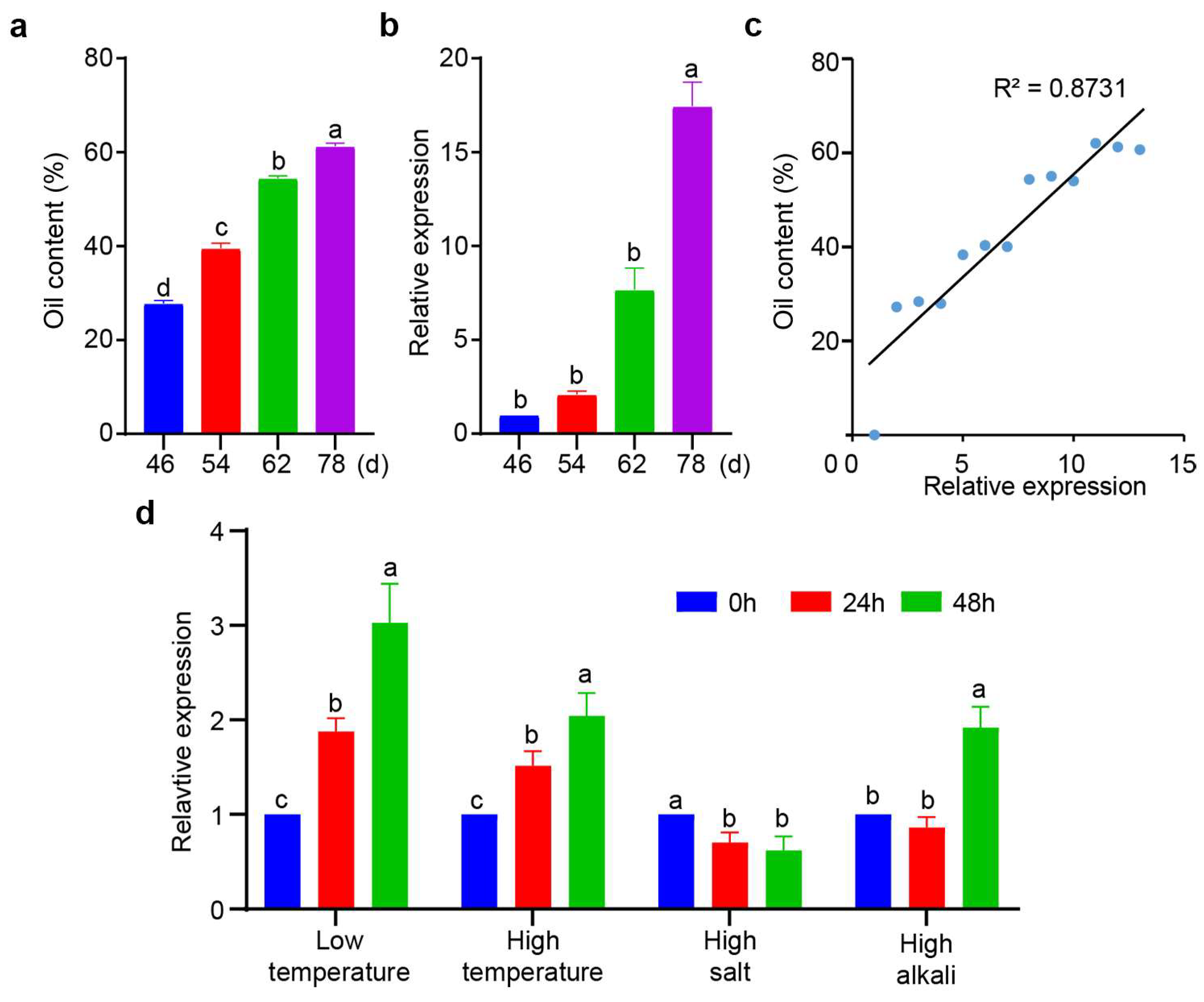
To further understand the role of XsPDAT1 in oil biosynthesis of X. sorbifolium, we analyzed the correlation between gene expression and oil content throughout the development of the seed kernel. Oil content increased linearly during seed kernel development, reaching 61.2% at maturity (78 d after anthesis) (Figure 2a). Similarly, the expression of XsPDAT1 increased as the seed kernel developed, reaching its highest at the ripening stage, where it was up-regulated 17.5-fold compared to the young fruiting stage (46 d after anthesis) (Figure 2b). Correlation analysis revealed a significant positive relationship between the expression pattern of XsPDAT1 and the oil content of the seed kernel, with a correlation coefficient of R2 = 0.8731 (Figure 2c). The expression pattern of XsPDAT1 showed the same trend under low-temperature and high-temperature stress, both of which showed a linear up-regulation and reached significant levels at 24 h and 48 h compared to control; it was down-regulated under salt stress, but showed a “down-up” trend under alkali stress, implying that the role of XsPDAT1 may differ in response to different stresses (Figure 2d).

3.3. Genetic Transformation and Screening

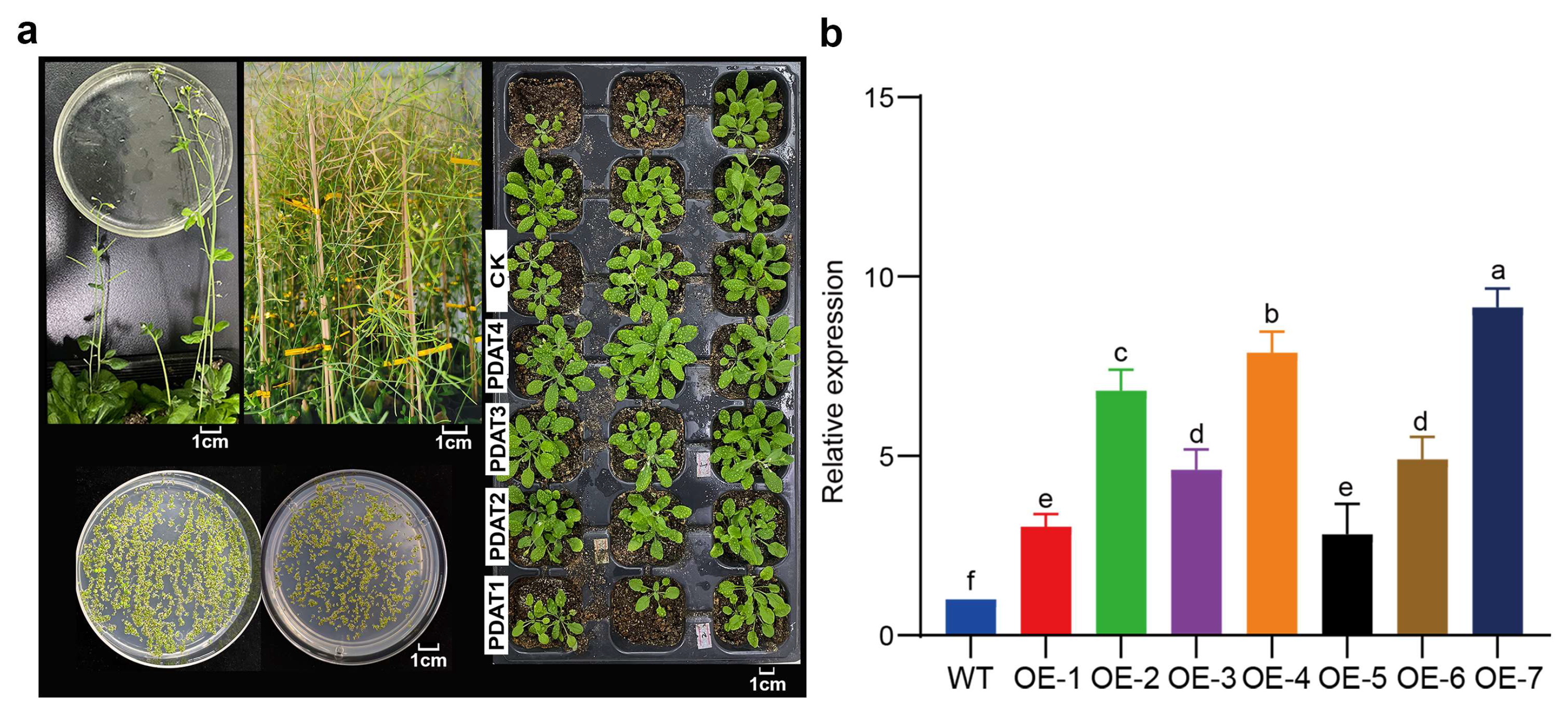
To investigate the function of XsPDAT1, the pCAMBIA1300-XsPDAT1 recombinant plasmid was transferred into Agrobacterium tumefaciens and A. thaliana was infected using the floral-dip method to obtain transgenic plants (Figure 3a). Ten A. thaliana seedlings were selected after resistance screening, and their genomic DNA was extracted; PCR detection revealed bright and correct target bands, indicating that XsPDAT1 had been stably transformed into seven A. thaliana plants (Figure S2). In order to obtain the highly expressed lines, qRT-PCR analysis was performed on these transgenic lines, which showed that the expression levels of all transgenic OE lines were higher than those of wild-type lines (Figure 3b). The higher-expressed OE-2, OE-4, and OE-7 were used for the subsequent experiments.

3.4. Changes in Morphology of Transgenic A. thaliana Lines

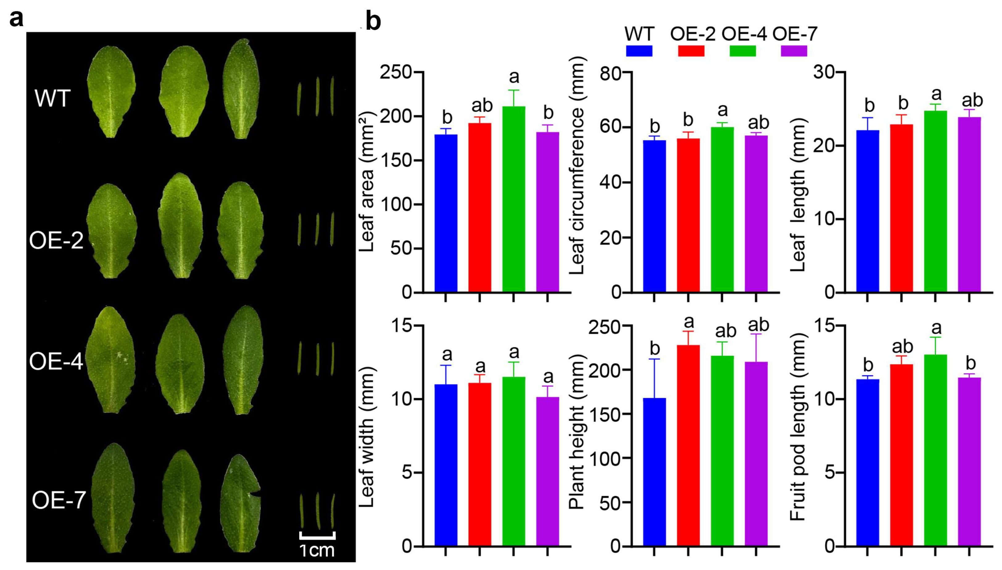
The transgenic and wild-type A. thaliana were comparatively analyzed to investigate whether XsPDAT1 affects the phenotypic changes of A. thaliana. Compared to the wild-type lines, transgenic lines showed better growth characteristics, such as enhancing plant height, leaf size, and fruit pod length (Figure 4a,b). OE-4 showed the most obvious changes. OE-4 differed significantly from the wild type in terms of leaf area, leaf circumference, leaf length, plant height, and fruit pod size, while no significant difference in leaf width was observed.

3.5. XsPDAT1 Confers Low-Temperature Resistance in Transgenic A. thaliana

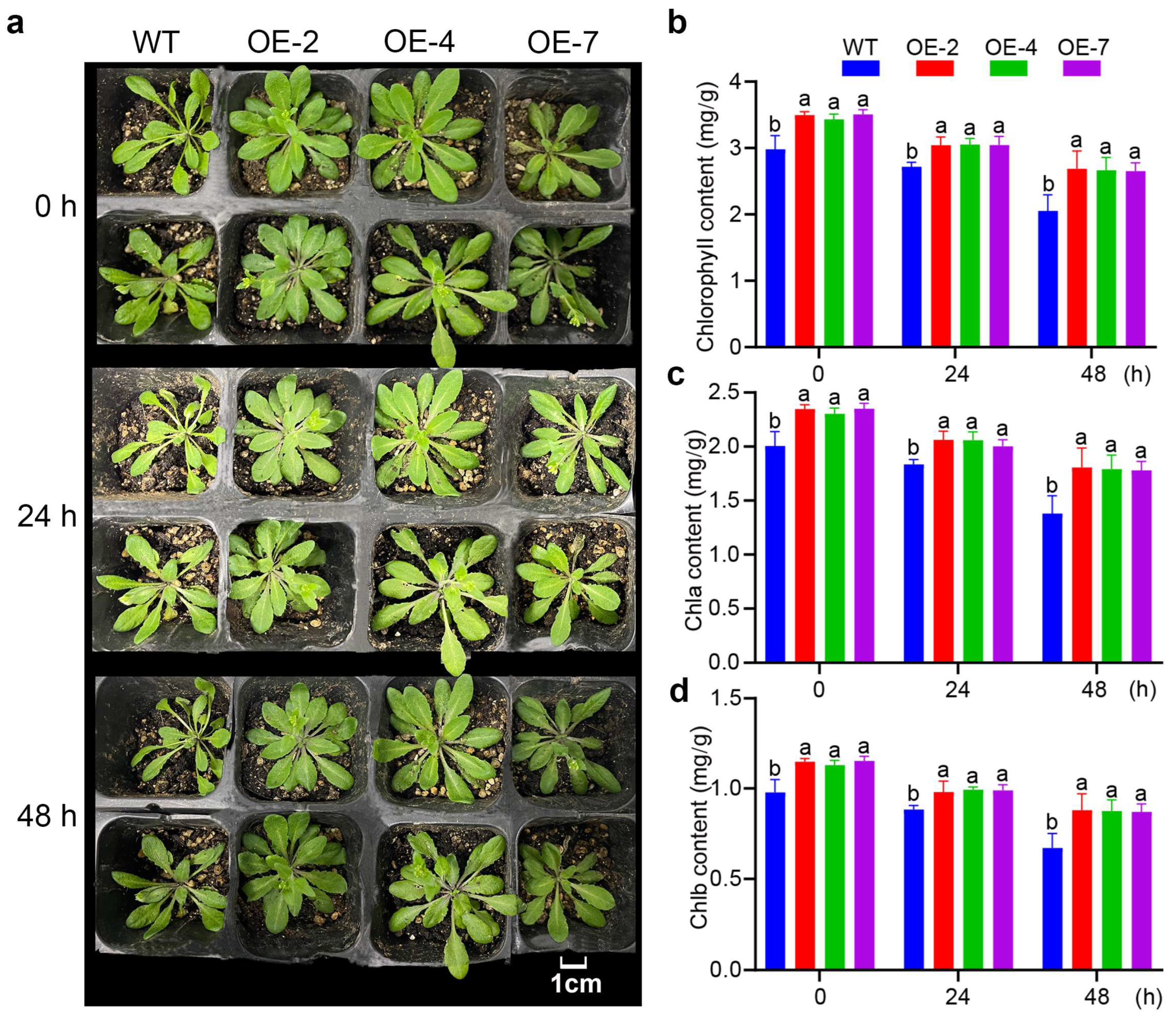
Observation of the growth changes of A. thaliana seedlings indicated minimal variations in leaf morphology between the transgenic and wild-type lines during low-temperature stress at 4 °C. The wild-type seedlings began to wilt at 24 h of low temperature, and wilted more pronouncedly at 48 h, with water loss and discoloration. The transgenic seedlings begin to wilt after 48 h. Under low-temperature stress, wild-type lines exhibited more significant leaf alterations than transgenic lines (Figure 5a).
As the duration of low-temperature treatment increased, the chlorophyll a, b, and total content of all A. thaliana lines decreased by varying magnitudes. However, the chlorophyll content in the transgenic lines was significantly higher than that of the wild type at 0 h, 24 h, and 48 h of low-temperature treatment (Figure 5b–d). The low-temperature stress resulted in the decomposition of chlorophyll in plants, which was lower in transgenic lines than in wild-type lines, indicating that XsPDAT1 mitigated the reduction of chlorophyll in A. thaliana (Figure 5b–d).

3.6. XsPDAT1 Enhances Cellular Homeostasis of A. thaliana in Response to Low-Temperature Stress

There was no significant difference in POD activity between transgenic and wild-type lines before low-temperature stress. The increase in POD activity of wild-type lines was smaller than that of transgenic lines, and the increase in three transgenic lines was approximately the same amount. After 48 h of continuous treatment, OE-7 had the highest POD activity (2476.99 U/g), representing a 400% increase. The POD activity in the transgenic lines was significantly higher than that in the wild-type lines after 24 h and 48 h of low-temperature treatment, indicating that the XsPDAT1 gene improved the adaptive ability of A. thaliana (Figure 6a).
The free proline content of all A. thaliana lines increased gradually with the prolongation of low temperature, and the proline content of the transgenic lines was significantly higher than that of the wild-type lines at different treatment durations. Under 48 h of low-temperature stress, the increase in the proline content of the wild-type lines was significantly lower than that of three transgenic lines, with OE-2 and OE-7 showing the largest increase, increasing by 131.64% and 127.86%, respectively (Figure 6b). Therefore, the XsPDAT1 gene promoted the accumulation of free proline in A. thaliana.
The relative conductivity of both the transgenic and the wild-type lines increased with the prolongation of the low-temperature treatment, and the relative conductivity of the wild-type lines was significantly higher than that of the transgenic lines (Figure 6c). At 48 h of low-temperature treatment, the relative conductivity of wild-type, OE-2, OE-4, and OE-7 increased by 112.32%, 59.81%, 88.46%, and 55.49%, respectively, compared to their levels before low-temperature stress. It can be seen that the XsPDAT1 gene alleviated the damage to the A. thaliana membrane caused by low temperature.

3.7. Analysis of Fatty Acid Composition of Transgenic A. thaliana Seeds

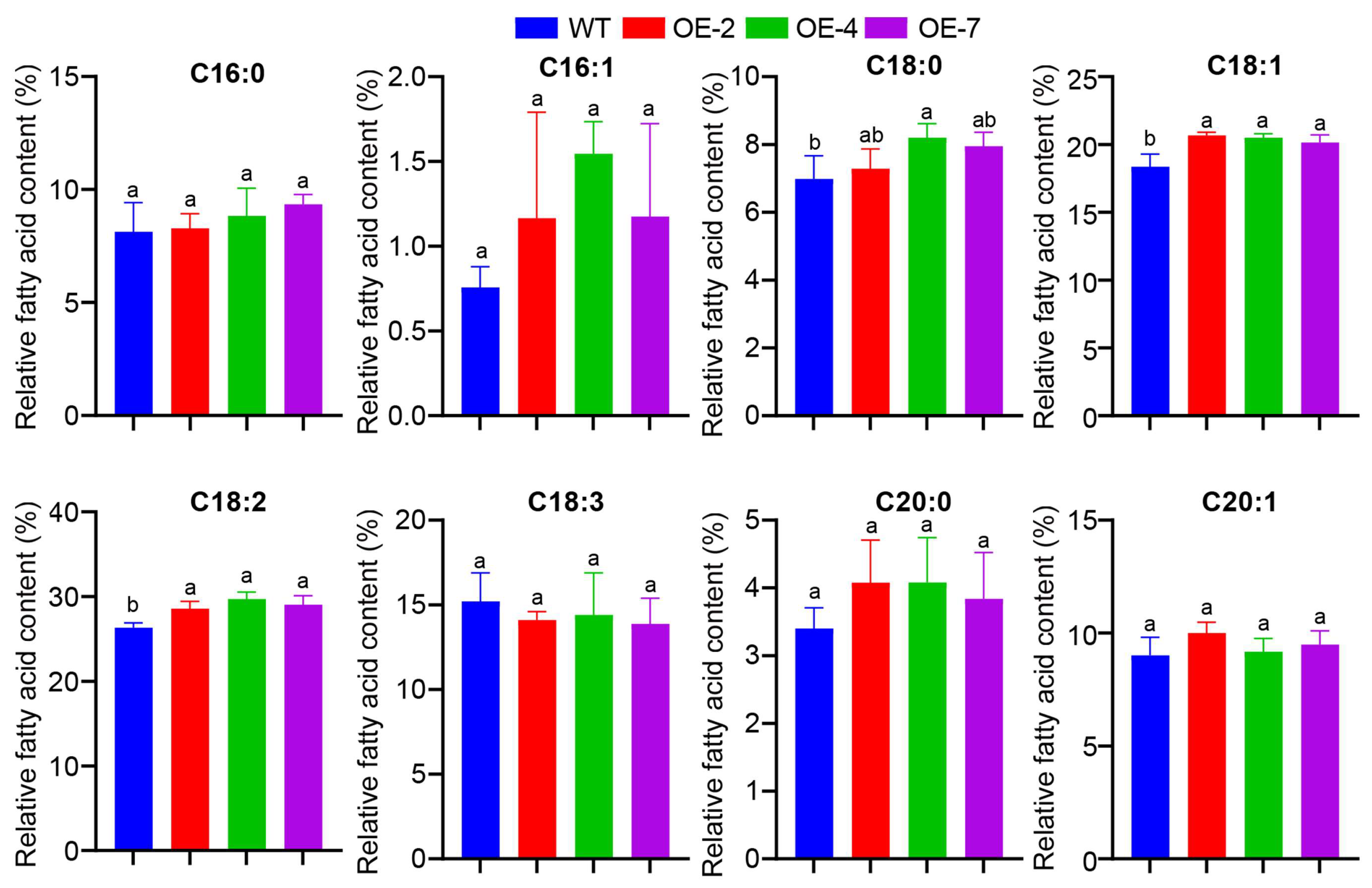
To detect the effect of XsPDAT1 on plant fatty acid fractions and their relative content, various fatty acid compositions of wild-type and transgenic A. thaliana seeds were examined by GC-MS (Figure 7). The fatty acid composition of all the lines was consistent, which was mainly composed of eight types of fatty acids, including palmitic acid (C16:0), palmitoleic acid (C16:1), stearic acid (C18:0), oleic acid (C18:1), linoleic acid (C18:2), linolenic acid (C18:3), arachidic acid (C20:0), and eicosenoic acid (C20:1), among which linoleic acid and oleic acid content was relatively high. Using seeds of wild-type lines as a control, the relative amounts of linoleic acid, oleic acid, and stearic acid in the seeds of transgenic lines increased by 12.7%, 12.6%, and 11.8%, respectively, and linolenic acid decreased by 7.3%, while the other fatty acids showed no significant changes.

4. Discussion

PDAT catalyzes the reaction between phospholipids and diacylglycerols to produce lysophospholipids and TAG, which is one of the key enzymes for TAG synthesis in plants. Meanwhile, it is also involved in the regulation of plant responses to abiotic stresses. In this study, XsPDAT1 was successfully isolated from the seed kernel of X. sorbifolium, and homology alignment of protein sequence and expression pattern analysis were performed. The PDAT gene shows different expression patterns in many plant species, including Camelina sativa [16], Camellia oleifera [31], Ricinus communis [32], and Olea europaea [33]. For example, the relative expression level of CoPDAT1 in Camellia oleifera showed an “up-down-up” pattern in developing seeds, with the highest expression at 42 weeks after flowering [31]. In Camelina sativa, the expression of CsPDAT1 during seed development was positively correlated with oil accumulation, and the expression patterns were different under cold, salt, drought, and ostomic treatments [16]. We also demonstrated that XsPDAT1 had different expression patterns under low temperature, high temperature, salt, and alkali stresses, and that XsPDAT1 had a positive regulatory effect on temperature stress. During the development of the X. sorbifolium seed kernel, the expression of XsPDAT1 gradually increased and was positively correlated with oil accumulation, indicating that XsPDAT1 could promote oil accumulation in the X. sorbifolium seed kernel.
Currently, the Agrobacterium-mediated plant infection is one of the main ways to promote plant genetic change, which offers several advantages, including low cost, simple operation, and high transformation efficiency [28]. A. thaliana is the most commonly used model plant in plant genetic engineering. The A. thaliana inflorescence was first infected using the Agrobacterium-mediated method, and then antibiotic screening was performed to obtain stable genetically modified plants. The GhPDAT1 and GhPDAT2 genes from cotton and the CePDAT3 gene from Cyperus esculentus were transformed into A. thaliana using the Agrobacterium-mediated floral-dip method, respectively, and transgenic lines with high expression and stable inheritance were obtained [17,34]. In this study, XsPDAT1 was also transferred into A. thaliana using the same method, and ten positive seedlings were obtained after resistance screening, of which seven transgenic plants were successfully transfected with the XsPDAT1 gene after the genomic DNA extraction and PCR assay.
Based on previous reports, overexpression of PDAT can increase the growth rate and make an A. thaliana seedling larger compared to the wild type [35,36]. In our experiment, transgenic A. thaliana overexpressing XsPDAT1 outperformed the wild type in various phenotypic indexes of leaf, plant height, and fruit pod. Low-temperature stress for 48 h resulted in wilting and other symptoms in both transgenic and wild-type lines, but transgenic lines exhibited a more cold-resistant phenotype. Chlorophyll content reflects plant photosynthetic capacity. Under low-temperature stress, a higher rate of chlorophyll breakdown indicates weaker cold tolerance, while a slower rate of degradation implies stronger cold tolerance. The increase of POD activity in plant cells can remove excessive ROS and reduce damage to plant cells, thus alleviating the damage caused by low temperature and other adversities. Free proline can protect the protoplasmic layer of plant cells by regulating the osmotic pressure inside the plant cells, and also has antioxidant properties that can increase the antioxidant reserves of plants, thus helping plants to resist unfavorable conditions. Low-temperature stress affects the components of the plant cell membrane system, increasing the selective permeability of the cell membrane, leading to electrolyte extravasation and tissue fluid efflux, and thus increasing relative conductivity, which is negatively correlated with the cold tolerance of plants. Therefore, indicators such as relative conductivity, chlorophyll content, free proline, and POD activity can be used to determine the cold resistance of plants. In this study, XsPDAT1 transgenic A. thaliana lines showed less cell membrane damage, a lower rate of chlorophyll decomposition, and a greater increase in free proline content and POD activity than wild-type lines after being subjected to low-temperature stress, suggesting that overexpression of XsPDAT1 in A. thaliana enhanced the cold hardiness of plants. This is consistent with the results of a recent study which found that overexpression of sesame SiPDAT1 in A. thaliana improved tolerance to low temperature compared to the wild type [36].
Fatty acids are a class of compounds composed of three elements, including carbon, hydrogen, and oxygen, which can be categorized according to the number of saturated bonds in the hydrocarbon chain into saturated fatty acid (SFA), monounsaturated fatty acid (MUFA) and polyunsaturated fatty acid (PUFA). It has been shown that the PDAT gene can affect the ratios of different fatty acids in plants. Overexpression of AtPDAT1 affected the oil content as well as the ratio of fatty acid composition in A. thaliana leaves. HpPDAT of Haematococcus Pluvialis was overexpressed in tobacco, and lipid composition analysis revealed that the HpPDAT gene affected the fatty acid ratios of tobacco leaves and showed a preference for saturated fatty acids in the order C18:0 > C20:0 > C16:0 [37]. The CsPDAT1a gene of Camelina sativa showed a preference for unsaturated fatty acids [16]. Overexpression of AtPDAT in Brassica napus resulted in reduced unsaturation of phospholipids and TAG [38]. Unlike the HpPDAT of H. Pluvialis, XsPDAT1 showed a preference for unsaturated fatty acids, with a significant increase in linoleic acid (C18:2) and oleic acid (C18:1), suggesting that XsPDAT1 was involved in the fatty acid synthesis of the seed and promoted the enrichment of oleic acid and linoleic acid.
Under low-temperature stress, plants improve membrane fluidity by increasing the concentration of unsaturated fatty acids in cell membrane lipids to adapt to changes in external temperature, thereby enhancing cold resistance [39]. Thus, the amounts of unsaturated fatty acids can be used as an index to identify the cold resistance of plants. Oleic acid and linoleic acid are the main unsaturated fatty acids and play an important role in improving tolerance to various environmental stresses [40]. In this study, the overexpression of the XsPDAT1 gene in A. thaliana increased the amount of unsaturated fatty acids (oleic acid and linoleic acid) in the membrane lipid of transgenic plants, which reduced the degree of membrane damage and improved the cold resistance of A. thaliana. Therefore, the role of XsPDAT1 in enhancing plant stress-resistance is based on its function of regulating fatty acid composition, which is an interrelated process.

5. Conclusions

The XsPDAT1 gene was cloned and the pCAMBIA1300-XsPDAT1 plant vector was constructed, which was successfully implanted into A. thaliana using the Agrobacterium-mediated floral-dip method, and seven transgenic plants were obtained. Under low-temperature stress, the results of various physiological indexes of wild-type and transgenic lines revealed that overexpression of the XsPDAT1 gene could improve the cold tolerance of plants. The results of lipid composition analysis in transgenic A. thaliana seeds showed a significant increase in the amounts of linoleic (C18:2) and oleic (C18:1) unsaturated fatty acids. This research demonstrated that the XsPDAT1 gene played a dual role in the regulation of plant lipid composition and low-temperature stress, which has important scientific application value for molecular genetic improvement of X. sorbifolium.

Supplementary Materials

The following supporting information can be downloaded at: https://www.mdpi.com/article/10.3390/f15101822/s1. Figure S1 The sequence of nucleotide and translated amino acids in the XsPDAT1 gene of X. sorbifolium. Figure S2 PCR identification of transgenic A. thaliana lines. Table S1 Primer sequences.

Author Contributions

J.G. designed the experiment; J.W. and H.R. performed research, designed the methodology, and finished writing and editing; W.Z., X.W. and Z.S. helped with the data and results; S.L., F.O.P. and X.B. critically polished the manuscript. All authors have read and agreed to the published version of the manuscript.

Funding

This work was funded by Shanxi Agricultural University Doctoral Research Initiation Project (2021BQ17); Shanxi Province doctoral graduates and postdoctoral researchers come to work in Shanxi Province to reward the fund scientific research project (SXBYKY2021061); Basic Research Program of Shanxi Province (202103021224168).

Data Availability Statement

The datasets for this study can be found in the Supplementary Material.

Conflicts of Interest

The authors declare no conflicts of interest.

References

  1. Yang, C.Y.; Ha, W.; Lin, Y.; Jiang, K.; Yang, J.L.; Shi, Y.P. Polyphenols isolated from Xanthoceras sorbifolia husks and their anti-tumor and radical-scavenging activities. Molecules 2016, 21, 1694. [Google Scholar] [CrossRef] [PubMed]
  2. Chen, X.; Lei, Z.; Cao, J.; Zhang, W.; Wu, R.; Cao, F.; Guo, Q.; Wang, J. Traditional uses, phytochemistry, pharmacology and current uses of underutilized Xanthoceras sorbifolium bunge: A review. J. Ethnopharmacol. 2022, 283, 114747. [Google Scholar] [CrossRef] [PubMed]
  3. Zheng, Y.; Gao, P.; Wang, S.; Ruan, Y.; Zhong, W.; Hu, C.; He, D. Comparison of different extraction processes on the physicochemical properties, nutritional components and antioxidant ability of Xanthoceras sorbifolia Bunge kernel oil. Molecules 2022, 27, 4185. [Google Scholar] [CrossRef]
  4. Ruan, C.J.; Yan, R.; Wang, B.X.; Mopper, S.; Guan, W.K.; Zhang, J. The importance of yellow horn (Xanthoceras sorbifolia) for restoration of arid habitats and production of bioactive seed oils. Ecol. Eng. 2017, 99, 504–512. [Google Scholar] [CrossRef]
  5. Liang, Q.; Liu, J.N.; Fang, H.; Dong, Y.; Wang, C.; Bao, Y.; Hou, W.; Zhou, R.; Ma, X.; Gai, S.; et al. Genomic and transcriptomic analyses provide insights into valuable fatty acid biosynthesis and environmental adaptation of yellowhorn. Front. Plant Sci. 2022, 13, 991197. [Google Scholar] [CrossRef]
  6. Li, N.; Wang, Y.; Li, X.; Zhang, H.; Zhou, D.; Wang, W.; Li, W.; Zhang, X.; Li, X.; Hou, Y.; et al. Bioactive phenols as potential neuroinflammation inhibitors from the leaves of Xanthoceras sorbifolia Bunge. Bioorg. Med. Chem. Lett. 2016, 26, 5018–5023. [Google Scholar] [CrossRef]
  7. Dahlqvist, A.; Stahl, U.; Lenman, M.; Banas, A.; Lee, M.; Sandager, L.; Ronne, H.; Stymne, S. Phospholipid: Diacylglycerol acyltransferase: An enzyme that catalyzes the acyl-CoA-independent formation of triacylglycerol in yeast and plants. Proc. Natl. Acad. Sci. USA 2000, 97, 6487–6492. [Google Scholar] [CrossRef]
  8. Liu, X.Y.; Ouyang, L.L.; Zhou, Z.G. Phospholipid: Diacylglycerol acyltransferase contributes to the conversion of membrane lipids into triacylglycerol in Myrmecia incisa during the nitrogen starvation stress. Sci. Rep. 2016, 6, 26610. [Google Scholar] [CrossRef]
  9. Mueller, S.P.; Unger, M.; Guender, L.; Fekete, Á.; Mueller, M.J. Phospholipid: Diacylglycerol acyltransferase-mediated triacylglyerol synthesis augments basal thermotolerance. Plant Physiol. 2017, 175, 486–497. [Google Scholar] [CrossRef]
  10. Niazian, M.; Sadat-Noori, S.A.; Tohidfar, M.; Mortazavian, S.M.M.; Sabbatini, P. Betaine aldehyde dehydrogenase (BADH) vs. Flavodoxin (Fld): Two important genes for enhancing plants stress tolerance and productivity. Front. Plant Sci. 2021, 12, 650215. [Google Scholar] [CrossRef]
  11. Yoon, K.; Han, D.; Li, Y.; Sommerfeld, M.; Hu, Q. Phospholipid: Diacylglycerol acyltransferase is a multifunctional enzyme involved in membrane lipid turnover and degradation while synthesizing triacylglycerol in the unicellular green microalga Chlamydomonas reinhardtii. Plant Cell 2012, 24, 3708–3724. [Google Scholar] [CrossRef] [PubMed]
  12. Klińska-Bachor, S.; Kdzierska, S.; Demski, K.; Banaś, A. Phospholipid: Diacylglycerol acyltransferase1-overexpression stimulates lipid turnover, oil production and fitness in cold-grown plants. BMC Plant Biol. 2023, 23, 370. [Google Scholar] [CrossRef] [PubMed]
  13. Stahl, U.; Carlsson, A.S.; Lenman, M.; Dahlqvist, A.; Huang, B.; Banaś, W.; Banaś, A.; Stymne, S. Cloning and functional characterization of a phospholipid: Diacylglycerol acyltransferase from Arabidopsis. Plant Physiol. 2004, 135, 1324–1335. [Google Scholar] [CrossRef]
  14. Van Erp, H.; Bates, P.D.; Burgal, J.; Shockey, J.; Browse, J. Castor phospholipid: Diacylglycerol acyltransferase facilitates efficient metabolism of hydroxy fatty acids in transgenic Arabidopsis. Plant Physiol. 2011, 15, 683–693. [Google Scholar] [CrossRef]
  15. Pan, X.; Siloto, R.M.P.; Wickramarathna, A.D.; Mietkiewska, E.; Weselake, R.J. Identification of a pair of phospholipid: Diacylglycerol acyltransferases from developing flax (Linum usitatissimum L.) seed catalyzing the selective production of trilinolenin. J. Biol. Chem. 2013, 288, 24173–24188. [Google Scholar] [CrossRef]
  16. Yuan, L.; Mao, X.; Zhao, K.; Ji, X.; Ji, C.; Xue, J.; Li, R. Characterisation of phospholipid: Diacylglycerol acyltransferases (PDATs) from Camelina sativa and their roles in stress responses. Biol. Open 2017, 6, 1024–1034. [Google Scholar]
  17. Zang, X.; Geng, X.; Ma, L.; Wang, N.; Pei, W.; Wu, M.; Zhang, J.; Yu, J. A genome-wide analysis of the phospholipid: Diacylglycerol acyltransferase gene family in Gossypium. BMC Genom. 2019, 20, 402. [Google Scholar] [CrossRef]
  18. Zhou, B.; Fei, W.; Yang, S.; Yang, F.; Qu, G.; Tang, W.; Ou, J.; Peng, D. Alteration of the fatty acid composition of Brassica napus L. via overexpression of phospholipid: Diacylglycerol acyltransferase 1 from Sapium sebiferum (L.) Roxb. Plant Sci. 2020, 298, 110562. [Google Scholar] [CrossRef]
  19. Voelker, T. Secrets of palm oil biosynthesis revealed. Proc. Natl. Acad. Sci. USA 2011, 108, 12193–12194. [Google Scholar] [CrossRef]
  20. Guo, H.; Li, Q.; Wang, T.; Hu, Q.; Deng, W.; Xia, X.; Gao, H. XsFAD2 gene encodes the enzyme responsible for the high linoleic acid content in oil accumulated in Xanthoceras sorbifolia seeds. J. Sci. Food Agric. 2014, 94, 482–488. [Google Scholar] [CrossRef]
  21. Guo, H.H.; Wang, T.T.; Li, Q.Q.; Zhao, N.; Zhang, Y.; Liu, D.; Hu, Q.; Li, F.L. Two novel diacylglycerol acyltransferase genes from Xanthoceras sorbifolia are responsible for its seed oil content. Gene 2013, 527, 266–274. [Google Scholar] [CrossRef] [PubMed]
  22. Lu, M.; Han, S.; Yang, Q.; Wang, J.; Guo, H. Cloning and expression analysis of LEC1 gene from Xanthoceras sorbifolia embryos. J. Beijing For. Univ. 2018, 40, 8–16. [Google Scholar]
  23. Wang, J.; Zhang, Y.; Yan, X.; Guo, J. Physiological and transcriptomic analyses of yellow horn (Xanthoceras sorbifolia) provide important insights into salt and saline-alkali stress tolerance. PLoS ONE 2020, 15, e0244365. [Google Scholar] [CrossRef]
  24. Wang, J.; Guo, J.; Zhang, Y.; Yan, X. Integrated transcriptomic and metabolomic analyses of yellow horn (Xanthoceras sorbifolia) in response to cold stress. PLoS ONE 2020, 15, e0236588. [Google Scholar] [CrossRef]
  25. Wang, J.; Hu, H.F.; Liang, X.Z.; Qamar, M.T.; Zhang, Y.X.; Zhao, J.G.; Ren, H.Q.; Yan, Y.R.; Ding, B.P.; Guo, J.P. High-quality genome assembly and comparative genomic profiling of yellowhorn (Xanthoceras sorbifolia) revealed environmental adaptation footprints and seed oil contents variations. Front. Plant Sci. 2023, 14, 1147946. [Google Scholar] [CrossRef]
  26. Ding, B.; Li, X.; Lin, Y.; Hu, C.; Yang, R.; Wei, B.; Cao, Y.; Bai, Y.; Yan, R.; Li, L. Study on cloning and bioinformatics of auxin nicotinamide synthase gene GH3 in ‘Yuluxiangli’. J. Shanxi Agric. Univ. (Nat. Sci.) 2024, 44, 14–23. [Google Scholar]
  27. Ding, B.; Hu, H.; Cao, Y.; Xu, R.; Lin, Y.; Muhammad, T.U.Q.; Song, Y.; He, G.; Han, Y.; Guo, H.; et al. Pear genomes display significant genetic diversity and provide novel insights into the fruit quality traits differentiation. Hortic. Plant J. 2024, 10, 1274–1290. [Google Scholar] [CrossRef]
  28. Clough, S.J.; Bent, A.F. Floral dip: A simplified method for Agrobacterium-mediated transformation of Arabidopsis thaliana. Plant J. 1998, 16, 735–743. [Google Scholar] [CrossRef]
  29. Jiang, L.; Li, X.X.; Lv, K.; Wang, H.; Li, Z.Y.; Qi, W.; Zhang, L.; Cao, Y.P. Rosaceae phylogenomic studies provide insights into the evolution of new genes. Horti. Plant J. 2024, in press. [Google Scholar] [CrossRef]
  30. Zhao, J.; Wu, S.; Du, W.; Yue, A.; Li, G.; Ding, Q. The relationship between variation and quality and the accumulation of protein and fat with different-maturing soybean cultivars. Acta Agric. Boreali-Sin. 2004, 19, 33–35. [Google Scholar]
  31. Zhao, G.; Song, Z.B.; Liu, M.L.; Long, H.X.; Li, Z.; Zhang, L. Cloning and expression analysis of a phospholipid: Diacylglycerol acyltransferase (PDAT) gene in Camellia oleifera. Plant Physiol. 2017, 53, 1619–1628. [Google Scholar]
  32. Kim, H.U.; Lee, K.R.; Go, Y.S.; Jung, J.H.; Suh, M.C.; Kim, J.B. Endoplasmic reticulum-located PDAT1-2 from Castor bean enhances hydroxy fatty acid accumulation in transgenic plants. Plant Cell Physiol. 2011, 52, 983–993. [Google Scholar] [CrossRef] [PubMed]
  33. Hernández, M.L.; Moretti, S.; Sicardo, M.D.; García, Ú.; Pérez, A.; Sebastiani, L.; Martínez-Rivas, J.M. Distinct physiological roles of three phospholipid: Diacylglycerol acyltransferase genes in olive fruit with respect to oil accumulation and the response to abiotic stress. Front. Plant Sci. 2021, 12, 751959. [Google Scholar] [CrossRef] [PubMed]
  34. Ren, W.; Guo, D.; Xing, G.; Yang, C.; Zhang, Y.; Yang, J.; Niu, L.; Zhong, X.; Zhao, Q.; Cui, Y.; et al. Complete chloroplast genome sequence and comparative and phylogenetic analyses of the cultivated Cyperus esculentus. Diversity 2021, 13, 405. [Google Scholar] [CrossRef]
  35. Bana’s, W.; Carlsson, A.S.; Bana´s, A. Effect of overexpression of PDAT gene on Arabidopsis growth rate and seed oil content. J. Agric. Sci. 2014, 6, 65–79. [Google Scholar] [CrossRef]
  36. Demski, K.; Łosiewska, A.; Jasieniecka-Gazarkiewicz, K.; Klińska, S.; Banaś, A. Phospholipid: Diacylglycerol acyltransferase1 overexpression delays senescence and enhances post-heat and cold exposure fitness. Front. Plant Sci. 2020, 11, 611897. [Google Scholar] [CrossRef]
  37. Zhao, C.; Zhang, H.; Zang, D.; Cui, H.; Li, R. Identification and expression analysis of phospholipid-diacylglycerol acyltransferase gene (HpPDAT) from Haematococcus pluvialis. J. Shanxi Agric. Sci. 2022, 50, 319–326. [Google Scholar]
  38. Liao, P.; Lechon, T.; Romsdahl, T.; Woodfield, H.; Fenyk, S.; Fawcett, T.; Wallington, E.; Bates, R.E.; Chye, M.L.; Chapman, K.D.; et al. Transgenic manipulation of triacylglycerol biosynthetic enzymes in B. napus alters lipid-associated gene expression and lipid metabolism. Sci. Rep. 2022, 12, 3352. [Google Scholar] [CrossRef]
  39. Liu, F.; Yu, S.; Liu, G. Mechanism and research progress of plant lipid regulation under biotic and abiotic stresses. Chin. J. Oil Crop. Sci. 2023, 45, 1062–1072. [Google Scholar]
  40. Saini, R.; Kumar, S. Genome-wide identification characterization and insilico profiling of genes encoding FAD (fatty acid desaturase) proteins in chickpea (Cicer arietinum L.). Plant Gene 2019, 18, 100180. [Google Scholar] [CrossRef]
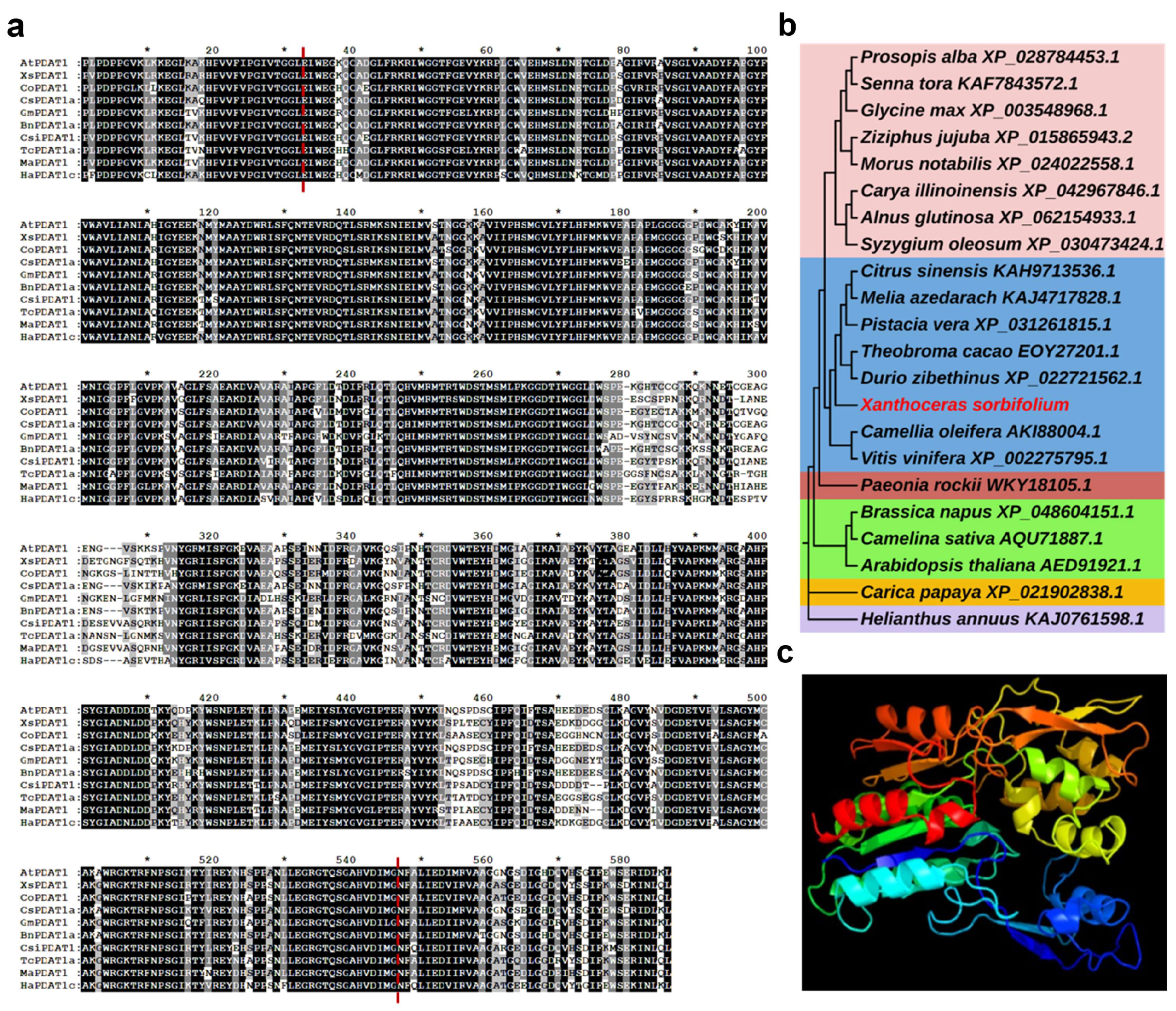
Figure 1. (a) Amino acid sequence alignments of PDAT1s in different plants. Black parts are highly homologous regions, grey parts are partially homologous regions, white parts are variable regions, and the regions between the red dotted lines are conserved structural domains of the lecithin-acyltransferase superfamily, and * represents an interval of ten amino acids. (b) Phylogenetic tree of PDATs from X. sorbifolium and other plants. (c) Predicted tertiary structure of XsPDAT1 protein.
Forests 15 01822 g001
Figure 2. Expression pattern of XsPDAT1 gene. (a) The oil content in different developmental stages of the seed kernel (46 d, 54 d, 62 d, and 78 d after anthesis). (b) Expression levels of XsPDAT1 gene in different developmental stages of the seed kernel (46 d, 54 d, 62 d, and 78 d after anthesis). (c) Oil accumulation correlated with the expressions of XsPDAT1. (d) Expression levels of XsPDAT1 before and after different abiotic stresses. Note: different lowercase letters in the figure indicate significant differences (p < 0.05).
Forests 15 01822 g002
Figure 3. Genetic transformation and screening of XsPDAT1-overexpressing A. thaliana. (a) The transgenic process of A. thaliana with the floral-dip method, including infestation, seed harvest, screening, and transplantation. (b) Relative expression levels of XsPDAT1 in the transgenic A. thaliana lines (OE-1–OE-7). Note: different lowercase letters in the figure indicate significant differences (p < 0.05).
Forests 15 01822 g003
Figure 4. Morphology analysis of the wild-type (WT) and transgenic A. thaliana lines (OE-2, OE-4 and OE-7). (a) Phenotypes of leaf and fruit pod. (b) Phenotypic indexes, including leaf area, leaf circumference, leaf length, leaf width, plant height, and fruit pod length. Note: different lowercase letters in the figure indicate significant differences (p < 0.05).
Forests 15 01822 g004
Figure 5. Changes in morphology and chlorophyll content of the wild-type (WT) and transgenic A. thaliana lines (OE-2, OE-4, OE-7) under low-temperature stress for 0 h, 24 h, and 48 h. (a) Morphology of all the lines under different treatments. (b) Total chlorophyll content of all A. thaliana lines under different treatments. (c) Chlorophyll a content of all A. thaliana lines under different treatments. (d) Chlorophyll b content of all A. thaliana lines under different treatments. Note: different lowercase letters in the figure indicate significant differences (p < 0.05).
Forests 15 01822 g005
Figure 6. Analysis of physiological indexes of wild-type (WT) and transgenic A. thaliana lines (OE-2, OE-4, OE-7) under low-temperature stress for 0 h, 24 h, and 48 h. (a) POD activity of all A. thaliana lines under different treatments. (b) Free proline content of all A. thaliana lines under different treatments. (c) Relative conductivity of all A. thaliana lines under different treatments. Note: different lowercase letters in the figure indicate significant differences (p < 0.05).
Forests 15 01822 g006
Figure 7. Fatty acid compositions of wild-type (WT) and transgenic A. thaliana lines (OE-2, OE-4, OE-7), including palmitic acid (C16:0), palmitoleic acid (C16:1), stearic acid (C18:0), oleic acid (C18:1), linoleic acid (C18:2), linolenic acid (C18:3), arachidic acid (C20:0), and eicosenoic acid (C20:1). Note: different lowercase letters in the figure indicate significant differences (p < 0.05).
Forests 15 01822 g007
Disclaimer/Publisher’s Note: The statements, opinions and data contained in all publications are solely those of the individual author(s) and contributor(s) and not of MDPI and/or the editor(s). MDPI and/or the editor(s) disclaim responsibility for any injury to people or property resulting from any ideas, methods, instructions or products referred to in the content.