Article

Genome-Wide Identification and Expression Profiling of the Response Regulator (RR) Gene Family in Pecan Reveals Its Possible Association with Callus Formation during Grafting

1
Institute of Botany, Jiangsu Province and Chinese Academy of Sciences, Nanjing 210014, China
2
Jiangsu Key Laboratory for the Research and Utilization of Plant Resources, Nanjing 210014, China
3
Jiangsu Engineering Research Center for the Germplasm Innovation and Utilization of Pecan, Nanjing 210014, China
*
Author to whom correspondence should be addressed.
These authors contributed equally to this work.
Forests 2024, 15(3), 473; https://doi.org/10.3390/f15030473
Submission received: 3 February 2024 / Revised: 26 February 2024 / Accepted: 1 March 2024 / Published: 3 March 2024
(This article belongs to the Special Issue Advances in Tree Germplasm Innovation and High-Efficiency Propagation)

Abstract

:
Response regulator (RR) is the core component of cytokinin (CK) signaling, and it regulates the expression of numerous downstream CK-responsive genes. However, the knowledge regarding the pecan RR (CiRR) gene family is still limited. In this study, we first monitored trans-zeatin riboside (tZR) content in the graft union 0, 7, 14, and 32 days after grafting and then conducted genome-wide analysis and expression profiling of the CiRR gene family using an available genome sequence and RNA-seq dataset, aiming to better understand the roles of CK during pecan grafting. The dynamic contents of tZR showed an increased trend during the specific period for both the scion and rootstock. There were 20 CiRRs in the pecan genome, including 12 type A CiRRs, 5 type B members, and 3 type C genes. All members contained a receiver domain and type B CiRRs possessed an additional Myb-like DNA-binding domain. Promoter analysis showed that the CiRR gene family contained cis-elements associated with growth and development, hormones, and stress. A total of 10 genes, including CiRR18/9/4a/14a/12c/5/12b/14b/2b/2a, were abundantly expressed in the samples of different tissues, drought stress, and kernel development. There were 12 genes (CiRR5/18/4a/12b/2b/12c/14b/2a/14a/4b/9/11a) showing active expressions during grafting, and weighted gene co-expression network analysis (WGCNA) grouped them into six modules. Among them, CiRR14a and CiRR12b were the hub genes for the turquoise and brown modules, respectively. Functional annotation indicated that the turquoise module was associated with gene transcription and translation, while the brown module was related to cell proliferation. Our results suggest that the CiRR gene family central to CK signaling is probably involved in callus formation during pecan grafting.

1. Introduction

Cytokinin is a key hormone that governs a broad array of biological processes associated with plant growth and development. It has functions in regulating cell division, inducing meristem activity, controlling leaf senescence, and many more processes [1]. Apart from its roles in growth and development, cytokinin responds to various abiotic and biotic stresses. The kinds of stress stimuli indicate that there is short-term accumulation/production of cytokinin, and this initial induction may be sustained under some adverse conditions, especially severe stress [2]. Graft union formation is a successful connection between graft partners, which is an intricate process involving both developmental and stress responses [3]. The developmental stages of graft union formation generally include necrotic layer production, callus formation, cambium differentiation, and vascular bundle connection [4], while the oxidative stresses imposed on graft union often consist of mechanical damage, hypoxia, and pathogen attack [5]. Given its pivotal roles in both normal development and stress, cytokinin has also been proven to be associated with grafting [6,7].
Various studies have indicated that cytokinin is a positive regulator during graft union formation. In melon (Cucumis melo)/squash (C. moschata) grafts, zeatin riboside (ZR) was highly induced in the graft interface at callus formation and vascular bridge stages [8]. For grapevine (Vitis vinifera) grafting, exogenous application of benzyladenine (BA) or kinetin (Ki) on the graft partners significantly enhanced callus formation and graft survival rates [9]. Another study conducted on grapevine showed that dipping the graft partners in a suitable concentration of benzylaminopurine (BAP) before grafting would enhance antioxidant activities and reduce oxidative stress, resulting in an increased successful grafting rate [10]. In walnut (Juglans regia) minigrafting, applying BA at an optimal concentration to the rootstocks could increase the rate of successful grafts [11]. During the graft formation of tomato (Solanum lycopersicum), three types of cytokinin (trans-zeatin, N6-(Δ2-isopentenyl)adenine, and cis-zeatin) were found to greatly accumulate at certain stages of grafting, and exogenous use of 6-BA and indole-3-acetic acid promoted xylem and phloem pattern formation [12].
The signal of cytokinin is transduced via a multistep phosphorelay system [13]. For eukaryotes, there are three major components in the system, including histidine kinase (HK), histidine-containing phosphotransfer protein (HP), and response regulator (RR) [14]. HK binds to cytokinin free bases, and this induces their auto-phosphorylation; HP senses the phosphoryl group from HK and transfers it to RR; phosphorylated RR regulates the transcriptional activation of downstream cytokinin-responsive genes, among which, many are transcription factors [15]. Given its pivotal role in cytokinin signaling, the RR gene family has been systematically explored in different kinds of plants, such as Arabidopsis thaliana [16], Populus trichocarpa [17], and hickory (Carya cathayensis) [18].
The bioinformatics, structural, and genetic analyses for the model plant of Arabidopsis have provided relatively detailed information regarding the RR gene family. According to their sequence and domain structure, the 24 Arabidopsis RR members (ARRs) can be divided into three types: type A (10 genes: ARR3–9, 15–17), type B (12 genes: ARR1–2, 10–14, 18–21, 23), and type C (2 genes: ARR22, 24) [19,20]. Type A RRs consist of a receiver domain and a short variable extension at the C-terminus, and these members are probably the primary targets for cytokinin [19]. Functional analysis has shown that type A RRs play distinctive roles in various aspects of plant growth and development, such as circadian rhythm regulation, meristem development, and root growth [21]. Type B RR members are composed of a receiver domain and a long C-terminal extension carrying a Myb-like DNA-binding domain (also named the GARP domain) [22]. Type B RR members act as key modulators for the transcription of cytokinin-responsive genes, including type A RRs [23]. Multiple mutations in type-B RRs showed insensitivity to exogenous cytokinins and various developmental defects [24]. Type C RRs have a domain structure identical to type A RRs, while their expressions are not obviously induced by cytokinin [19,20]. One of the type C RRs (ARR22) has been proven to be a positive regulator in stress tolerance response, including dehydration and cold [20].
Pecan (C. illinoinensis) is a widely cultivated nut tree around the world; propagation of this species is heavily depended on grafting. For the commonly used bud grafting (budding) technique, its survival rate is varied with cultivars, and some cultivars present a low and unsteady graft-take ratios [25]. There may be a need to repeat the process several times when grafting fails, which would increase the spending for grafting and hamper the introduction process for elite cultivars. Understanding the molecular mechanism behind successful grafting would lay a basis for the highly efficient propagation of pecan. Nowadays, the involvement of cytokinin in graft union formation has been confirmed; however, its association with pecan grafting is still elusive. With an aim to understand the possible roles of cytokinin in grafting, we first detected the dynamics of cytokinin content during graft union formation and then systematically performed genome-wide identification and expression profiling of pecan RR family genes (CiRRs). The results obtained in our study will preliminarily dissect the roles of cytokinin during pecan grafting and provide a foundation for the future exploration of the molecular mechanism of RR-mediated graft union formation.

2. Materials and Methods

2.1. Detection of Trans-Zeatin Riboside (tZR) during Grafting

Patch budding was performed at our experimental farm in July 2022. Scions were the branches of ‘Jinhua’ (a typically difficult-to-survive cultivar) on the current season’s growth, and rootstocks were the seedlings of the ‘Shaoxing’ cultivar at two years of age. Samples were the detached scion and rootstock tissues at 0, 7, 14, and 32 days after grafting (DAG; Figure S1 showing the representative samples during collection). For each time point, there were three biological replicates, and each replication was the pooled tissues consisting of at least five individual grafts. After collection, tZR contents in the graft partners were monitored using high-performance liquid chromatography–triple quadrupole mass spectrometry (HPLC-MS), and the detection was identical to a previous report [26].

2.2. Genome-Wide Identification of CiRR Members

The RR sequences of Arabidopsis thaliana and Populus trichocarpa were downloaded from the UniProt database (https://www.uniprot.org/, accessed on 20 April 2023), and those amino acid sequences were used as queries for searches against the pecan reference genome (https://data.jgi.doe.gov/refine-download/phytozome?organism=CillinoinensisPawnee, accessed on 8 January 2021). The hits were checked manually to confirm the existence of domains using the NCBI conserved domain database (CDD, http://www.ncbi.nlm.nih.gov/cdd, accessed on 21 April 2023) and the Pfam database (http://pfam.xfam.org/search, accessed on 21 April 2023).

2.3. Physio-Chemical and Phylogenetic Analyses of the CiRR Gene Family

The predicted molecular weight (MW) and theoretical isoelectric point (PI) of CiRRs were analyzed using an online tool, ExPASy (https://web.expasy.org/protparam/, accessed on 26 April 2023). The subcellular localization of CiRR was predicted with Plant-mPLoc (http://www.csbio.sjtu.edu.cn/bioinf/plant-multi/#, accessed on 26 April 2023) [27]. To validate the subcellular localization prediction, the open reading frames (ORFs) without stop codons of three CiRRs (CiRR4a, CiRR12b, and CiRR12c) were cloned upstream of the enhanced green fluorescent protein (eGFP) gene in the p2GWF7.0 vector. The used primers are provided in Table S1. Fusion expression was performed in tobacco (Nicotiana benthamiana) using Agrobacterium tumefaciens GV3101. An empty vector carrying the 35S::GFP fusion protein was applied as the control. Transformation of the vector into tobacco leaf epidermal cells was conducted as previously reported [28]. The fluorescence signal was monitored after two days of transformation via a confocal laser scanning microscope (Zeiss, Jena, German).
A phylogenetic tree was built to reveal the relationship of RR proteins between pecan and Arabidopsis. The analysis was conducted with MEGA software v.5 with the following parameters: neighbor-joining method, Poisson model, pairwise deletion, and 1000 bootstrap replications. The CiRRs were named according to the corresponding orthologs in Arabidopsis.

2.4. Exploring the Structure, Duplication Event, Selective Pressure, and Cis-Element of CiRRs

Based on the GFF3-formatted annotation file, the exon–intron structure of CiRRs was displayed via TBtools v2.003 [29]. The conserved motif was identified using the MEME online tool (http://meme-suite.org/tools/meme, accessed on 2 May 2023), and the setting was as follows: zero or one occurrence per sequence; motif width, 6~50; maximum motif number, 8. The detected motifs were annotated with Pfam. The duplicate gene pair of CiRRs was determined by using blastp, with an E-value of 1 × 10⁻⁵ and the top three matches. The collinear (segmental) and tandem gene pairs were discovered using the MCScanX program v.1.1 [30], and the result was visualized using TBtools v2.003. For selective pressure analysis, all of the coding sequences (CDS) of duplicate CiRR gene pairs were extracted to compute the substitution rates of non-synonymous (Ka) to synonymous (Ks) in TBtools v2.003. For cis-element prediction, promoter areas, which are the 2000 bp sequences upstream from the ATG translation start codon, were extracted and analyzed using PlantCARE (http://bioinformatics.psb.ugent.be/webtools/plantcare/html/, accessed on 14 May 2023).

2.5. Expression Profile Analysis of CiRRs in Different Tissues and Response to Stress and Development

Publicly available RNA-seq datasets concerning different tissues, drought stress, pistillate flower development, and kernel development were downloaded from the NCBI SRA database. The PRJNA799663 (accession number) project gave the sequencing data of different tissues including pistillate flowers, staminate flowers, leaves, fruits, seeds, and roots [31]. This project also supplied the transcriptomes of drought stress, and the samples were the leaves collected from 1-year-old ‘Pawnee’ trees after 0, 3, 6, 9, 12, and 15 days (d) of water deficit. The PRJNA533506 project provided the raw data regarding the pistillate flower of ‘Pawnee’ at five stages: flower bud 1 (FB1), FB2, FB3, flower 1 (FL1), and FL2 in timing order [32]. The PRJNA792564 project offered the RNA-seq raw data of a developing kernel that collected from ‘YLC28’ trees 100, 114, 121, 135, and 150 d after pistillate flower full bloom [33]. The downloaded raw data were filtered via standard quality control to generate clean reads for mapping. The aligned clean reads were calculated and normalized to transcripts per million (TPM) for gene expression level assessment. Based on the TPM value, genes are considered to be low (TPM < 10) or high (TPM ≥ 10) expression [34]. Genes showing at least two-fold change were recognized to be differentially expressed between two separate conditions.

2.6. Investigating the Potential Role of CiRRs during Grafting

Our previous RNA-seq project (accession no. PRJNA1033822) gave a total of 36 sequencing libraries, which were collected from the two graft partners (scion and rootstock) 0, 2, 7, 14, 22, and 32 DAG in three biological replicates per time point. After conventional transcriptome processing, TPM values were produced. Four CiRR genes were selected for RT-qPCR to verify the RNA-seq data. The same RNA samples applied for RNA-seq were reversed transcribed by using a Prime-Script™II First Strand cDNA synthesis kit (Takara, Dalian, China). Actin was selected as the internal control [35]. Gene quantitation detection was conducted on a Bio-Rad CFX Opus 96 Real-Time PCR System (Bio-Rad, Hercules, CA, USA) through the use of a TB Green Premix Ex Taq™ II kit (Takara, Dalian, China).
Weighted gene co-expression network analysis (WGCNA) was conducted to identify CiRR-associated co-expression genes. To ensure the accuracy of co-expression module construction, only genes with TPM > 4 and median absolute deviation (MAD) > 2.15 were selected for analysis. In total, 16,403 genes (Table S2) met this criterion and were further used as input for the WGCNA package. The input genes were grouped into various co-expression networks (modules) using the following parameters: power = 12, minModuleSize = 100, networkType = unsinged, TOMType = unsinged, mergeCutHeight = 0.25. The eigengene-based connectivity (kME) for module genes was exported by the signedKME function. The modules, where CiRRs were hub genes (absolute kME ≥ 0.85), were functionally annotated using Gene Ontology (GO) enrichment analysis. The R package clusterProfiler was used for calculation, and all of the genes in the pecan genome that could be annotated in the GO database were employed as the background input. Only the ontology regarding the biological process was analyzed, and terms with adjusted p (Padj) values < 0.05 were considered to be over-represented.

3. Results

3.1. Dynamic Changes of tZR during Grafting

As for the scion, the tZR contents increased at both 7 and 14 DAG when compared with that at 0 DAG, and its concentration returned to the starting value 30 DAG. As for the rootstock, the contents of tZR decreased after grafting with respect to its initial level (0 DAG), while an increased trend was seen from 7 to 14 DAG (Figure 1).

3.2. Total Members for the CiRR Family

With the query sequences from model plants, 20 non-redundant CiRRs were detected from the pecan genome (Table S3). CiRRs were renamed according to the closest homologs in A. thaliana. For the CiRR family genes, the open reading frame (ORF) varied from 459 (CiRR24) to 2148 bp (CiRR12d), the molecular weight was in the range of 16.73~78.27 Kd, and the predicted isoelectric points (pI) ranged between 4.46 (CiRR22b) to 9.03 (CiRR16). Subcellular localization was predicted to locate in the nucleus for all of the members. Experimental validation suggested that CiRR4a, CiRR12b, and CiRR12c both act in the nucleus (Figure 2).

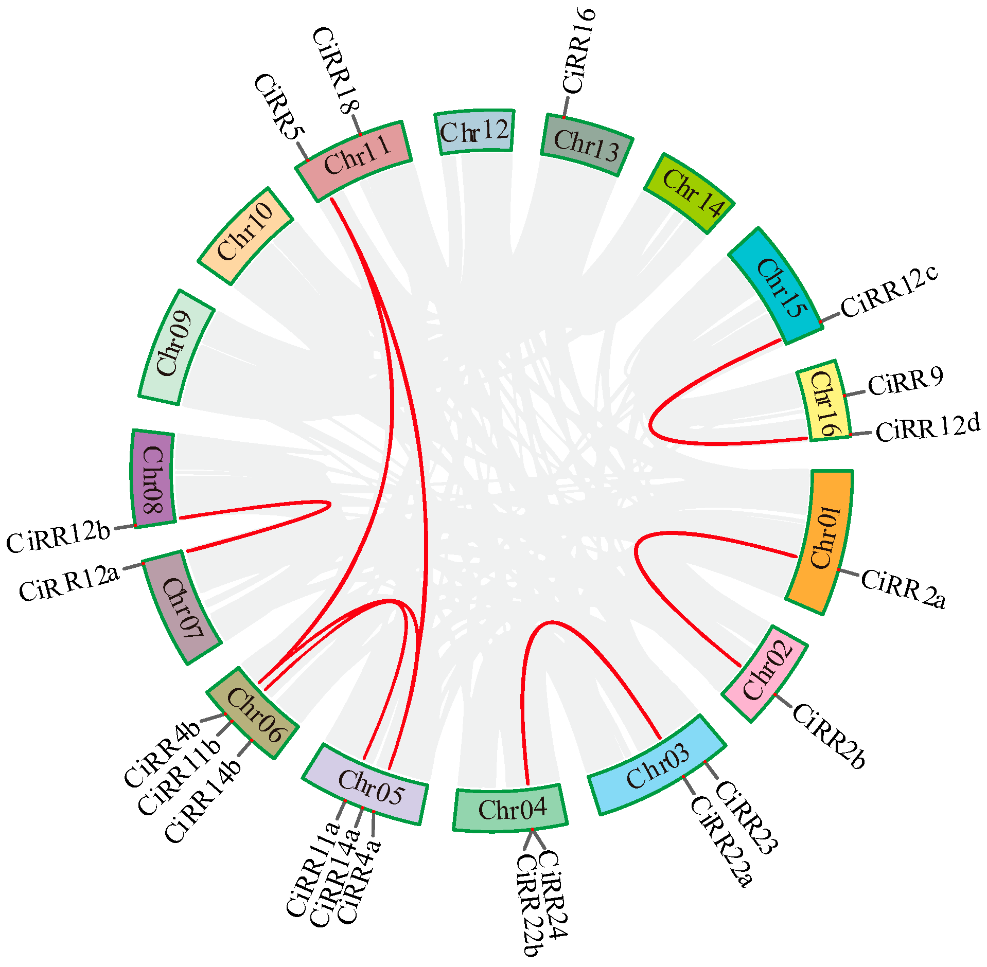
3.3. Phylogenic Relationship and Duplicated Event among CiRRs

A phylogenetic tree was constructed to dissect the evolutionary relationship of RR proteins between pecan and Arabidopsis. As shown in Figure 3, RR family members could be classified into three types, designated type A, B, and C. Among these classes, type B was the largest, containing 24 RR proteins (12 from pecan; 10 from Arabidopsis). Type A was formed with 15 RR members: 5 from pecan and 10 from Arabidopsis. There were only five type C members, with three from pecan and two from Arabidopsis.
Duplicate analysis detected eight segmental gene pairs: including CiRR2a-CiRR2b, CiRR22a-CiRR24, CiRR11a-CiRR11b, CiRR4a-CiRR4b, CiRR4a-CiRR5, CiRR4b-CiRR5, CiRR12a-CiRR12b, and CiRR12c-CiRR12d (Figure 4). One tandem-duplicated gene pair (CiRR24 and CiRR22b) was identified in chromosome 1 (Chr01). The nine duplicates were used to calculate ka/ks ratios, and the values ranged from 0.1220 to 0.4327 (Table S4).

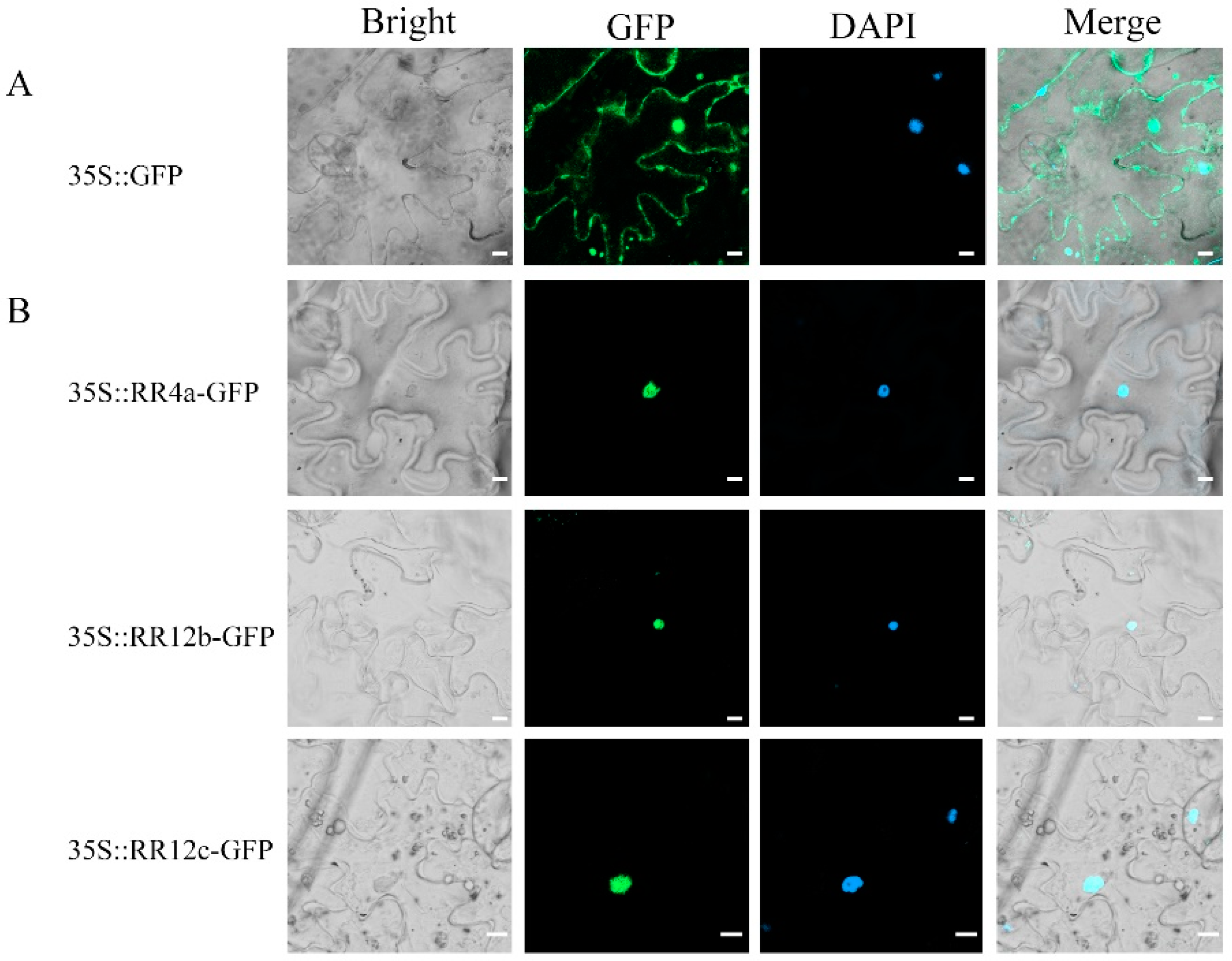
3.4. Structural Characteristics of CiRRs

According to the gene structure analysis (Figure 5), the exon–intron patterns in type B genes were variable, with exon numbers varying from five (CiRR23) to eleven (CiRR18). Most of the type B members (eight out of twelve) contained six exons, which were divided by five introns. For the genes within the type C group, the number of exons in the ORFs was conserved, both spanning two exons. As for the type A genes, they showed similar exon–intron patterns, which were all composed of five exons and four introns. The domain detection result indicate that all of the CiRR proteins embraced receiver (REC) superfamily domains, and the specific domains for the type A, B, and C members were separately REC_typeA_ARR, REC_typeB_ARR-like, and REC_hyHK_CKI1_RcsC-like (Figure 5B).
A conserved motif was detected via the MEME website, and eight motifs were found for the 20 CiRRs (Figure 5C). The number of motifs in type B CiRRs was the most abundant (4~7), followed by type A CiRRs (3) and type C CiRRs (2). Annotation of the conserved motif showed that motifs 1, 2, 3, and 5 had hits in the Pfam database (Table S5). Among them, motifs 1, 3, and 5 both annotated as the REC domain. The REC domains of type B proteins all comprised motifs 3 and 1, except for CiRR23, which consisted of motifs 5 and 1. Type C members both had a short REC domain, and only motif 5 constituted this domain. For the type A CiRRs, their REC domains were all made up of motifs 5 and 1. Motif 2 belonged to the Myb-like DNA-binding domain and was specifically located in type B CiRRs. Motifs 6 and 8 were also group-specific and only existed in some type B CiRRs. Motif 4 is highly conserved for the CiRRs, which was located in all of the members except CiRR23.
To explore the responsiveness of the CiRR gene family, cis-elements were analyzed using the PlantCARE online tool. As shown in Figure 6, CiRRs contained elements associated with growth and development, hormones, and stress. Among these, the hormone-responsive elements were the most abundant, followed by stress responsiveness. Elements relating to growth and development were present in most of the members (17 out of 20). A few genes (nine out of twenty) possessed the elements concerning flavonoid biosynthesis and/or zein metabolism.

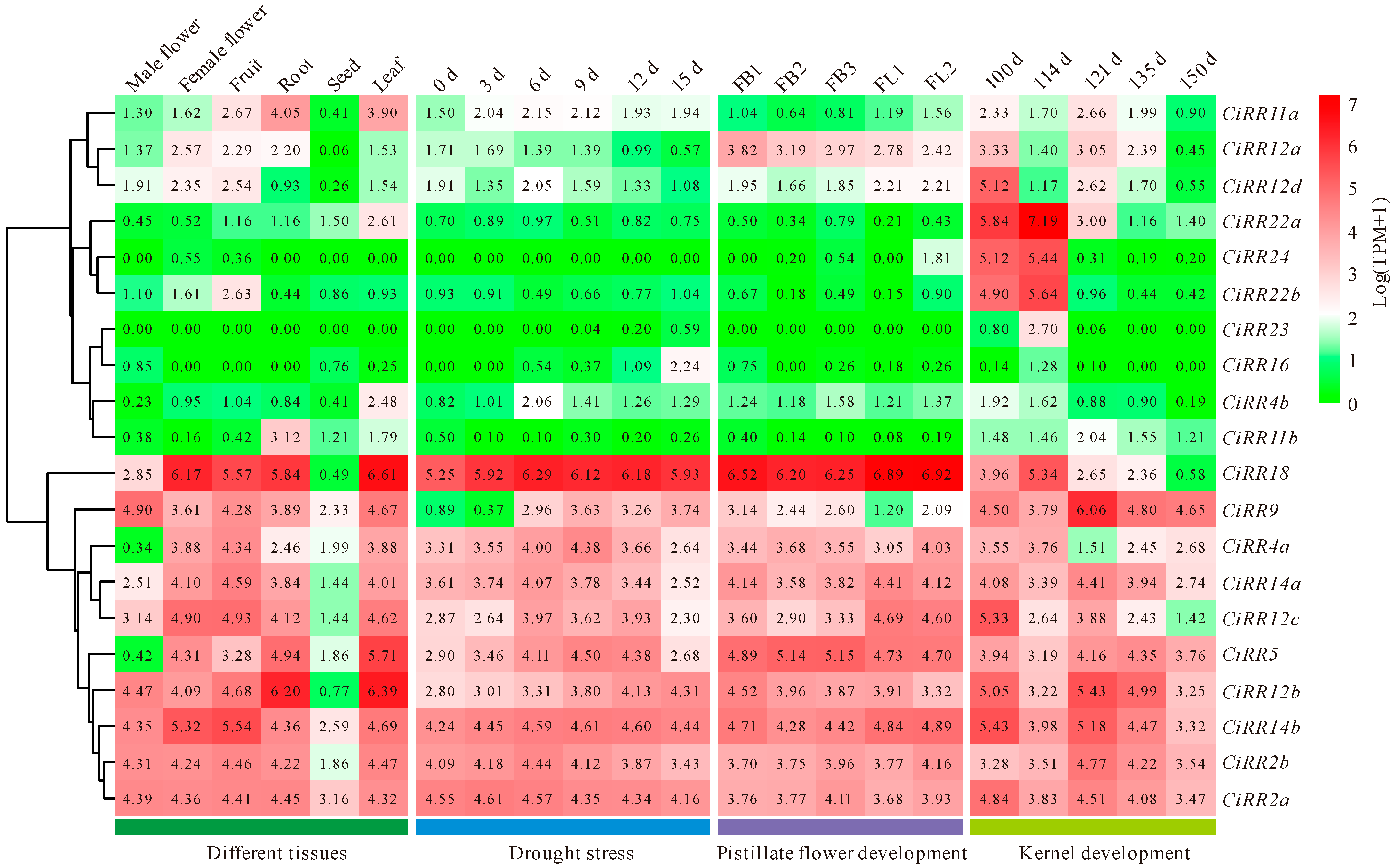
3.5. CiRR Expression Patterns in Various Tissues and during Drought Stress and Kernel Development

The expression patterns of CiRRs in various conditions were explored (Figure 7). Genes with TPM ≥ 10, which is equal to log(TPM + 1) > 3.46, were recognized as highly expressed. According to this criteria, 10 genes including CiRR18/9/4a/14a/12c/5/12b/14b/2b/2a were generally abundantly expressed in all of the detected samples. Beside this, there were no additional genes showing high expression during drought stress; however, one (CiRR12a) and four extra genes (CiRR12d/22a/24/22b) were specifically highly transcribed at certain stages of pistillate flower and kernel development, respectively. Among the ten genes with high expression, six (CiRR18/9/4a/12c/5/12b) were upregulated at the certain time points following drought treatment with respect to the control (0 d).

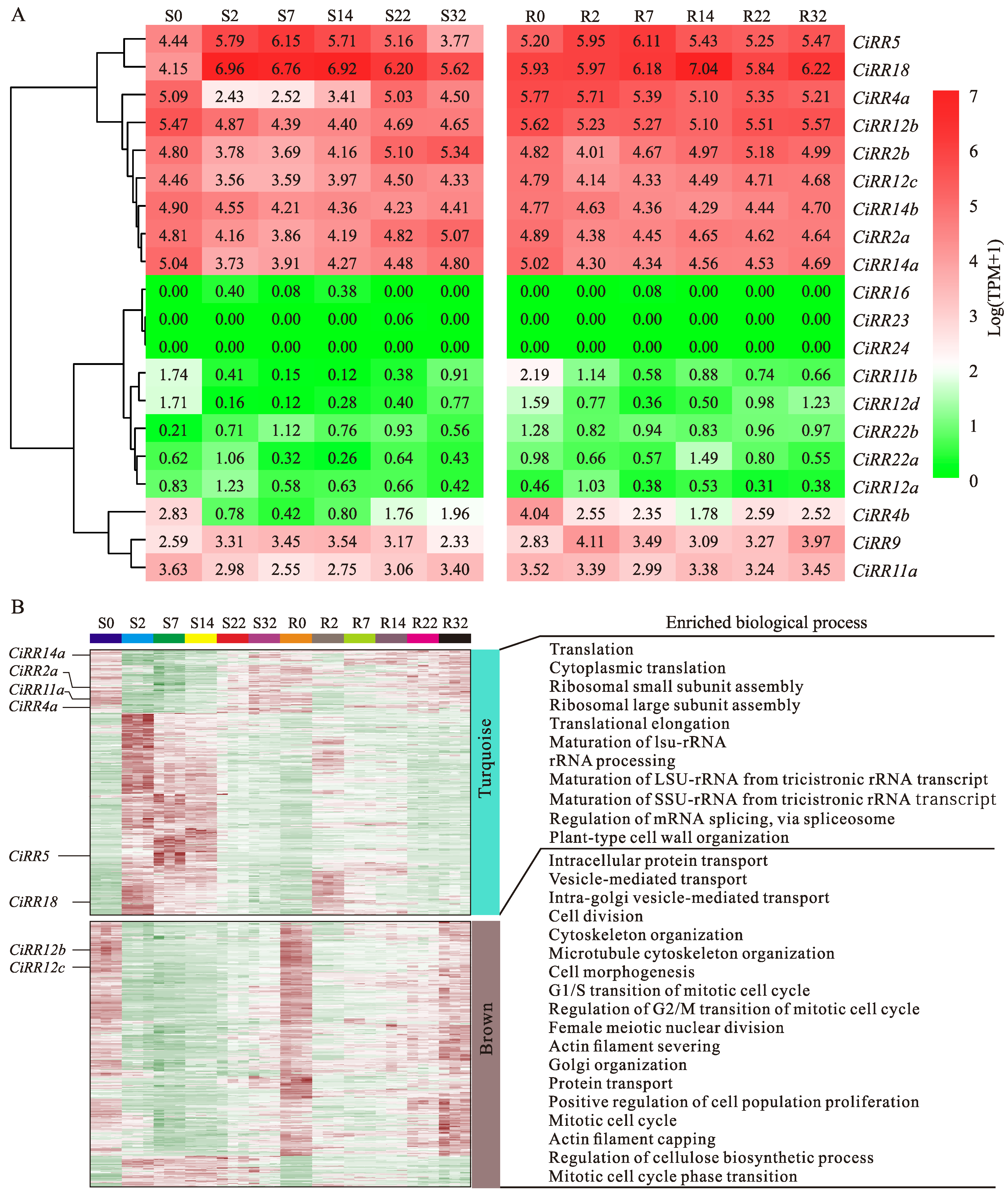
3.6. CiRR Transcriptional Dynamics during Grafting

To reveal its possible role during grafting, the expression pattern of the CiRR gene family was analyzed using RNA-seq. As shown in Figure 8A, 12 genes including CiRR5/18/4a/12b/2b/12c/14b/2a/14a/4b/9/11a were actively expressed at specific time points or throughout the whole period of grafting. The expressions of four genes were further validated using RT-qPCR, and the results were generally consistent the RNA-Seq data (Figure S2). The 12 highly expressed CiRR genes were distributed in six modules, including turquoise, brown, blue, black, green, and yellow. The absolute kME values of the CiRRs ranged from 0.53 to 0.87 (Table S6). The two hub genes, CiRR14a and CiRR12b, were separately located in turquoise and brown. GO enrichment suggested that the turquoise module, where most of the genes were more highly expressed in the scion 2~14 DAG and the rootstock 2 DAG, was over-represented in the terms relevant for gene transcription and translation (such as ‘translation’, ‘cytoplasmic translation’, ‘translational elongation’, and ‘Regulation of mRNA splicing, via spliceosome’) (Figure 8B). The brown module, in which most of the genes were greatly expressed in the scion 0 DAG and the rootstock 0, 14~32 DAG, was enriched in the categories mainly associated with cell proliferation, such as ‘cell division’, ‘positive regulation of cell population proliferation’, and ‘mitotic cell cycle’.

4. Discussion

Cytokinin regulates different kinds of developmental and stress response processes in plants. In our research, increased trends of tZR (an active form of CK) for certain time periods in both scion and rootstock were detected (Figure 1), demonstrating a possible positive role of CK during pecan grafting. However, its exact roles during grafting are not yet well understood. A previous study highlighted the importance of CK on callus formation when a wound was produced [36]. As grafting inevitably induces wound generation, CK is likely to function on callus proliferation during grafting [37]. On the other hand, CK was inferred to promote vascular reconnection during grafting, as this hormone could stimulate vessel regeneration in the healing process of stem wounds [7]. Given that the roles of CK during grafting were generally deduced from wound healing instead of graft healing, we analyzed the CK signaling-related RR gene family, aiming to indirectly dissect the functions of this phytohormone during pecan grafting.
For the RR gene family, a total of 20 members were detected in pecan. This number was less than that of Rhododendron delavayi (33) [38], Malus domestica (34) [39], and Citrus clementina (29) [40] and closely equivalent to the size in Fragaria vesca (18) [41], Jatropha curcas (14) [42], and hickory (17) [18]. The RR gene family has been reported to be expanded greatly by duplication events [43]. In our study, nine duplicated CiRR gene pairs were identified (Table S4), which also suggested that duplication, especially segmental duplication, was the driving force for RR gene family expansion. Despite the fact that the CiRR gene family has undergone expansion, it is relatively small. For the duplicated CiRR gene pairs, their Ka/Ks values were both less than one (Table S4). Since Ka/Ks < 1 is an indicator of negative selection [44], the CiRR gene pairs might experience limited functional divergence after duplication.
RR proteins are defined by the presence of a REC domain, which functions as a phosphorylation-mediated switch [45]. In the current research, both domain and motif analysis revealed the existence of a REC domain for the CiRRs, suggesting the precise identification for the gene family. Plenty of reports have divided RR genes into three groups [46,47,48], and the CiRRs in our study were also categorized into three types: type A, B, and C. Previous studies concluded that type B RR proteins possessed longer sequences and additional motifs with respect to the other two type RRs [18,49], and our study also showed similar result. For the three types of CiRRs, the number of motifs were different, and the presence/absence of specific motifs may contribute to function diversification for CiRRs.
The cis-acting element participates in the regulation of gene expression and is closely associated with the function of the gene [50]. Since the existence of abundant hormone-responsive elements (Figure 6), it is plausible that CiRR gene family mediates the crosstalk between CK and other phytohormones. There are reports suggesting that RR genes respond to different kinds of hormones besides CK. For potato (S. tuberosum), when it was cultivated on tuberizing medium, one type A RR gene (StRR9d) was significantly upregulated by auxin in the stems and tubers [51]. In Arabidopsis, sucrose nonfermenting1-related kinases (SnRKs), which are the critical kinases of ABA signaling, interact directly with and phosphorate ARR5 to mediate drought tolerance [16]; TGA3, a SA response factor, interacts with ARR2 and recruits it to the promoter of a defense-related gene Pr1, resulting in enhanced disease resistance [52]. Additionally, the presence of stress and development related cis-elements indicated the involvement of the RR gene family in the stress response as well as growth and developmental processes, consistent with the observations in Gossypium hirsutum [53], Sorghum bicolor [54], and Vigna radiata [55].
An expression profile is a reflection of a gene’s functional characteristic. The numbers of CiRRs with high expression during flower and kernel development were both greater than that in drought stress (Figure 7). This demonstrated that the CiRR gene family might be prone to action during growth and developmental processes in pecan. Six drought-inducible CiRRs were also abundantly expressed in developmental processes (Figure 7), suggesting possible multi-functions for those genes. Similar results can be found in the model plant of Arabidopsis. For CiRR18, its Arabidopsis counterpart ARR18 has been shown to be a positive regulator of drought stress and cell proliferation [56,57]. ARR5, one of the most highly cytokinin-induced genes [58], responds to various stresses including cold, dehydration, and salinity [59]. On the other hand, ARR5 has been reported to control shoot and root apical meristem development [60]. Since successful grafting is a process involving both oxidative detoxification and tissue regeneration [4,61], the induced or high expression of CiRRs in response to stress and the developmental process indicated that this gene family is likely to play critical roles during grafting.
WGCNA is a systems biology method that facilitates the dissection of the underlying biological mechanism for a co-expressed gene set [62]. In our study, CiRR14a and CiRR12b were separately the hub genes for the turquoise and brown modules (Figure 8), suggesting that they directly or indirectly controlled mounting genes in the corresponding modules. The turquoise module was related to transcription and translation. Since CiRR14a showed a reverse expression profile compared with most of the genes in the module, it might play a negative role in regulating gene expression during grafting. For the brown module, the enriched terms such as ‘mitotic cell cycle’, ‘cell division’, and ‘microtubule cytoskeleton organization’ were proven to be associated with cell proliferation [36,63,64]. During the formation of a graft union, callus guarantees the nutrients and signal transduction between the graft partners, which is an essential prerequisite for successful grafting [65]. As CiRR12b exhibited a similar expression pattern compared to most of the module genes, it was likely to play a positive role in callus formation. A similar function for its homolog could be seen in a previous study, which showed that the mutants of Arabidopsis arr12 heavily inhibited callus formation under various induction conditions [66]. As no stress-responsive processes were detected, the CiRR gene family might mainly participate in callus formation during pecan grafting.

5. Conclusions

There were 20 RR family genes in pecan, and their encoding proteins both contained a REC domain. Cis-element and expression profile analyses suggested that the CiRR gene family was likely to regulate the crosstalk between CK and other phytohormones involved in stress response as well as cellular growth and development. 12 CiRR genes were highly expressed during graft union formation; among them, CiRR12b might control various genes associated with cell proliferation. As for pecan grafting, CK probably plays a positive role, and the CiRR gene family might mediate the developmental process of callus formation instead of stress response.

Supplementary Materials

The following supporting information can be downloaded at: https://www.mdpi.com/article/10.3390/f15030473/s1. Table S1: The primers used in this study; Table S2: Genes used for WGCNA; Table S3: Physio-chemical characteristics of CiRR family genes; Table S4: Ka/Ks analysis for duplicated gene pairs of CiRRs; Table S5: Details of conserved motifs for CiRRs; Table S6: The eigengene-based connectivity (kME) for CiRR genes; Figure S1: Pecan budding and representative samples for each time point; Figure S2: Verification of RNA-seq data using RT-qPCR.

Author Contributions

Conceptualization, Z.M.; methodology, Y.Z. and Z.J.; formal analysis, Z.M. and Z.J.; investigation, L.H.; resources, M.Z.; data curation, Y.Z., G.W. and M.H.; writing—original draft preparation, Y.Z. and Z.M.; writing—review and editing, Y.Z., Z.J. and Z.M.; funding acquisition, Z.M. and J.X. All authors have read and agreed to the published version of the manuscript.

Funding

This work was supported by the Central Government Demonstration Project of Forestry Science and Technology (su[2022]TG11), the National Natural Science Foundation of China (31901347), and the Key Research and Development Program of Jiangxi Province (20223BBF61014).

Data Availability Statement

Data are contained within the article or Supplementary Materials.

Conflicts of Interest

The authors declare no conflicts of interest.

References

  1. Cortleven, A.; Leuendorf, J.E.; Frank, M.; Pezzetta, D.; Bolt, S.; Schmülling, T. Cytokinin action in response to abiotic and biotic stresses in plants. Plant Cell Environ. 2019, 42, 998–1018. [Google Scholar] [CrossRef]
  2. Zwack, P.J.; Rashotte, A.M. Interactions between cytokinin signalling and abiotic stress responses. J. Exp. Bot. 2015, 66, 4863–4871. [Google Scholar] [CrossRef]
  3. Mo, Z.; Zhang, Y.; Hu, L.; Zhai, M.; Xuan, J. Genome-wide identification and expression analysis of auxin response factor (ARF) gene family in pecan indicates its possible roles during graft union formation. Sci. Hortic. 2023, 322, 112401. [Google Scholar] [CrossRef]
  4. Mo, Z.; He, H.; Su, W.; Peng, F. Analysis of differentially accumulated proteins associated with graft union formation in pecan (Carya illinoensis). Sci. Hortic. 2017, 224, 126–134. [Google Scholar] [CrossRef]
  5. Mo, Z.; Jiang, X.; Zhang, Y.; Zhai, M.; Hu, L.; Xuan, J. Weighted gene co-expression network analysis reveals key pathways and hub genes associated with successful grafting in pecan (Carya illinoinensis). Forests 2023, 14, 835. [Google Scholar] [CrossRef]
  6. Qiu, L.; Jiang, B.; Fang, J.; Shen, Y.; Fang, Z.; Rm, S.K.; Yi, K.; Shen, C.; Yan, D.; Zheng, B. Analysis of transcriptome in hickory (Carya cathayensis), and uncover the dynamics in the hormonal signaling pathway during graft process. BMC Genom. 2016, 17, 935. [Google Scholar] [CrossRef] [PubMed]
  7. Sharma, A.; Zheng, B. Molecular responses during plant grafting and its regulation by auxins, cytokinins, and gibberellins. Biomolecules 2019, 9, 397. [Google Scholar] [CrossRef]
  8. Xu, C.; Zhang, Y.; Zhao, M.; Liu, Y.; Xu, X.; Li, T. Transcriptomic analysis of melon/squash graft junction reveals molecular mechanisms potentially underlying the graft union development. PeerJ 2021, 9, e12569. [Google Scholar] [CrossRef] [PubMed]
  9. Köse, C.; Güleryüz, M. Effects of auxins and cytokinins on graft union of grapevine (Vitis vinifera). N. Z. J. Crop Hort. 2006, 34, 145–150. [Google Scholar] [CrossRef]
  10. Bidabadi, S.S.; Afazel, M.; Sabbatini, P. Iranian grapevine rootstocks and hormonal effects on graft union, growth and antioxidant responses of Asgari seedless grape. Hortic. Plant J. 2018, 4, 16–23. [Google Scholar] [CrossRef]
  11. Farsi, M.; Fatahi Moghadam, M.R.; Zamani, Z.; Hassani, D. Effects of scion cultivar, rootstock age and hormonal treatment on minigrafting of Persian walnut. Int. J. Hortic. Sci. Technol. 2018, 5, 185–197. [Google Scholar]
  12. Cui, Q.; Xie, L.; Dong, C.; Gao, L.; Shang, Q. Stage-specific events in tomato graft formation and the regulatory effects of auxin and cytokinin. Plant Sci. 2021, 304, 110803. [Google Scholar] [CrossRef]
  13. Ferreira, F.J.; Kieber, J.J. Cytokinin signaling. Curr. Opin. Plant Biol. 2005, 8, 518–525. [Google Scholar] [CrossRef]
  14. Mira-Rodado, V. New insights into multistep-phosphorelay (MSP)/two-component system (TCS) regulation: Are plants and bacteria that different? Plants 2019, 8, 590. [Google Scholar] [CrossRef]
  15. Argueso, C.T.; Raines, T.; Kieber, J.J. Cytokinin signaling and transcriptional networks. Curr. Opin. Plant Biol. 2010, 13, 533–539. [Google Scholar] [CrossRef] [PubMed]
  16. Huang, X.; Hou, L.; Meng, J.; You, H.; Li, Z.; Gong, Z.; Yang, S.; Shi, Y. The antagonistic action of abscisic acid and cytokinin signaling mediates drought stress response in Arabidopsis. Mol. Plant 2018, 11, 970–982. [Google Scholar] [CrossRef]
  17. Ramírez-Carvajal, G.A.; Morse, A.M.; Davis, J.M. Transcript profiles of the cytokinin response regulator gene family in Populus imply diverse roles in plant development. New Phytol. 2008, 177, 77–89. [Google Scholar] [CrossRef] [PubMed]
  18. Tao, S.; Zhao, L.; Mei, J.; Abbas, F.; Xie, X.; Yang, Y.; Huang, Q.; Wang, J.; Yuan, H.; Sharma, A. Genome-wide identification and expression analysis of response regulators family genes in chinese hickory (Carya cathayensis) suggests their potential roles during grafting. J. Plant Growth. Regul. 2023, 42, 5099–5115. [Google Scholar] [CrossRef]
  19. Hwang, I.; Chen, H.C.; Sheen, J. Two-component signal transduction pathways in Arabidopsis. Plant Physiol. 2002, 129, 500–515. [Google Scholar] [CrossRef]
  20. Kang, N.Y.; Cho, C.; Kim, J. Inducible expression of Arabidopsis response regulator 22 (ARR22), a type-C ARR, in transgenic Arabidopsis enhances drought and freezing tolerance. PLoS ONE 2013, 8, e79248. [Google Scholar] [CrossRef]
  21. Ren, B.; Liang, Y.; Deng, Y.; Chen, Q.; Zhang, J.; Yang, X.; Zuo, J. Genome-wide comparative analysis of type-A Arabidopsis response regulator genes by overexpression studies reveals their diverse roles and regulatory mechanisms in cytokinin signaling. Cell Res. 2009, 19, 1178–1190. [Google Scholar] [CrossRef] [PubMed]
  22. Zubo, Y.O.; Schaller, G.E. Role of the cytokinin-activated type-B response regulators in hormone crosstalk. Plants 2020, 9, 166. [Google Scholar] [CrossRef] [PubMed]
  23. Kim, K.; Ryu, H.; Cho, Y.H.; Scacchi, E.; Sabatini, S.; Hwang, I. Cytokinin-facilitated proteolysis of ARABIDOPSIS RESPONSE REGULATOR 2 attenuates signaling output in two-component circuitry. Plant J. 2012, 69, 934–945. [Google Scholar] [CrossRef] [PubMed]
  24. Argyros, R.D.; Mathews, D.E.; Chiang, Y.H.; Palmer, C.M.; Thibault, D.M.; Etheridge, N.; Argyros, D.A.; Mason, M.G.; Kieber, J.J.; Schaller, G.E. Type B response regulators of Arabidopsis play key roles in cytokinin signaling and plant development. Plant Cell 2008, 20, 2102–2116. [Google Scholar] [CrossRef] [PubMed]
  25. Mo, Z.; Yang, X.; Hu, L.; Zhai, M.; Xuan, J. Histological, physio-biochemical, and transcriptomic analyses reveal the potential limiting factors for successful grafting of pecan. J. For. Res. 2024, 35, 35. [Google Scholar] [CrossRef]
  26. Movahedi, A.; Wei, H.; Pucker, B.; Ghaderi-Zefrehei, M.; Rasouli, F.; Kiani-Pouya, A.; Jiang, T.; Zhuge, Q.; Yang, L.; Zhou, X. Isoprenoid biosynthesis regulation in poplars by methylerythritol phosphate and mevalonic acid pathways. Front. Plant Sci. 2022, 13, 968780. [Google Scholar] [CrossRef]
  27. Chou, K.C.; Shen, H.B. Plant-mPLoc: A top-down strategy to augment the power for predicting plant protein subcellular localization. PLoS ONE 2010, 5, e11335. [Google Scholar] [CrossRef]
  28. Zhu, K.; Jin, Q.; Samma, M.K.; Lin, G.; Shen, W. Molecular cloning and characterization of a heme oxygenase1 gene from sunflower and its expression profiles in salinity acclimation. Mol. Biol. Rep. 2014, 41, 4109–4121. [Google Scholar] [CrossRef]
  29. Chen, C.; Wu, Y.; Li, J.; Wang, X.; Zeng, Z.; Xu, J.; Liu, Y.; Feng, J.; Chen, H.; He, Y. TBtools-II: A “one for all, all for one” bioinformatics platform for biological big-data mining. Mol. Plant 2023, 16, 1733–1742. [Google Scholar] [CrossRef] [PubMed]
  30. Wang, Y.; Tang, H.; DeBarry, J.D.; Tan, X.; Li, J.; Wang, X.; Lee, T.; Jin, H.; Marler, B.; Guo, H. MCScanX: A toolkit for detection and evolutionary analysis of gene synteny and collinearity. Nucleic Acids Res. 2012, 40, e49. [Google Scholar] [CrossRef] [PubMed]
  31. Zhu, K.; Fan, P.; Liu, H.; Tan, P.; Ma, W.; Mo, Z.; Zhao, J.; Chu, G.; Peng, F. Insight into the CBL and CIPK gene families in pecan (Carya illinoinensis): Identification, evolution and expression patterns in drought response. BMC Plant Biol. 2022, 22, 221. [Google Scholar] [CrossRef]
  32. Wang, M.; Xi, D.; Chen, Y.; Zhu, C.; Zhao, Y.; Geng, G. Morphological characterization and transcriptome analysis of pistillate flowering in pecan (Carya illinoinensis). Sci. Hortic. 2019, 257, 108674. [Google Scholar] [CrossRef]
  33. Zhang, C.; Ren, H.; Yao, X.; Wang, K.; Chang, J. Comparative transcriptome analysis reveals differential regulation of flavonoids biosynthesis between kernels of two pecan cultivars. Front. Plant Sci. 2022, 13, 804968. [Google Scholar] [CrossRef] [PubMed]
  34. Luo, G.; Shen, L.; Zhao, S.; Li, R.; Song, Y.; Song, S.; Yu, K.; Yang, W.; Li, X.; Sun, J. Genome-wide identification of seed storage protein gene regulators in wheat through coexpression analysis. Plant J. 2021, 108, 1704–1720. [Google Scholar] [CrossRef] [PubMed]
  35. Mo, Z.; Chen, Y.; Lou, W.; Jia, X.; Zhai, M.; Xuan, J.; Guo, Z.; Li, Y. Identification of suitable reference genes for normalization of real-time quantitative PCR data in pecan (Carya illinoinensis). Trees-Struct. Funct. 2020, 34, 1233–1241. [Google Scholar] [CrossRef]
  36. Ikeuchi, M.; Iwase, A.; Rymen, B.; Lambolez, A.; Kojima, M.; Takebayashi, Y.; Heyman, J.; Watanabe, S.; Seo, M.; De Veylder, L. Wounding triggers callus formation via dynamic hormonal and transcriptional changes. Plant Physiol. 2017, 175, 1158–1174. [Google Scholar] [CrossRef] [PubMed]
  37. Nanda, A.K.; Melnyk, C.W. The role of plant hormones during grafting. J. Plant Res. 2018, 131, 49–58. [Google Scholar] [CrossRef] [PubMed]
  38. Peng, L.; Li, X.; Gao, Y.; Xie, W.; Zhang, L.; Song, J.; Li, S.; Zhao, Z. Genome-wide identification of the RR gene family and its expression analysis in response to TDZ induction in Rhododendron delavayi. Plants 2023, 12, 3250. [Google Scholar] [CrossRef] [PubMed]
  39. Li, Y.; Zhang, D.; Zhang, L.; Zuo, X.; Fan, S.; Zhang, X.; Shalmani, A.; Han, M. Identification and expression analysis of cytokinin response-regulator genes during floral induction in apple (Malus domestica Borkh). Plant Growth Regul. 2017, 83, 455–464. [Google Scholar] [CrossRef]
  40. Zhang, M.; Wang, F.; Wang, X.; Feng, J.; Yi, Q.; Zhu, S.; Zhao, X. Mining key genes related to root morphogenesis through genome-wide identification and expression analysis of RR gene family in citrus. Front. Plant Sci. 2022, 13, 1068961. [Google Scholar] [CrossRef]
  41. Yang, Y.; Jiang, Y.; Mi, X.; Gan, L.; Gu, T.; Ding, J.; Li, Y. Identification and expression analysis of cytokinin response regulators in Fragaria vesca. Acta Physiol. Plant. 2016, 38, 198. [Google Scholar] [CrossRef]
  42. Geng, X.; Zhang, C.; Wei, L.; Lin, K.; Xu, Z.F. Genome-wide identification and expression analysis of cytokinin response regulator (RR) genes in the woody plant Jatropha curcas and functional analysis of JcRR12 in Arabidopsis. Int. J. Mol. Sci. 2022, 23, 11388. [Google Scholar] [CrossRef]
  43. Qiang, Y.; He, X.; Li, Z.; Li, S.; Zhang, J.; Liu, T.; Tursunniyaz, M.; Wang, X.; Liu, Z.; Fang, L. Genome-wide identification and expression analysis of the response regulator gene family in alfalfa (Medicago sativa L.) reveals their multifarious roles in stress response. Front. Plant Sci. 2023, 14, 1149880. [Google Scholar] [CrossRef] [PubMed]
  44. Wei, D.; Li, N.; Zhang, N.; Liu, F.; Wu, J.; Zhao, S.; Shen, J.; Wang, Z.; Peng, L.; Fan, Y. Selective modes affect gene feature and function differentiation of tetraploid Brassica species in their evolution and domestication. Front. Plant Sci. 2023, 14, 1142147. [Google Scholar] [CrossRef] [PubMed]
  45. Bourret, R.B. Receiver domain structure and function in response regulator proteins. Curr. Opin. Microbiol. 2010, 13, 142–149. [Google Scholar] [CrossRef] [PubMed]
  46. Bhaskar, A.; Paul, L.K.; Sharma, E.; Jha, S.; Jain, M.; Khurana, J.P. OsRR6, a type-A response regulator in rice, mediates cytokinin, light and stress responses when over-expressed in Arabidopsis. Plant Physiol. Bioch. 2021, 161, 98–112. [Google Scholar] [CrossRef] [PubMed]
  47. He, L.; Zhang, F.; Wu, X.; Hu, Y.; Dong, L.; Dewitte, W.; Wen, B. Genome-wide characterization and expression of two-component system genes in cytokinin-regulated gall formation in Zizania latifolia. Plants 2020, 9, 1409. [Google Scholar] [CrossRef] [PubMed]
  48. Bian, L.; Shi, B.; Yu, K.; Guo, D.; Ji, X.; Ni, P.; Yang, Y.; Zhang, G.; Yu, Y. Genome-wide characterization of cytokinin response regulator in grape and expression analyses during berry set process. Russ. J. Plant Physl. 2022, 69, 46. [Google Scholar] [CrossRef]
  49. Zubo, Y.O.; Blakley, I.C.; Yamburenko, M.V.; Worthen, J.M.; Street, I.H.; Franco-Zorrilla, J.M.; Zhang, W.; Hill, K.; Raines, T.; Solano, R. Cytokinin induces genome-wide binding of the type-B response regulator ARR10 to regulate growth and development in Arabidopsis. Proc. Natl. Acad. Sci. USA 2017, 114, E5995–E6004. [Google Scholar] [CrossRef] [PubMed]
  50. Hernandez-Garcia, C.M.; Finer, J.J. Identification and validation of promoters and cis-acting regulatory elements. Plant Sci. 2014, 217, 109–119. [Google Scholar] [CrossRef]
  51. Kolachevskaya, O.O.; Myakushina, Y.A.; Getman, I.A.; Lomin, S.N.; Deyneko, I.V.; Deigraf, S.V.; Romanov, G.A. Hormonal regulation and crosstalk of auxin/cytokinin signaling pathways in potatoes in vitro and in relation to vegetation or tuberization stages. Int. J. Mol. Sci. 2021, 22, 8207. [Google Scholar] [CrossRef] [PubMed]
  52. Choi, J.; Huh, S.U.; Kojima, M.; Sakakibara, H.; Paek, K.H.; Hwang, I. The cytokinin-activated transcription factor ARR2 promotes plant immunity via TGA3/NPR1-dependent salicylic acid signaling in Arabidopsis. Dev. Cell 2010, 19, 284–295. [Google Scholar] [CrossRef] [PubMed]
  53. Zhao, L.; Guo, L.; Lu, X.; Malik, W.A.; Zhang, Y.; Wang, J.; Chen, X.; Wang, S.; Wang, J.; Wang, D. Structure and character analysis of cotton response regulator genes family reveals that GhRR7 responses to draught stress. Biol. Res. 2022, 55, 27. [Google Scholar] [CrossRef] [PubMed]
  54. Zameer, R.; Sadaqat, M.; Fatima, K.; Fiaz, S.; Rasul, S.; Zafar, H.; Qayyum, A.; Nashat, N.; Raza, A.; Shah, A.N. Two-component system genes in Sorghum bicolor: Genome-wide identification and expression profiling in response to environmental stresses. Front. Genet. 2021, 12, 794305. [Google Scholar] [CrossRef] [PubMed]
  55. Afzal, M.A.; Azeem, F.; Afzal, S.; Afzal, N.; Rizwan, M.; Seo, H.; Shah, A.A.; Nawaz, M.A. Comparative omics-based identification and expression analysis of a two-component system in Vigna radiata in drought stress. Agronomy 2023, 13, 989. [Google Scholar] [CrossRef]
  56. Djeghdir, I.; Chefdor, F.; Bertheau, L.; Koudounas, K.; Carqueijeiro, I.; Cruz, P.L.; Courdavault, V.; Depierreux, C.; Larcher, M.; Lamblin, F. Evaluation of type-B RR dimerization in poplar: A mechanism to preserve signaling specificity? Plant Sci. 2021, 313, 111068. [Google Scholar] [CrossRef] [PubMed]
  57. Ikeuchi, M.; Shibata, M.; Rymen, B.; Iwase, A.; Bågman, A.M.; Watt, L.; Coleman, D.; Favero, D.S.; Takahashi, T.; Ahnert, S.E. A gene regulatory network for cellular reprogramming in plant regeneration. Plant Cell Physiol. 2018, 59, 770–782. [Google Scholar] [CrossRef] [PubMed]
  58. Rashotte, A.M.; Carson, S.D.; To, J.P.; Kieber, J.J. Expression profiling of cytokinin action in Arabidopsis. Plant Physiol. 2003, 132, 1998–2011. [Google Scholar] [CrossRef]
  59. O’Brien, J.A.; Benková, E. Cytokinin cross-talking during biotic and abiotic stress responses. Front. Plant Sci. 2013, 4, 451. [Google Scholar] [CrossRef]
  60. Hutchison, C.E.; Kieber, J.J. Cytokinin signaling in Arabidopsis. Plant Cell 2002, 14, S47–S59. [Google Scholar] [CrossRef]
  61. Xie, L.; Dong, C.; Shang, Q. Gene co-expression network analysis reveals pathways associated with graft healing by asymmetric profiling in tomato. BMC Plant Biol. 2019, 19, 373. [Google Scholar] [CrossRef] [PubMed]
  62. Giulietti, M.; Occhipinti, G.; Principato, G.; Piva, F. Weighted gene co-expression network analysis reveals key genes involved in pancreatic ductal adenocarcinoma development. Cell. Oncol. 2016, 39, 379–388. [Google Scholar] [CrossRef] [PubMed]
  63. Komaki, S.; Sugimoto, K. Control of the plant cell cycle by developmental and environmental cues. Plant Cell Physiol. 2012, 53, 953–964. [Google Scholar] [CrossRef] [PubMed]
  64. Luo, J.; Li, H.; Liu, Z.; Li, C.; Wang, R.; Fang, J.; Lu, S.; Guo, J.; Zhu, X.; Wang, X. Integrative analyses of gene expression profile reveal potential crucial roles of mitotic cell cycle and microtubule cytoskeleton in pulmonary artery hypertension. BMC Med. Genom. 2020, 13, 86. [Google Scholar] [CrossRef]
  65. Liu, Y.; Liu, H.; Zhang, T.; Liu, J.; Sun, X.; Sun, X.; Wang, W.; Zheng, C. Interactions between rootstock and scion during grafting and their molecular regulation mechanism. Sci. Hortic. 2023, 308, 111554. [Google Scholar] [CrossRef]
  66. Iwase, A.; Mitsuda, N.; Koyama, T.; Hiratsu, K.; Kojima, M.; Arai, T.; Inoue, Y.; Seki, M.; Sakakibara, H.; Sugimoto, K. The AP2/ERF transcription factor WIND1 controls cell dedifferentiation in Arabidopsis. Curr. Biol. 2011, 21, 508–514. [Google Scholar] [CrossRef]
Figure 1. Dynamic changes of trans-zeatin riboside (tZR) during grafting. S0, S7, S14, and S32 represent the scion tissues collected 0, 7, 14, and 32 days after grafting. R0, R7, R14, and R32 indicate the rootstock tissues collected 0, 7, 14, and 32 days after grafting. The significance of the differences was calculated individually for both tissues, and different letters indicate significant differences between means.
Forests 15 00473 g001
Figure 2. Subcellular localization of CiRR proteins based on transient expression in tobacco leaves. (A) 35S::GFP, (B) 35S::CiRR4a-GFP, 35S::CiRR12b-GFP, and 35::CiRR12c-GFP. The green fluorescence indicates the localization of the green fluorescent protein (GFP)-fusion protein. Blue fluorescence suggests the DAPI-stained nucleus. The overlap of the bright, green, and blue channels is shown as a merge. Scale bars = 10 μm.
Forests 15 00473 g002
Figure 3. Phylogenetic relationship of the response regulator (RR) between Arabidopsis and pecan. The phylogenetic tree was built using MEGA software according to the neighbor-joining method with 1000 bootstrap replications. RR proteins were classified into three types: A, B, and C.
Forests 15 00473 g003
Figure 4. Chromosomal distribution and segmental gene pairs of CiRRs. Gray lines indicate all segmental blocks in the pecan genome. Red lines suggest segmental CiRR gene pairs.
Forests 15 00473 g004
Figure 5. Structure and conserved domain analysis of CiRRs. (A) Phylogenetic relationship among the CiRRs and (B) exon–intron structure of the CiRRs. The black line stands for the intron. Green, yellow, red, light blue, and dark blue boxes represent the untranslated region, the exon region excluding the conserved domain, the REC_typeB_ARR-like domain, the REC_hyHK_CKI1_RcsC-like domain, and the REC_typeA_ARR domain, respectively. (C) Motifs identification for CiRR proteins. Different colored boxes represent different conserved motifs.
Forests 15 00473 g005
Figure 6. Cis-regulatory element analysis of CiRR promoters. Cis-elements are grouped into four functional categories, including growth and development, hormones, stress, and metabolism. The number represent the total number of cis-acting element in the promoter regions.
Forests 15 00473 g006
Figure 7. Expression patterns of CiRRs. The different tissues include male flower, female flower, fruit, root, seed, and leaf. Drought stress samples were collected at 0, 3, 6, 9, 12, and 15 days (d) of water deficit. The pistillate flower development samples are the five stages of flower bud 1 (FB1), FB2, FB3, flower 1 (FL1), and FL2 in timing order. The kernel development samples are the developing kernels collected 100, 114, 121, 135, and 150 d after pistillate flower full bloom. The expression value is normalized as transcripts per million (TPM), and the heatmap is constructed based on log(TPM + 1).
Forests 15 00473 g007
Figure 8. Expression profile of CiRRs and their associated co-expression genes during grafting. (A) Expressions patterns of CiRRs during grafting. The expression value is normalized as transcripts per million (TPM), and the heatmap is constructed based on log(TPM + 1). (B) Co-expression networks centered around CiRRs. The gene pattern is the Z-score normalized. S0, S2, S7, S14, S22, and S32 represent the scion tissues collected 0, 2, 7, 14, 22, and 32 days after grafting. R0, R2, R7, R14, R22, and R32 indicate the rootstock tissues collected 0, 2, 7, 14, 22, and 32 days after grafting.
Forests 15 00473 g008
Disclaimer/Publisher’s Note: The statements, opinions and data contained in all publications are solely those of the individual author(s) and contributor(s) and not of MDPI and/or the editor(s). MDPI and/or the editor(s) disclaim responsibility for any injury to people or property resulting from any ideas, methods, instructions or products referred to in the content.

