Article

Phylogenetic Diversity, Host Specificity, and Distribution of the Wood-Decaying Fungus Phellinotus teixeirae in Western Colombia’s Seasonally Dry Tropical Forest

by
Ana C. Bolaños-Rojas
1,
Jorge M. Londoño-Caicedo
1,2,
Andrés J. Cortés
3,4,*,† and
Viviana Motato-Vásquez
1,5,*
1
Grupo de Investigación en Biología de Plantas y Microorganismos (BPM), Departamento de Biología, Facultad de Ciencias Naturales y Exactas, Universidad del Valle, Calle 13 No. 100-00, Cali 760032, Colombia
2
Grupo de Investigación en Biodiversidad y Biotecnología (GIBUQ), Universidad del Quindío, Armenia 630002, Colombia
3
Corporación Colombiana de Investigación Agropecuaria (AGROSAVIA)-C.I. La Selva, Km 7 Vía Rionegro-Las Palmas, Rionegro 054048, Colombia
4
Facultad de Ciencias Agrarias-Departamento de Ciencias Forestales, Universidad Nacional de Colombia-Sede Medellín, Medellín 050034, Colombia
5
Calima, Fundación para la Investigación de la Biodiversidad en el Trópico, Cali 760043, Colombia
*
Authors to whom correspondence should be addressed.
Secondary Address: Department of Plant Breeding, Swedish University of Agricultural Sciences, 23053 Alnarp, Sweden.
Forests 2024, 15(6), 1008; https://doi.org/10.3390/f15061008
Submission received: 28 March 2024 / Revised: 28 May 2024 / Accepted: 31 May 2024 / Published: 8 June 2024
(This article belongs to the Special Issue Diversity, Abundance, and Distribution of Wood-Decay Fungi)

Abstract

:
Phellinotus (Polyporales) is a common genus of wood-decay fungi in tropical and subtropical areas, endemic to the Seasonally Dry Tropical Forest (SDTF) biome. However, Phellinotus diversity remains unexplored, despite being a major threat to living trees. Therefore, this study is aimed at confirming and characterizing through morphological and molecular data the first isolates of Phellinotus teixeirae in Pithecellobium dulce (Fabaceae) trees (locally referred to as ‘Chiminango’) from the endangered Colombian SDTF biome. Fifteen fungal specimens were recovered from living P. dulce trees, in the urban area and at the Universidad del Valle campus, and classified as P. teixeirae based on taxonomical descriptors. Phylogenetic relationships were inferred from a four-loci dataset (ribosomal and gene-coding regions), including 82 taxa covering 3991 nucleotide positions. The analysis recovered seven highly supported (>90% bootstrapping) monophyletic taxa of the ‘Phellinotus Clade’, and confirmed the new distribution range of P. teixeirae (100% bootstrap support), which extends approx. 1000 km north in the Neotropics. Hierarchical stratified Analysis of MOlecular VAriance (AMOVA) provided a clear genetic distinction between species (70% of variation, p-value = 0.001) and low differentiation among country of origin within species (11%, p-value = 0.044). Discriminant Analysis for Principal Components (DAPC) indicated complex clustering including closely related species, probably a signal of recent radiation and weak species boundaries. Median-joining haplotype network analysis identified unique haplotypes, which may correlate with new host colonization and population expansion (Tajima’s D ≤ −0.5). In conclusion, this study provides the first assessment of the genetic diversity of P. teixeirae in a novel geography (SDTP) and host tree (P. dulce). However, increasing the number of isolates remains critical to understand further the genus’ distribution patterns and drivers of genetic diversity.

1. Introduction

Some of the most common wood-decay fungi in tropical and subtropical regions belong to the Phellinotus (Drechsler-Santos, Robledo and Rajchenb.) genus. Phellinotus is a genus of wood-decay fungi distributed in America and Australia’s tropical and subtropical climatic zones, growing on living members of various angiosperms, mainly of the Fabaceae family [1,2]. The genus was initially described as comprising two species, P. neoaridus Drechsler-Santos and Robledo and P. piptadeniae (Teixeira) Drechsler-Santos and Robledo [3]. Currently, Phellinotus includes six additional species referred to as P. magnoporatus Salvador-Mont. and Drechsler-Santos, P. teixeirae Salvador-Mont., Elias and Drechsler-Santos, and P. xerophyticus Robledo, Urcelay and Drechsler-Santos, described from phylogenetic analyses; and P. badius (Cooke) Salvador-Mont., Popoff and Drechsler-Santos, P. resinaceus (Kotl. & Pouzar) Salvador-Mont. and Drechsler-Santos, and P. scaber (Berk.) Salvador-Mont., Drechsler-Santos and Popoff, the three of which were included in the genus based on morphological analysis [2].
Species of Phellinotus are morphologically characterized by perennial basidiomata with dark lines and a mycelial core, a monomitic hyphal system in the context and dimitic in the tubes, and yellowish basidiospores with a flattened side that becomes chestnut brown in KOH solution [3,4,5]. Phellinotus is phylogenetically related to Arambarria Rajchenb and Pildain, Inocutis Fiasson and Niemelä, Fomitiporella Murrill, Fulvifomes Murrill, Phylloporia Murrill, and Rajchenbergia Salvador-Montoya, Popoff and Drechsler-Santos, as well as other taxonomically unresolved lineages within the ‘Phellinotus Clade’ [1,2,3,6,7,8]. This clade groups poroid Hymenochaetaceae taxa with thick-walled and colored basidiospores and without setae or setal hyphae. Additionally, the adaxially flattened basidiospores seem to be a distinctive taxonomic feature and are probably phylogenetically significant for the clade [2]. The taxonomy of Hymenochaetales comprises the largest species radiation of wood-decaying fungi [9], and its classification has been improved in recent years (e.g., [10,11,12,13,14,15,16,17]). However, specimens in this order remain understudied and under-collected in the Neotropics, especially in the Seasonally Dry Tropical Forest (SDTF) biome [3,4,18]. The SDTF is considered one of the world’s most endangered biomes [19,20]. In Colombia, the SDTF matches regions with intense human pressure, been subjected to intense transformation [21].
The characterization of Hymenochaetales biodiversity in the SDTF ecosystem is therefore considered incipient due to the limited studies conducted for the understanding of its taxonomy and phylogenetics, in addition to the fact that some of these efforts remain unpublished. Recent works have described multiple species recorded in the country, reporting approximately 113 species of Hymenochaetales [22,23], but none of them have been phylogenetically corroborated. To bridge this gap, during a survey of poliporoid and corticioid fungi in the SDTF of Valle del Cauca, Colombia, in urban areas and at the Universidad del Valle campus, several specimens were collected from Pithecellobium dulce trees and tentatively identified as Phellinotus sp. The tree species P. dulce, classified in the subfamily Mimosoideae–Fabaceae, is widely distributed in Colombia’s endangered SDTP biome. The tree is native to the Pacific coast (locally known as ‘Chiminango’), and the foothills of the highlands in Mexico, Central America and northern South America, yet it is present too in India, the Philippines, and East Africa. Despite the wide distribution of P. dulce, wood-decay fungal pathogens associated with living trees of the species have been poorly characterized in Neotropical areas, partly because of the complexity in the antagonistic biotic interaction. Therefore, the goal of this study was to comparatively assess the morphological and phylogenetic diversity of such fungal collections and discuss their taxonomical classification, geographic distribution, and host specialization on Fabaceae trees. This effort will inform knowledge of fungal diversity in the SDTF, which so far has been understudied despite being a major threat for living trees, including the iconic ‘Chiminango’ (P. dulce).

2. Materials and Methods

2.1. Morphological Analyses

A total of 15 specimens were collected in the SDTF biome at the Universidad del Valle campus and the urban perimeter vegetation of Cali, Colombia, in a time-lapse from 2017 to 2022. Basidiomes growing on living trees of Pithecellobium dulce were photographed, removed from different heights in the trees, placed in paper bags, and taken to the laboratory. The macro-morphological features included the size, shape, pileal surface, context, tubes, pores, and dissepiments, as well as the number of pores per millimeter and color of basidioma. Culture isolation included small portions of contextual tissue of basidioma and/or small portions of the associated wood rot in a substrate of 2% malt extract agar medium.
The isolates were preserved in distilled water [24] and deposited in the in vitro tissue collection of the Universidad del Valle herbarium following standardized CUVC coding. For microscopic analysis, free-hand sections of basidiomes were mounted on microscope slides and examined. All microscopical structures were measured with the aid of an eyepiece micrometer, with a subjective accuracy of 0.1 µm, using Cotton Blue (abbreviated CB, Merck 1275; Kenilworth, New Jersey, USA) prepared in lactic acid, following Miettinen et al. [25], with ×1000 magnification and phase contrast illumination. Melzer’s reagent (IKI) was used to determine the presence/absence of amyloid or dextrinoid or a negative reaction. Additionally, a 3% KOH solution was used for microscopy. Observations were then marked as CB+ for cyanophilic, CB− for acyanophilic, or IKI− meaning neither amyloid nor dextrinoid reaction.
Further morphological confirmation required at least 20 hyphae from the subiculum and hymenophoral trama and five basidia, and at least 30 basidiospores were measured per specimen. When variation of hyphal width and basidiospore size were observed, the 20% and 5% extreme tails were respectively reported (different thresholds are advised when hyphal width variation is larger than spore size variation [26]). Sections of the tramal and context of the basidioma were incubated in 3% (v/w) NaOH solution for 48 h at 40 °C, and carefully dissected under a stereomicroscope to accurately describe the hyphal system [3]. Microscopic descriptions were abbreviated as follows: L for basidiospore length, W for basidiospore width, Q’ for length/width ratio of individual spores, and Q—L/W for number (n) of basidiospores measured per specimen.

2.2. DNA Extraction, Amplification and Sequencing of Barcodes

Total DNA was extracted from dried herbarium samples of basidiomata or mycelia growing in agar cultures. Sample tissue was cut out with a scalpel and then homogenized with a pestle in a 1.5 mL microcentrifuge tube. Further steps were performed according to the kit manufacturer’s protocol of the E.Z.N.A. Forensic DNA kit (Omega Bio-tek, Norcross, GA, USA).
The primers ITS5 and ITS4 were used for amplifying and sequencing the nuclear ribosomal DNA Internal Transcribed Spacer (ITS1–5.8S–ITS2) [27]. The nuclear ribosomal Large SUbunit (nLSU) region was amplified with the primers CTB6 (as UNI in Haynes et al. [28]) and LR7, and sequenced with the same primers or combining CTB6–LR5 [29]. The primers 983F and 1567R were used for amplifying and sequencing the translation elongation factor 1-α (TEF1-α) [30].
Polymerase chain reactions (PCRs) used BlastTaqTM 2X MasterMix (abm) in a 25 µL volume reaction and were carried out on a thermal cycler (C1000 Touch™ Thermal Cycler Bio-Rad; Hercules, CA, USA). For ITS and nLSU regions, the cycling parameters described by Oghenekaro et al. [31] were followed, while for TEF1-α, the Miettinen et al. [26] protocol was followed. PCR products were visualized in 1.5% agarose gel via electrophoresis. Amplified products were purified and sequenced in both directions on an Applied Biosystem 3730xl DNA analyzer (MacroGen Ltd.; Seoul, Republic of Korea).
The electropherograms of forward and reverse sequences were assembled, visualized, and edited using the Consed/PhredPhrap package v.20 [32,33,34] The electropherograms were visually inspected to ensure good sequence quality, and ambiguous sequence reads were discarded. Double peaks were interpreted as true base ambiguities when they were detected in both forward and reverse sequencing electropherograms. Consensus sequences were queried against the entire GenBank database using BLAST+ 2.15.0 algorithm (http://blast.ncbi.nlm.nih.gov/ accessed on 30 June 2023), and hits’ pairwise identity was recorded. All newly generated consensus sequences were made available in GenBank.

2.3. Phylogenetic Analysis

The consensus sequences generated in this study and the sequences chosen based on Salvador-Montoya et al. [2] were downloaded from GenBank (www.ncbi.nih.gov/genbank/, accessed on June 30 2023 Table 1).
Sequences were aligned using MAFFT v.7.299 [35]. The ITS and nLSU regions were aligned using the L-INS-I strategy (command line: mafft—localpair-maxiterate 1000). The coding regions were aligned using the E-INS-I strategy with no cost for opening gaps and equal cost for transformations (command line: mafft -genafpair-maxiterate 1000). After alignment, sequences were translated and checked for stop codons using Aliview v.1.18 [36]. The combined dataset had 77 sequences of ITS, 68 of nLSU, 32 of TEF1-α, and 20 of RPB2. The dataset was subdivided into nine data partitions (ITS, nLSU, TEF1-α-1st, -2nd, and -3rd codon positions, TEF1-α-introns, and RPB2-1st, -2nd and -3rd codon positions).
Phylogenetic relationships were inferred in a maximum likelihood as implemented in IQTREE v.2.0 [37]. Phellinus igniarius (L.) Quél. was used as outgroup. ModelFinder [38] was used to select the optimal partition scheme and Markovian substitution models. The ultrafast Bootstrap [39] and the Shimodaira–Hasegawa approximate likelihood-ratio test support (SH aLRT, [40]) were subsequently carried out with the command: iqtree -s concat.nex -spp partition.nex. best_scheme.nex -B 1000 -alrt 1000 -pers 0.2 -nstop 1000.

2.4. Data Analysis

The analyses of the patterns of nucleotide diversity were conducted from curated datasets aligned in Bioedit® v.7.1.11 [41], including DNA sequences previously reported (Table 1), recovering 993 bp of ITS, 1016 bp of nLSU, 1241 bp of RPB2, and 1120 bp of TEF1-α-1 gene regions. Summary statistics were conducted in R Studio with the packages ape and pegas (R Core Team). The concatenated dataset (ITS and nLSU) covered 2011 nucleotide sites. Downstream analyses included a boxplot and clustered heatmap for pairwise FST, with 1000 bootstrap replicates for each case, using the R packages poppr, and adegenet (R Core Team). An additional concatenated dataset was considered, including only the Phellinotus species to identify intra-genus relationships.
In terms of clustering, Analysis of MOlecular VAriance (AMOVA) was performed in R packages poppr and adegenet [42] using hierarchical and stratified data corresponding to the species levels and country of origin. Discriminant Analysis for Principal Components (DAPC) was carried out for each dataset independently with 1000 bootstrap replicates using the R packages poppr, pegas–ape, and adegenet [42]. Finally, to identify unique and shared alleles, Venn diagrams were drawn using the web-based tool InteractiVenn [43], available at http://www.interactivenn.net/ accessed on 30 June 2023. Median-joining haplotype networks were built for ITS and nLSU in the R package pegas–ape (R Core Team).

3. Results

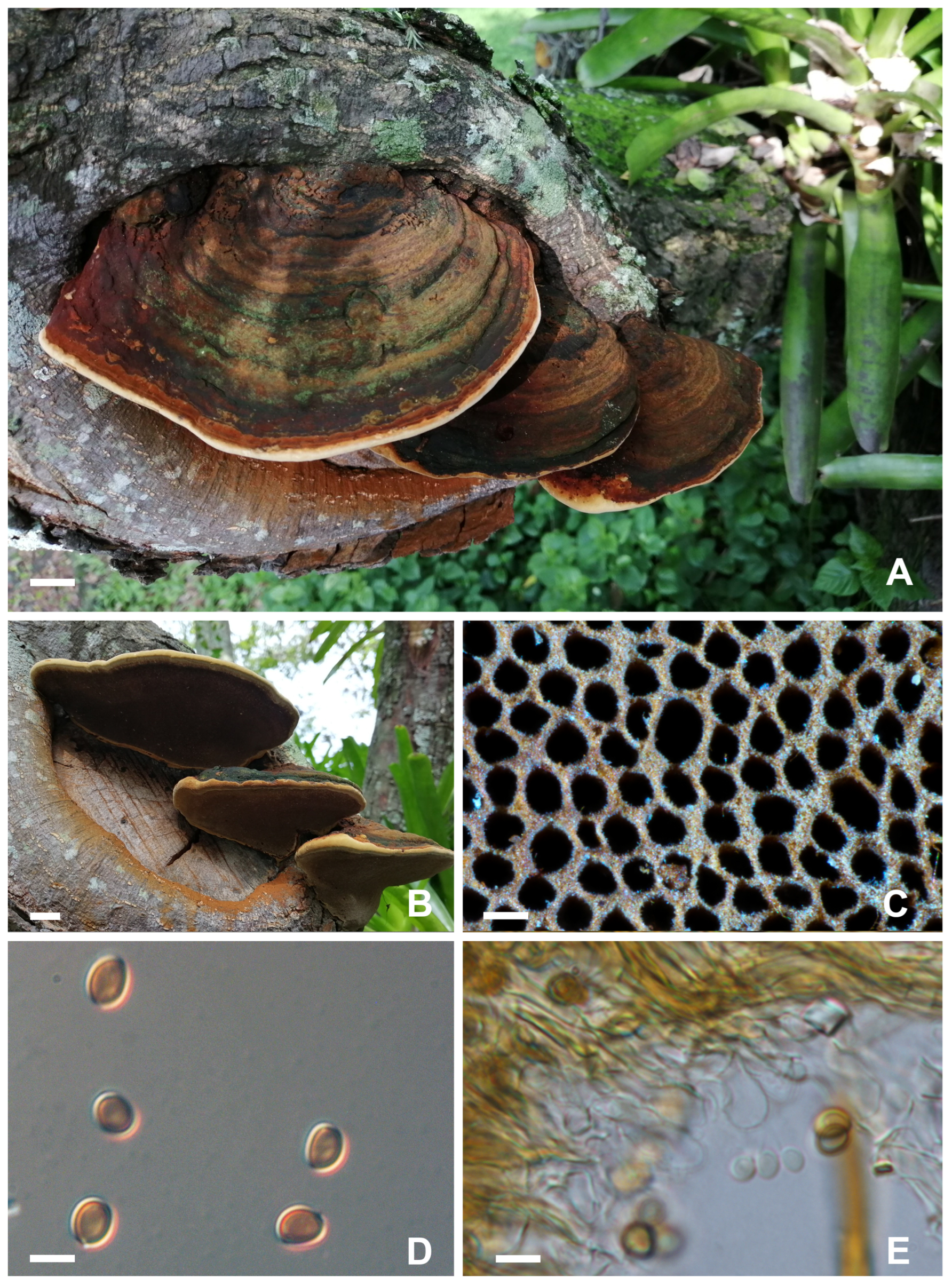
3.1. Taxonomy–Record Description for Phellinotus Teixeirae Salvador-Montoya, Elias and Drechsler-Santos [MycoBank MB840997]

Basidioma was mostly perennial, pileate, up to 105 mm long, 74 mm wide, 50 mm thick at the base, sessile, applanate to triquetrous, sometimes with a basal umbo, woody hard, solitary, or in groups with many basidiomata widespread along the substrate. Margins were convex, entire, thick, pubescent, always distinct from older parts of the basidioma, yellowish when young, then turning brown with age, or when beaten.
Pileus fulvous was brown to black greyish, dull, azonate, sulcate, and strongly cracked. The context was zonate, with black lines below the upper surface of the pilei, a granular core variably present, pale to dark yellowish, becoming black with KOH, and a thick black crust above the context, up to 8 mm thick in mature specimens. Tubes were stratified, with/without contextual tissue layer between them, dark reddish brown, up to 42 mm long. The hymenophore was poroid, with round regular pores, (3-)4-6 per mm, fulvous brown to dark brown, and dissepiments were entire.
The hyphal structure was pseudo-dimitic, with skeletal hyphae restricted to the trama of the tube layer; skeletal hyphae were reddish dark brown, thin- to thick-walled, with a visible lumen, almost solid, (3.5-)3.8-5.5(-6.5) µm wide, some with small aborted ramifications. Generative hyphae dominated in the context, with a simple septate, pale yellow to reddish dark brown, thin- to thick-walled, branched, (2-)2.5-4(-6.2) µm wide. Generative hyphae in the trama were thin- to thick-walled, with a simple septate, hyaline to pale yellow, becoming sclerified in some portions. Setae or other sterile elements were absent. Basidia were (9-)10-12.5(-14) × 4.5-5.8(-6.2) µm. Basidiospores were broadly ellipsoid to ellipsoid, adaxially flattened, smooth, thick-walled, yellow in water to chestnut brown in KOH, (4.5-)5-6.8(-7) × 4–5(-5.5) µm, L = 5.7 W = 4.5 µm, Q’ = 1-1.1(-1.5) µm, Q = 1.2 (n = 354/14), IKI-, CB+.
Regarding habitat, distribution, and host, the species was previously recorded in the SDTF of the Andean valley in northern Peru and southern Ecuador and on the Pacific coast of Ecuador and northern Peru. In this study, P. teixeirae was recorded for the first time in southwest Colombia, growing on living trees of Pythecelobium dulce, a new host. Previously, the species was recorded growing on other species of living trees of Fabaceae such as Pythecelobium excelsum, Libidibia glabrata, and Pityrocarpa moniliformis, in addition to Acacia and Piptadenia species [2].
The material examined, in the Colombian province of Valle del Cauca, Cali (urban perimeter) was from living trees of Pythecelobium dulce, 12 November 2017, leg. Bolaños-Rojas, A.C., Chaurra, A. and Aguilar, G., ACB381 (CUVC 75577); from the Universidad del Valle campus Melendez, from living trees of Pythecelobium dulce, ACB432 (CUVC 75578), 30 November 2018, leg. Bolaños-Rojas, A.C., ACB448 (CUVC 75579), ACB449 (CUVC 75580), ACB455 (CUVC 75581), ACB458 (CUVC 75582), 4 December 2018, leg. Bolaños-Rojas, A.C. and Ramos, A., ACB460 (CUVC 75583), ACB462 (CUVC 75584), 22 January 2019, leg. Bolaños-Rojas, A.C., Gonzalez, L. and Ramos, A., ACB463 (CUVC 75585), ACB496 (CUVC 75586), ACB497 (CUVC 75587, ACB498 (CUVC 75588), ACB842 (CUVC 75589), and 15 March 2021, leg. Motato-Vásquez, V., MV1109 (CUVC 75590).
Notes (Figure 1)—Phellinotus teixeirae was characterized with applanate to ungulate basidiomata with cracked and dark greyish brown pileal surfaces, hymenophore with (3-)4-6 pore per mm, dark lines in the context, a monomitic hyphal system in the context and dimitic in the tubes, absence of setae, and broadly ellipsoid to ellipsoid basidiospores, (4.5-)5-6.8(-7) × 4-5(-5.5) µm, with a flattened side turning darker in KOH solution. The species differs from P. piptadeniae s.s., which has a lobulate and cracked olive grey pileal surface, with deep concentric furrows and wide lobes. Morphological patterns of the pileal surface are useful to split species in Hymenochaetaceae [2]. Phylogenetics corroborate this classification (Figure 2), as detailed in the following section.

3.2. Phylogenetic Relationships

A total of four loci were sequenced for seven Phellinotus isolates including the widely used barcoding ribosomal nuclear regions ITS and nLSU, the coding genes RNA polymerase II second largest subunit (RPB2), and the translation elongation factor (TEF1-α) (Table 1). Twenty, new sequence records were generated comprising seven for ITS, three for nLSU, six for TEF1-α, and four for RPB2. GenBank accession numbers as in Table 1.
The combined dataset with four loci for the ‘Phellinotus Clade’ (ITS + nLSU + TEF1-α-1st + -2nd + -3rd + TEF1-α-introns + RPB2-1st + -2nd + -3rd) included 82 taxa, resulting in an alignment with 3991 positions, of which 2177 were conserved, 441 varied, and 1373 were parsimony-informative. The best tree inferred in a maximum likelihood framework from ten independent runs had a log-likelihood = −50,925.2117. The selected best-fit models were TN + F + I + G4 for ITS, TIM2 + F + R3 for nLSU, GTR + F + I + G4 for TEF1-α in all codons, HKY + F + R2 for TEF1-α-introns, and TIM2+F + I + G4 for RPB2 in all codon positions. The phylogenetic analysis (Figure 2) was agreed with previous studies [2,3,6,7].
Seven genera were recovered as monophyletic lineages with strong support, all recognized as the ‘Phellinotus Clade’: Arambarria (BS = 100, SH alRT = 100), Inocutis (BS = 100, SH alRT = 100), Fomitiporella s.s. (BS = 98, SH alRT = 90), Fulvifomes (BS = 100, SH alRT = 100), Phellinotus (BS = 96, SH alRT = 92.6), Phylloporia (BS = 100, SH alRT = 100), and Rajchenbergia (BS = 99, SH alRT = 95.3), and the reminiscent groups of Fomitiporella s.l.
Both phylogenetic and morphological analyses confirmed that the collected specimens, growing on living trees of Pithecellobium dulce (Roxb.) Benth in the SDTF of Valle del Cauca, Colombia, corresponded to the species Phellinotus teixeirae (BS = 100, SH alRT = 100). Therefore, this is the first record of the species and the genus in Colombia. This result extends the distribution range of P. teixeirae approximately 1000 km north in the Neotropics. The new host, P. dulce, classified in the subfamily Mimosoideae–Fabaceae, is a species widely distributed in the SDTF of Colombia [44]. The tree species is native to the Pacific coast, the foothills of the highlands in Mexico, Central America, and northern South America [45], yet, it is present too in India, the Philippines, and East Africa [46,47,48].

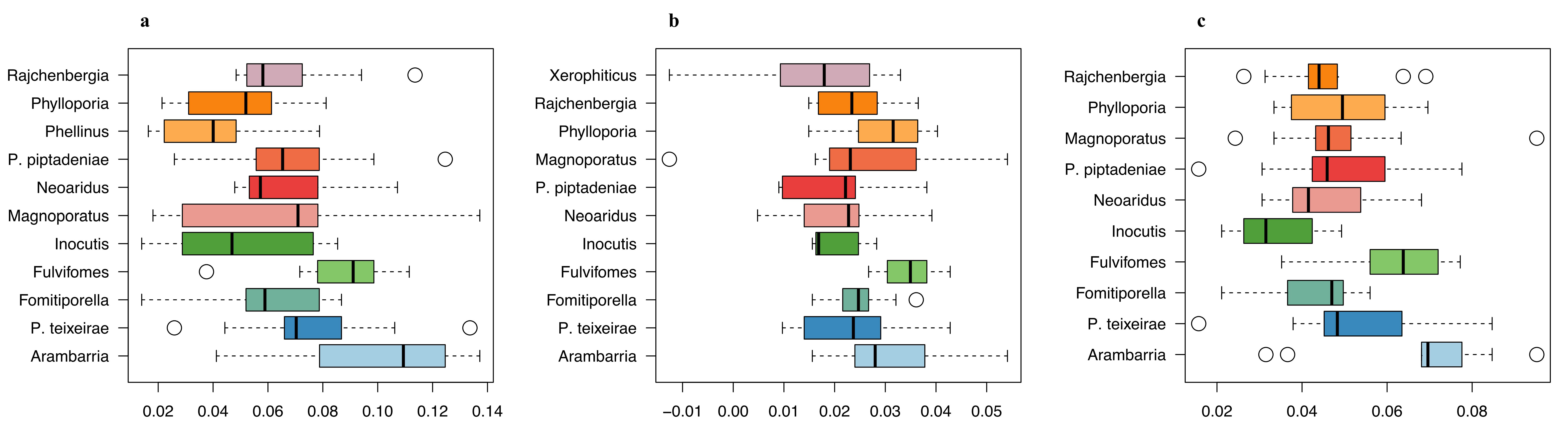
3.3. Patterns of Nucleotide Diversity

Considering the limiting information available in the databases for alternative loci (i.e., TEF1-α, RPB2) in the ‘Phellinotus Clade’, most of the analyses focused on ITS and nLSU loci. Nucleotide parameters are summarized in Table 2. The calculated value of Tajima’s D was significantly negative at ITS for P. piptadeniae; P. neoaridus, P. piptadeniae, and P. teixeirae also had highly significant negative values at the LSU locus, indicating possible signals of population expansion. Coding DNA genes RBP2 and TEF exhibited positive and negative Tajima’s D values, respectively, although these were non-significant.
Fixation indices were computed for each species in the ‘Phellinotus Clade’ using the total variance detected among the analyzed loci (i.e., ITS, nLSU, and ITS + nLSU). Boxplots for FST scores summarize pairwise species comparisons against one taxonomic group at a time (Figure 3a–c). Differences among boxplots, that is FST differentiation of classifications against the rest, were not statistically significant. The ITS locus displayed the highest FST median values (≥0.04), with Arambarria yielding moderate genetic differentiation (median ≥ 0.11) and wide variance in allele frequency (Figure 3a). Contrastingly, the nLSU locus contained low variation, both within and between species, with median values ranging from 0.02 to <0.04 (Figure 3b). The concatenated dataset differentiated Arambarria, with median values generally between 0.04 and 0.08, indicating small genetic differentiation among groups (Figure 3c).
Heatmap analysis, based on FST values in the ITS, LSU, and concatenated datasets, also clustered all isolates into the P. teixeirae clade (Figure 4a–c). Arambarria and Fulvifomes groups were distant in the ‘Phellinotus Clade’, whilst P. teixeirae and P. piptadeniae were genetically similar, based on the ITS (Figure 4a) and concatenated (Figure 4c) datasets, providing similar cluster topologies. LSU-based clustering indicated similar allele frequencies with low nucleotide variation, limiting the discriminatory power of this locus for recent evolutionary events within species (Figure 4b), yet with enough resolution for species discrimination within the ‘Phellinotus Clade’.

3.4. Analysis of Molecular Variance

Analysis of MOlecular VAriance (AMOVA) was performed to determine species boundaries, and the possible cluster differentiation was based on the region of origin. Through this process, we prepared a stratified hierarchical AMOVA categorizing species and country-of-origin levels. The final dataset contained four species groups and five localities regarding the country of origin (Argentina, Brazil, Colombia, Peru, and Uruguay). The independent and combinational effects were evaluated to understand the percentage of variation at significant p-value thresholds of 0.05 (Table 3). The results showed a high contribution of the species partition, with an explained variance of 70% and a p-value of 0.001, higher compared to the country of origin within species that only accounted for 28% of the variation (p-value 0.0009). Meanwhile, the locality level explained 11.1% of the within-species variation (p-value 0.04). In general, differences between species contributed to most of the distinction. As the variation within species was lower than between species, ribosomal loci offered a clear taxonomic distinction.

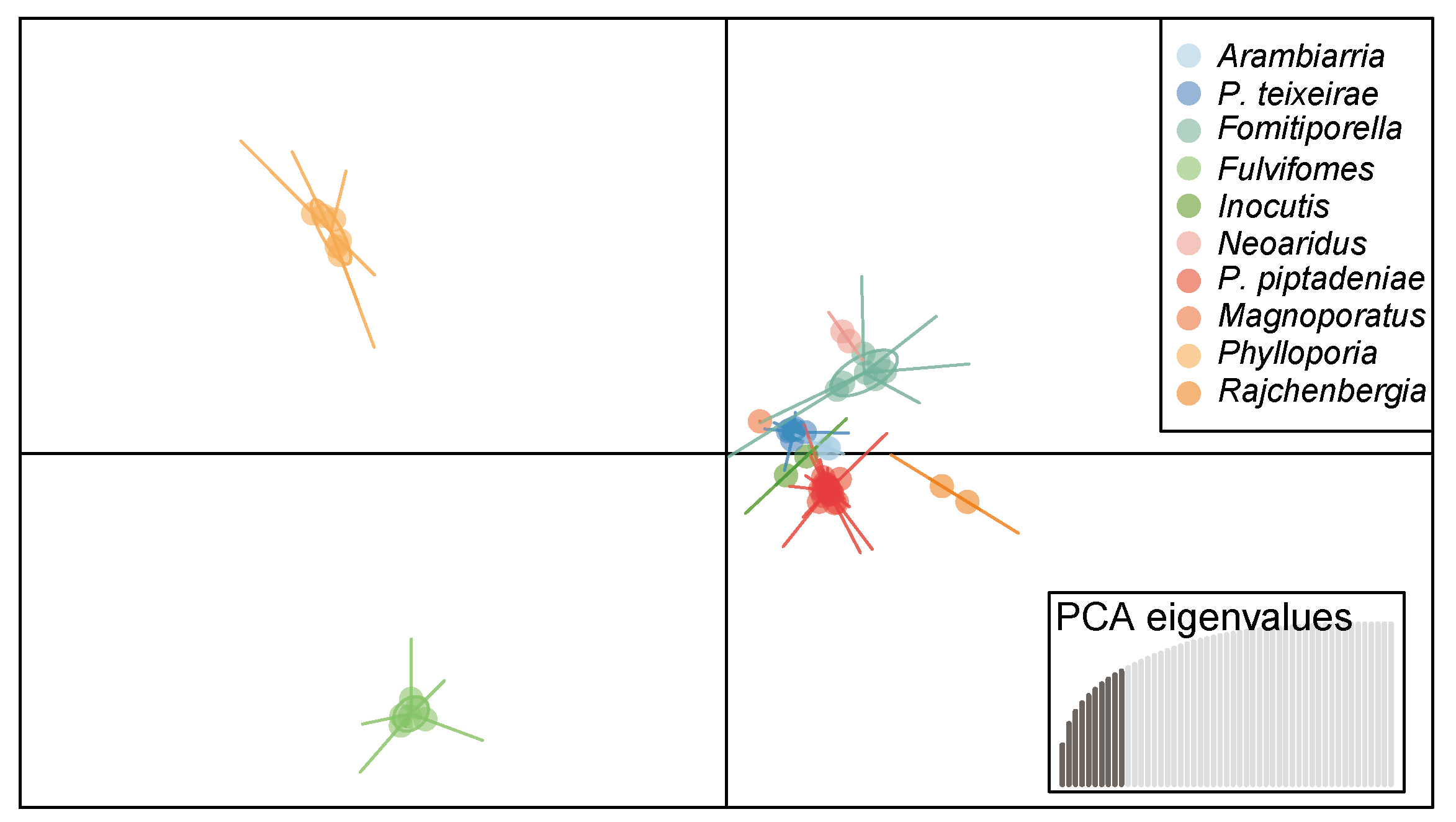
3.5. Discriminant Analysis of Principal Components

Discriminant Analysis for Principal Components (DAPC) was carried out separately for each dataset. Genetic variation was partitioned into between- and within-group components. The isolates obtained here were clustered with the P. teixeirae group for the three datasets, ITS, LSU, and concatenated, denoting similarity in allele frequencies.
The ITS locus explained > 60% of the genetic diversity by retaining the first 15 components, enabling discrimination of four groups and differentiating P. teixeirae, Fulvifomes sp., and Phylloporia sp. into the ‘Phellinotus Clade’. Additionally, the LSU locus explained > 80% of the genetic similarity condensed in the first 15 components. However, it provided lower discriminatory power, displaying only three well-differentiated groups, Fulvifomes sp. being the most divergent cluster. For the concatenated dataset, 10 components were retained that explained > 60% of the genetic diversity, discriminating between Fulvifomes sp. and Phylloporia sp. Most of the Phellinotus species were closely related according to the ITS analysis (Figure 5).

3.6. Patterns of SNP Allelic Distribution in Phellinotus Species

To investigate the number of SNP loci (unique and shared) in the different species analyzed here, comprising 26 samples distributed in four species, 189 SNP loci were retained at a Minor Allele Frequency (MAF) ≥ 0.05, computed in Tassel v5 [49], from 1738 nucleotide sites in the original concatenated dataset (ITS + nLSU). Most of the SNPs were recovered from the ITS locus (118), representing 62.4%. Large variation was observed between species. For instance, P. neoaridus contained the highest number of unique alleles (85), followed by P. magnoporatus (80), P. teixeirae, and P. piptadeniae (51) (Figure 6). Overall, the pattern of shared alleles showed large variation among the four Phellinotus species. Phellinotus piptadeniae and P. teixeirae had the most shared alleles (a total of 40), compared to P. magnoporatus and P. piptadeniae with nine alleles. Between all species, only 15 alleles were shared, corresponding to 7.93%. Overall, there were more shared alleles between P. teixeirae and P. piptadeniae than between any other compared species, whereas P. magnoporatus and P. neoaridus showed the lowest frequency of shared alleles across all analyzed species pairs.

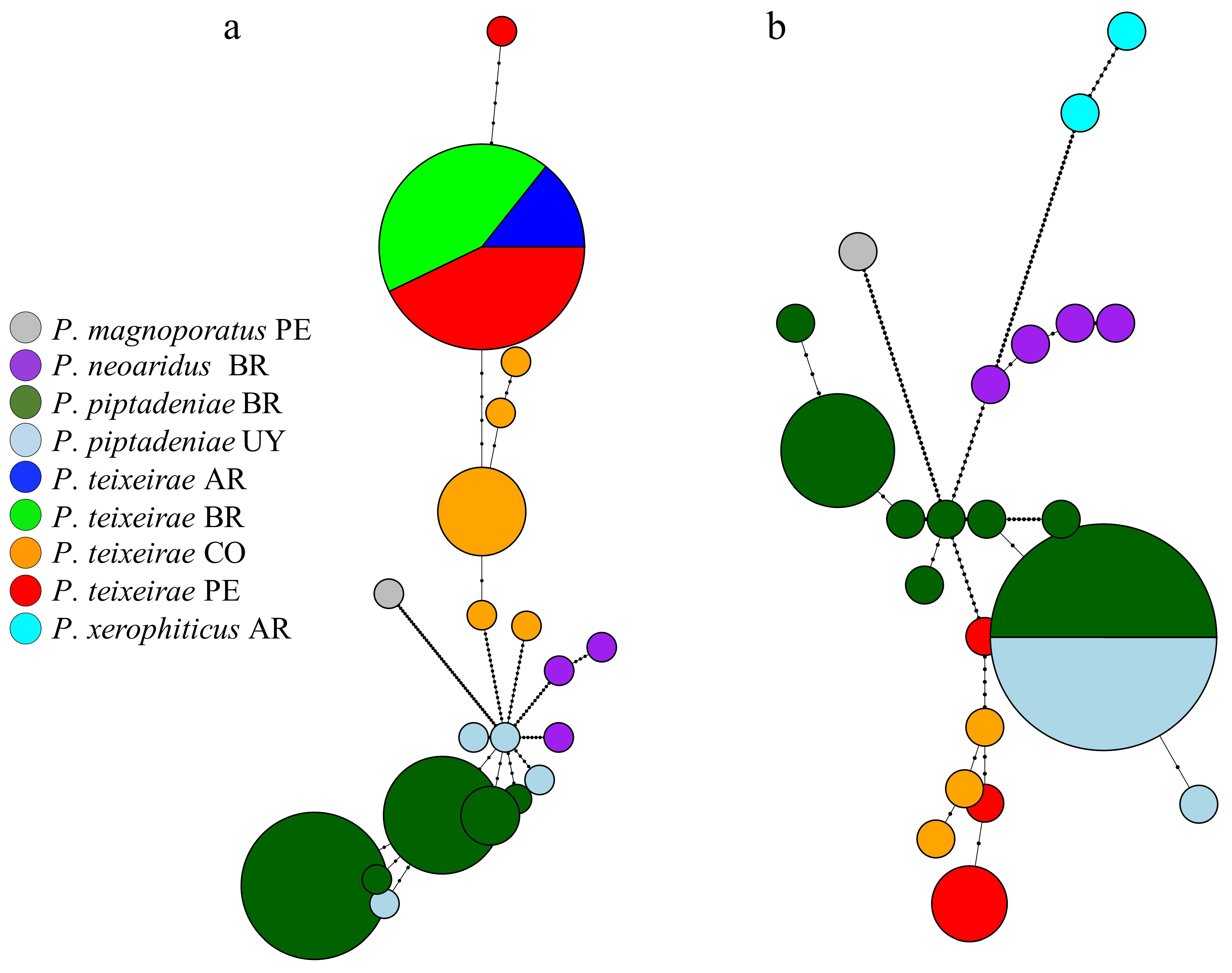
3.7. Network Analysis and Haplotype Distribution of Phellinotus Species

To detect the number of haplotypes per species and their distribution in the region, haplotype network reconstructions were made for ITS (Figure 7a) and nLSU (Figure 7b) separately marking the sampling origin of the four Phellinotus species. For ITS and LSU loci, the same species were included, except for LSU in which P. xerophiticus was additionally incorporated. A total of twenty haplotypes were identified for the ITS locus, distributed into nine haplotypes for P. piptadeniae, seven for P. teixeirae, three for P. neoaridus, and one for P. magnoporatus. For the LSU locus, twenty-two haplotypes were recognized, including nine for P. piptadeniae, six for P. teixeirae, four for P. neoaridus, two for P. xerophiticus, and one for P. magnoporatus. Haplotype distribution for ITS and LSU indicated a high frequency across countries for P. piptadeniae and P. teixeirae. Regarding the country of origin, the identified notes displayed a clear distinction between Brazilian and Uruguayan isolates for P. piptadeniae at the ITS locus. For P. teixeirae, the Colombian isolates were discriminated through the two loci, generating five nodes for ITS and three nodes for LSU. Interestingly, by following the haplotype connections (i.e., mutational events between pairs of nodes) over the geographic regions, it was possible to identify that the isolates of P. teixeirae registered for Colombia in the present study were closely related to the haplotypes reported in Peru and Brazil according to the ITS, and to the ones from Peru, based on the LSU.

4. Discussion

Our phylogenetic analysis of the combined dataset across four loci (ITS, nLSU, TEF1-α, and RPB2) recovered the ‘Phellinotus Clade’ genera (i.e., Arambarria, Inocutis, Fomitiporella s.s., Fulvifomes, Phylloporia and Rajchenbergia) as a monophyletic group with high support, corroborating the hypothesis proposed by Pildain et al. [6] and Salvador-Montoya et al. [2]. Yet, in the current study the genus Phellinotus was discovered to form a sister clade with Rajchenbergia, which also presents a dark line and mycelial core in the context, but differs through having a monomitic hyphal structure in the basidiomata [7]. In contrast, Salvador-Montoya et al. [2] showed that Phellinotus forms a sister clade with Rajchenbergia + Fomitiporella s.s., which indicates that it is still necessary to add different molecular markers for future analysis to further elucidate the underlying phylogenetic relationships of Phellinotus within the clade.
The genus Phellinotus is considered a pathogenic polypore, commonly living on members of the family Fabaceae. Morphologically speaking, species of Phellinotus are classified and can be recognized through the presence of a dark line, a mycelial core, a dimitic hyphal structure with skeletal hyphae only present in the trama of the tube layer and adaxially flattened, ellipsoid, and thick-walled, the absence of setae or setal hyphae, and pale yellow basidiospores that turn chestnut in KOH solution [2]. In Hymenochaetaceae, Phellinotus was traditionally considered a genus with a Neotropical distribution based on phylogenetic and phylogeographic analyses [3]. However, Salvador-Montoya et al. [2], based on morphological analysis of P. badius (Berk. ex Cooke) Salvador-Montoya, Popoff and Dreschler-Santos, P. resinaceus (Kotl. & Pouzar) Salvador-Montoya and Dreschler-Santos, and P. scaber (Berk.) Salvador-Montoya, Dreschler-Santos and Popoff, reported the expanded distribution of the genus further into the tropical and subtropical climatic zones of America and Australia.
Species of Phellinotus have been widely studied in South America (i.e., [1,2,3,7], mainly P. Piptadeniae). Recently, Salvador-Montoya et al. [2] showed that P. teixeirae conforms an entirely different entity from P. piptadeniae s.s., and showed that the former is distributed within the limits of SDTFs in South America, growing on living tree species of the Fabaceae family such as Pithecellobium excelsum, Libidibia glabrata, and Pityrocarpa moniliformis. These three species are distributed within the limits of SDTFs, which makes them ecologically related to some degree [50]. Hence, several authors have considered P. teixeirae as an SDTF host specialist. In this study, P. teixeirae was recorded growing on Pithecellobium dulce, one of the most representative trees in the SDTF biome in the inter-Andean valley of Valle del Cauca, Colombia. Pithecellobium dulce is commonly and locally known as ‘Chiminango’. It is a tree that has several uses, such as for living fences, posts, shade, medicine, utensils, firewood [51], edible fruits, construction, fodder [52,53], and in the urban arborization of many cities in Colombia.
Morphologically, P. teixeirae differs from P. piptadeniae s.s. because the latter presents a lobulated and cracked olive-gray pileal surface with deep concentric furrows and broad lobes. The morphological patterns of the pileal surface have been documented as useful characteristics to distinguish species in the Hymenochaetaceae (i.e., Fulvifomes species; [8]). Furthermore, P. piptadeniae s.s. is distributed both in dry and wet forests in eastern South America, unlike P. teixeirae, considered as an SDTF host habitat specialist (i.e., it grows restrictedly in SDTF habitats [2]). The current results reinforce that specimens and taxa from other unexplored regions of the world should continue being included in phylogenetic analysis to understand in more detail the diversity of the genus and the natural classification of Hymenochaetaceae.

4.1. Patterns and Processes of Genetic Diversity in Phellinotus Species

Due to uncertainties in the species classification within the ‘Phellinotus Clade’, the application of molecular markers has provided a more precise assessment of species assignation and diversity [6], especially considering recently discovered species [3], and the developing understanding of the ecology and evolution of these complex taxa for which morphological validation is often lacking. The phylogenetic studies on the ‘Phellinotus Clade’ [1,2,3,5,54] demonstrated instability, although the application of several morphological and molecular descriptors has provided more robust alternative inferences. The application of multi-locus molecular markers is a tool that can potentially further unveil fungal phylogenetic patterns [6]. Here, we have reported an alternative perspective on genetic diversity in the ‘Phellinotus Clade’ by screening the most common loci used for fungal molecular systematic analysis [55].
Multiple molecular approaches have been utilized to understand the diversity of the recently discovered genus Phellinotus [3], which is proposed to be part of a species complex including still unresolved taxa (i.e., Fomitiporella, Fulvifomes, Inocutis, and Phylloporia). Fortunately, highly supported phylogenetic nodes in our multi-locus reconstructions reinforce the monophyletic hypothesis of the clade [6]. The specimens all coincided in showing high variability at the surveyed loci, as previously reported in other phylogenetic analyses [3]. These molecular markers provide robust species delimitations for P. piptadeniae and P. teixeirae. To continue unveiling the patterns of genetic diversity and identification of new P. teixeirae strains, an alternative analytical approach, not only for P. teixeirae but also for the ‘Phellinotus Clade’, was incorporated, As detailed below.
Interestingly, high nucleotide and haplotype diversity, and inflated Tajima’s D scores at the rDNA loci are indicative of divergent selection as part of the population expansion towards the northern Andes while involving colonization and adaptation to new hosts [56]. These levels of nucleotide diversity could, in turn, harness species delimitation between the closely related taxa P. piptadeniae and P. teixeirae. Similarly, the high nucleotide diversity found in P. piptadeniae indicates that clusters of divergent sequences occur within the species, possibly as a consequence of geographic distance, environmental local adaptation (considering its disjunct distribution [1]), divergent host interactions, or ancient intraspecific rDNA polymorphisms [57,58]. For P. teixeirae, the nucleotide variation was very low between our isolates and previous reports, despite the new host interactions with Pithecellobium dulce. These contrasting outcomes have been identified in eukaryotic genomes [59,60,61,62,63], particularly coupled with considerable variation in gene copy number at rDNA loci in fungal species [57,58,64,65] and intragenomic variation at these loci [58,64,65]. The dynamics of these variations complicate the analysis of rDNA amplicons, being a critical factor for species delimitation, especially for microbial communities in heterogeneous environments [6]. Additionally, the persistence of rDNA variants through lineages that exhibit optimal fitness to spread over new hosts and distant geographic regions has been found to be the result of purifying selection and concerted evolution [60,61,66,67].
Through analyzing the haplotype networks of the most representative species in this study, i.e., P. piptadeniae and P. teixerae, it was found that P. piptadeniae was the basal haplotype. The reticulated network spread outwards from a single P. piptadeniae haplotype, involving few mutational points among P. piptadeniae isolates, yet considerably increasing from closely (i.e., P. teixeirae and P. neoaridus) to highly (P. magnoporatus and P. xerophiticus) divergent sequences. The patterns of the haplotype network found for P. teixeirae in Colombia are closely related to the isolates from Peru, exhibiting possible correlations with ecological signatures of new host colonization since the Colombian haplotypes are unique. The newly identified P. teixeirae isolates provide clues for the population expansion hypothesis towards the northern Andes following the geographic equivalency [1] associated with the SDTF [5]. The identification of dominant, widespread haplotypes for P. teixeirae and P. piptadeniae correlates with the assumption of new variant acquisition with potential adaptation to novel hosts and environments [68], especially in highly tree-species-diverse Neotropical ecosystems [69].
Although some of these fungal species have been reported to parasite specific hosts [5,54], with a narrow distribution range [3], this perspective is changing based on the integration of new species into the genus [2], host records [1,5], and the identification of new strains supporting its potential broad distribution. New Phellinotus records indicate a broad spectrum of tree species serving as hosts, with a predominant parasitic behavior for species of the Fabaceae family, probably a result of its geographical predominance in the SDTF biome.

4.2. Perspectives

More intense sampling is necessary to support the population expansion hypothesis and the possible genetic adaptation to new hosts. Considering the defense mechanisms of plants driven via secondary metabolites [70], these fungal species are particularly interesting as model organisms to study the metabolic strategies underlying parasitism on novel hosts. Also, mapping the genetic variation responsible for host colonization could provide a more detailed comprehension of the population diversity patterns in Phellinotus species, and whether they are congruent with selection pressures acting on de novo mutations or recruiting standing adaptive variation. Assessing these scenarios would help build a more cohesive understanding of the mechanisms favoring new host colonization in the face of climate change and the rapid niche variation in the SDTF [71].
The application of novel sequencing technologies further provides an alternative method to study the genetic traits under selection during a co-evolving parasitic biotic interaction [72], with potential detection of species-specific genes, metabolites, and interspecific gene transfer among closely related species [70,73]. Although the application of barcoding genes has been useful for species identification in genetically structured populations [74], more notably for some groups of fungi than others [75], exploration of additional loci may also have potential applications for sub-generic-level identification [76] in intricate taxa, in addition to accounting for [77] and reconstructing [78] the complexity of the underlying demographic histories.

5. Conclusions

Pithecellobium dulce (Fabaceae) as a new host tree of the wood-decay fungus P. teixeirae in the northern Andes broadens the perspective of the potential distribution of the recently discovered Phellinotus genus in the Neotropics, extending its distribution range 1000 km north. These results suggest that Phellinotus species could be undergoing population expansion following the geographic equivalency associated with the SDTF biome, making the group phylogenetically complex, and potentially subject to multiple local selection pressures driving adaptation. The incorporation of new technologies in the study of this taxa would provide new insights for the discovery of new species and their potential adaptation to new environments.

Author Contributions

A.C.B.-R. conceived and designed the project, directed all field sampling, and collected fungal material. A.C.B.-R. and V.M.-V. carried out all the laboratory procedures including strain isolation, morphological characterization, and molecular evaluation. A.C.B.-R., V.M.-V. and J.M.L.-C. analyzed all sequence data, and together with A.J.C., performed the interpretation of the genetic results. A.C.B.-R., V.M.-V., J.M.L.-C., and A.J.C. contributed to the overall discussion of the results, as well as to the writing of the manuscript. A.C.B.-R., in her role as principal investigator (PI), provided all the resources required to accomplish the research, and supervised the execution of the project. A.J.C. also arranged funding resources to cover the articles’ APC. All authors have read and agreed to the published version of the manuscript.

Funding

The research was partly supported by the “Fondo Nacional de Financiamiento para la Ciencia, la Tecnología y la Innovación, Francisco José de Caldas” from MinCiencias under contract number 6482020 (Universidad del Valle project number 71257), and project title “Hongos corticioides (Russulales, Basidiomycota) degradadores de madera: diversidad, filogenia y potencial enzimático en ecosistemas amenazados del Valle del Cauca, Colombia”. A.J.C. acknowledges support from Vetenskapsrådet and Kungliga Vetenskapsakademien through grants 2016-00418/2022-04411 and BS2017-0036, respectively.

Institutional Review Board Statement

Not applicable.

Informed Consent Statement

Not applicable.

Data Availability Statement

R codes are available upon request.

Acknowledgments

We acknowledge to the “Fondo Nacional de Financiamiento para la Ciencia, la Tecnología y la Innovación, Francisco José de Caldas” from MinCiencias for funding the project “Hongos corticioides (Russulales, Basidiomycota) degradadores de madera: diversidad, filogenia y potencial enzimático en ecosistemas amenazados del Valle del Cauca, Colombia”. We are also grateful to Andrés Ramos, Laura González, and Maria Fernanda Hincapie for their valuable help during fieldwork. Special thanks to Universidad del Valle for providing all laboratory support to fulfill this study. Authors acknowledge three independent readers for the valuable comments and suggestions to improve the paper.

Conflicts of Interest

Authors declare that the research was conducted in the absence of any commercial or financial relationships that could be constructed as a potential conflict of interest.

References

  1. Elias, S.G.; Salvador-Montoya, C.A.; Costa-Rezende, D.H.; Guterres, D.C.; Fernandes, M.; Olkoski, D.; Klabunde, G.H.F.; Drechsler-Santos, E.R. Studies on the biogeography of Phellinotus piptadeniae (Hymenochaetales, Basidiomycota): Expanding the knowledge on its distribution and clarifying hosts relationships. Fungal Ecol. 2020, 45, 100912. [Google Scholar] [CrossRef]
  2. Salvador-Montoya, C.A.; Elias, S.G.; Popoff, O.F.; Robledo, G.L.; Urcelay, C.; Góes-Neto, A.; Martínez, S.; Drechsler-Santos, E.R. Neotropical Studies on Hymenochaetaceae: Unveiling the Diversity and Endemicity of Phellinotus. J. Fungi 2022, 8, 216. [Google Scholar] [CrossRef] [PubMed]
  3. Drechsler-Santos, E.R.; Robledo, G.L.; Limajúnior, N.C.; Malosso, E.; Reck, M.A.; Gibertoni, T.B.; De Queiroz Cavalcanti, M.A.; Rajchenberg, M. Phellinotus, a new neotropical genus in the Hymenochaetaceae (Basidiomycota, Hymenochaetales). Phytotaxa 2016, 261, 218–239. [Google Scholar] [CrossRef]
  4. Drechsler-Santos, E.R.; Santos, P.J.P.; Gibertoni, T.B.; Cavalcanti, M.A.Q. Ecological aspects of Hymenochaetaceae in an area of Caatinga (semi-arid) in Northeast Brazil. Fungal Divers. 2010, 42, 71–78. [Google Scholar] [CrossRef]
  5. Salvador-Montoya, C.A.; Robledo, G.L.; Cardoso, D.; Borba-Silva, M.A.; Fernandes, M.; Drechsler-Santos, E.R. Phellinus piptadeniae (Hymenochaetales: Hymenochaetaceae): Taxonomy and host range of a species with disjunct distribution in South American seasonally dry forests. Plant Syst. Evol. 2015, 301, 1887–1896. [Google Scholar] [CrossRef]
  6. Pildain, M.B.; Cendoya, R.R.; Ortiz-Santana, B.; Becerra, J.; Rajchenberg, M. A discussion on the genus Fomitiporella (Hymenochaetaceae, hymenochaetales) and first record of F. americana from southern South America. MycoKeys 2018, 38, 77–91. [Google Scholar] [CrossRef] [PubMed]
  7. Salvador-Montoya, C.A.; Popoff, O.F.; Góes-Neto, A.; Drechsler-Santos, E.R. Global phylogenetic and morphological reassessment of Fomitiporella s.l. (Hymenochaetales, Basidiomycota): Taxonomic delimitation of Fomitiporella s.s. and segregation of Rajchenbergia, gen. nov. Plant Syst. Evol. 2020, 306, 1–27. [Google Scholar] [CrossRef]
  8. Salvador-Montoya, C.A.; Martínez, M.; Drechsler-Santos, E.R. Taxonomic update of species closely related to Fulvifomes robiniae in America. Mycol. Prog. 2022, 21, 95. [Google Scholar] [CrossRef]
  9. Floudas, D.; Binder, M.; Riley, R.; Barry, K.; Blanchette, R.A.; Henrissat, B.; Martínez, A.T.; Otillar, R.; Spatafora, J.W.; Yadav, J.S.; et al. The paleozoic origin of enzymatic lignin decomposition reconstructed from 31 fungal genomes. Science 2012, 336, 1715–1719. [Google Scholar] [CrossRef]
  10. Larsson, K.H.; Parmasto, E.; Fischer, M.; Langer, E.; Nakasone, K.K.; Redhead, S.A. Hymenochaetales: A molecular phylogeny for the hymenochaetoid clade. Mycologia 2006, 98, 926–936. [Google Scholar] [CrossRef]
  11. Zhou, L.W.; Dai, Y.C. Phylogeny and taxonomy of Phylloporia (Hymenochaetales): New species and a worldwide key to the genus. Mycologia 2012, 104, 211–222. [Google Scholar] [CrossRef] [PubMed]
  12. He, S.H.; Dai, Y.C. Taxonomy and phylogeny of Hymenochaete and allied genera of Hymenochaetaceae (Basidiomycota) in China. Fungal Divers. 2012, 56, 77–93. [Google Scholar] [CrossRef]
  13. Amalfi, M.; Decock, C. Fomitiporia castilloi sp. nov. and multiple clades around F. apiahyna and F. texana in meso-and South America evidenced by multiloci phylogenetic inferences. Mycologia 2013, 105, 873–887. [Google Scholar] [CrossRef] [PubMed]
  14. Tian, X.M.; Yu, H.Y.; Zhou, L.W.; Decock, C.; Vlasák, J.; Dai, Y.C. Phylogeny and taxonomy of the Inonotus linteus complex. Fungal Divers. 2013, 58, 159–169. [Google Scholar] [CrossRef]
  15. Zhou, L.W.; Qin, W.M. Phylogeny and taxonomy of the recently proposed genus phellinopsis (Hymenochaetales, Basidiomycota). Mycologia 2013, 105, 689–696. [Google Scholar] [CrossRef] [PubMed]
  16. Zhou, L.-W. Fulvifomes hainanensis sp. nov. and F. indicus comb. nov. (Hymenochaetales, Basidiomycota) evidenced by a combination of morphology and phylogeny. Mycoscience 2014, 55, 70–77. [Google Scholar] [CrossRef]
  17. Zhu, L.; Song, J.; Zhou, J.L.; Si, J.; Cui, B.K. Species diversity, phylogeny, divergence time, and biogeography of the genus sanghuangporus (Basidiomycota). Front. Microbiol. 2019, 10, 812. [Google Scholar] [CrossRef] [PubMed]
  18. Neves, M.A. Guide to the common fungi of the semiarid region of Brazil. In Common Fungi of the Semiarid Region of Brazil; Neves, M.A., Baseia, I., Dreschler-Santos, E., Góes-Neto, A., Eds.; TECC Editora: Florianópolis, Brazil, 2013; p. 131. [Google Scholar]
  19. Riggio, J.; Baillie, J.E.M.; Brumby, S.; Ellis, E.; Kennedy, C.M.; Oakleaf, J.R.; Tepe, T.; Theobald, D.M.; Venter, O.; Watson, J.E.M.; et al. Global human influence maps reveal clear opportunities in conserving Earth’s remaining intact terrestrial ecosystems. Glob. Chang. Biol. 2020, 26, 4344–4356. [Google Scholar] [CrossRef]
  20. Ocón, J.P.; Ibanez, T.; Franklin, J.; Pau, S.; Keppel, G.; Rivas-Torres, G.; Shin, M.E.; Gillespie, T.W. Global tropical dry forest extent and cover: A comparative study of bioclimatic definitions using two climatic data sets. PLoS ONE 2021, 16, 0252063. [Google Scholar] [CrossRef]
  21. Etter, A.; Andrade, A.; Saavedra, K.; Cortés, J. Actualización de la Lista Roja de los Ecosistemas Terrestres de Colombia: Conocimiento del riesgo de ecosistemas como herramienta para la gestión. In Estado y Tendencias de la Biodiversidad Continental de Colombia; Moreno, L.A., Rueda, C., Andrade, G.I., Eds.; Instituto de Investigación de Recursos Biológicos Alexander von Humboldt: Bogotá, DC, USA, 2017. [Google Scholar]
  22. Gómez-Montoya, N.; Ríos Sarmiento, C.; Zora-Vergara, B.; Benjumea-Aristizabal, C.; Santa-Santa, D.J.; Zuluaga-Moreno, M.; Franco-Molano, A.E. Diversidad de macrohongos (Basidiomycota) de Colombia: Listado de especies. Actual. Biológicas 2022, 44, 1–94. [Google Scholar] [CrossRef]
  23. ColFungi. Useful Fungi of Colombia. Facil by R Bot Gard Kew Publ Internet. Available online: https://colfungi.org/2023 (accessed on 10 July 2023).
  24. Castellani, A. Maintenance and cultivation of common pathogenic fungi in man in sterile distilled water: Further resaerches. J. Trop. Med. Hyg. 1967, 70, 181–184. [Google Scholar]
  25. Miettinen, O.; Niemelä, T.; Spirin, W. Northern Antrodiella species: The identity of A. semisupina, and type studies of related taxa. Mycotaxon 2006, 96, 211–239. [Google Scholar]
  26. Miettinen, O.; Vlasák, J.; Rovoire, B.; Spirin, V. Fungal Systematics and Evolution VOLUME. Fungal Syst. Evol. 2018, 1, 169–215. [Google Scholar]
  27. White, T.J.; Bruns, T.; Lee, S.; Taylor, J. Amplification and Direct Sequencing of Fungal Ribosomal Rna Genes for Phylogenetics. In PCR Protocols; Innis, M.D.H.G., Sninsky, J., White, T., Eds.; Academic Press: Cambridge, MA, USA, 1990; pp. 315–322. [Google Scholar] [CrossRef]
  28. Haynes, K.A.; Westerneng, T.J.; Fell, J.W.; Moens, W. Rapid detection and identification of pathogenic fungi by polymerase chain reaction amplification of large subunit ribosomal DNA. Med. Mycol. 1995, 33, 319–325. [Google Scholar] [CrossRef] [PubMed]
  29. Vilgalys, R.; Hester, M. Rapid Genetic Identification and Mapping of Enzymatically Amplified Ribosomal DNA from SeveralCryptococcus Species. J. Bacteriol. 1990, 172, 4238–4246. [Google Scholar] [CrossRef] [PubMed]
  30. Brandon Matheny, P.; Wang, Z.; Binder, M.; Curtis, J.M.; Lim, Y.W.; Henrik Nilsson, R.; Hofstetter, V.; Ammirati, J.F.; Schoch, C.L.; Langer, E.; et al. Contributions of rpb2 and tef1 to the phylogeny of mushrooms and allies (Basidiomycota, Fungi). Mol. Phylogenet. Evol. 2007, 43, 430–451. [Google Scholar] [CrossRef] [PubMed]
  31. Oghenekaro, A.O.; Miettinen, O.; Omorusi, V.I.; Evueh, G.A.; Farid, M.A.; Gazis, R.; Asiegbu, F.O. Molecular phylogeny of Rigidoporus microporus isolates associated with white rot disease of rubber trees (Hevea brasiliensis). Fungal Biol. 2014, 118, 495–506. [Google Scholar] [CrossRef] [PubMed]
  32. Ewing, B.; Green, P. Base-calling of automated sequencer traces using phred. II. Error probabilities. Genome Res. 1998, 8, 186–194. [Google Scholar] [CrossRef] [PubMed]
  33. Gordon, D.; Abajian, C.; Green, P. Consed: A Graphical Tool for Sequence Finishing. Genome Res. 1998, 8, 195–202. [Google Scholar] [CrossRef]
  34. Gordon, D.; Green, P. Consed: A graphical editor for next-generation sequencing. Bioinformatics 2013, 29, 2936–2937. [Google Scholar] [CrossRef]
  35. Katoh, K.; Standley, D.M. MAFFT multiple sequence alignment software version 7: Improvements in performance and usability. Mol. Biol. Evol. 2013, 30, 772–780. [Google Scholar] [CrossRef]
  36. Larsson, A. AliView: A fast and lightweight alignment viewer and editor for large datasets. Bioinformatics 2014, 30, 3276–3278. [Google Scholar] [CrossRef] [PubMed]
  37. Nguyen, L.T.; Schmidt, H.A.; Von Haeseler, A.; Minh, B.Q. IQ-TREE: A fast and effective stochastic algorithm for estimating maximum-likelihood phylogenies. Mol. Biol. Evol. 2015, 32, 268–274. [Google Scholar] [CrossRef] [PubMed]
  38. Kalyaanamoorthy, S.; Minh, B.Q.; Wong, T.K.F.; Von Haeseler, A.; Jermiin, L.S. ModelFinder: Fast model selection for accurate phylogenetic estimates. Nat. Methods 2017, 14, 587–589. [Google Scholar] [CrossRef] [PubMed]
  39. Hoang, D.T.; Chernomor, O.; von Haeseler, A.; Minh, B.Q.; Vinh, L.S. UFBoot2: Improving the Ultrafast Bootstrap Approximation. Mol. Biol. Evol. 2018, 35, 518–522. [Google Scholar] [CrossRef] [PubMed]
  40. Guindon, S.; Dufayard, J.F.; Lefort, V.; Anisimova, M.; Hordijk, W.; Gascuel, O. New algorithms and methods to estimate maximum-likelihood phylogenies: Assessing the performance of PhyML 3.0. Syst. Biol. 2010, 59, 307–321. [Google Scholar] [CrossRef] [PubMed]
  41. Hall, T.A. BioEdit: A user friendly biological seque. Nucleid Acids Symp. Ser. 1999, 41, 95–98. [Google Scholar]
  42. Jombart, T.; Devillard, S.; Balloux, F. Discriminant analysis of principal components: A new method for the analysis of genetically structured populations. BMC Genet. 2010, 11, 94. [Google Scholar] [CrossRef] [PubMed]
  43. Heberle, H.; Meirelles, V.G.; da Silva, F.R.; Telles, G.P.; Minghim, R. InteractiVenn: A web-based tool for the analysis of sets through Venn diagrams. BMC Bioinform. 2015, 16, 169. [Google Scholar] [CrossRef]
  44. Forero, E.; Romero, C. Estudios de Leguminosas en Colombia; Academia Colombiana de Ciencias Exactas, Físicas y Naturales: Bogotá, Colombia, 2005; Volume 25. [Google Scholar]
  45. Duke, J.A.; Wain, K.K. Medicinal plants of the world. Computer index with more than 85000 entries. In Handbook of Medicinal Herbs; Longman gr.: Boca Raton, FL, USA, 1981. [Google Scholar]
  46. Manimaran, P.; Sanjay, M.R.; Senthamaraikannan, P.; Yogesha, B.; Barile, C.; Siengchin, S. A new study on characterization of Pithecellobium dulce fiber as composite reinforcement for light-weight applications. J. Nat. Fibers 2020, 17, 359–370. [Google Scholar] [CrossRef]
  47. Misra, G.; Nigam, S.K. Pithecolobium duke: A tree of commercial importance. Pharm. Biol. 1978, 16, 158–162. [Google Scholar] [CrossRef]
  48. Preethi, S.; Marysaral, A. GC-MS Analysis of Microwave Assisted Ethanolic Extract of Pithecellobium dulce. Malaya J. Biosci. 2014, 1, 242–247. [Google Scholar]
  49. Bradbury, P.J.; Zhang, Z.; Kroon, D.E.; Casstevens, T.M.; Ramdoss, Y.; Buckler, E.S. TASSEL: Software for association mapping of complex traits in diverse samples. Bioinformatics 2007, 23, 2633–2635. [Google Scholar] [CrossRef] [PubMed]
  50. Pennington, R.; Prado, D.; Pendry, C. Neotropical seasonally dry forests and quaternary vegetation changes. J. Biogeogr. 2000, 27, 261–273. [Google Scholar]
  51. Pinto-Ruiz, R.; Hernández, D.; Gómez, H.; Cobos, M.; Quiroga, R.; Pezo, D. Árboles forrajeros de tres regiones ganaderas de Chiapas, México: Usos y características nutricionales. Univ. Cienc. 2010, 26, 19–31. [Google Scholar]
  52. De la Puente, J.; Villa, A.; Astie, W. Inventario Forestal del Gobierno de Jalisco; Secretaría de Agricultura y Ganadería, Subsecretaría Forestal y de la Fauna, Dirección general del Inventario Nacional Forestal: Mexico City, Mexico, 1970; pp. 1–64. [Google Scholar]
  53. Carabias, J.; Arizpe, L. El deterioro ambiental: Cambios nacionales, cambios globales. In Desarrollo Sustentable: Hacia una Política Ambiental; Azuela, A., Carabias, J., Provencio, E., Quadri, G., Eds.; Universidad Nacional Autónoma de México, Coordinación de Humanidades: Ciudad de México, Mexico, 1993; pp. 43–59. [Google Scholar]
  54. Nogueira-Melo, G.S.; Santos, P.J.P.; Gibertoni, T.B. Host-exclusivity and host-recurrence by wood decay fungi (Basidiomycota-Agaricomycetes) in Brazilian mangroves. Acta Bot. Bras. 2017, 31, 566–570. [Google Scholar] [CrossRef]
  55. He, M.Q.; Zhao, R.L.; Hyde, K.D.; Begerow, D.; Kemler, M.; Yurkov, A.; McKenzie, E.H.C.; Raspé, O.; Kakishima, M.; Sánchez-Ramírez, S.; et al. Notes, outline and divergence times of Basidiomycota. Fungal Divers. 2019, 99, 105–367. [Google Scholar] [CrossRef]
  56. Wakeley, J. Coalescent Theory: An Introduction. In Greenwood Village and Colo, 1st ed.; Roberts & Co. Publishers: Greenwood Village, CO, USA, 2009; pp. 1–326. [Google Scholar]
  57. Iwanaga, T.; Anzawa, K.; Mochizuki, T. Variations in ribosomal DNA copy numbers in a genome of Trichophyton interdigitale. Mycoses 2020, 63, 1133–1140. [Google Scholar] [CrossRef] [PubMed]
  58. Paloi, S.; Luangsa-ard, J.J.; Mhuantong, W.; Stadler, M.; Kobmoo, N. Intragenomic variation in nuclear ribosomal markers and its implication in species delimitation, identification and barcoding in fungi. Fungal Biol. Rev. 2022, 42, 1–33. [Google Scholar] [CrossRef]
  59. Averbeck, K.T.; Eickbush, T.H. Monitoring the mode and tempo of concerted evolution in the Drosophila melanogaster rDNA locus. Genetics 2005, 171, 1837–1846. [Google Scholar] [CrossRef]
  60. Ganley, A.R.D.; Kobayashi, T. Highly efficient concerted evolution in the ribosomal DNA repeats: Total rDNA repeat variation revealed by whole-genome shotgun sequence data. Genome Res. 2007, 17, 184–191. [Google Scholar] [CrossRef] [PubMed]
  61. Keller, I.; Chintauan-Marquier, I.C.; Veltsos, P.; Nichols, R.A. Ribosomal DNA in the grasshopper Podisma pedestris: Escape from concerted evolution. Genetics 2006, 174, 863–874. [Google Scholar] [CrossRef] [PubMed]
  62. McTaggart, S.J.; Dudycha, J.L.; Omilian, A.; Crease, T.J. Rates of recombination in the ribosomal DNA of apomictically propagated Daphnia obtusa lines. Genetics 2007, 175, 311–320. [Google Scholar] [CrossRef] [PubMed]
  63. Londoño-Caicedo, J.M.; Uribe-Londoño, M.; Buitrago-Bitar, M.A.; Cortés, A.J.; Muñoz-Flórez, J.E. Molecular Identification and Phylogenetic Diversity of Native Entomopathogenic Nematodes, and Their Bacterial Endosymbionts, Isolated from Banana and Plantain Crops in Western Colombia. Agronomy 2023, 13, 1373. [Google Scholar] [CrossRef]
  64. Lavrinienko, A.; Jernfors, T.; Koskimäki, J.J.; Pirttilä, A.M.; Watts, P.C. Does Intraspecific Variation in rDNA Copy Number Affect Analysis of Microbial Communities? Trends Microbiol. 2021, 29, 19–27. [Google Scholar] [CrossRef] [PubMed]
  65. Simon, U.K.; Weiß, M. Intragenomic variation of fungal ribosomal genes is higher than previously thought. Mol. Biol. Evol. 2008, 25, 2251–2254. [Google Scholar] [CrossRef] [PubMed]
  66. Long, E.; Dawid, B. Drosophila of Ribosomal melanogaster DNA Insertions in Drosohpila melanogaster. Cell 1979, 18, 1185–1196. [Google Scholar] [CrossRef] [PubMed]
  67. Eickbush, T.H.; Eickbush, D.G. Finely orchestrated movements: Evolution of the ribosomal RNA genes. Genetics 2007, 175, 477–485. [Google Scholar] [CrossRef] [PubMed]
  68. Cortés, A.J.; López-Hernández, F.; Osorio-Rodriguez, D. Predicting Thermal Adaptation by Looking Into Populations’ Genomic Past. Front. Genet. 2020, 11, 1–14. [Google Scholar] [CrossRef]
  69. Vasco-Palacios, A.; Lücking, R.; Moncada, B.; Palacio, M.; Motato-Vásquez, V. A Critical Assessment of Biogeographic Distribution Patterns of Colombian Fungi. In Ctalogue Fungi Colombia, 1st ed.; De Almedia, R., Lüking, R., Vasco-Palacios, A., Gaya, E., Diazgranados, M., Eds.; Kew Royal Botanical Garden: Richmond, UK, 2022; pp. 120–137. [Google Scholar]
  70. Vesth, T.C.; Nybo, J.L.; Theobald, S.; Frisvad, J.C.; Larsen, T.O.; Nielsen, K.F.; Hoof, J.B.; Brandl, J.; Salamov, A.; Riley, R.; et al. Investigation of inter- and intraspecies variation through genome sequencing of Aspergillus section Nigri. Nat. Genet. 2018, 50, 1688–1695. [Google Scholar] [CrossRef]
  71. Cortés, A.J.; Restrepo-Montoya, M.; Bedoya-Canas, L.E. Modern Strategies to Assess and Breed Forest Tree Adaptation to Changing Climate. Front. Plant Sci. 2020, 11, 583323. [Google Scholar] [CrossRef] [PubMed]
  72. Guevara-Escudero, M.; Osorio, A.N.; Cortés, A.J. Integrative pre-breeding for biotic resistance in forest trees. Plants 2021, 10, 2022. [Google Scholar] [CrossRef] [PubMed]
  73. Li, S.; Wang, Z.; Gao, M.; Li, T.; Cui, X.; Zu, J.; Sang, S.; Fan, W.; Zhang, H. Intraspecific Comparative Analysis Reveals Genomic Variation of Didymella arachidicola and Pathogenicity Factors Potentially Related to Lesion Phenotype. Biology 2023, 12, 476. [Google Scholar] [CrossRef] [PubMed]
  74. Shnyreva, A.; Badalyan, S.; Shnyreva, A. Analysis of Intraspecies Genetic Variability among Collections of Medicinal Red Belt Conk Mushroom, Fomitopsis pinicola (Agaricomycetes). Int. J. Med. Mushrooms 2019, 21, 291–300. [Google Scholar] [CrossRef] [PubMed]
  75. Nilsson, R.H.; Kristiansson, E.; Ryberg, M.; Hallenberg, N.; Larsson, K.H. Intraspecific ITS variability in the Kingdom Fungi as expressed in the international sequence databases and its implications for molecular species identification. Evol. Bioinform. 2008, 4, 193–201. [Google Scholar] [CrossRef] [PubMed]
  76. Bolaños, A.C.; Ramos, V.L.; Londoño, J.M.; Castillo, A.; Vitali, V.M.; Mello, A.; Muñoz, J.E. Detecting Manganese Peroxidase (MnP) Gene in Ganoderma Species Detecting Manganese Peroxidase (MnP) gene in Ganoderma species. Cryptogamie 2018, 39, 325–340. [Google Scholar] [CrossRef]
  77. Arenas, S.; Cortés, A.J.; Mastretta, A.; Jaramillo, J.P. Evaluating the accuracy of genomic prediction for the management and conservation of relictual natural tree populations. Tree Genet. Genomes 2021, 17, 12. [Google Scholar] [CrossRef]
  78. Cortés, A.J. On the origin of the common bean. Am. J. Plant Sci. 2013, 4, 1998–2000. [Google Scholar] [CrossRef]
Figure 1. Fresh basidiomata and microscopic characteristics of wood-decay P. teixeirae (ACB462). (A,B) Basidiomata of P. teixeirae growing in situ on P. dulce trees. (C) Poroid hymenophore. (D) Basidiospores. (E) Basidia. Scale bars: (AC) = 1 cm, (D,E) = 5 µm. Photos by Bolaños-Rojas, AC.
Forests 15 01008 g001
Figure 2. Phylogenetic relationships in wood-decay Phellinotus species inferred from a combined dataset of ITS + 28S + TEF + RPB2 created using IQ-TREE optimal tree (log-likelihood = −21,181.5918). All sequences generated in this study are indicated in bold. Values at nodes indicate ultrafast bootstrap (left) and the Shimodaira–Haegawa approximate likelihood-ratio test (right); (–) indicates bootstrap values lower than 60%. (T) corresponds to type specimens. Two codes after voucher specimens denote country and province of origin. Horizontal lengths indicate the number of expected substitutions per position.
Forests 15 01008 g002
Figure 3. Boxplot of the summarized pairwise FST distributions (i.e., FST differentiation of each classification against the rest) in the (a) ITS, (b) LSU, and (c) concatenated (ITS—LSU) datasets analyzed at the genera level for the wood-decay ‘Phellinotus Clade’. Boxplots are colored according to taxonomy (genera and species names are indicated in the left axis).
Forests 15 01008 g003
Figure 4. Clustered heatmap of the pairwise FST distributions in the (a) ITS, (b) LSU, and (c) concatenated (LSU–ITS) datasets analyzed at the wood-decay ‘Phellinotus Clade’ level.
Forests 15 01008 g004
Figure 5. Discriminant Analysis for Principal Components (DAPC [42]) of genetic polymorphism in the ITS–LSU concatenated dataset of wood-decay Phellinotus species. The lower right inset corresponds to the principal components retained in the analysis. Dots are colored according to genera.
Forests 15 01008 g005
Figure 6. Multi-dimensional Venn diagram (s.l. Euler diagram) including four wood-decay Phellinotus species analyzed using 189 SNP loci recovered in Tassel v5 [49] from the concatenated dataset (ITS–nLSU) at MAF ≥ 0.05. Ellipses are colored according to species. Intersections indicate the number of SNPs shared among groups. Unique SNPs are in the no-intersection zones.
Forests 15 01008 g006
Figure 7. Haplotype networks of genetic polymorphism in the (a) ITS and (b) LSU ribosomal DNA sequences in the ‘Phellinotus Clade’. Four species were included for ITS and five for LSU networks, respectively (insets). Nodes represent haplotypes and their size is relative to their frequency. Small black dots in the connecting lines between pairs of nodes represent mutational events among haplotypes. Nodes are colored according to taxonomy and origin. P. magnoporatus (grey), P. neoaridus (purple), P. piptadeniae (dark green—Brazil, light blue—Uruguay), P. teixeirae (blue—Argentina, light green—Brazil, red—Peru, orange—Colombia), and P. xerophiticus (cyan).
Forests 15 01008 g007
Table 1. Taxa of wood-decay fungi sampled in this study and used in phylogenetic analyses. For each collected sampled, the species name, voucher, and GenBank accession number are provided. Passport data include both the fungi scientific name, as well as the scientific name of the corresponding tree host species. The reference collection keeping the fungi voucher is also indicated. Missing information is indicated with an n-dash (–). The GenBank accession numbers of sequences generated in this study are highlighted in bold, as compared to already available data download from the GenBank repository. Finally, (T) corresponds to type specimens and (*) mark the generic types.
Passport DataGenBank Accession Number
GeneraFungi SpeciesCountry (ISO)Reference
Collection
Tree HostITSnLSUtef1rpb2
Arambarria Rajchenb. and PildainA. destruens Rajchenb. and Pildain *ARCIEFAPcc 347Diostea junceaKP347538KP347523KY907666
A. cognata (Bres.) Rajchnb. PildainUYCGP473Dodonaea
viscosa
KY907683KY907687KY907675
A. cognataUYCGP474Dodonaea
viscosa
KY907682KY907692KY907676
Fomitiporella MurrillF. austroasiana Y.C. Dai, X.H. Ji and J. Vlasák (T)CNDai 16244On fallen angiosperm trunkMG657328MG657320
F. caryophylli (Racib.) T. Wagner and M. Fisch.INCBS 448.76Shorea robustaAY558611AY059021
F. chinensis (Pilát) Y.C. Dai, X.H. Ji and J. Vlasák CNCui 11230Quercus sp.KX181309KY693759KY693958
F. inermis (Ellis & Everh.) MurrillUSJV 1009/56Ilex mucronataKX181306KX181347
F. subinermis Y.C. Dai, X.H. Ji and VlasákCNDai 15114On root of angispoerm treeKX181308KX181344
F. umbrinella (Bres.) Murrill *USJV 0509/114UnknownKX181314KX181336
F. umbrinella (Bres.) MurrillBRFLOR 51648 MK802943MK802941
Fulvifomes MurrillF. elaeodendri Tchotet, M.P.A. Coetzee, Rajchenb. and Jol. RouxZACMW 47825Elaeodendron croceumMH599094MH599134MT108964
F. centroamericanus Y.C. Dai, X.H. Ji and J. Vlasák (T)GTJV 0611_IIIOn living angiosperm treeKX960763KX960764
F. fastuosus (Lév.) Bondartseva and S. HerreraPHCBS 213.36Gliricidia sepiumAY558615AY059057
F. nilgheriensis (Mont.) Bondartseva and S. HerreraUSCBS 209.36On dead deciduous woodAY558633AY059023
F. robiniae (Murrill) Murrill USCBS 211.36Robinia
pseudoacacia
AY558646AY059038
F. squamosus Salvador-Montoya and Drechsler-SantosPEUSM258361Acacia macracanthaMF479267MF479266
Inocutis Fiasson and NiemeläI. dryophilus (Berk.) Fiasson and NiemeläUSDLL 2012-001Quercus albaKU139186KU139255KU139317
I. jamaicensis (Murrill) A.M. Gottlieb, J.E. Wright and MoncalvoUSRLG 15819Quercus
arizonica
KY907684KY907703
I. rheades (Pers.) Fiasson and Niemelä *RUCBSMH866581
Inonotus P. Karst.I. cuticularis (Bull.) P. Karst. *JV 1109/89-JKF446595KF446610
I. griseus L.W. ZhouCNDai 13436KX674583KX832925KY693959KX364919
I. vitis A.A. Brown, D.P, Lawr. And K. Baumgartner (T)USOC1/CBS 1453555Vitis viniferaMN108118MN113944MN114509MN104164
Phellinotus Drechsler-Santos, Robledo and Rajchenb.P. magnoporatus Salvador-Mont. And Drechsler-SantosPEUSM 250523Ocotea
aurantiodora
MZ954859MZ964981OK000625
P. neoaridus Drechsler-Santos and Robledo * (T)BRURM 80362Caesalpinia sp.KM211294KM211286
P. neoaridusBRURM77673MZ954857
P. neoaridusBRURM83203MZ954858MZ964977
P. neoaridusBRHUEFS 122186Cenostigma
pyramidale
MZ964976
P. neoaridusBRURM 80579Caesalpinia sp.MZ964978
P.piptadeniae (Teixeira) Drechsler-Santos and RobledoBRURM 80345Senegalia sp.KM211291KM211283
P. piptadeniaeBRURM 80322Mimosa sp.KM211290KM211282
P. piptadeniae (T)BRURM 80361Mimosa sp.KM211288KM211280
P. piptadeniaeBRURM 80768Piptadenia
stipulaceae
KM211289KM211281
P. piptadeniaeBRURM 80766Mimosa sp.KM211293KM211285
P. piptadeniaeBRURM 80360Mimosa sp.KM211292KM211284
P. piptadeniaeBRFLOR 51451Piptadenia
gonoacantha
MZ954839MZ964964
P. piptadeniaeBRFLOR 63105Piptadenia
gonoacantha
MZ954847MZ964971OK000617
P. piptadeniaeBRFLOR 63111Piptadenia
gonoacantha
MZ954845MZ964969OK000618
P. piptadeniaeBRFLOR 62129MZ954840MZ964965
P. piptadeniaeBRFLOR 62132MZ954841MZ964966
P. piptadeniaeBRFLOR 63627Piptadenia
gonoacantha
KP412305KP412282
P. piptadeniaeBRFLOR 63101Piptadenia
gonoacantha
MZ954846MZ964970OK000619
P. piptadeniaeUYMVHC 5756Calliandra tweedieiMZ954842MZ964968
P. piptadeniaeUYMVHC 5754Calliandra tweedieiMZ954843MZ964967
P. piptadeniaeUYMVHC 5561Calliandra tweedieiMZ954844KT266877
P. piptadeniaeUYMVHC 5562Calliandra tweedieiKT266876KT266878
P. piptadeniaeBRFLOR 39572Piptadenia
gonoacantha
MZ954848
P. teixeirae Salvador-Mont., Elias and Drechsler-Santos
(T: corresponds to type specimens)
PEUSM 250528Pithecellobium excelsumMZ954853MZ964972
Phellinotus Drechsler-Santos, Robledo and Rajchenb.P. teixeiraePEUSM 258362Libidibia glabrataMZ954854MZ964975OK000621OK000626
P. teixeiraePEUSM 278225Libidibia glabrataMZ954855MZ964974OK000622OK000627
P. teixeiraeBRURM 80889Pytyrocarpa
moniliformis
MZ954852
P. teixeiraeBRURM 80403Piptadenia sp.MZ054849
P. teixeiraeBRURM 80636Pytyrocarpa
moniliformis
MZ954850
P. teixeiraePEUSM 258366Libidibia glabrataMZ954856MZ964973
P. teixeiraeARCTES 515266MZ954851
P. teixeiraeCOACB 431Pithecellobium dulce OR205894 OR186199 OR209163
P. teixeiraeCOACB 432Pithecellobium dulce OR205895 OR186200 OR209164 OR204685
P. teixeiraeCOACB 433Pithecellobium dulce OR205896 OR209165 OR204686
P. teixeiraeCOACB 460Pithecellobium dulce OR205897 OR209166 OR204687
P. teixeiraeCOACB 463Pithecellobium dulce OR205898 OR209167 OR204688
P. teixeiraeCOACB 553Pithecellobium dulce OR205899 OR186201 OR209168
P. teixeiraeCOACB 848Pithecellobium dulce OR205900
P. xerophyticus Robledo, Urcelay and Drechsler-Santos (T)ARCORD 3551Prosopis sp.MZ964979OK000624OK000629
P. xerophyticusARCORD 3552Prosopis sp.MZ964980OK000623OK000628
Phylloporia MurrillP. crataegi L.W. Zhou and Y.C. DaiCNDai 18133Crataegus spp.MH151191MH165865MH167431MH161224
P. elegans Ferreira-Lopes, Robledo and Drechsler-SantosBRFLOR 51178KJ639049KJ631408
P. gabonensis Decock and Yombiy.GAMUCL 55571Dichostema glaucescensKU198355KU198353
P. nodostipitata Ferreira-Lopes, Robledo and Drechsler-SantosBRFLOR 51153On living rootsKJ639057KJ631414
P. parasitica Murrill *ARLeif Ryvarden 19843KU198361
P. pectinata (Klotzsch) RyvardenAUVoucher 113MH151181MH165867MH167421MH161213
P. pseudopectinata Yuan Y. Chen and B.K. CuiCNCui 13749AngiospermMF410323KX242356MH167429MH161222
Rajchenbergia Salvador-Montoya, Popoff & Drechsler-SantosR. mangrovei (Y.C. Dai, X.H. Ji and J. Vlasák) Salvador-Mont. Drechsler-Santos and PopoffFRJV1612/25-JConocarpus
erectus
MG657325MG657331
R. pertenuis (Xavier de Lima and Oliveira-Filho) Salvador-Mont. Popoff and Drechsler-SantosBRPPT111On dead woodMG806100MG806101
R. tenuissima (H.Y. Yu, C.L. Zhao and Y.C. Dai) Salvador-Mont. Drechsler-Santos and PopoffCNDai12245AngiospermKC999902KC456242
Sanghuangporus Sheng H. Wu, L.W. Zhou and Y.C. DaiS. baumii (Pilát) L.W. Zhou and Y.C. DaiCNDai 16900MF772785MF772802MF977782MF973476
S. sanghuang (Sheng H. Wu, T. Hatt and Y.C. Dai) Sheng H. Wu, L.W. Zhouand Y.C. Dai *CNCui 14419MF772789MF772810MF977790MF973483
S. vaninii (Ljub) L.W. Zhou and Y.C. DaiUSDMR-95-1-TPopulus
tremuloides
KU139198KU139258KU139380KU139318
S. zonatus (Y.C. Dai and X.M. Tian) L.W. Zhou and Y.C. DaiCNDai 10841Living angiosperm treeJQ860306KP030775MF977797MF973490
Tropicoporus L.W. Zhou, Y.C. Dai and Sheng H. WuT. dependens (Murrill) L.W. Zhou, Y.C. Dai and VlasákUSJV 0409/12-JAngiosperm woodKC778777MF772818MF977799MF973492
T. drechsleri Salvador-Mont. and PopoffARCTES 570140Patagonula americanaMG242439MG242444
T. excentrodendri L.W. Zhou and Y.C. Dai * (T)CNYuan 6232Excentrodendron tonkinenseKP030790
T. texanus A.A. Brown, D.P. Lawr. and K. Baumgartner (T)USTX9-CBS 145.357Vitis viniferaMN108124MN113950MN114515MN104178
RootP. igniarius (L.) Quél. *CZJV 9411/5KR013061KR013092
Table 2. Summary statistics of the site frequency spectrum for RBP2 and TEF-encoding genes, and ITS and LSU ribosomal DNA regions for the wood-decay ‘Phelinotus Clade’ species. Abbreviations: S: number of segregating sites; π: nucleotide diversity; and θW: Theta of Watterson per site from S.
Gene NameTaxaLengthSπθWTajima’s D
ITSAll7372460.03259.32−2.935 **
ITSP. piptadeniae457380.00211.24−3.663 ***
ITSP. teixeirae628130.0054.08−0.561
LSUAll9921700.02842.9−2.424 *
LSUP. neoaridus74640.00132.181−5.07 ***
LSUP. piptadeniae557440.001813.26−3.87 ***
LSUP. teixeirae726160.0026.53−3.049 ***
RBP2All1247610.02223.520.289
RBP2P. teixeirae120150.0022.1890.196
TEFAll5041170.0638.74−1.134
* p < 0.05, ** p < 0.02, *** p < 0.001.
Table 3. Analysis of MOlecular VAriance (AMOVA) considering species and country of origin of wood-decay Phellinotus species. Df = degrees of freedom; Sum Sq = sum of squares; Mean Sq = mean of squares; % = percentage variation. Significant p-values are in bold (p-value < 0.05) and underlined (p-value ≤ 0.01).
Species/Locality LevelDfSum SqMean SqSigma%p-Value
Between species31812.04604.0179.2870.090.0019
Between samples within spp.4148.8137.201.110.980.117
Between localities28916.1432.7232.7228.930.0009
Total35287782.20113.11100
Species level
Between species31812.04604.0179.8470.580.0009
Between samples321064.9633.2333.2329.42
Total35287782.20113.12100
Locality level
Between localities3410.58136.879.6411.110.044
Between samples322466.4277.0877.07688.89
Total35287782.2086.71100
Disclaimer/Publisher’s Note: The statements, opinions and data contained in all publications are solely those of the individual author(s) and contributor(s) and not of MDPI and/or the editor(s). MDPI and/or the editor(s) disclaim responsibility for any injury to people or property resulting from any ideas, methods, instructions or products referred to in the content.

