Article

New Role in the 5-HT Receptor: The Sex Attracting of Bursaphelenchus mucronatus

College of Forestry and Biotechnology, Zhejiang A & F University, Hangzhou 311300, China
*
Author to whom correspondence should be addressed.
These authors contributed equally to this work.
Forests 2024, 15(7), 1115; https://doi.org/10.3390/f15071115
Submission received: 31 May 2024 / Revised: 18 June 2024 / Accepted: 25 June 2024 / Published: 27 June 2024
(This article belongs to the Section Forest Health)

Abstract

:
Bursaphelenchus mucronatus is the sibling species of B. xylophilus, which causes pine wilt disease. Sex pheromone-mediated mating behavior underlies the development of B. xylophilus populations. The study of the molecular mechanism of sex pheromone receptor genes is vital for the control of B. xylophilus. The pivotal role of the 5-HT receptor ser-1 in Caenorhabditis elegans’s mating has been demonstrated, but there is little known in B. mucronatus and B. xylophilus. In our study, the molecular features and biological functions of Bmu-ser-1 are explored. qPCR results showed that Bmu-ser-1 was expressed at all ages, especially at J2 and J4, with significantly high expression. Notably, the expression levels in males were significantly higher than that in females. The results of in situ hybridization suggest that the Bmu-ser-1 gene was expressed at the site of the digestive system during the embryonic stage, the whole body during the J2 stage, and mainly at the end of the germinal primordium during the J3 stage, as well as centrally in the vulva of females and in the gonads and tail of males during the J4 and adult stages. The RNAi results indicate a significant decrease in hatchability and stagnation at the J1 stage after interference. Treated J2 had reduced motility and stunted growth. Males after interference showed mismatch and females showed spawning difficulties. Sexual arousal experiments further validated Bmu-ser-1 as a receptor gene for males receiving female sex pheromones. Collectively, our findings strongly suggest that the Bmu-ser-1 gene has a classical role, the control of nematode growth; and a novel role involved in mating. The study on the molecular mechanisms of growth and reproduction in B. mucronatus could provide a reference for understanding the population expansion and disease epidemics of B. xylophilus.

1. Introduction

Pine wilt disease (PWD), caused by Bursaphelenchus xylophilus, has led to significant ecological and economic losses worldwide [1,2]. B. mucronatus is the sister species of B. xylophilus, and together with it forms the pine wood nematode complex group [3]. There are clear similarities in morphological characteristics, vector hosts, and physiological activities between the two species [4]. Consequently, studying the development and reproductive mechanisms of B. mucronatus is pivotal as a reference for investigating the epidemiology of PWD.
G protein-coupled receptors (GPCRs) have an impact on diverse physiological processes and represent the largest gene family encoding transmembrane proteins (TMs) [5,6]. The neurotransmitter 5-hydroxytryptamine (5-HT) serves as a key regulator of mood, sleep, reproduction, and locomotion in organisms, and it is primarily mediated by GPCRs [7]. Subtypes of 5-HT receptors, including 5-HT1Ce, 5-HT2Ce, and 5-HT7Ce receptors, are encoded by ser-4, ser-1, and ser-7 genes, respectively [8]. The ser-1 gene plays a central role in feeding and locomotion in Caenorhabditis elegans [9,10]. ser-1 has further been indicated to regulate female oviposition and male mating behavior in C. elegans [11]. Moreover, the combined actions of ser-1 with ser-7 significantly promote nematode muscle activity and egg-laying behavior, with the absence of these genes resulting in impaired egg-laying [8]. The role of ser-1 in mating among B. xylophilus and B. mucronatus has not been thoroughly explored, and has its potential as a sex pheromone receptor has also not been tested. The mating behavior, being the foundation of biological reproduction, significantly impacts the population expansion of B. xylophilus. The mating behavior of B. xylophilus has recently been investigated in preliminary studies. Kiyohara discovered that males are attracted to volatile cues from females but not volatile cues from mated or pregnant females [12]. Unmated females of B. xylophilus not only attract males but also emit a volatile pheromone that attracts both mated and unmated females [13]. Meng et al. suggested that ascarosides contribute to competitive displacement between two nematode species, which may explain observations in forests infested by B. xylophilus, where B. mucronatus was prevalent before B. xylophilus invaded [14]. However, the mechanisms underlying the sex pheromones of these two nematodes, along with the significant genes regulating these processes, have not yet been elucidated.
Therefore, in this study, we functionally analyzed the Bmu-ser-1 gene of B. mucronatus in order to unravel the mechanisms of intraspecific mating and reproduction. The evolutionary status of the B. mucronatus gene was explained using a bioinformatics analysis. Expression characteristics of the gene were clarified using in situ hybridization and qPCR. The biological functions of Bmu-ser-1 were investigated in B. xylophilus via RNAi knockdown experiments. Furthermore, this study demonstrates for the first time that the Bmu-ser-1 gene is a sex pheromone receptor gene from both a behavioral and molecular biological perspective. It can provide a reference for further investigations of the function of G-protein signaling in nematodes, as well as a molecular basis for unraveling the mechanism of interspecific competition between the two species of nematodes.

2. Materials and Methods

2.1. Nematode Samples and Culture Conditions

Specimen ANL7 of B. mucronatus was collected from Pinus massoniana infected in Xinling, Nanling County, Anhui Province, China. The nematodes were cultured at 25 °C in the dark on fungal mats of Botrytis cinerea for 4–5 days. Gravid females, obtained using the Baermann funnel method, were placed on glass Petri dishes in the dark for 1–2 h to collect mixed-stage nematodes. After removing nematodes, synchronized embryos were obtained. These embryos developed into synchronized second-stage juveniles (J2) in water over approximately 29 h. The J2 were then cultured on B. cinerea for 35, 62, and 82 h to develop into synchronized J3, J4, and adults. Synchronized unmated adult males and females were obtained from J4 stages, separated by their gender characteristics, and cultured for an additional 20 h [15].

2.2. Gene Cloning

Total RNA was extracted from nematodes spanning different ages utilizing TRIZOL (Thermo Fisher Scientific Inc., Waltham, USA). First-strand cDNA was synthesized via reverse transcription utilizing the PrimeScript RT Reagent Kit with gDNA Eraser (Takara Bio Inc., Shiga, Japan). Cloning primers (Table 1) employed for PCR amplification were designed based on Bmu-ser-1’s nucleic acid sequence (Bmu009835). The amplification process, product purification, ligation, and transformation were referenced to the method of Zhou et al. [16].

2.3. Bioinformatic Analysis of Bmu-ser-1

The full-length Bmu-ser-1 sequences were subjected to open reading frame (ORF) analysis and protein sequence prediction (www.ncbi.nlm.nih.gov/genbank/, accessed on 17 November 2023). The amino acid sequences were analyzed using BLASTp in NCBI for homology comparison, and sequences with high homology were downloaded. Phylogenetic analysis of BMU-SER-1 from B. mucronatus with SER-1 from other species was performed using PhyloSuite software (Version 1.2.10) [17].

2.4. Quantitative Real-Time PCR

RT-qPCR was employed to analyze Bmu-ser-1’s expression levels in various ages and sexes of nematodes. The gene 18S (Table 1) of B. mucronatus was selected as the internal control for normalization. All primers for qPCR were designed by Primer 5 (Premier Biosoft International, San Francisco, CA, USA), and all primer sequences are provided in Table 1. The qPCR amplification procedure was as follows: 30 s at 95 °C, followed by 35 cycles of 3 s at 95 °C, 30 s at 60 °C, and 15 s at 95 °C.

2.5. In Situ Hybridization of Bm-ser-1

Whole-mount in situ hybridization was employed to detect Bmu-ser-1’s expression location, which refers to the method of Zhou et al. [17]. Single-stranded RNA (ssRNA) of Bmu-ser-1’s forward and backward probes was preformed utilizing the DIG-RNA marker kit (Roche Gmbh, Mannheim, Germany). Finally, photographs of nematodes were taken with a Zeiss microscope (Carl Zeiss, Oberkochen, Germany) to observe the stain sites.

2.6. RNAi of Bm-ser-1

T7 promoter sequences were inserted into the end of target cloning primers of genes for the synthesis of dsRNA (Table 1). The Bm-ser-dsRNA fragment was synthesized using the MEGAscript® T7 High Yield Transcription Kit (Thermo Fisher Scientific Inc., Waltham, USA). Nematodes were soaked in a solution for 30 h, containing 3 μL of M9 buffer and 10 μg of Bm-ser-1-dsRNA. Nematodes soaked in M9 buffer without dsRNA served as a negative control. All treatment solutions were made up to a total volume of 20 μL, with each treatment having three replicates.
Synchronized embryos were soaked for 30 h, corresponding to the incubation period of B. mucronatus embryos. The embryo hatching rate is calculated by the ratio of hatched J2 to the total number of treated embryos. Hatched J2 motility was observed under a microscope, measured by head swing frequency. A head swing was recorded when the nematode’s head movement reached or exceeded an angle of 45°.
Synchronized J2 were collected and soaked for 30 h. A microscope was used to observe and photograph the nematodes’ developmental morphology. Then J2 body length was measured using Image J software (Version 1.49). In total, 30 nematodes in each treatment group were randomly recorded, and each treatment had three replicates. The soaked J2 were transferred to B. cinerea for culturing for about 80 h, until the nematodes in the control group reached adulthood. About 100 nematodes were randomly picked from each group, and every treatment had three replicates. The developmental progress of nematodes was evaluated by the adult ratio (1), in which ratio is positively correlated with the developmental progress.
T h e   a d u l t   r a t i o % = A d u l t   n e m a t o d e s T h e   t o t a l   t r e a t e d   n e m a t o d e s   × 100
Virgin adult male and female nematodes were collected and soaked in a dsRNA buffer for 30 h. To assess their mating and reproductive capabilities, one pair of nematodes (1 ♀ + 1 ♂) was placed in a single drop of water. Three cross-mating combinations were designed: CK♀ + CK♂, RNAi♀ + CK♂, and CK♀ + RNAi♂. Each group consisted of 20 pairs of nematodes, and the experiment was repeated three times. During the nematodes’ mating peak period (within 8 h), the mating duration and frequency of the nematodes were recorded. After 24 h, the number of offspring (embryos) was counted. Five parameters ((2)–(6)) were calculated to evaluate the mating and reproductive capabilities. We define “correct mating behavior” as the male copulatory spicule accurately entering into the female vulva for at least 1 min.
T h e   m a t i n g   r a t e % = T h e   n u m b e r   o f   m a t i n g   p a i r s T h e   n u m b e r   o f   e x p e r i m e n t a l   p a i r s × 100
T h e   a v e r a g e   o f   m a t i n g   t i m e s = T h e   s u m   o f   t o t a l   m a t i n g   t i m e s T h e   n u m b e r   o f   m a t i n g   p a i r s
T h e   a v e r a g e   o f   m a t i n g   d u r a t i o n = T h e   s u m   o f   m a t i n g   d u r a t i o n T h e   n u m b e r   o f   m a t i n g   p a i r s
T h e   e f f e c t i v e   m a t i n g   r a t e % = T h e   n u m b e r   o f   e g g p r o d u c t i o n   p a i r s T h e   n u m b e r   o f   m a t i n g   p a i r s × 100
T h e   a v e r a g e   o f   e g g   p r o d u c t i o n = T h e   s u m   o f   e g g   p r o d u c t i o n T h e   n u m b e r   o f   e f f e c t i v e   m a t e d   p a i r s

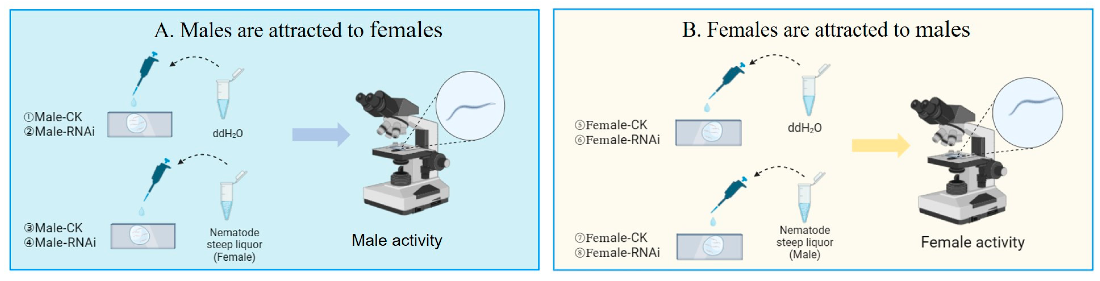
2.7. Sexual Arousal Experiments

Pheromone substances released by nematodes can stimulate the activity of nematodes of opposite sex [13]. To verify whether the Bmu-ser-1 gene is a sex pheromone receptor gene and whether it is differentially expressed between males and females, eight experimental groups were designed (Figure 1, Table 2). In this study, Groups 1 and 5 were considered as the control groups. For Group 1, 100 μL of ddH2O was added to a slide, and 1000 virgin males were transferred to the water drop. In Group 2, 1000 virgin males were incubated in Bmu-ser-1 dsRNA in the dark for 24 h, then transferred to the ddH2O drop. In Group 3, 1000 virgin females were placed in a centrifuge tube with 1 mL ddH2O and kept in the dark for 12 h. Then, 100 μL of this solution was pipetted onto slides, and 1000 virgin males were transferred to the solution. In Group 4, 1000 virgin males were soaked in Bmu-ser-1 dsRNA before being transferred to female steep liquor. Conversely, to examine the role of the ser-1 gene in female attraction to males, the males and females in the above experiments were swapped with each other (Table 2). In Group 5, 1000 virgin females were transferred to the ddH2O water drop, while in Group 6, 1000 dsRNA-treated virgin females were transferred to the water drop. In Group 7, 1000 virgin females were transferred to the male steep liquor, while in Group 8, 1000 dsRNA-treated virgin females were transferred to the male steep liquor. The frequency of head swings of 30 nematodes in each group was recorded randomly under the microscope. Finally, to detect the gene expression levels of Bmu-ser-1, the nematodes from all groups were collected for qPCR assay. The experiments were performed with three biological replicates for every sample.

2.8. Data Analysis

The mean ± standard error (SE) of triplicate measurements for every group was calculated using Microsoft Excel software (Office Excel 2016, Microsoft Corp, Redmond, WA, USA). We used Prism 5.0 (IBM, Armonk, NY, USA) for statistical analyses. The data underwent analysis using one-way ANOVA followed by Tukey’s multiple comparisons test (three or more groups) or t-test (two groups), with p < 0.05 considered significant. The relative expression levels were obtained with the 2−∆∆Ct method [18].

3. Results

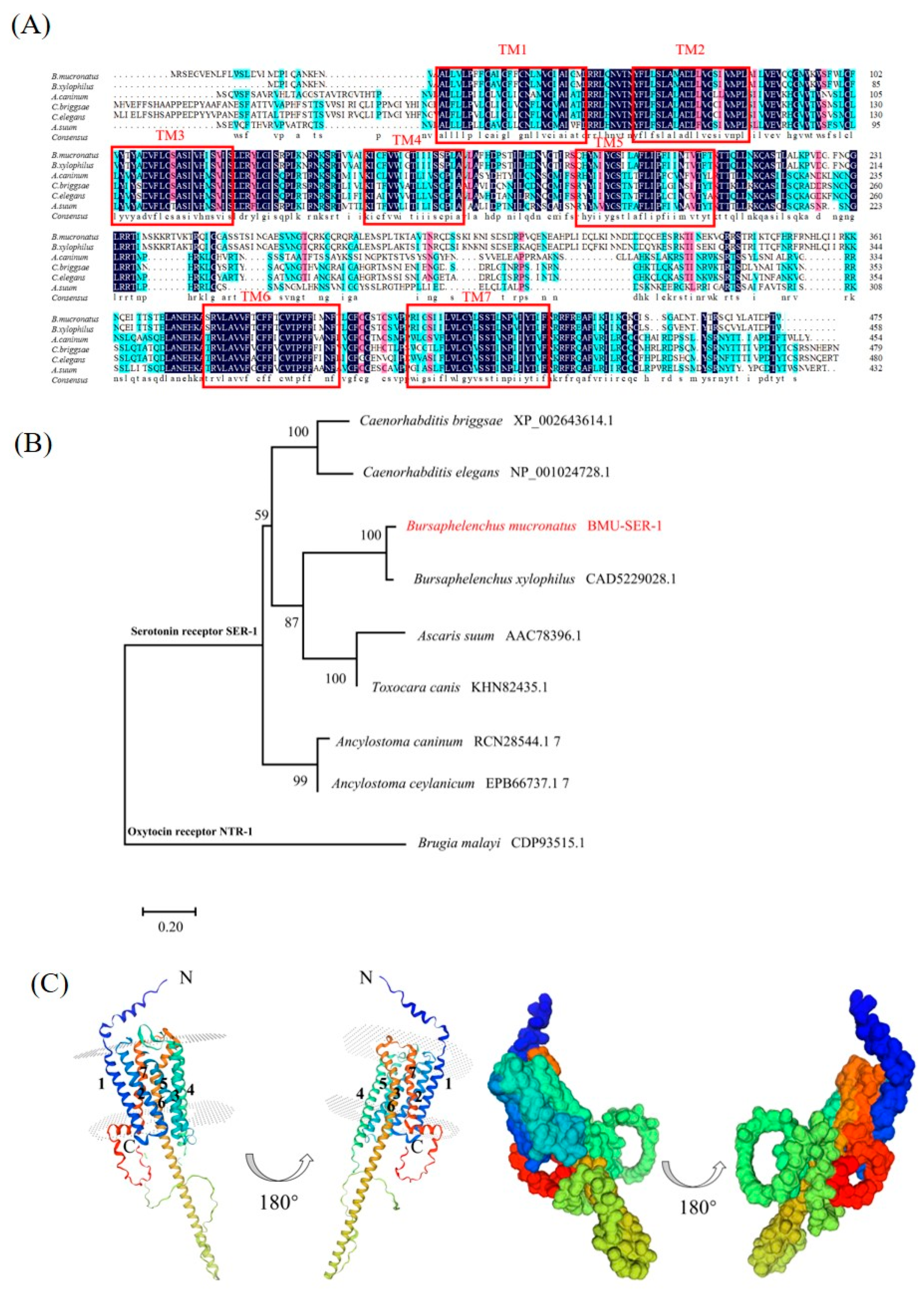
3.1. Sequence Analysis of Bm-ser-1

The results of bioinformatics analysis suggested that the sequence of Bmu-ser-1 is 1428 bp, encoding 475 amino acids. It has a seven-transmembrane helix structure and belongs to the 5-HT receptor family in the typical 7TM-GPCRs superfamily (Figure 2A). The results of multiple comparisons showed that the amino acid sequence of SER-1 in B. mucronatus was highly conserved with those of other nematodes, especially in seven structural domains. The phylogenetic tree included genera such as Bursaphelenchus, Caenorhabditis, and Ancylostoma, covering multiple species. The BMU-SER-1 amino acid sequence from B. mucronatus clustered with the SER-1 sequences from other organisms, distinct from the oxytocin receptor NTR-1, another GPCR. And the SER-1 of B. mucronatus and B. xylophilus were clustered on the same sister branch (Figure 2B). The BMU-SER-1 protein has seven transmembrane structures and belongs to the family of GPCRs. The gene encodes a protein without a signal peptide. The N-terminus was outside the membrane, and the C-terminus was inside the membrane (Figure 2C).

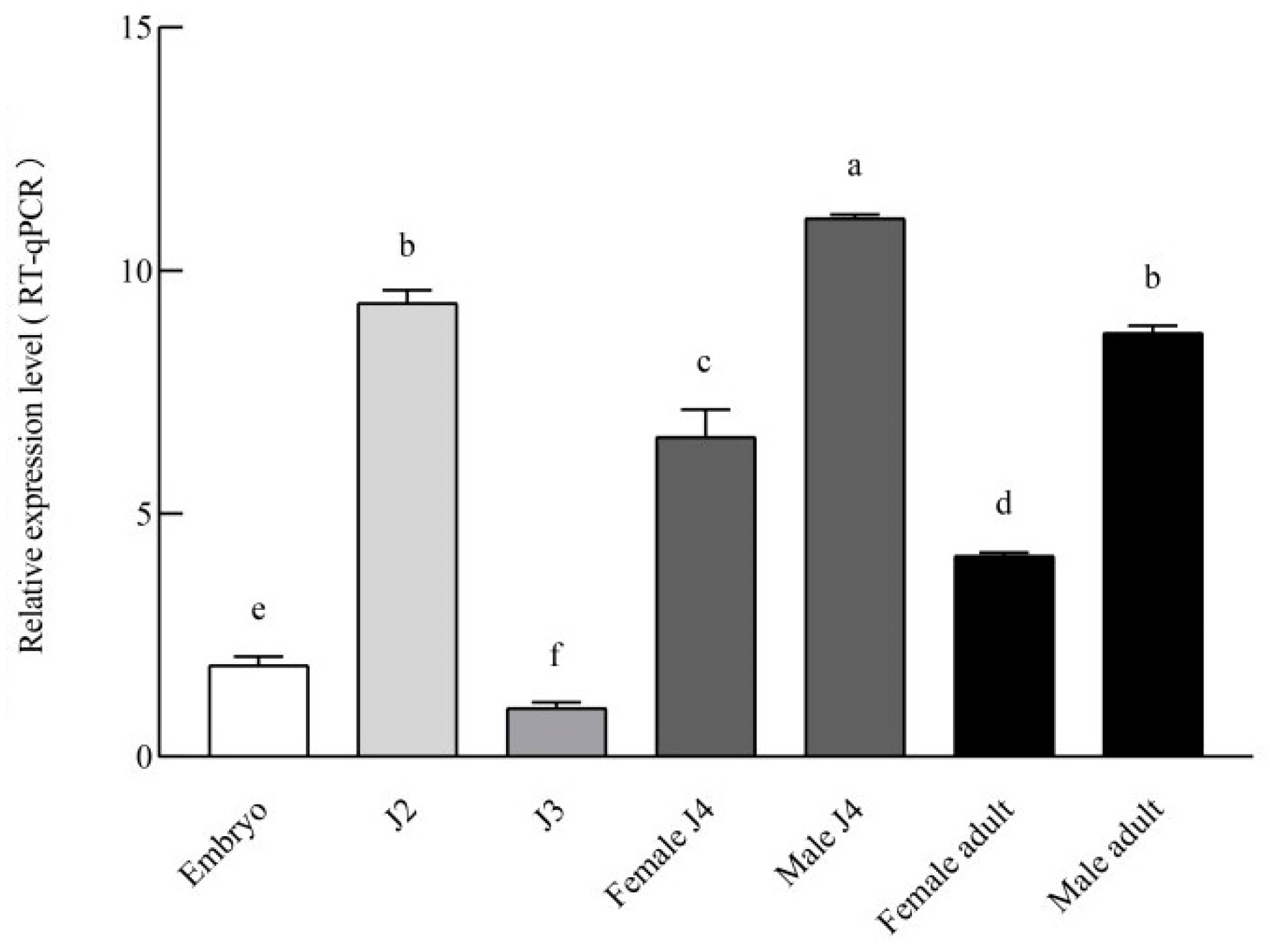
3.2. Expression Pattern of Bmu-ser-1

Bmu-ser-1 was expressed at all developmental stages. The expression level of J3 was the lowest and was set to 1. J2 and J4 males exhibited significantly higher gene expression levels compared to other stages, these being 9.33 and 11.07 times that of J3, respectively. In embryos, the expression level of Bmu-ser-1 was relatively low, at 1.65 times that of J3. Expression was moderately high in adult males and females, at 8.72 and 4.13 times that of J3, respectively. Notably, gene expression was significantly higher in males than in females (p < 0.05) (Figure 3).

3.3. In Situ Hybridization of Bm-ser-1

The staining site of the embryo is located in the mid-upper abdomen, and this site will develop into the labial region, the pharyngeal gland, and intestinal tract (Figure 4A). The gene is widely expressed during the J2 period (Figure 4C). It is expressed mainly in the end of the nematode’s gonadal primordium during the J3 period (Figure 4D). During the J4 period, it is expressed centrally in the vulva of females and the gonads of males (Figure 4E,F). The gene is expressed in the vulva of females, and the spermatogonia and primary spermatocytes of males was expressed in the adult stage (Figure 4G,H). The results are consistent with the results of previous in situ hybridizations.

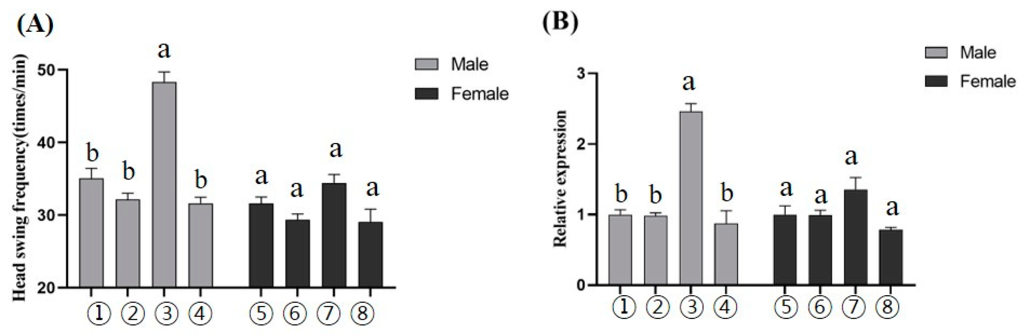
3.4. RNAi for the Bm-ser-1 Gene

The hatching rate of the control group was 95.68%, while the hatching rate of Bmu-ser-1-dsRNA-treated embryos was only 79.04%, and the variance between the two reached a significant level (Figure 5A). The frequency of head shaking was significantly lower in embryos than in the control group after development from J2 (Figure 5B). Unhatched embryos lagged in J1 and eventually died in the eggshell (Figure 5C); 18.7% of J2 larvae showed enlarged and deformed intestinal regions. In addition, the body length of normal J2 was 251.49 μm, as well as the body length of J2 larvae in the interference group was only 232.94 μm (Figure 5D). Continuing the immersion in sterile water for 2 h, it was found that the nematode remained rigid. Its head swing frequency was about 5.27 ± 1.18, which was significantly lower than that of the control nematodes (23.70 ± 1.90 per min) (Figure 5E). After 82 h of cultivation, 98.86%±0.76% of normal nematodes developed into adults, compared to only 85.31% ± 0.44% in the interference group (Figure 5F). The females that developed from J2 after the treatment showed impaired oviposition and eventually died of difficult labor (Figure 5G).
The mating results of adult individuals developed from dsRNA-treated J2 are shown in Table 3. The mating rate of males treated with dsRNA (78.13 ± 1.70) was significantly lower than that of the control group (82.29 ± 2.25), but the mating rate of females treated with dsRNA (83.33 ± 1.70) was not significantly different. The mating duration of the treated males (8.00 ± 0.49) was significantly lower than that of the control group (14.34 ± 0.45), while there was no significant difference in treated females (13.21.00 ± 0.44). And 22.10% ± 4.24 nematodes showed the wrong mating behavior in the treated male group, which was significantly higher than that in the control (5.77% ± 3.12) group and the treated female group (9.67 ± 4.05). The effective mating rate of treated males was only 34.10 ± 2.41, which was much lower than that in the control group (73.50 ± 1.09). However, there was no significant difference in the effective mating rate between females (63.73 ± 1.09) and controls. The number of eggs in dsRNA-treated females and males ranged from 5.27 to 6.12, which was significantly lower than that of the control group (10.24 ± 0.21).

3.5. The Bm-ser-1 Gene Is a Potential Sex Pheromone Receptor Gene

In the present study, the results suggested that the head shaking frequency in the control group (Group 1) was 35.07±1.37 (Figure 6A), and its expression level of the Bm-ser-1 gene was set to 1 (Figure 6B). Compared to Group 1, there were few changes in the head shaking frequency or the expression level of the Bm-ser-1 gene after RNAi treatment in Groups 2 and 4 (Figure 6A). Notably, the frequency of head swinging (48.31 ± 1.41) and the expression level of Bm-ser-1 (2.46) in Group 3 were significantly higher than those in Groups 1 (Figure 6A,B).
To compare the functional differences of the Bm-ser-1 gene between male and female of B. mucronatus, four treatment groups with females of B. mucronatus were simultaneously tested. The results showed that the head swing frequency and Bm-ser-1 gene expression levels in the control group (Groups 5) were 31.6 ± 0.89 and 1, respectively. Females in other treatment groups (Groups 6, 7, and 8) did not show significant differences (Figure 6A,B). Group 7 showed slightly higher expression levels compared to the other groups.

4. Discussion

The expression pattern and biological functions of Bmu-ser-1 in B. mucronatus were studied. Embryo hatchability was significantly reduced after treatment and embryos stagnated at the J1 stage. The observed larvae were trapped inside the shell, with less movement, and eventually died. Normal larvae break the shell via rapid writhing, with continuous tumbling and twisting inside the eggshell and by squeezing the eggshell, through the keratinized lip area [19]. It was assumed that the treated embryos would show restricted muscle development in the labial region and reduced shell-breaking ability, thus failing to break the shell. The motility of J2 was reduced after treatment. 5-HT5-HT1ADro and 5-HT1BDro could regulate the speed and direction of movement in Drosophila [20]. The ser-1 gene of C. elegans regulates the speed and direction of movement by modulating neurons in the head [14]. Therefore, it is assumed that the 5-HT receptor genes play an essential role in the locomotor function of B. mucronatus. Meanwhile, the gut and body wall muscles of dsRNA-treated J2 became enlarged and the body length was significantly lower than control. Release of 5-HT from ADF neurons could stimulate the diet of C. elegans [21]. In addition, the ser-1 gene has been reported to regulate the nematode pharyngeal pump and thus affect feeding [21]. It is obvious that ser-1 act as a vital role in regulating the development of pharyngeal pump and body wall muscles in B. mucronatus.
Notably, Bmu-ser-1 is involved in egg laying in females and mating behavior in males of B. mucronatus. Although dsRNA-treated females were able to mate, egg-laying was difficult. The vulva, which serves as the passageway for laying eggs, is composed of 22 cells that originate from the successive divisions of three vulval precursor cells (VPCs): P5.p, P6.p, and P7.p [22]. If the cells of the vulva are unable to migrate, ectopic invagination will occur, which is likely to cause difficulties in spawning [23]. It is possible that the silencing of Bmu-ser-1 causes the abnormal formation of vulval cells, resulting in the inability of females to ovulate. Treated males were mating-impaired and could not accurately locate the female’s vulva. During the normal mating process of B. mucronatus, the core step is for the male to actively seek, contact, and locate the vulva of the female, insert the copulatory spicule, and mate [24]. As shown in the experimental data (Table 3), the knockdown of the Bmu-ser-1 gene in males resulted in a longer search for females, a greater probability of not being able to accurately locate a female’s pubic area, and a greater rate of mismatches. The ser-1 mutant of C. elegans showed great resistance to exogenous 5-HT, with a significant reduction in egg production in both sexes and males developing difficulties in tail curling and turning, leading to mating failure [15,25]. According to previous studies and the results of the spatiotemporal expression, it was revealed that Bmu-ser-1 is involved in the reproductive behavior of B. mucronatus.
As shown in Figure 6A, the knockdown of the Bmu-ser-1 gene led to a decrease in the frequency of head swinging and the expression level of the Bmu-ser-1 gene (Figure 6A,B). These findings directly suggested that the Bmu-ser-1 gene is a possible sex pheromone receptor gene that plays a role in regulating neurons of pheromone transmission. These neurons primarily regulate physiological activities in coordination with corresponding receptor genes. Upon induction by ascr#10, C. elegans activates sensory neurons related to ADL and chemosensory neuronal processes related to ASI for signal transduction. This activation leads to the expression of major mod-1 [26] and rh-1 receptor genes [27]. Replacing the experimental subjects with females, we found that the gene expression levels at all stages were low, indicating that the gene mainly regulates the reception of sex pheromones in males. From the multiple functions of the Bmu-ser-1 gene, it can be hypothesized that 5-HT receptor gene (Bmu-ser-1) may also be a sex pheromone gene of males.
Currently, Bmu-ser-1 is known to be a sex pheromone receptor gene, but further research is needed on the ligand-encoding genes, neurons in this signaling pathway (Figure 7), and additional unknown key genes. The above studies can be referenced in the future to explore how the Bmu-ser-1 gene regulates sex pheromones specific to B. mucronatus. This study is the first to propose the use of behavioral and molecular means to validate sex pheromone receptor genes. This study has elucidated the molecular mechanisms controlling B. mucronatus population density from a mating perspective, and it can inform management practices in forestry development.
In summary, the Bmu-ser-1 gene, as a sex pheromone gene, plays an important role in regulating nematode development, mating, and reproduction. This contributes to our understanding of growth mechanisms in the nematode population and provides new ideas for the control of B. xylophilus and B. mucronatus.

5. Conclusions

In our study, the expression pattern and biological functions of Bmu-ser-1 in B. mucronatus were explored. The results indicated that Bmu-ser-1 was expressed in all nematode stages, with differences in the sites of expression in males and females. Bmu-ser-1 plays a crucial role in regulating nematode growth, mating, and reproduction. Bmu-ser-1 is involved in the population reproduction and development of nematodes; this contributes to our understanding of the physiological activities of Pinus sylvestris and provides new ideas for the population expansion of B. xylophilus.

Author Contributions

W.L. and R.M. conceived the study; W.L. and R.M. performed the experiments; J.C., Q.L. and K.G. contributed materials and analyzed the data; H.S. and J.H. secured funding; H.S. wrote the manuscript. All authors have read and agreed to the published version of the manuscript.

Funding

This study was funded by the National Natural Science Foundation of China (32371892) and the Launching Funds for Talents of Zhejiang A & F University (2023LFR117).

Data Availability Statement

The data presented in this study are available on request from the corresponding author. The data are not publicly available due to privacy restrictions.

Conflicts of Interest

The authors declare no conflicts of interest.

References

  1. Kikuchi, T.; Cotton, J.A.; Dalzell, J.J.; Hasegawa, K.; Kanzaki, N.; McVeigh, P.; Takanashi, T.; Tsai, I.J.; Assefa, S.A.; Cock, P.J.A.; et al. Genomic insights into the origin of parasitism in the emerging plant pathogen Bursaphelenchus xylophilus. PLOS Pathog. 2011, 7, e1002219. [Google Scholar] [CrossRef] [PubMed]
  2. Zhao, L.; Zhang, X.; Wei, Y.; Zhou, J.; Zhang, W.; Qin, P.; Chinta, S.; Kong, X.; Liu, Y.; Yu, H.; et al. Ascarosides coordinate the dispersal of a plant-parasitic nematode with the metamorphosis of its vector beetle. Nat. Commun. 2016, 7, 12341. [Google Scholar] [CrossRef]
  3. Webster, J.M.; Anderson, R.V.; Baillie, D.L.; Beckenbach, K.; Rutherford, T.A. DNA probes for differentiating isolates of the pinewood nematode species complex. Rev. Nématologie 1990, 13, 255–263. [Google Scholar] [CrossRef]
  4. Mamiya, Y.; Enda, N.J.N. Bursaphelenchus mucronatus n. sp.(Nematoda: Aphelenchoididae) from pine wood and its biology and pathogenicity to pine trees. Nematologica 1979, 25, 353–361. [Google Scholar] [CrossRef]
  5. Howard, A.D.; McAllister, G.; Feighner, S.D.; Liu, Q.; Nargund, R.P.; Van der Ploeg, L.H.T.; Patchett, A.A. Orphan G-protein-coupled receptors and natural ligand discovery. Trends Pharmacol. Sci. 2001, 22, 132–140. [Google Scholar] [CrossRef] [PubMed]
  6. Taylor, K.P. Regulation of Behavioral Arousal and Quiescence in C. elegans. Genetics 2016, 199, 523–531. [Google Scholar]
  7. Bockaert, J.; Pin, J.j. Molecular tinkering of G protein-coupled receptors: An evolutionary success. EMBO J. 1999, 18, 1723–1729. [Google Scholar] [CrossRef] [PubMed]
  8. Anganathan, R.; Cannon, S.C.; Horvitz, H.R.J.N. MOD-1 is a serotonin-gated chloride channel that modulates locomotory behaviour in C. elegans. Nature 2000, 408, 470–475. [Google Scholar] [CrossRef] [PubMed]
  9. Churgin, M.A.; McCloskey, R.J.; Peters, E.; Fang-Yen, C.J.J.o.N. Antagonistic serotonergic and octopaminergic neural circuits mediate food-dependent locomotory behavior in Caenorhabditis elegans. J. Neurosci. 2017, 37, 7811–7823. [Google Scholar] [CrossRef]
  10. Dernovici, S.; Starc, T.; Dent, J.A.; Ribeiro, P.J.D. The serotonin receptor SER-1 (5HT2ce) contributes to the regulation of locomotion in Caenorhabditis elegans. Dev. Neurobiol. 2007, 67, 189–204. [Google Scholar] [CrossRef]
  11. Carnell, L.; Illi, J.; Hong, S.W.; McIntire, S.L. The G-protein-coupled serotonin receptor SER-1 regulates egg laying and male mating behaviors in Caenorhabditis elegans. J. Neurosci. 2005, 25, 10671–10681. [Google Scholar] [CrossRef]
  12. Kiyohara, T. Sexual Attraction in Bursaphelenchus xylophilus. Jpn. J. Nematol. 1982, 11, 7–12. [Google Scholar] [CrossRef]
  13. Shinya, R.; Chen, A.; Sternberg, P.W. Sex attraction and mating in Bursaphelenchus okinawaensis and B. xylophilus. J. Nematol. 2015, 47, 176–183. [Google Scholar] [CrossRef]
  14. Meng, J.; Wickham, J.D.; Ren, W.; Zhao, L.; Sun, J. Species displacement facilitated by ascarosides between two sympatric sibling species: A native and invasive nematode. J. Pest Sci. 2020, 93, 1059–1071. [Google Scholar] [CrossRef]
  15. Zhou, L.; Liu, W.; Bai, L.; Liu, H.; Wang, J.; Ma, X.; Huang, L.; Guo, K.; Yu, H.; Hu, J. Female’s war: A story of the invasion and competitive displacement between two xylophilus group nematode species. J. Pest Sci. 2023, 96, 1301–1311. [Google Scholar] [CrossRef]
  16. Zhou, L.; Ma, X.; Zhu, N.; Zou, Q.; Guo, K.; Bai, L.; Yu, H.; Hu, J. The role of mab-3 in spermatogenesis and ontogenesis of pinewood nematode, Bursaphelenchus xylophilus. Pest Manag. Sci. 2021, 77, 138–147. [Google Scholar] [CrossRef] [PubMed]
  17. Kalyaanamoorthy, S.; Minh, B.Q.; Wong, T.K.F.; von Haeseler, A.; Jermiin, L.S. ModelFinder: Fast model selection for accurate phylogenetic estimates. Nat. Methods 2017, 14, 587–589. [Google Scholar] [CrossRef]
  18. Urwin, P.E.; Lilley, C.J.; Atkinson, H.J. Ingestion of double-stranded RNA by preparasitic juvenile cyst nematodes leads to RNA interference. Mol. Plant-Microbe Interact. 2002, 15, 747–752. [Google Scholar] [CrossRef]
  19. Wang, J.; Liu, H.; Guo, K.; Yu, H.; Ye, J.; Hu, J. Molecular characterization and functional analysis of Bxy-octr-1 in Bursaphelenchus xylophilus. Gene 2022, 823, 146350. [Google Scholar] [CrossRef] [PubMed]
  20. Saudou, F.; Boschert, U.; Amlaiky, N.; Plassat, J.L.; Hen, R.J.E.J. A family of Drosophila serotonin receptors with distinct intracellular signalling properties and expression patterns. J. Entomol. Soc. Br. 1992, 11, 7–17. [Google Scholar] [CrossRef]
  21. Cunningham, K.A.; Hua, Z.; Srinivasan, S.; Liu, J.; Lee, B.H.; Edwards, R.H.; Ashrafi, K.J.C.M. AMP-activated kinase links serotonergic signaling to glutamate release for regulation of feeding behavior in C. elegans. Cell Metab. 2012, 16, 113–121. [Google Scholar] [CrossRef] [PubMed]
  22. Ranawade, A.V.; Cumbo, P.; Gupta, B.P. Caenorhabditis elegans histone deacetylase hda-1 is required for morphogenesis of the vulva and LIN-12/Notch-mediated specification of uterine cell fates. Genes Genomes Genet. 2013, 3, 1363–1374. [Google Scholar] [CrossRef]
  23. Dufourcq, P.; Victor, M.; Gay, F.; Calvo, D.; Hodgkin, J.; Shi, Y. Functional Requirement for Histone Deacetylase 1 in Caenorhabditis elegans Gonadogenesis. Mol. Cell. Biol. 2002, 22, 3024–3034. [Google Scholar] [CrossRef] [PubMed]
  24. Zhu, N.; Bai, L.; Schütz, S.A.; Liu, B.; Liu, Z.; Zhang, X.Y.; Yu, H.; Hu, J. Observation and quantification of mating behavior in the pinewood nematode, Bursaphelenchus xylophilus. J. Vis. Exp. 2016, 118, 54842. [Google Scholar] [CrossRef]
  25. Dempsey, C.M.; Mackenzie, S.M.; Gargus, A.; Blanco, G.; Sze, J.Y. Serotonin (5HT), Fluoxetine, imipramine and dopamine target distinct 5HT receptor signaling to modulate Caenorhabditis elegans egg-laying behavior. Genetics 2005, 169, 1425–1436. [Google Scholar] [CrossRef] [PubMed]
  26. Aprison, E.Z.; Ruvinsky, I. The roles of several sensory neurons and the feedback from egg laying in regulating the germline response to a sex pheromone in C. elegans hermaphrodites. Micropublication Biol. 2022. [Google Scholar] [CrossRef]
  27. Park, J.; Oh, H.; Kim, D.Y.; Cheon, Y.; Park, Y.J.; Hwang, H.; Neal, S.J.; Dar, A.R.; Butcher, R.A.; Sengupta, P.; et al. CREB mediates the C. elegans dauer polyphenism through direct and cell-autonomous regulation of TGF-β expression. PLoS Genet. 2021, 17, e1009678. [Google Scholar] [CrossRef]
Figure 1. The ser-1 gene’s role in sexual attraction behavior. (A) ① One thousand virgin males were added to ddH2O. ② The virgin males treated by Bmu-ser-1 dsRNA were added to ddH2O. ③ Virgin males were added to female steep liquor. ④ Virgin males treated by Bmu-ser-1 dsRNA were added to female steep liquor. (B) ⑤ The virgin females were added to ddH2O. ⑥ The virgin females treated by Bmu-ser-1 dsRNA were added to ddH2O. ⑦ The virgin females were added to male steep liquor. ⑧ The virgin females treated by Bmu-ser-1 dsRNA were added to male steep liquor.
Forests 15 01115 g001
Figure 2. Amino acid sequence analysis of Bmu-ser-1. (A) Multiple alignments of Bmu-ser-1 with the other seven nematodes SER-1 were constructed using ClustalW. Red, green and blue shading indicate invariant, highly conserved and moderately conserved amino acid residues, respectively. Seven transmembrane domains (TM1-7) of SER-1 are enclosed by red boxes. (B) Phylogenetic analysis of SER-1 conserved domain proteins was performed with the neighbor-joining method. Oxytocin receptors NTR-1 were selected as an outgroup. The percentage nodal support based on 1000 bootstrap replicates is shown; the scale bar indicates evolutionary distance (0.2 substitutions per position). (C) The three-dimensional structure of Bmu-ser-1. The two diagrams on the left are cartoon models, and the two diagrams on the right are surface models. TM1-7 were labeled in the cartoon model. N, N-terminal, indicates the extramembrane direction.
Forests 15 01115 g002
Figure 3. The relative expression levels of Bmu-ser-1 in B. mucronatus at different developmental stages. Values represent the mean ± SE of three independent biological samples. Different letters on the error bars indicate statistically significant differences determined using Tukey’s (HSD) multiple range test (p < 0.05).
Forests 15 01115 g003
Figure 4. The in situ hybridization of Bmu-ser-1 in B. mucronatus at different developmental stages. (A) embryo; (B) embryo in the control group; (C) J2; (D) J3; (E) the female J4; (F) the male J4; (G) the female adult; (H) the male adult. m, metacorpus; an, anus; v, vulva; sp, spicules. the scale bars are marked at the bottom right corner of the worm.
Forests 15 01115 g004
Figure 5. RNAi of Bmu-ser-1. (A) The hatching rate of embryos after RNAi for 24 h via the soaking method. (B) The head swing frequency of hatched J2 after RNAi embryo. (C) The hatched J2 was crouched and inactive after RNAi. (D) The phenotype of J2 after RNAi for 24 h, which showed an enlarged bowel and shorter body length. (E) The body length of J2 after RNAi. (F) Proportion of adults in the RNAi group when the almost control larvae developed into adults (82 h, 98.86% ± 0.76%). (G) Phenotype of female in the RNAi group, where embryos were unable to expel from the female’s vulva, causing the female to die. White arrows indicate areas of abnormal traits. The values represent the mean ± SE of three independent biological samples and were analyzed by t-test. Asterisks indicate statistically significant differences (** p < 0.01, *** p < 0.001, **** p < 0.0001).
Forests 15 01115 g005
Figure 6. Behavioral and qPCR functional validation of the Bmu-ser-1 gene. (A) Rectangular images of head bobbing in the different groups. (B) Expression of the Bmu-ser-1 gene in different groups. The revelation of ①–⑧ is the same as that of Figure 1. The values represent mean ± SE of three independent biological samples. Different letters indicate significant differences among treatments at p < 0.05 based on Turkey’s (HSD).
Forests 15 01115 g006
Figure 7. Sex pheromone pathway map for mating in B. mucronatus.
Forests 15 01115 g007
Table 1. Primers used in the present study.
PrimerSequence (5′-3′)
CloningBmu-ser-1-C-FTTAGCGATGGCAGATTTA
Bmu-ser-1-C-RTTCGGGTAGTCAACACTC
RT-qPCR18S-FTTCTGACCGTAAACGA TGCCAACT
18S-RCCTCACCTCGAACGCCGAATTA
Bmu-ser-1-Q-FGGACCGACGGCTTCAAAATG
Bmu-ser-1-Q-RCGGAAACCGGTATAACCAGC
RNAiT7 promoterTAATACGACTCACTATAGGG
Table 2. Experimental design to explore the role of ser-1 gene in sexual attraction behavior.
GroupNematodeAdded Solution
1Male-CKddH2O
2Male-RNAiddH2O
3Male-CKFemale steep liquor
4Male-RNAiFemale steep liquor
5Female-CKddH2O
6Female-RNAiddH2O
7Female-CKMale steep liquor
8Female-RNAiMale steep liquor
Table 3. Mating results of adult individuals grown from dsRNA-treated J2.
GroupMating Rate (%)Mating Time/minWrong Mating Rate (%)Effective Mating Rate (%)Brood Size
CK♀ + CK♂82.29 ± 2.25 a *14.34 ± 0.45 a5.77 ± 3.12 b73.50 ± 1.09 a10.24 ± 0.21 a
RNAi♀ + CK♂83.33 ± 1.70 a13.21 ± 0.44 a9.67 ± 4.05 b63.73 ± 0.93 a5.27 ± 0.42 b
CK♀ + RNAi♂78.13 ± 0.47 b8.00 ± 0.49 b22.10 ± 4.24 a34.10 ± 2.41 b6.12 ± 0.42 b
* Values represent the mean ± SE of three independent biological samples. Different letters denote statistical significance determined using Tukey’s (HSD) multiple range test (p < 0.05).
Disclaimer/Publisher’s Note: The statements, opinions and data contained in all publications are solely those of the individual author(s) and contributor(s) and not of MDPI and/or the editor(s). MDPI and/or the editor(s) disclaim responsibility for any injury to people or property resulting from any ideas, methods, instructions or products referred to in the content.

