Article

Evaluation of Tolerance and Selection of Heat-Tolerant Woody Plants against Heat Stress

1
Department of Variety Examination, National Forest Seed and Variety Center, Chungju 27495, Republic of Korea
2
Division of Environmental Forest Science, Gyeongsang National University, Jinju 52828, Republic of Korea
3
Institute of Agriculture of Life Science, Gyeongsang National University, Jinju 52828, Republic of Korea
*
Author to whom correspondence should be addressed.
Forests 2024, 15(8), 1366; https://doi.org/10.3390/f15081366
Submission received: 14 June 2024 / Revised: 14 July 2024 / Accepted: 23 July 2024 / Published: 5 August 2024
(This article belongs to the Section Forest Ecophysiology and Biology)

Abstract

:
Heat-tolerant species have become increasingly important because of climate change; however, a selection system for woody plants is not well established. This study was conducted to establish a selection system for heat-tolerant woody plant species and to select heat-tolerant species. After selecting heat-tolerant woody plants and applying heat stress to 27 species, the electrolyte leakage index (ELI) was measured. The ELI of the heat-tolerant species was lower than that of the non heat-tolerant species, and they survived well after heat stress treatment. For the preselected species, the degree of cell death was measured by Evans blue staining method, and the heat stress recovery ability was measured by a 3,3′-diaminobenzidine (DAB) staining method. The species showed less cell death even after heat treatment, and oxidative stress was low after recovery from heat stress. Traditional screening methods are mainly performed through field screening; however, this is difficult because it requires many samples and considerable time. The results of this study are relatively rapid, reproducible, and highly sensitive, so it is judged to be a method that can complement the existing traditional method as a heat-tolerant plant selection system. The results of this study can be widely used for the selection and breeding of heat-tolerant plants in preparation for climate change.

1. Introduction

An increase in temperature due to human activities has affected forest ecosystems because forest plants that are sensitive to high temperatures are often at risk. Global surface temperature in the first two decades of the 21st century (2001–2020) increased by 0.99 (0.84 to 1.10) C compared to 1850–1900 [1]. Furthermore, the temperature rise is going to be accelerated. According to the climate response to five illustrative scenarios based on Shared Socio-economic Pathways (SSPs), in very low and very high green house gas (GHG) emissions scenarios (SSP1-2.6 and SSP5-8.5), the average temperature may increase by 2.0 and 7.1 C more than present in Korean peninsula in 2100 [2]. At the global warming of 1.5 C, 3%–14% of the tens of thousands of species assessed in terrestrial ecosystems will likely face very high risk of extinction [1]. The response of plants to heat stress, especially in Arabidopsis and other crops, has been well documented. Common physiological response to heat stress in plants include decreased photosynthetic efficiency, reactive oxygen species (ROS) accumulation, and cell membrane dysfunction [3]. When leaves are exposed to high temperature, RuBisCO activation decreases and photosystem II activity is inhibited, resulting in the inhibition of net photosynthesis [4,5,6]. Heat stress also induces oxidative stress in leaves, where an increase in ROS concentration and antioxidant enzyme activity is observed after exposure to high temperature [7,8]. Molecular chaperone proteins were well known for preventing protein aggregation due to heat stress by functioning as holdases or foldases [9,10,11]. Molecular chaperones such as HSP70, HSP101, and smHSP are highly conserved in plants. Antioxidant proteins are also upregulated to protect the cell membrane from oxidization by ROS [12]. The cellular mechanism of the unfolded protein response is to reduce stress [13]. This series of processes for maintaining plant homeostasis has been revealed under controlled conditions. Controlled heat-stress conditions have been used to breed heat-tolerant genotypes. Various laboratory-scale heat stress treatment schemes have been developed to assess plant heat tolerance [14]. The heat treatment of 44 C for 4 h was employed to evaluate the heat tolerance of tomato plants [15]. However, an efficient heat stress treatment for screening heat-tolerant native Korean woody plants has not yet been reported. Several physical-biochemical properties contribute to the abiotic stress tolerance of plants. The application of rapid, nondestructive, sensitive, and efficient screening techniques in breeding programs for abiotic stress tolerance has become very important. In previous studies on abiotic stress tolerance screening, screening was arduous because of the large sample size and time required. Plant responses to heat stress can be measured based on the physiological changes after heat application [16]. Research on the measurement of stress, including heat, has been conducted for a long time. Since the 1930s, plant tolerance to various environmental stressors (including heat) has been estimated based on the measurement of stress-induced ion leakage from plant tissues [17,18,19]. Plant cell membrane permeability is a key indicator for assessing the tolerance of plants to abiotic stresses such as drought, heat, cold, and salt [20]. This can be evaluated by measuring electrolyte leakage in the leaves of each plant species. It is widely accepted that the more stressed plants are, the more their cell membranes degrade, reflecting a subsequent increase in ion flux from the cytoplasm to the water bath. In addition, heat stress affects chlorophyll fluorescence (Fv/Fm) in plants. This decreased after heat stress treatment, reflecting the damaging effect of thermal stress on the structure and function of the photosynthetic apparatus [21]. Heat stress also affects the activities of antioxidant enzymes (CAT, GPOX, and SOD), proline content, and sugar content in leaves, all of which increase when seedlings of the moth bean (Vigna aconitifolia) are exposed to heat stress [22]. However, it is difficult to determine plant responses to heat stress accurately. One experimental method may not be sufficient for identifying heat-tolerant plants. The evaluation of heat tolerance has been conducted mainly in wheat cultivars and other herbaceous plants, and observation of leaf wilting and measurement of biomass and grain yield were the main methods used for evaluation [23,24]. Moreover, physiological assays such as the malondialdehyde (MDA) and soluble sugar content assays have been applied in some studies [25]. These methods have also been used to evaluate the heat tolerance of woody plants such as chestnuts [26] and pines [27]. Woody plants have a longer life cycle than herbaceous plants; therefore, non-destructive and efficient methods are needed to evaluate not only their heat tolerance, but also their ability to recover from heat stress. However, there has been limited research on establishing effective methods for evaluating the heat tolerance of woody plants and their ability to heat stress. Moreover, Evans blue and 3,3’-diaminobenzidine (DAB) staining assays, which can be used to visualize and measure the response of plants to heat stress, have rarely been used to evaluate the heat tolerance and recovery abilities of plants.
This study was conducted to provide an efficient method for measuring the heat tolerance of woody plants and to select heat-resistant tree species. First, heat-resistant woody plants were preselected through electrolyte leakage analysis of 27 native woody species in Korea. The degree of cell death due to heat stress was measured using Evans blue staining, and the recovery ability of plants subjected to heat stress was tested using DAB staining. Finally, heat-resistant woody plant species prepared for climate change were selected using a series of methods.

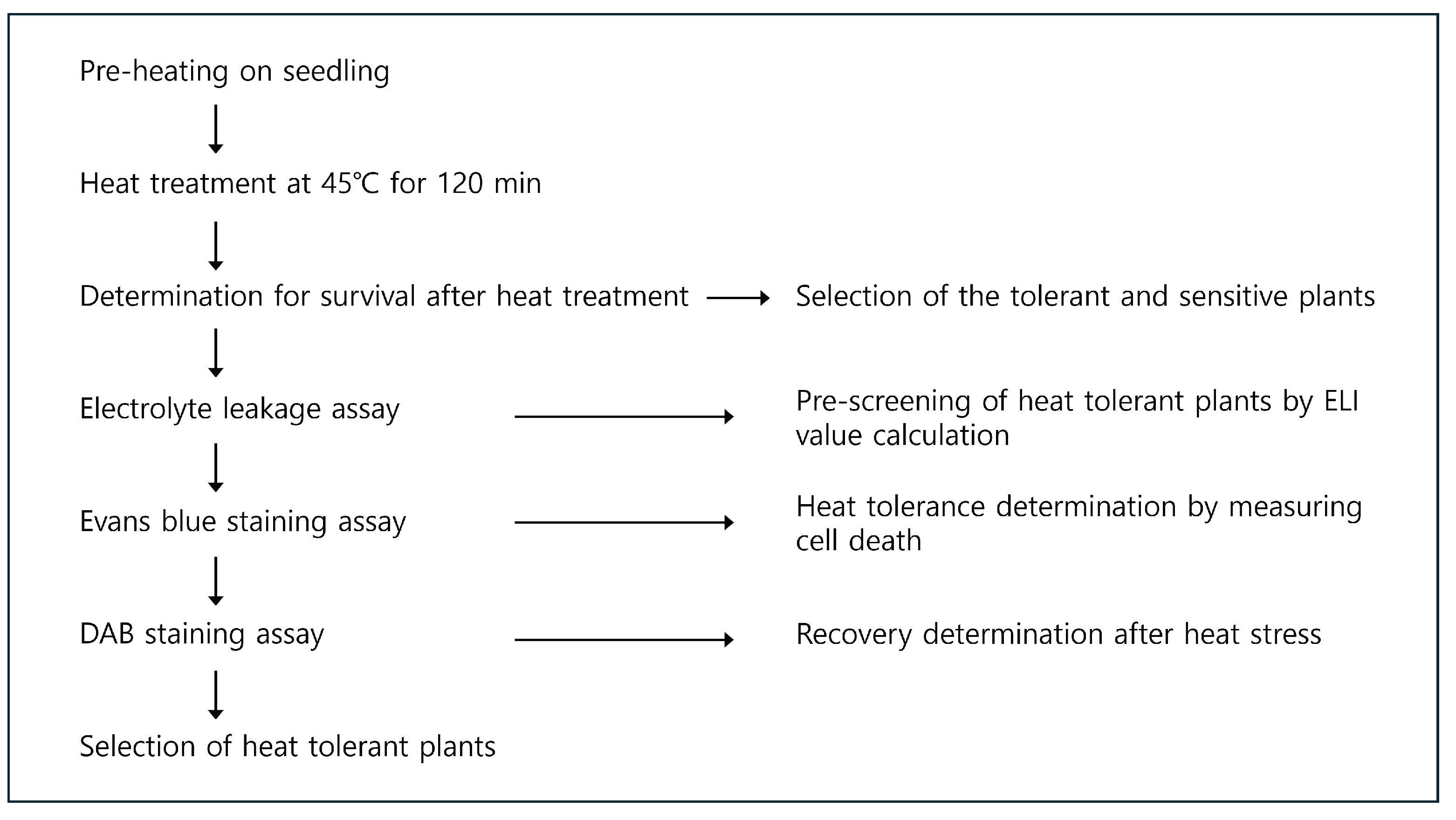
2. Materials and Methods

2.1. Plant Materials

To establish a heat-tolerant plant selection method, and to select heat-tolerant tree species using the method, 27 woody plants were used (Table 1). These plant species were collected from the Gyeongsangnam-do Forest Environment Research Institute in Jinju, Republic of Korea and the Korean National Arboretum in Pocheon. Woody plants, all of which were 1-year-old, were obtained from the experimental forest at Gyeongsang National University. Plug cell trays (diameter, 60/45 mm; depth, 93 mm; volume, 210 cc) and commercial potting soil were used in this study. To avoid experimental errors, similarly grown seedlings were then selected for further experiments and acclimated in the growth chamber (SH-301, Seyeong Science Co., Ulsan, Republic of Korea) under the condition of 25 ± 1 C, 50 ± 5% humidity, and a photoperiod of 16 h of light with 37 µmol m−2s−1 (about 3000 lux)/8 h of the dark.

2.2. Heat Stress Treatment

To establish optimal heat stress conditions for testing heat tolerance, six species (Q. myrsinaefolia, Q. glauca, C. obtusa, A. julibrissin, L. cyrtobotrya, and R. pseudoacacia) were chosen based on the results of a preliminary test, in which the heat tolerance of 27 woody plant species was evaluated. Evaluation was conducted using the electrolyte leakage method described in the following paragraph. The seedlings were subjected to 37 C for 120 min in an incubator, and heat tolerance of each species was evaluated. Three species (Q. myrsinaefolia, Q. glauca, and C. obtusa) with the highest tolerance and three species (A. julibrissin, L. cyrtobotrya, and R. pseudoacacia) with the highest sensitivity were selected. These species were considered heat-tolerant or heat-sensitive plants, and were used to determine the optimal heat stress conditions. Plants were subjected to heat stress using a modified version of the scheme described by Yeh et al. [14] (Figure S1). 1-year-old woody plant seedlings were placed into an incubator which was set to 37 C for 120 min for pre-heating, and then they were recovered in the 25 C chamber for 60 min. The pre-heated plants were incubated under high-temperature conditions (40, 45, 50, 55 C) for 120 min to observe their responses under heat stress. The temperatures and durations of heat treatment were chosen based on previous studies that evaluated the heat tolerance of plants [14,23,24,25,26,27].
Differences in phenotypic response of each seedling were clearly shown under 45 C for 120 min (Figure S2). Under these heat-stress conditions, Q. myrsinaefolia, Q. glauca, and C. obtusa survived well, whereas A. julibrissin, L. cyrtobotrya, and R. pseudoacacia showed wilt symptoms. No adverse effects on the seedlings were shown under heat stress of 40 C whereas all seedlings were lethal in the heat-stress conditions of 50 C and 55 C. Thus, the condition of 45 C for 120 min was used for screening heat-tolerant woody plants.

2.3. Measuring Electrolyte Leakage Indexes

Electrolyte leakage from leaf disks was measured to assess cell membrane stability under heat stress. Five leaf disks (Ø 5 mm) were collected from the top 2–4 leaves of each plant. The leaf disks were heated in a conical tube containing 5 mL of deionized water at 50 C for 120 min to assess cell membrane stability on the heat. As the positive control, other conical tubes containing five leaf disks were autoclaved under 121 C for 15 min. As the negative controls, the other conical tubes containing five leaf disks were placed at 25 C for 120 min. All tubes were shaken and incubated at 100 rpm for 30 min, and the amount of electrolyte in the water was measured using a conductivity probe (VE 4810, Korea Scientific, Seoul, Rpublic of Korea). Electrolyte leakages index (ELI) was calculated using Equation (1).
E L I = E l e c t r o l y t e l e a k a g e H E l e c t r o l y t e l e a k a g e C E l e c t r o l y t e l e a k a g e C
Electrolyte leakage = Electrical conductivity of the treatment/electrical conductivity of positive control, Electrolyte  l e a k a g e H : Electrolyte leakage of the heat-stressed sample, Electrolyte l e a k a g e C : Electrolyte leakage of negative control
Pre-screening was performed to determine the tolerance of woody plants to heat stress. ELI values of 27 woody plants were determined after heat treatment at 45 C for 60 min. After the ELI was obtained, as described above, plant species with values similar to or lower than those of C. obtusa, Q. myrsinaefolia and Q. glauca, which had the lowest ELI, were selected. Eight species with low ELI values (C. sinensis, T. japonica, I. cornuta, I. crenata, N. sericea, Q. acuta, Q. phillyraeoides and Q. salicina) were selected. The phenotypic responses of the pre-screened heat-tolerant candidates under heat stress were observed to confirm heat tolerance. For testing the heat tolerance of candidates, the pre-screened species were exposed to the heat stress condition of 45 C for 120 min and ELI value was measured every 30 min. The experiment was conducted in 3 replication per treatment.

2.4. Investigation of Dead Tissue by Evans Blue Staining

Cell death in the eight species after heat stress was evaluated using Evans blue staining. Leaf samples (1 cm2) prepared from each plant species and incubated under normal conditions or heat treatment were treated with Evans blue solution (1%) for an hour. For positive controls, leaf samples collected from each plant species were heated in boiling water for 1 h and stained under the conditions described above. The leaf samples were then extensively washed with distilled water to remove the unbound Evans blue dye. Photographs of the leaf samples were taken using a microscope (BH-2; Olympus, Tokyo, Japan) at 200× magnification. To assess dead cells, the stained dye was dissolved in 2 mL of 50% ethanol solution containing 2% sodium dodecyl sulfate (SDS). The absorbance of the dye dissolved in the leaves was measured at 600 nm. The cell death rates were calculated using Equation (2).
C e l l d e a t h r a t e ( % ) = A b s H A b s N C A b s P C A b s N C × 100 ( % )
A b s H : Absorbance value of the heat-stressed sample, A b s N C : Absorbance value of Negative Control, A b s P C : Absorbance value of Positive Control

2.5. Evaluation of Recovery of Heat-Stressed Plants by DAB Staining

A DAB staining assay was performed on the eight species to evaluate their recovery ability after heat stress. The fresh 3,3′-Diaminobenzidine solution was prepared by the protocol reported by Daudi and O’Brien [28]. Leaf samples were collected from plant species that were incubated during heat treatment. Leaf samples from each plant species that were incubated under normal conditions were used as negative controls. As a positive control, 30% (v/v) hydrogen peroxide was taken up from leaf samples by gentle vacuum-infiltration for 10 min. The DAB solution was added to all leaf samples including the treatment and control groups, by gently vacuum-infiltrating the leaves for 10 min. The leaf samples were then placed on a shaker for 4 h at 100 rpm. The DAB solution was replaced with a bleaching solution (ethanol: acetic acid: glycerol = 3:1:1), and then leaf samples were boiled at 95 C for 30 min to remove chlorophyll. Photographs of the bleached leaf samples were obtained using a microscope (BH-2; Olympus, Tokyo, Japan) at 200× magnification.

2.6. Statistical Analysis

Data are expressed as the average of five separate experiments. Bars indicate standard deviation of the mean for each replicate treatment. Data were subjected to statistical analysis using SPSS software (version 27.0). A one-way analysis of variance (ANOVA) was conducted, and the means were compared using Duncan’s multiple range test (DMRT) at a probability level of 0.05. Additionally, Pearson’s correlation analysis was conducted to measure the correlations between the methods for evaluating heat tolerance.

3. Results

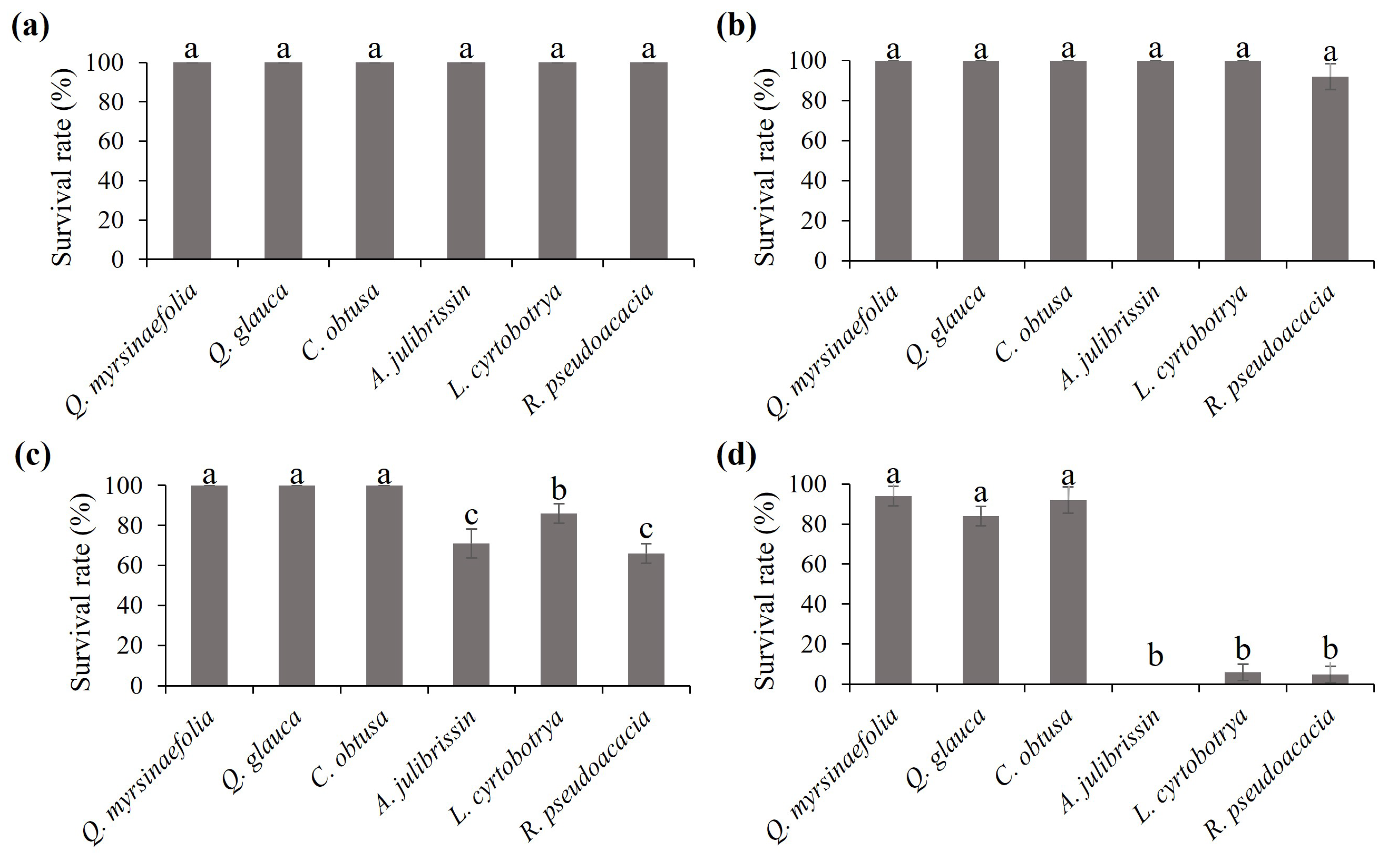
3.1. Measurement of the Response of Heat-Tolerant and Sensitive Woody Plants

Under the condition of 45 C for 120 min, phenotypic responses of heat-tolerant and sensitive woody plants were observed. Seedlings of Q. myrsinaefolia, Q. glauca, C. obtusa, A. julibrissin, L. cyrtobotrya, and R. pseudoacacia showed different phenotypic response under heat stress of 45 C for 120 min (Figure 1). There was no significant difference in the survival rate between the species after 30 and 60 min of heat treatment (p > 0.05). However, the differences between the survival rates of the six species were obvious at 90 and 120 min (p < 0.001). The seedlings of Q. myrsinaefolia, Q. glauca, and C. obtusa tolerated heat stress. In contrast, A. julibrissin, L. cyrtobotrya and R. pseudoacacia exhibited sensitive phenotypes under heat stress. After 120 min of heat stress, the survival rates of Q. myrsinaefolia (97.3%), Q. glauca (86.7%), and C. obtusa (93.3%) were higher than those of A. julibrissin (0.0%), L. cyrtobotrya (6.7%) and R. pseudoacacia (6.7%). These results indicate that Q. myrsinaefolia, Q. glauca, and C. obtusa are more tolerant to heat stress than A. julibrissin, L. cyrtobotrya, and R. pseudoacacia.
Damage caused by heat stress to the leaves of tolerant and sensitive plants was assessed by Evans blue staining assay (Figure 2). None of the plant leaves were stained with Evans blue dye under normal conditions. However, after heat stress, the leaf tissues of A. julibrissin, L. cyrtobotrya, and R. pseudoacacia were highly stained, whereas the leaf tissues were slightly stained in Q. myrsinaefolia, Q. glauca, and C. obtusa. There were obvious differences in the results of the Evans blue assay between the tolerant phenotypes (Q. myrsinaefolia, Q. glauca, and C. obtusa) and sensitive phenotypes (A. julibrissin, L. cyrtobotrya, and R. pseudoacacia). The cell death rates of the tolerant phenotypes were significantly lower than those of the sensitive phenotypes (p < 0.001). The lowest cell death rate was observed in Q. myrsinaefolia (6.9%), followed by C. obtusa (7.2%), Q. glauca (14.3%), L. cyrtobotrya (70.0%), A. julibrissin (70.2%) and R. pseudoacacia (71.7%). These results indicated that heat-tolerant phenotypes were less damaged under heat stress. To assess the tolerance to heat stress, an electrolyte leakage assay was performed on the six woody plant species (Figure 3). As the duration of heat stress increased, the ELI values for all plant species increased. However, the ELI of A. julibrissin, L. cyrtobotrya, and R. pseudoacacia increased more rapidly than those of Q. myrsinaefolia, Q. glauca, and C. obtusa. The ELI of A. julibrissin, L. cyrtobotrya, and R. pseudoacacia were significantly higher than those of the other species after 60 min of heat treatment. Therefore, heating at 45 C for 60 min was considered to be the efficient method for screening heat-tolerant woody plants.

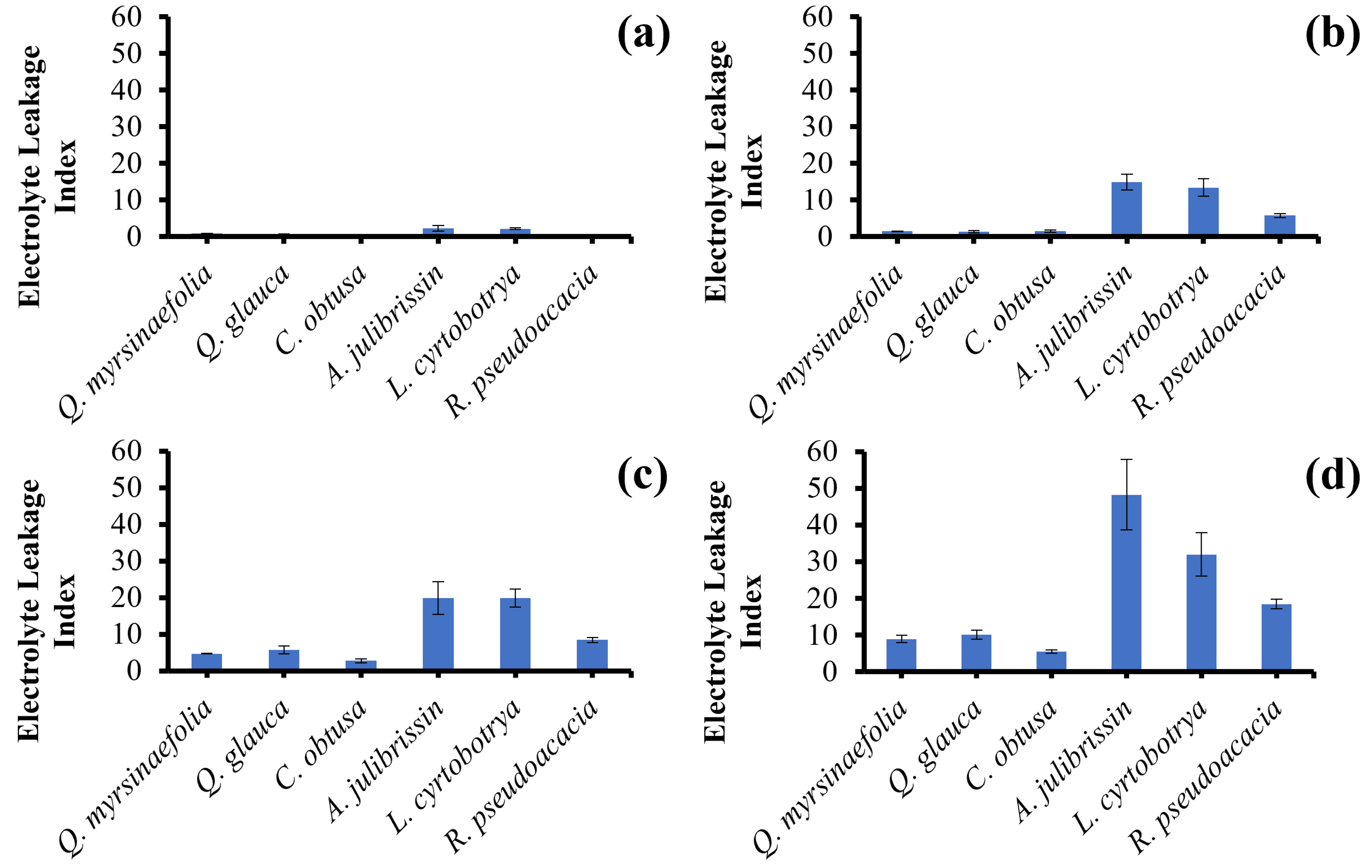
3.2. Pre-Screening of Heat-Tolerant Woody Plants Using Electrolyte Leakage Assay

To conduct pre-screening, ELI values of 27 woody plants were determined under 45 C heat stress for 60 min (Figure 4). Among the 27 species, C. obtusa, Q. glauca, Q. myrsinaefolia, I. cornuta, I. crenata, N. sericea, Q. acuta, Q. phillyraeoides, Q. salicina, T. japonica, and C. sinensis showed lower ELI values than other species. The lowest ELI value was observed in T. japonica (1.15 ± 0.03), followed by Q. salicina (1.32 ± 0.14), Q. phillyraeoides (1.34 ± 0.37), Q. acuta (1.36 ± 0.21), Q. glauca (1.38 ± 0.28), N. sericea (1.43 ± 0.37), C. sinensis (1.62 ± 0.17), I. cornuta (1.65 ± 0.24) and I. crenata (1.99 ± 0.13). None of the species showed higher ELI values than A. julibrissin, L. cyrtobotrya, and R. pseudoacacia, which were sensitive to heat stress. Among the species with low ELI values, eight candidates were selected for heat tolerance testing. To confirm heat tolerance, the candidates of 8 heat-tolerant woody plants were exposed to the heat stress condition of 45 C for 120 min. The heat-tolerant candidates well survived. No obvious damage due to heat stress was observed in the plantlets of any of the prescreened species. These results indicate that these species can endure heat stress, whereas A. julibrissin, L. cyrtobotrya, and R. pseudoacacia are sensitive to heat stress.

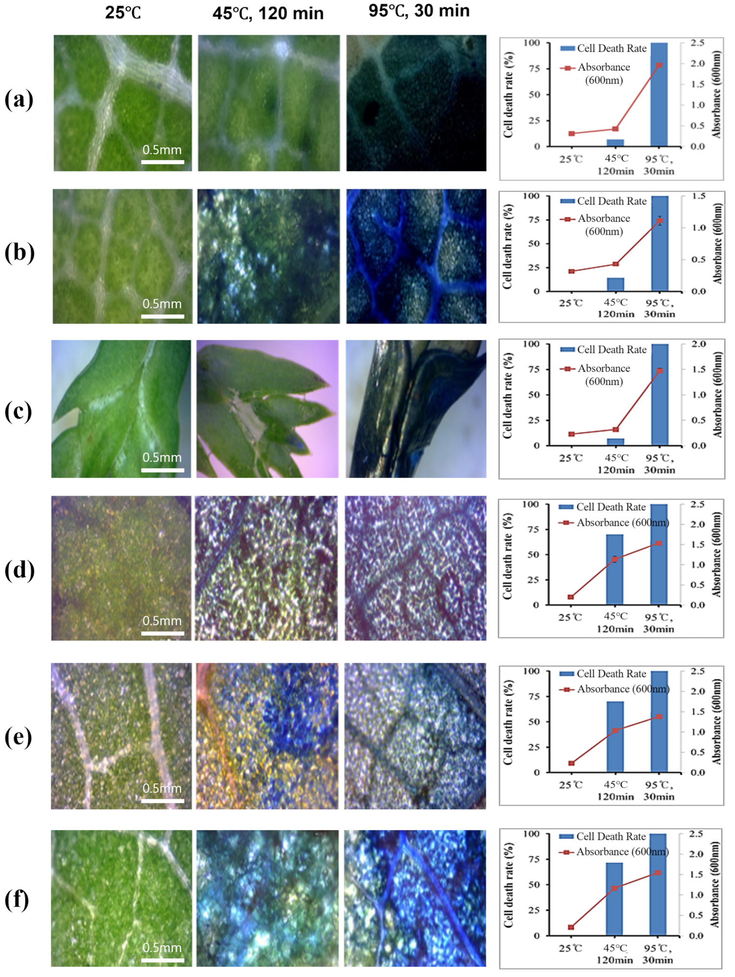
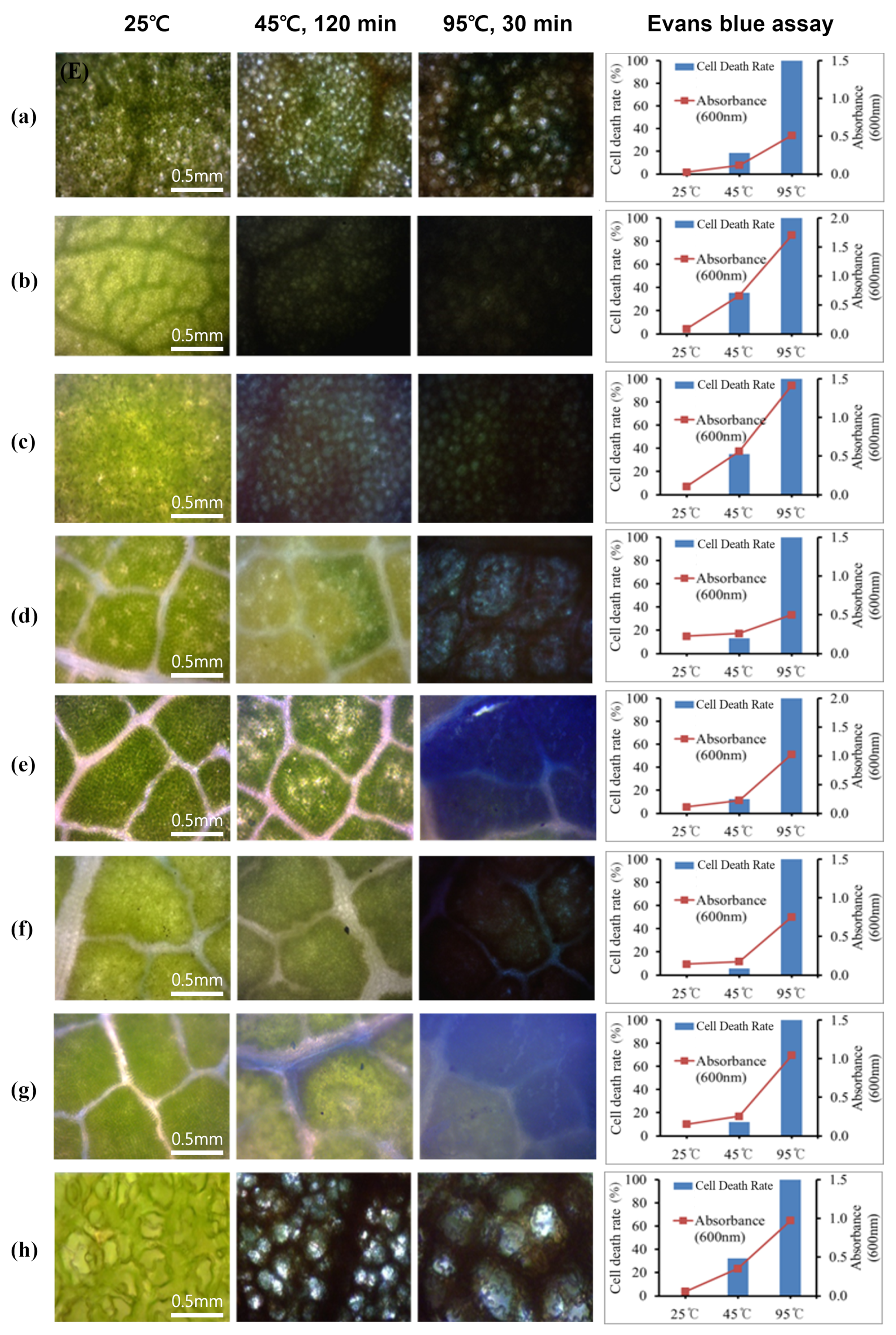
3.3. Verification of Heat Tolerance Using Evans Blue Staining and DAB Staining

The damage to the leaves of woody plants under heat stress was visualized and assessed using Evans blue staining (Figure 5). Under heat stress for 120 min, slightly stained tissue was observed in five tolerant candidate species: N. sericea, C. sinensis, Q. acuta, Q. phillyraeoides, and Q. salicina. However, more stained tissue was observed in the leaves of the other pre-screened species: I. cornuta, I. crenata and T. japonica. The cell death rates of the tolerant phenotypes were significantly lower than those of the sensitive phenotypes (p < 0.001). As the results of Evans blue assay, the lowest cell death rate was shown in the leaf of Q. phillyraeoides (5.6%), followed by Q. acuta (7.9%), Q. salicina (8.1%), N. sericea (13.2%), C. sinensis (18.5%), T. japonica (32.5%), I. crenata (34.9%) and I. cornuta (35.5%). The cell death rates of the heat-tolerant candidates were generally lower than those of the heat-sensitive woody plants.
The cell membrane permeability of each pre-screened 8 species of woody plant species was assessed by measuring the ELI under heat stress (Figure 6). The ELI values of all the plants were maximized after 120 min of heat stress. The ELI values of the heat-tolerant candidates were significantly lower than those of the heat-sensitive plants. Under heat-stress for 120 min, the lowest ELI value was observed in N. sericea (3.37 ± 0.37), followed by C. sinensis (4.04 ± 0.27), Q. acuta (5.29 ± 0.31), Q. salicina (5.73 ± 0.71), Q. phillyraeoides (5.75 ± 1.13), T. japonica (7.22 ± 0.39), I. cornuta (10.33 ± 0.50) and I. crenata (10.90 ± 1.24). It was previously shown that the ELI values of A. julibrissin, L. cyrtobotrya, and R. pseudoacacia were much higher than those of heat-tolerant candidates under the same conditions. These results indicated that the heat stress tolerance of the prescreened heat-tolerant candidates was qualified.

3.4. Plant Recovery Ability of Selected Woody Plant Species under Heat Stress

Using DAB staining methods, in situ hydrogen peroxide induced by heat stress was detected (Figure 7). The leaves of the heat-tolerant and sensitive woody plants under heat stress were collected every 30 min for 120 min, and then the plantlets were transferred to normal condition (25 C) for recovery. During the recovery phase, leaves were collected, and plant-induced hydrogen peroxide levels were observed by DAB staining. In the leaves of I. cornuta, the dye started to appear dark after 30 min, and the reactants were observed. The degree of staining increased after 120 min, and the entire leaf was widely stained. However, these reactions almost disappeared after 30 min in the recovery phase. Heat stress in I. crenata leaves appeared after 60 min of heat treatment but almost recovered after 240 min. N. sericea leaves reacted 30 min after heat stress treatment, and then changed more intensely after longer heat treatments. However, reactions in N. sericea leaves were not observed after 60 min of recovery. In Q. acuta leaves, the staining reaction appeared after 120 min of heat treatment, and it took 60 min for the leaves to show their original color. In Q. phillyraeoides, the peroxide reaction was observed after 60 min of heat stress. Under recovery conditions, the reactants almost disappeared after 120 min and completely disappeared after 240 min. In Q. salicina leaves under heat stress, dark staining occurred at 120 min, but disappeared quickly and did not appear after 30 min under recovery conditions. In T. japonica leaves, a peroxide reaction was observed after 90 min under heat stress, and no reactants were detected after 120 min during the recovery phase. In the leaves of C. sinensis, a peroxide reaction appeared after 120 min of heat stress and was not detected after 30 min of recovery.
On the other hand, the hydrogen induction level and recovery time in the heat-sensitive woody plants were different from those in heat-tolerant woody plants. In each leaf of the three species of heat-sensitive woody plants, a peroxide reaction was detectable after 30 min, which increased with longer periods of heat treatment. The induction levels were also much higher than those in the heat-tolerant woody plants. Furthermore, heat-induced hydrogen peroxide was still detectable after 480 min under recovery conditions in all the sensitive woody plants. These results indicate that hydrogen peroxide is induced later, and the induction level is much lower in heat-tolerant woody plants than in heat-sensitive woody plants.

3.5. Correlation Analysis of the Methods for Heat Tolerance Evaluation

A correlation analysis was performed to investigate the relationship between ELI, cell death rate, and survival rate after heat treatment, depending on the heating time (Table 2). The cell death rate significantly and positively correlated with ELI at all treatment times (p < 0.01). In contrast, the cell death rate was negatively correlated with the survival rate but was significant only at 90 and 120 min (p < 0.01). A negative correlation was generally observed between the survival rate and ELI, and the survival rate at 120 min showed a negative correlation with ELI, regardless of time (p < 0.01).
The results showed that an increase in ELI and cell death rate is related to a decrease in survival rate; therefore, electrolyte leakage and Evans blue staining are considered useful methods for evaluating heat tolerance.

4. Discussion

Adequate heat stress conditions were established to screen heat-tolerant native Korean plants. For screening heat-tolerant woody plants, the heat condition of 45 C for 120 min was applied. Under these conditions, plantlets of C. obtusa, Q. glauca, and Q. myrsinaefolia showed tolerant phenotypes whereas plantlets of A. julibrissin, L. cyrtobotrya, and R. pseudoacacia showed sensitive phenotypes (Figure 3 and Figure 4). Under the heat stress condition, the leaf tissue of the tolerant plants was less damaged than that of the sensitive plants (Figure 3). Therefore, the adequate heat stress conditions are 45 C for 120 min for woody plants, and these conditions can be applied for screening other heat-tolerant plants among Korean natives.
Heat treatment temperature is important for the selection of heat-tolerant plants. For this experiment, they were incubated at 45 C for 2 h. When plants are subjected to heat stress, heat shock proteins (HSP) are biosynthesized in response to heat stress. Transferring the soybean Glycine max var. Wayne seedlings from their normal growing temperature of 28 C to a maximum of 40 C (heat shock) results in dramatic changes in protein synthesis, resulting in the creation of new proteins known as heat shock proteins (HSPs) and a significant decrease in protein synthesis [29]. It was reported that HSP was synthesized even after short exposure to 45 C for 10 min. It is important to choose appropriate heating conditions to compare the heat resistances of different plants. The heat treatment temperature and time were appropriate for this study.
In the present study, an efficient method for screening heat stress-tolerant plants was established using an electrolyte leakage assay. The high heat tolerance group showed the lowest ELI, whereas the heat-sensitive group showed the highest ELI [30]. Bhattarai et al. [31] used the electrolyte leakage method to select tomato varieties with excellent heat tolerance when grown in a high-temperature environment in southern Texas. Therefore, to select heat-tolerant plant species, it is preferable to select plant species with low ELI values. Under environmental stress, reactive oxygen species (ROS) accumulate in cells and weaken the cell membrane through their oxidizing activities. Because of damage to the cell membrane, cellular molecules can leak. Cell damage and hardiness can be estimated by comparing the conductivity of leaked contents from injured and uninjured tissues in water [32]. However, this method is time-consuming, is not suitable for optimizing the conditions for all plants investigated, and may tend to overestimate heat resistance [18].
The ELI values after heat treatment differed between heat-tolerant and heat-sensitive plants. In this screening condition, ELI values of I. cornuta, I. crenata, N. sericea, T. japonica, C. sinensis, Q. acuta, Q. phillyraeoides, and Q. salicina were similiar to those of Q. myrsinaefolia, Q. glauca, and C. obtusa and their phenotypic response was also tolerant under 45 C for 120 min. However, A. julibrissin, L. cyrtobotrya, and R. pseudoacacia, which showed sensitive phenotypes in the screening condition, showed high ELI values in the heat-stress condition of 45 C for 120 min. ELI has also been used as an effective physiological indicator to determine cold tolerance by observing the degree of damage to the cell membranes of plants exposed to a short period of low temperature [33]. Heidarvand et al. [34] selected cold-tolerant strains using ELI analysis after inducing chilling stress in various strains of chickpea.
High temperatures impair the activity of proteins and fluid membrane lipids, affecting the activity of chloroplast- and mitochondrial-based enzymes and membrane integrity. Long-term exposure to severe heat stress and moderately high temperatures can result in cell damage and cell death [35]. Heat stress induces programmed cell death in plant cells and it is triggered by ROS [36]. During the early stages of stress, ROS are induced and function as cellular signaling compounds for adaptation. However, the excessive accumulation of ROS disturbs cellular homeostasis [10]. ROS are induced by extreme heat damage to plant cellular membrane [37,38]. Because of the loss of membrane integrity due to oxidative stress, electrolytes in the membrane leak [39,40]. Thus, ROS levels in plants indicate the stress status under stress conditions [41]. In this study, Evans blue staining assay and DAB staining method were used to test for ROS caused by heat.
Evans blue staining was used to assess the tolerance of woody plants to heat stress. In previous studies, Evans blue staining was used to determine cell viability [42,43,44]. It has been also reported that the Evans blue staining method can be applied to determine cell death caused by heavy metal treatment as an abiotic stress factor [45]. However, Evans blue staining assay has rarely been used to assess the tolerance/sensitivity of plants under heat stress. In the present study, the results of the Evans blue staining assay corresponded to the phenotypic responses of tolerant or sensitive plants under heat stress. Therefore, this method not only provides a visual image of regions damaged by heat stress, but also helps quantify its intensity.
It is important to assess the recovery ability of heat-tolerant plants after heat treatment; however, this has been overlooked in previous studies. In the case of herbaceous plants, there have been few reports on the physiological factors associated with recovery. Wang and Huang [46] evaluated the ability of Kentucky Bluegrass to recover from drought and heat stress by measuring cell membrane stability, leaf Fv/Fm, lipid peroxidation, and SOD and CAT activities. These methods provide information regarding the extent to which plants recover from stress. However, they are not suitable for visualizing the extent to which plants recover from stress within a short time interval. In this study, the DAB staining method was used, which is considered to be a very good assay. DAB staining has been used to detect in situ hydrogen peroxide [47,48,49]. This led to an assessment of the amount of heat-induced hydrogen peroxide accumulation under heat stress and how quickly it was removed from the leaf tissue of each screened plant species under recovery conditions. By monitoring heat-induced hydrogen peroxide using DAB staining, we observed differences between heat-tolerant and heat-sensitive plant species. Less hydrogen peroxide accumulated in the leaves of heat-tolerant plants (I. cornuta, I. crenata, N. sericea, Q. acuta, Q. phillyraeoides, Q. salicina, T. japonica, and C. sinensis) than in those of heat-sensitive plant species (A. julibrissin, L. cyrtobotrya, and R. pseudoacacia). Under recovery conditions, the time points at which heat-induced hydrogen peroxide was undetectable were shorter in the leaves of heat-tolerant plants than in those of heat-sensitive plants. These results indicate that heat-tolerant plants experience less oxidative stress than heat-sensitive plants and recover from stress more rapidly. Moreover, these results suggested that lower levels of oxidative stress cause less electrolyte leakage and damage to heat-tolerant plants. Despite the usefulness of DAB staining method, quantification was not performed in this study. If the measurement of DAB staining values is supported in the future, strong evidence of the stress state and recovery ability of plant species or varieties can be provided.
Heat tolerance differed among the species. Among the tested species, C. obtusa, Q. glauca, and Q. myrsinaefolia exhibited strong heat tolerance. C. obtusa, a coniferous tree, has long been used in construction and furniture. Essential oils extracted from the leaves and twigs have been used as functional additives or fragrances in soap, toothpaste, and cosmetics. Studies on abiotic stress in C. obtusa have not been conducted, except for those on drought tolerance. Ikei et al. [50] studied the response of C. obtusa to drought stress in six mountainous regions in Korea. Total lysates, carotenoids, and H2O2 were measured to select drought-tolerant cypress trees. Quercus spp. have proven to be useful for carbon storage and are widely used in building materials, furniture materials, and appliances. Evergreen oaks have been reported to be a promising tree species that can prepare for climate change because they are tolerant to abiotic stresses such as heat and drought. Park et al. [51] reported that Quercus spp. (Q. acuta, Q. glauca, Q. myrsinaefoila, and Q. salicina) were tested for cold tolerance when selecting cold-tolerant individuals. When Q. myrsinaefolia seedlings were exposed to low-temperature stress, their survival rate was higher than that of the other species. These tree species could be used for reforestation in response to climate change in Northeast Asia including Korea.

5. Conclusions

Recently, owing to climate change caused by global warming, interest in breeding plants with abiotic stress tolerance has increased significantly. The existing abiotic stress tolerance screening methods mainly measure plant growth in the field. Therefore, screening is difficult because it requires many samples and is time consuming. The results of this study are relatively rapid, reproducible, and highly sensitive, so it is judged to be a method that can supplement the existing traditional method as a heat-tolerant plant selection system. It is difficult to select heat-tolerant species by evaluating plant heat tolerance alone. In this study, heat tolerance evaluation methods for various domestic woody plants were established and plants with strong heat tolerance were selected. In this study, instead of using a specific method to measure the heat tolerance of plants, the degree of electrolyte leakage was measured and cell death was observed using histological methods, such as Evans blue staining. In addition, recovery from heat stress was confirmed by DAB staining (Figure 8). Using this method, heat-tolerant woody plants were selected by measuring electrolyte leakage indices and were proven to have strong heat tolerance in the results of histological examination and recovery tests. This indicates that our method is highly effective for selecting heat-tolerant plant species. The results of this study can be widely used for the selection and breeding of heat-tolerant plants in response to climate change.

Supplementary Materials

The following supporting information can be downloaded at: https://www.mdpi.com/article/10.3390/f15081366/s1, Figure S1: Schemes of heat stress treatments. This scheme was designed by modifying the method from the report of Yeh et al. [14].; Figure S2: Appearance of six species of woody plants heat-treated at 40, 45, 50, and 55 C for 120 min. (a) Q. myrsinaefolia, (b) Q. glauca, (c) C. obtusa, (d) A. julibrissin, (e) L. cyrtobotrya, and (f) R. pseudoacacia.

Author Contributions

Conceptualization, M.S.C. and S.H.Y.; methodology, D.J.P., K.B.P. and D.H.K.; resources, J.-H.L. and S.A.C.; writing—review and editing, S.A.K., S.H.Y. and M.S.C.; visualization, S.H.Y.; funding acquisition, M.S.C. All authors have read and agreed to the published version of the manuscript.

Funding

This study was carried out with the support of “Training of talent tailored to the forest industry field project(R&D)” of the Korea Forestry Promotion Institute (Short-term Income Forest Products Governance Education Center(SFPEC), Project No. RS-2024-040519661382116530101) in 2024.

Data Availability Statement

Data is contained within the article or Supplementary Materials.

Conflicts of Interest

The authors declare no conflict of interest.

References

  1. IPCC. Climate Change 2023: Synthesis Report. Contribution of Working Groups I, II and III to the Sixth Assessment Report of the Intergovernmental Panel on Climate Change [Core Writing Team, H. Lee and J. Romero (eds.)]; IPCC: Geneva, Switzerland, 2023; pp. 42–74. [Google Scholar] [CrossRef]
  2. Climate Change Situation Map (Korea Meteorological Administration). Available online: http://www.climate.go.kr/atlas/dashboard (accessed on 12 June 2024).
  3. Zhao, J.; Lu, Z.; Wang, L.; Jin, B. Plant Responses to Heat Stress: Physiology, Transcription, Noncoding RNAs, and Epigenetics. Int. J. Mol. Sci. 2021, 22, 117. [Google Scholar] [CrossRef] [PubMed]
  4. Crafts-Brandner, S.J.; Salvucci, M.E. Sensitivity of Photosynthesis in a C4 Plant, Maize, to Heat Stress. Plant Physiol. 2002, 129, 1773–1780. [Google Scholar] [CrossRef] [PubMed]
  5. Camejo, D.; Rodríguez, P.; Morales, M.A.; Dell’Amico, J.M.; Torrecillas, A.; Alarcón, J.J. High temperature effects on photosynthetic activity of two tomato cultivars with different heat susceptibility. J. Plant Physiol. 2005, 165, 281–289. [Google Scholar] [CrossRef] [PubMed]
  6. Havaux, M. Characterization of thermal damage to the photosynthetic electron transport system in potato leaves. Plant Sci. 1993, 94, 19–33. [Google Scholar] [CrossRef]
  7. Yin, H.; Chen, Q.; Yi, M. Effects of short-term heat stress on oxidative damage and responses of antioxidant system in Lilium longiflorum. Plant Growth Regul. 2008, 54, 45–54. [Google Scholar] [CrossRef]
  8. Xu, S.; Li, J.; Zhang, X.; Wei, H.; Cui, L. Effects of heat acclimation pretreatment on changes of membrane lipid peroxidation, antioxidant metabolites, and ultrastructure of chloroplasts in two cool-season turfgrass species under heat stress. Environ. Exp. Bot. 2006, 56, 274–285. [Google Scholar] [CrossRef]
  9. Sarkar, N.K.; Kundnani, P.; Grover, A. Functional analysis of Hsp70 superfamily proteins of rice (Oryza sativa). Cell Stress Chaperones 2013, 18, 427–437. [Google Scholar] [CrossRef] [PubMed]
  10. Vickers, C.E.; Gershenzon, J.; Lerdau, M.T.; Loreto, F. A unified mechanism of action for volatile isoprenoids in plant abiotic stress. Nat. Chem. Biol. 2009, 5, 283–291. [Google Scholar] [CrossRef] [PubMed]
  11. Wang, M.; Zou, Z.; Li, Q.; Sun, K.; Chen, X.; Xinghui, L. The CsHSP17.2 molecular chaperone is essential for thermotolerance in Camellia sinensis. Sci. Rep. 2017, 7, 1237. [Google Scholar] [CrossRef] [PubMed]
  12. Asthir, B. Protective mechanisms of heat tolerance in crop plants. J. Plant Interact. 2015, 10, 202–210. [Google Scholar] [CrossRef]
  13. Moreno, A.A.; Orellana, A. The physiological role of the unfolded protein response in plants. Biol. Res. 2011, 44, 75–80. [Google Scholar] [CrossRef] [PubMed]
  14. Yeh, C.H.; Kaplinsky, N.J.; Hu, C.; Charng, Y. Some like it hot, some like it warm: Phenotyping to explore thermotolerance diversity. Plant Sci. 2012, 195, 10–23. [Google Scholar] [CrossRef] [PubMed]
  15. Hameed, M.; Keitel, C.; Ahmad, N.; Mahmood, T.; Trethowan, R. Screening of tomatoes germplasm for heat stress tolerance under controlled conditions. Procedia Environ. Sci. 2015, 29, 173–174. [Google Scholar] [CrossRef]
  16. Bibi, A.C.; Oosterhuis, D.M.; Gonias, E.G. Photosynthesis, quantum yield of photosystem II, and membrane leakage as affected by high temperature in cotton genotypes. Cotton Sci. 2008, 12, 150–159. [Google Scholar]
  17. Dexter, S.T.; Tottingham, W.E.; Graber, L.F. Preliminary results in measuring the hardiness of plants. Plant Physiol. 1930, 5, 215–223. [Google Scholar] [CrossRef] [PubMed]
  18. Ilík, P.; Špundováet, M.; Šicner, M.; Melkovičová, H.; Kučerová, Z.; Krchňák, P.; Fürst, T.; Večeřová, K.; Panzarová, K.; Benediktyová, Z.; et al. Estimating heat tolerance of plants by ion leakage: A new method based on gradual heating. New Phytol. 2018, 218, 1278–1287. [Google Scholar] [CrossRef] [PubMed]
  19. Pérez-Harguindeguy, N.; Díaz, S.; Garnier, E.; Lavorel, S.; Poorter, H.; Jaureguiberry, P.; Bret-Harte, M.S.; Cornwell, W.K.; Craine, J.M.; Gurvich, D.E.; et al. New handbook for standardised measurement of plant functional traits worldwide. Aust. J. Bot. 2013, 61, 167–234. [Google Scholar] [CrossRef]
  20. Mansour, M.M.F. Plasma membrane permeability as an indicator of salt tolerance in plants. Biol. Plant. 2013, 57, 1–10. [Google Scholar] [CrossRef]
  21. Murkowski, A. Heat stress and spermidine: Effect on chlorophyll fluorescence in tomato plants. Biol. Plant. 2001, 44, 53–57. [Google Scholar] [CrossRef]
  22. Harsh, A.; Sharma, Y.K.; Joshi, U.; Rampuria, S.; Singh, G.; Kumar, S.; Sharma, R. Effect of short-term heat stress on total sugars, proline and some antioxidant enzymes in moth bean (Vigna aconitifolia). Ann. Agric. Sci. 2016, 61, 57–64. [Google Scholar] [CrossRef]
  23. Li, Q.; Wang, Z.; Li, D.; Wei, J.; Qiao, W. Meng, X.; Sun, S.; Li, H.; Zhao, M.; Chen, X.; Zhao, F. Evaluation of a new method for quantification of heat tolerance in different wheat cultivars. J. Integr. Agric. 2018, 17, 786–795. [Google Scholar] [CrossRef]
  24. Hossain, A.; Sarker, M.A.Z.; Saifuzzaman, M.; Teixeira da Silva, J.A.; Lozovskaya, M.V.; Akhter, M.M. Evaluation of growth, yield, relative performance and heat susceptibility of eight wheat (Triticum aestivum L.) genotypes grown under heat stress. Int. J. Plant Prod. 2013, 7, 615–636. [Google Scholar] [CrossRef]
  25. Wassie, M.; Zhang, W.; Zhang, Q.; Ji, K.; Chen, L. Effect of Heat Stress on Growth and Physiological Traits of Alfalfa (Medicago sativa L.) and a Comprehensive Evaluation for Heat Tolerance. Agronomy 2019, 9, 597. [Google Scholar] [CrossRef]
  26. Dorado, F.J.; Solla, A.; Alcaide, F.; Martín, M.Á. Assessing heat stress tolerance in Castanea sativa. Forestry 2022, 95, 667–677. [Google Scholar] [CrossRef]
  27. Marias, D.E.; Meinzer, F.C.; Woodruff, D.R.; McCulloh, K.A. Thermotolerance and heat stress responses of Douglas-fir and ponderosa pine seedling populations from contrasting climates. Tree Physiol. 2017, 37, 301–315. [Google Scholar] [CrossRef] [PubMed]
  28. Daudi, A.; O’Brien, J.A. Detection of hydrogen peroxide by DAB staining in Arabidopsis leaves. Bio-Protocol 2012, 2, e263. [Google Scholar] [CrossRef]
  29. Lin, C.Y.; Roberts, J.K.; Key, J.L. Acquisition of thermotolerance in soybean seedlings: Synthesis and accumulation of heat shock proteins and their cellular localization. Plant Physiol. 1984, 74, 152–160. [Google Scholar] [CrossRef] [PubMed]
  30. Jin, E.J.; Cho, M.G.; Bae, E.J.; Park, J.H.; Lee, K.S.; Choi, M.S. Physiological responses to drought stress of seven evergreen hardwood species. J. Korean For. Soc. 2017, 106, 397–407. [Google Scholar] [CrossRef]
  31. Bhattarai, S.; Harvey, J.T.; Djidonou, D.; Leskovar, D.I. Exploring morpho-physiological variation for heat stress tolerance in tomato. Plants 2021, 10, 347. [Google Scholar] [CrossRef] [PubMed]
  32. McNabb, K.; Takahashi, E. Freeze Damage to Loblolly Pine Seedlings as Indicated by Conductivity Measurements and Outplanting Survival; Research Report 00-4; Auburn University Southern Forest Nursery Management Cooperative: Auburn, AL, USA, 2000. [Google Scholar]
  33. Orlova, I.V.; Serebriiskaya, T.S.; Popov, V.; Merkulova, N.; Nosov, A.M.; Trunova, T.I.; Tsydendambaev, V.D.; Los, D.A. Transformation of Tobacco with a gene for the thermophilic Acyl-lipid desaturase enhances the chilling tolerance of plants. Plant Cell Physiol. 2003, 44, 447–450. [Google Scholar] [CrossRef] [PubMed]
  34. Heidarvand, L.; Maali Amiri, R.; Naghavi, M.R.; Farayedi, Y.; Sadeghzadeh, B.; Alizadeh, K. Physiological and morphological characteristics of chickpea accessions under low temperature stress. Russ. J. Plant Physiol. 2011, 58, 157–163. [Google Scholar] [CrossRef]
  35. Wahid, A.; Gelani, S.; Ashraf, M.; Foolad, M.R. Heat tolerance in plants: An overview. Environ. Exp. Bot. 2007, 61, 199–223. [Google Scholar] [CrossRef]
  36. Petrov, V.; Hille, J.; Mueller-Roeber, B.; Gechev, T.S. ROS-mediated abiotic stress-induced programmed cell death in plants. Front. Plant Sci. 2015, 6, 69. [Google Scholar] [CrossRef] [PubMed]
  37. Hemantaranjan, A.; Bhanu, A.N.; Singh, M.N.; Yadav, D.K.; Patel, P.K.; Singh, R.; Katiyar, D. Heat stress responses and thermotolerance. Adv. Plants Agric. Res. 2014, 1, 62–70. [Google Scholar] [CrossRef]
  38. Zhu, J.K. Abiotic stress signaling and responses in plants. Cell 2008, 167, 313–324. [Google Scholar] [CrossRef] [PubMed]
  39. Breusegem, F.V.; Dat, J.F. Reactive oxygen species in plant cell death. Plant Physiol. 2006, 141, 384–390. [Google Scholar] [CrossRef] [PubMed]
  40. Demidchik, V.; Straltsova, D.; Medvedev, S.S.; Pozhvanov, G.A.; Sokolik, A.; Yurin, V. Stress-induced electrolyte leakage: The role of K+-permeable channels and involvement in programmed cell death and metabolic adjustment. J. Exp. Bot. 2014, 65, 1259–1270. [Google Scholar] [CrossRef] [PubMed]
  41. Kim, C.H.; Meskauskiene, R.; Apel, K.; Laloi, C. No single way to understand singlet oxygen signalling in plants. EMBO Rep. 2008, 9, 435–439. [Google Scholar] [CrossRef] [PubMed]
  42. Huang, C.N.; Cornejo, M.J.; Bush, D.S.; Jones, R.L. Estimating viability of plant protoplasts using double and single staining. Protoplasma 1986, 135, 80–87. [Google Scholar] [CrossRef]
  43. Silva, F.D.; Menedez-Yuffa, A. Viability in protoplasts and cell suspensions of Coffea arabica cv. Catimor. Electron. J. Biotechn. 2006, 9, 593–597. [Google Scholar] [CrossRef]
  44. Castro-Concha, L.A.; Escobedo, R.M.; Miranda-Ham, M.L. Measurement of cell viability in in vitro cultures. In Plant Cell Culture Protocols. Methods in Molecular Biology™; Loyola-Vargas, V.M., Vázquez-Flota, F., Eds.; Humana Press: Totowa, NJ, USA, 2006; Volume 318, pp. 71–76. [Google Scholar] [CrossRef]
  45. Gonzalez-Mendoz, D.; Quiroz-Moreno, A.; Medrano, R.E.; Grimaldo-Juarez, O.; Zapata-Perez, O. Cell viability and leakage of electrolytes in Avicennia germinans exposed to heavy metals. Z. Naturforsch. 2008, 64, 391–394. [Google Scholar] [CrossRef] [PubMed]
  46. Wang, Z.; Huang, B. Physiological recovery of Kentucky bluegrass from simultaneous drought and heat stress. Crop Sci. 2004, 44, 1729–1736. [Google Scholar] [CrossRef]
  47. Yan, Y.; Guo, X. Overexpression of GhWRKY27a reduce tolerance to drought stress and resistance to Rhizoctonia solani infection in transgenic Nicotiana benthamiana. Front. Physiol. 2015, 6, 265. [Google Scholar] [CrossRef] [PubMed]
  48. Nakabayashi, R.; Yonekura-Sakakibara, K.; Urano, K.; Suzuki, M.; Yamada, Y.; Nishizawa, T.; Matsuda, F.; Kojima, M.; Sa-kakibara, H.; Shinozaki, K.; et al. Enhancement of oxidative and drought tolerance in Arabidopsis by overaccumulation of antioxidant flavonoids. Plant J. 2014, 77, 367–379. [Google Scholar] [CrossRef] [PubMed]
  49. Pellinen, R.I.; Korhonen, M.S.; Tauriainen, A.A.; Palva, E.T.; Kangasjärvi, J. Hydrogen peroxide activates cell death and defense gene expression in birch. Plant Physiol. 2002, 130, 549–560. [Google Scholar] [CrossRef] [PubMed]
  50. Ikei, H.; Song, C.; Miyazaki, Y. Physiological effect of olfactory stimulation by Hinoki cypress (Chamaecyparis obtusa) leaf oil. J. Physiol. Anthropol. 2008, 34, 44. [Google Scholar] [CrossRef] [PubMed]
  51. Park, D.J.; Yong, S.H.; Choi, M.S. Establishment of selection method for cold-tolerant individuals through evaluating tolerance of evergreen Quercus spp. against cold stress. J. For. Environ. Sci. 2019, 35, 232–239. [Google Scholar] [CrossRef]
Figure 1. Survival rates of Korean native woody plants after heat stress of 45 C for 30 min to 120 min. Means with the same letters are not significantly different at p < 0.05 (Duncan’s multiple-range test). (a) 30 min, (b) 60 min, (c) 90 min, (d) 120 min.
Forests 15 01366 g001
Figure 2. Leaf tissue damage assessment of Korean native woody plants under heat stress by Evans blue staining. Leaf tissues were subjected to heat treatment at 45 C for 120 min. The photographs on the left shows the cell dead part of the leaves, which is dyed with Evans blue. The magnification is identical in the photographs. The graphs on the right shows how much the Evans blue was absorbed by leaf tissue, which was calculated by the absorbance at 600 nm. (a) Q. myrsinaefolia, (b) Q. glauca, (c) C. obtusa, (d) A. julibrissin, (e) L. cyrtobotrya, and (f) R. pseudoacacia.
Forests 15 01366 g002
Figure 3. Electrolyte leakage assay of Korean native woody plants under heat treatment at 45 C for various heating time. Means with the same letters are not significantly different at p < 0.05 (Duncan’s multiple-range test). (a) 30 min, (b) 60 min, (c) 90 min, (d) 120 min.
Forests 15 01366 g003
Figure 4. Electrolyte leakage index of Korean native plants after heat stress of 45 C for 60 min. Means of 3 replicates with the same letters are not significantly different at p < 0.05 (Duncan’s multiple-range test).
Forests 15 01366 g004
Figure 5. Leaf tissue damage assessment of pre-screened heat-tolerant woody plants under heat shock by Evans blue staining. Leaf tissues were subjected to heat treatment at 45 C for 120 min. The photographs on the left shows the dead part of the leaves, which is dyed with Evans blue. The magnification is identical in the photographs. The graphs on the right shows how much the Evans blue was absorbed by leaf tissue, which was calculated by the absorbance at 600 nm. (a) C. sinensis, (b) I. cornuta, (c) I. crenata, (d) N. sericea, (e) Q. acuta, (f) Q. phillyraeoides, (g) Q. salicina, (h) T. japonica.
Forests 15 01366 g005
Figure 6. Leaf tissue damage assessment of pre-screened heat-tolerant woody plants under heat stress by electrolyte leakage index. The leaf tissues were subjected to heat treatment of 45 C for 30 min to 120 min. (a) C. sinensis, (b) I. cornuta, (c) I. crenata, (d) N. sericea, (e) Q. acuta, (f) Q. phillyraeoides, (g) Q. salicina, and (h) T. japonica
Forests 15 01366 g006
Figure 7. Development of in situ hydrogen peroxide in the screened heat-tolerant woody plants by DAB staining. Heat treatment was performed at 45 C for 120 min at every 30 min. Recovery was observed at 25 C for 480 min. Negative control: incubated in normal condition, Positive control: soaked in 30% (v:v) hydrogen peroxide for 10 min (a) C. sinensis, (b) I. cornuta, (c) I. crenata, (d) N. sericea, (e) T. japonica, (f) Q. acuta, (g) Q. phillyraeoides, and (h) Q. salicina
Forests 15 01366 g007
Figure 8. Experimental schematic diagram for efficient heat-tolerant woody tree species selection.
Forests 15 01366 g008
Table 1. The list of Korean native woody plants used for testing heat tolerance.
Scientific NameFamilyCommon Name
Albizia julibrissin Durazz.FabaceaeSilk Tree
Amorpha fruticosa L.FabaceaeFalse indigo bush
Camellia sinensis L.TheaceaeTea camellia
Castanopsis sieboldii (Makino) Hatus.FabaceaeSiebold’s chinquapin
Chamaecyparis obtusa (Siebold and Zucc.)CupressaceaeJapanese false cypress
Cinnamomum yabunikkei H.OhbaLauraceaeJapanese camphor tree
Dendropanax morbiferus H. LévAraliaceaeKorean dendropanax
Eucommia ulmoides Oliv.EucommiaceaeGutta-percha tree
Ilex cornuta Lindl.AquifoliaceaeHorned holly
Ilex crenata Thunb.AquifoliaceaeBox-leaf holly
Indigofera kirilowii Maxim.FabaceaeKirilow’s indigo
Indigofera pseudotinctoria Matsum.FabaceaeDwarf false-indigo
Lespedeza bicolor Turcz.FabaceaeShrub lespedeza
Lespedeza cyrtobotrya Miq.FabaceaeLeafy lespedeza
Ligustrum japonicum Thunb.OleaceaeWax-leaf privet
Morus bombycis Koidz.MoraceaeKorean mulberry
Neolitsea sericea (Blume) Koidz.LauraceaeSericeous newlitsea
Pinus rigida Mill.PinaceaePitch pine
Quercus acuta Thunb.FagaceaeRed-wood evergreen oak
Quercus glauca Thunb.FagaceaeRing-cup oak
Quercus myrsinaefolia Bl.FagaceaeBamboo-leaf oak
Quercus phillyraeoides A. GrayFagaceaeUbame oak
Quercus salicina Bl.FagaceaeWillow-leaf evergreen oak
Robinia pseudoacacia L.FabaceaeBlack locust
Sorbaria sorbifolia var. stellipila Max.RosaceaeFalse spiraea
Spiraea prunifolia f. simpliciflora Nakai.RosaceaeSimple bridalwreath spiraea
Ternstroemia japonica Thunb.PentaphylacaceaeNaked-anther ternstroemia
Table 2. Correlation analysis of the methods for heat tolerance evaluation.
S 30 S 60 S 90 S 120 ELI 30 ELI 60 ELI 90 ELI 120 CD
S 30 - a--------
S 60 10.57 *0.350.230.070.110.06−0.34
S 90 10.88 **−0.25−0.53 *−0.43−0.54 *−0.87 **
S 120 1−0.59 **−0.81 **−0.76 **−0.76 **−0.99 **
E L I 30 10.87 **0.92 *0.88 **0.65**
E L I 60 10.98 **0.95 **0.80**
E L I 90 10.96 **0.78 **
E L I 120 10.77 **
C D 1
* Correlation is significant at the 0.05 level (2-tailed). ** Correlation is significant at the 0.01 level (2-tailed). a Cannot be calculated because S 30 has constant value (100). S 30 : survival rate after 30 min, S 60 : survival rate after 60 min, S 90 : survival rate after 90 min, S 120 : survival rate after 120 min, E L I 30 : electrolyte leakage indexes after 30 min, E L I 60 : electrolyte leakage indexes after 60 min, E L I 90 : electrolyte leakage indexes after 90 min, E L I 120 : electrolyte leakage indexes after 120 min, CD: cell death rate.
Disclaimer/Publisher’s Note: The statements, opinions and data contained in all publications are solely those of the individual author(s) and contributor(s) and not of MDPI and/or the editor(s). MDPI and/or the editor(s) disclaim responsibility for any injury to people or property resulting from any ideas, methods, instructions or products referred to in the content.

