Communication

Detection of Hymenoscyphus fraxineus in Leaf Rachises from European Ash (Fraxinus excelsior) in Germany

Thünen Institute of Forest Genetics, Eberswalder Chaussee 3a, 15377 Waldsieversdorf, Germany
*
Author to whom correspondence should be addressed.
Forests 2025, 16(1), 149; https://doi.org/10.3390/f16010149
Submission received: 21 November 2024 / Revised: 3 January 2025 / Accepted: 13 January 2025 / Published: 15 January 2025
(This article belongs to the Special Issue Management of Forest Pests and Diseases—2nd Edition)

Abstract

:
The ash dieback disease caused by Hymenoscyphus fraxineus is widespread in Germany and is the subject of intensive research efforts. The fungus identification is based on the genomic internal transcribed spacer (ITS) region, which can also be the site of intragenomic variability. In Germany, despite intense research efforts, only a few numbers of H. fraxineus sequence data are recorded. Therefore, this study aims to characterize H. fraxineus isolates obtained from diseased ash leaves in Brandenburg (Germany). Fungal isolates from infected ash leaf tissue were analyzed molecularly using species-specific primers and based on sequencing the ITS region of rDNA. The analysis of the two identified sequences revealed two base substitutionscompared to the reference sequences. Thus, they show an identity of 98.8%–100% to the reference sequences and support the assumption that H. fraxineus has multiple copies of the ITS region. The phylogenetic grouping with reference sequences did not show a distinct cluster for the European, particularly the German, sequences. This indicates that the evolvement of the genetic variability of the ITS region is still an ongoing process.

1. Introduction

Since the early 1990s, a severe disease has affected European ash (Fraxinus excelsior L.) trees, which is caused by the ascomycete Hymenoscyphus fraxineus (T. Kowalski), as discovered by Baral, Queloz, and Hosoya (syn. H. pseudoalbidus, anamorph: Chalara fraxinea T. Kowalski) [1,2,3]. In Europe, H. fraxineus was first described as a new pathogenic species in diseased ash trees in Poland, and from there, it spread to most other European countries [1,4,5]. Since, it has spread to Eastern and Western European countries, as listed in the EPPO Global Database “https://gd.eppo.int/taxon/CHAAFR/distribution (accessed on 14 October 2024)”. Its closely related and non-pathogenic sister species H. albidus (Roberge ex Desm.) W. Phillips is native to Europe and lives as a saprotroph without harming F. excelsior. Although H. fraxineus and H. albidus have a high degree of genetic similarity, the remaining differences enable H. fraxineus to adapt rapidly to its ecological niche and cause severe symptoms in F. excelsior [6].
European ash trees growing in the arboretum of the State Forest Enterprise Brandenburg Office (Germany) frequently show characteristic symptoms of ash dieback disease. Chlorosis of leaflet blades and necrotic lesions on leaf rachises as well as leaflet veins are considered as early visible symptoms that lead to wilting of the foliage and premature shedding. Finally, tips twigs and branches show dieback and shoot mortality in spring, especially if the ratio of leaves with necrosis at the end of the previous vegetation period was high [3,7,8,9]. Apothecia of H. fraxineus appear on ash leaf rachises in the summer after falling. The released ascospores are spread by wind and can infect ash trees [2,10,11]. The fungus colonizes not only tissue in the crown area but also the stem tissue and causes basal lesions, necrosis, and collar rots by interactions with other secondary pathogens [7,12,13]. Besides the visible damage on the host tree, an infection with H. fraxineus also impacts the composition of the leaf microbial network [14]. Interestingly, recent research reveals specific microbial communities that colonize the leaves of ash trees tolerant to ash dieback [15,16]. The features relevant to the potential to enhance tree tolerance are associated with genes for stress adaptation, as well as protein secretion, the synthesis of exopolysaccharides, and biofilm production [17].
In Germany, the ash disease has been known since 2002, and the first proof of infection of ash trees with H. fraxineus was documented in 2007 [18,19,20]. Subsequently, intense investigations at important provenances revealed an increasing severity of ash dieback and a high genetic variation in the susceptibility of the ash trees to the pathogen, too [13,21,22,23,24,25]. Recently, several aspects of ash dieback in Germany, such as disease epidemiology, the preservation of ash trees, and breeding strategies, have been investigated within the interdisciplinary research project FraxForFuture [26].
For fungal identification and phylogenetic analyses, the ITS region is commonly accepted as the primary fungal barcode [27]. It combines conserved and highly variable DNA areas, allowing us to distinguish between fungal species with high resolution [28,29]. Genome assemblies from more than 2000 fungal taxa revealed the internal transcribed spacers (ITS) as regions of even intragenomic variation. Multiple ITS copies can be found in over 25% of the evaluated taxa [30]. In particular, intragenomic variation is defined as a variation existing within individual genomes between multiple copies of the same sequence region [31]. So, intraspecific variation in the ITS region is generally considered widespread and is used to differentiate H. fraxineus from other leaf endophytes and pathogens, respectively. Therefore, H. fraxineus isolates cultivated on artificial media are commonly used [2,32,33,34,35].
Despite the wide distribution of H. fraxineus in all German federal states [13], only a few local molecular data on the pathogen are available. This study aims to isolate H. fraxineus strains from rachises of infected ash leaves and their molecular identification regarding the DNA metabarcoding region. Here, we contribute to the ITS sequence data collection about pathogens in German ash stands.

2. Materials and Methods

2.1. Material Sampling

European ash trees growing in Waldsieversdorf (federal state Brandenburg, Germany) frequently show characteristic ash dieback symptoms, such as necrotic lesions on leaf rachises, wilting of foliage, and shoot mortality (Figure 1B). In detail, 25 rachises with pseudosclerotial plates from the leaf litter were sampled in November 2020. In the consecutive year, rachises with apothecia have been found (Figure 1C).

2.2. Fungal Isolation

Immediately after collecting, the rachises were cut into 1 cm long segments. For surface sterilization, the segments were submerged for 15 s in a 0.05% AgNO3-solution with one drop of Tween®20 (Carl Roth GmbH & Co. KG, Karlsruhe, Germany), rinsed three times with sterile water, prayed with 75% ethanol, and dried under sterile conditions until the ethanol was evaporated. The segments obtained from the tissue of different rachises were labeled accordingly and placed onto Petri dishes (Ø 94 mm, Greiner Bio-One GmbH, Frickenhausen, Germany) containing malt extract agar (MEA, Duchefa Biochemie, Haarlem, The Netherlands). The Petri dishes were incubated at 20 °C in the dark. To obtain pure cultures, the growth and morphological characteristics of the initial colonies growing from the rachis segments were macroscopically checked 10 days after incubation for the first time followed by frequent inspections. Criteria for mycelia selection were growth rate, pigmentation, and radial growth. The colonies with mold-suspicious morphology were discarded. To transfer to fresh MEA, 1 × 1 cm agar blocks with fungal mycelia were cut and transferred to new Petri dishes. During the subculturing processes, the segments from which the next mycelia transfer was supposed to occur were selected based on their morphological characteristics and further cultivated as separate isolates, as described by Kowalski and Bartnik [36]. After the 4th subculture, mycelia were scraped cautiously from the solid media and stored at −20 °C for further DNA extraction.

2.3. DNA Extraction, PCR and Sequencing

For DNA extraction, putative H. fraxineus isolates were selected according to their morphologic characteristics [36]. Therefore, the DNeasy Plant Mini Kit (QIAGEN GmbH, Hilden, Germany) was used according to manufacturer instructions. The pellet was finally resuspended in 50 µL H2Obidest.
PCR analyses were conducted using the forward primer 5′-AGC TGG GGA AAC CTG ACT G-3′ and the reverse primer 5′-ACA CCG CAA GGA CCC TAT C-3′ [37,38]. The PCR reactions were performed using the Uno II Thermoblock Thermal Cycler (Biometra GmbH, Göttingen, Germany) with an AccuPrime™ Taq DNA Polymerase System (Invitrogen, Waltham, MA, USA) and 2 mM MgCl2 concentration. The PCR reactions were initialized at 94 °C for 3 min for 35 cycles starting at 95 °C for 30 s, 57 °C for 30 s, ending at 68 °C for 60 s, and finally 68 °C for 7 min. PCR products were visualized on a 1.2% TBE agarose gel, stained with GelRed® (Merck, Darmstadt, Germany) and purified using the PCR Purification Kit (QIAGEN GmbH, Hilden, Germany). PCR products were sent to Eurofins Genomics GmbH (Ebersberg, Germany) and sequenced bi-directionally by a standard Sanger sequencing protocol.
The sequences obtained from the 37 putative H. fraxineus isolates were assessed and aligned with the Clustal W algorithm [39] using the BioEdit Sequence Alignment Editor (version 7.7.1.). Minor corrections were performed manually. After the identification of single base substitutions, two consensus sequences were obtained and identified with BLASTN (version 2.16.0) [40].

2.4. Phylogeny

A maximum likelihood (ML) phylogenetic tree was performed with MEGA (version 11) [41], including 48 H. fraxineus reference sequences from Europe and Asia showing the highest ITS sequence similarities, in order to determine the phylogenetic placement of the two new German isolates. Sequences from H. scutula, H. caudatus, and H. fructigenus were included as outgroup references, and one sequence from H. albidus was included as the closest related species. The phylogenetic tree based on sequences of the ITS 1, 5.8S gene, and the ITS 2 region of the rDNA operon was generated by applying the BioNJ algorithm to a matrix of pairwise distances estimated using the maximum likelihood method and the Tamura–Nei model [37,42]. The nodal support for the individual branches was estimated by bootstrapping using 500 replicates.
Figure 1. Phylogenetic tree and isolates of Hymenoscyphus fraxineus (T. Kowalski). (A) Phylogenetic relationship of the H. fraxineus sequences from Waldsieversdorf (Germany, indexed by frame) compared to 48 H. fraxineus reference sequences (countries of origin are indicated); those from Germany are additionally indicated by location name (NR-WF: federal state of North Rhine Westphalia). Sequences from H. scutula (Pers.) W. Philips, H. caudatus (P. Kart.) Dennis, and H. fructigenus (Bull.) Gray serve as the outgroup. Additionally, one sequence from H. albidus (Roberge ex Desm.) W. Phillips is included. The phylogenetic tree based on sequences of the ITS1, 5.8S gene, and ITS2 region of the rDNA operon was generated by applying the BioNJ algorithm to a matrix of pairwise distances estimated using the maximum likelihood method and the Tamura–Nei model [42]. The percentage of replicate trees in which the associated taxa clustered together in the bootstrap test (500 replicates) are shown next to the branches. Evolutionary analyses were conducted in MEGA11 [41]. (B) wilting ash twig (arrow); (C) apothecia on ash leaf rachis; (D,E) cultured isolates of H. fraxineus on MEA medium.
Forests 16 00149 g001

3. Results

3.1. Fungal Isolates

The isolates cultured on the artificial MEA medium were checked frequently. The criteria for mycelia selection were growth rate, pigmentation, and radial growth (Figure 1D,E) [36,38]. Out of 132 obtained fungal isolates in total, 37 were macroscopically evaluated as putative H. fraxineus isolates.

3.2. Sequence Analysis
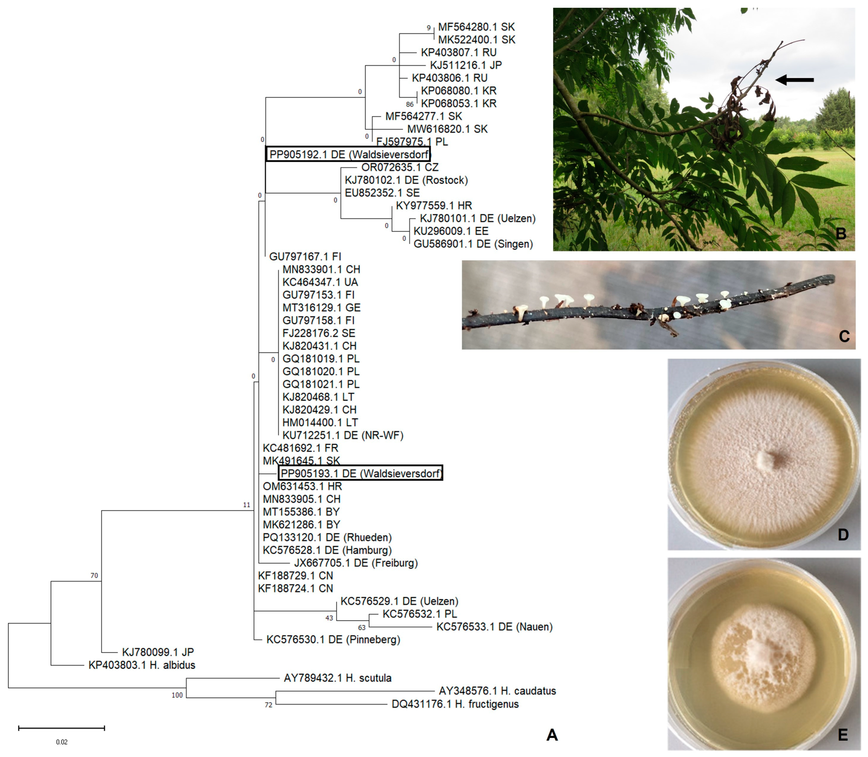
The molecular identification of the 37 isolates by the specific primer pair revealed the presence of the pathogen in 30 out of 37 tested samples. The alignment of all the sequences obtained from the Sanger sequencing revealed two distinct sequence variants with 474 bp in length (Wsd_c2 and Wsd_c3). So, these were considered as consensus sequences and deposited at the National Center for Biotechnology Information (NCBI) database under accession numbers PP905192.1 and PP905193.1.
The alignment of these two sequences with forty-six reference sequences of H. fraxineus from Europe and Asia shows two alterations in their base sequence. The isolate Wsd_c3 varies in its nucleotide sequence at two sites by substituting an adenine for guanine and a thymine for adenine. So, this sequence, partially covering the ITS 1, the 5.8S ribosomal RNA gene and the ITS 2 regions, matches between 98.8% and 99.5% of all aligned reference sequences. In contrast, Wsd_c2 shows a 99.0%–100% sequence identity. With the 10 reference isolates obtained from German locations, the sequences also show between 98.8% and 100% identity.
Future analyses of H. fraxineus isolates obtained from German locations would provide additional sequence information if the entire ITS 1 and ITS 2 regions with flanking ribosomal genes were mapped. Therefore, the primer pair ITS1-F and ITS4 can be applied [15,29].

3.3. Phylogeny

The phylogenetic analysis of the 12 isolates from Germany together with the sequences from Asia and Europe reveal that German isolates and those from Waldsieversdorf (PP905192.1, PP905193.1) in particular do not form a distinct cluster (Figure 1A).
Greater depth in the phylogenetic analysis could be achieved by using H. fraxineus-specific single-nucleotide polymorphism (SNP) or microsatellite (SSR) markers [24,35].

4. Discussion and Conclusions

This study contributes to the collection of ITS sequence data of H. fraxineus infecting ash in Germany and reveals an intragenomic variation within the German isolates.
Since the ash dieback disease has been documented to be widespread in Germany [43], the pathogen was sampled, molecularly detected, and characterized at a few different locations (Figure 1). Isolated not from necrotic ash tissue but from ash leaf rachises, the obtained fungal isolates in this study are supposed to represent a broad range of the natural population from the sampling location Waldsieversdorf [44]. Applying species-specific ITS primers, H. fraxineus was identified in 30 out of the isolated 37 fungal samples, and two sequence variants have been found (Supplementary Materials). The ribosomal ITS region is accepted as the standard barcode for fungi while allowing species identification with a high probability of correctness [27]. The level of intraspecific variation in this genomic region can be sorted into five categories, ranging between ‘no genomic variation’ and ‘high intragenomic variation’ [30]. The identities of the two ITS sequences in this study range between 98.8% and 100% compared to the reference sequences from Europe and Asia. In the catalog of ITS-correlated intragenomic variation, identities with <98% pairwise identity are rated as having ‘high intragenomic variation’ and those with 98%–99.99% pairwise identity as having ‘low intragenomic variation’, respectively [30]. Within the German isolates, the highest variation was found between the isolates from Uelzen (KC576533.1) and Pinneberg (KC576530.1), with 99.1% identity. Interestingly, the isolates Wsd_c2 and Wsd_c3 show the highest identity scores of 100% for sequences detected in Slovakia, Croatia, Sweden, and the Czech Republic.
As representative isolates for the sampling location, the sequences from the isolates Wsd_c2 and Wsd_c3 show an identity of 99.5% to each other. Although the two sequences only partially map the regions ITS 1 and ITS 2, including the complete sequence of the 5.8S ribosomal RNA gene, they reveal a low but noteworthy intragenomic variation in this region even between isolates of the same location. This indicates the presence of two copies of the ITS region for H. fraxineus in Waldsieversdorf. Multiple sequence variants can be found comparing the isolates from several European and Asian locations. Previous studies documented the presence of multiple copies of this rDNA region that occurred in 27% of the analyzed fungal genome assemblies [30,45]. Furthermore, levels of genetic variability are linked to environmental conditions and demographic processes. They are reflected by intragenomic variability [31,44]. Therefore, the quantity of rDNA copies of the H. fraxineus isolates from Germany and their phylogenetic sorting is supposed to represent an ongoing dynamic evolutionary adaptation process. The sequences included in the phylogenetic analysis do not explicitly show a geographic correlation between the clusters. Previous studies have already found a high gene flow between separate populations far located from each other [32,35,46]. This coincides with our results. In future investigations, microsatellite data can be used to obtain a deeper insight into the spatial distribution and phylogenetic relationships of distinct populations [35,44].
The findings in this study complement the recent collection of molecular ITS sequence data of H. fraxineus. These data will contribute to the decoding of evolving factors, driving the distribution of the pathogen within ash stands in Europe and particularly Germany. This will help to understand the mechanisms of pathogenic adaptation.

Supplementary Materials

The following supporting information can be downloaded at: https://www.mdpi.com/article/10.3390/f16010149/s1, S1_Alignment_H.frax_GermSequ S1: Alignment of German H. fraxineus-isolates.

Author Contributions

Conceptualization, A.-M.E.E.; Methodology and formal analysis, A.-M.E.E., B.B. and V.B.; Draft Preparation, A.-M.E.E.; Review and Editing, V.S.; Project Administration, V.S.; Funding Acquisition B.B. and V.S. All authors have read and agreed to the published version of the manuscript.

Funding

This research was funded by the German Federal Ministry of Food and Agriculture (BMEL) through the Fachagentur für Nachwachsende Rohstoffe e.V. (FNR), project ResEsche (funding code 22019915).

Data Availability Statement

The data presented in this study are openly available in “https://www.thuenen.de/en/institutes/forest-genetics/publications (accessed on 12 January 2025)” and in the repository Open Agrar “https://www.openagrar.de/receive/openagrar_mods_00001946 (accessed on 12 January 2025)”.

Acknowledgments

We extend our gratitude to Marlies Karaus for her excellent technical assistance.

Conflicts of Interest

The authors declare no conflicts of interest.

References

  1. Kowalski, T. Chalara fraxinea sp. nov. associated with dieback of ash (Fraxinus excelsior) in Poland. For. Pathol. 2006, 36, 264–270. [Google Scholar] [CrossRef]
  2. Kowalski, T.; Holdenrieder, O. The teleomorph of Chalara fraxinea, the causal agent of ash dieback. For. Pathol. 2009, 39, 304–308. [Google Scholar] [CrossRef]
  3. Gross, A.; Holdenrieder, O.; Pautasso, M.; Queloz, V.; Sieber, T.N. Hymenoscyphus pseudoalbidus, the causal agent of European ash dieback. Mol. Plant Pathol. 2014, 15, 5–21. [Google Scholar] [CrossRef]
  4. McMullan, M.; Rafiqi, M.; Kaithakottil, G.; Clavijo, B.J.; Bilham, L.; Orton, E.; Percival-Alwyn, L.; Ward, B.J.; Edwards, A.; Saunders, D.G.O.; et al. The ash dieback invasion of Europe was founded by two genetically divergent individuals. Nat. Ecol. Evol. 2018, 2, 1000–1008. [Google Scholar] [CrossRef]
  5. Carroll, D.; Boa, E. Ash dieback: From Asia to Europe. Plant Pathol. 2024, 73, 741–759. [Google Scholar] [CrossRef]
  6. Elfstrand, M.; Chen, J.; Cleary, M.; Halecker, S.; Ihrmark, K.; Karlsson, M.; Davydenko, K.; Stenlid, J.; Stadler, M.; Brandström Durling, M. Comparative analyses of the Hymenoscyphus fraxineus and Hymenoscyphus albidus genomes reveals potentially adaptive differences in secondary metabolite and transposable element repertoires. BMC Genom. 2021, 22, 503. [Google Scholar] [CrossRef] [PubMed]
  7. Lygis, V.; Vasiliauskas, R.; Larsson, K.H.; Stenlid, J. Wood-inhabiting fungi in stems of Fraxinus excelsior in declining ash stands of northern Lithuania, with particular reference to Armillaria cepistipes. Scand. J. For. Res. 2005, 20, 337–346. [Google Scholar] [CrossRef]
  8. Schumacher, J.; Kehr, R.; Leonhard, S.; Wulf, A. New details on the pathogenesis of ash dieback. J. Cultiv. Plants 2010, 62, 1–9. [Google Scholar]
  9. Marçais, B.; Giraudel, A.; Husson, C. Ability of the ash dieback pathogen to reproduce and to induce damage on its host are controlled by different environmental parameters. PLoS Pathog. 2024, 19, e1010558. [Google Scholar] [CrossRef]
  10. Kirisitis, T.; Cech, T.L. Beobachtungen zum sexuellen Stadium des Eschentriebsterben-Erregers Chalara fraxinea in Österreich. Forstsch. Aktuell 2009, 48, 21–25. [Google Scholar]
  11. Cleary, M.R.; Daniel, G.; Stenlid, J. Light and scanning electron microscopy studies of the early infection stages of Hymenoscyphus pseudoalbidus on Fraxinus excelsior. Plant Pathol. 2013, 62, 1294–1301. [Google Scholar] [CrossRef]
  12. Langer, G. Collar Rots in Forests of Northwest Germany Affected by Ash Dieback. Balt. For. 2017, 23, 4–19. [Google Scholar]
  13. Enderle, R.; Fussi, B.; Lenz, H.; Langer, G.; Nagel, R.; Metzler, B. Ash dieback in Germany: Research on disease development, resistance and management options. In Dieback of European Ash (Fraxinus spp.): Consequences and Guidelines for Sustainable Management; Vaisatis, R., Enderle, R., Eds.; SLU: Uppsala, Sweden, 2017; pp. 89–105. [Google Scholar]
  14. Griffiths, S.M.; Galambao, M.; Rowntree, J.; Goodhead, I.; Hall, J.; O’Brien, D.; Atkinson, N.; Antwis, R.E. Complex associations between cross—Kingdom microbial endophytes and host genotype in ash dieback disease dynamics. J. Ecol. 2020, 108, 291–309. [Google Scholar] [CrossRef]
  15. Becker, R.; Ulrich, K.; Behrendt, U.; Kube, M.; Ulrich, A. Analyzing ash leaf-colonizing fungal communities for their biological control of Hymenoscyphus fraxineus. Front. Microbiol. 2020, 11, 590944. [Google Scholar] [CrossRef]
  16. Ulrich, K.; Becker, R.; Behrendt, U.; Kube, M.; Ulrich, A. A Comparative Analysis of Ash Leaf-Colonizing Bacterial Communities Identifies Putative Antagonists of Hymenoscyphus fraxineus. Front. Microbiol. 2020, 11, 966. [Google Scholar] [CrossRef] [PubMed]
  17. Becker, R.; Ulrich, K.; Behrendt, U.; Schneck, V.; Ulrich, A. Genomic characterization of Aureimonas altamirensis C2P003—A specific member of the microbiome of Fraxinus excelsior trees tolerant to ash dieback. Plants 2022, 11, 3487. [Google Scholar] [CrossRef] [PubMed]
  18. Heydeck, P.; Bemmann, M.; Kontzog, H.G. Triebsterben an Gemeiner Esche (Fraxinus excelsior) im nordostdeutschen Tiefland. Forst Holz 2005, 60, 505–506. [Google Scholar]
  19. Wulf, A.; Schumacher, J. Die Waldschutzsituation 2005 in der Bundesrepublik Deutschland. Forst Holz 2005, 60, 503–505. [Google Scholar]
  20. Schumacher, J.; Wulf, A.; Leonhard, S. Erster Nachweis von Chalara fraxinea T. Kowalski sp. nov. in Deutschland-ein Verursacher neuartiger Schäden an Eschen. Nachrichtenblatt Dtsch. Pflanzenschutzd. 2007, 59, 121–123. [Google Scholar]
  21. Leonhard, S.; Straßer, L.; Nannig, A.; Blaschke, M.; Schumacher, J.; Immler, T. Neues Krankheitsphänomen an der Esche.Das von Chalara fraxinea verursachte Eschentriebsterben ist auch in Bayern nachgewiesen. LWF Aktuell 2009, 71, 60–63. [Google Scholar]
  22. Metzler, B.; Enderle, R.; Karopka, M.; Töpfner, K.; Aldinge, E. Entwicklung des Eschentriebsterbens in einem Herkunftsversuch an verschiedenen Standorten in Süddeutschland. Allg. Forst Jagdztg. 2012, 183, 168–180. [Google Scholar]
  23. Enderle, R.; Peters, F.; Nakou, A.; Metzler, B. Temporal development of ash dieback symptoms and spatial distribution of collar rots in a provenance trial of Fraxinus excelsior. Eur. J. For. Res. 2013, 132, 865–876. [Google Scholar] [CrossRef]
  24. Fussi, B.; Konnert, M. Genetic analysis of European common ash (Fraxinus excelsior L.) populations affected by ash dieback. Silvae Gen. 2014, 63, 198–212. [Google Scholar] [CrossRef]
  25. Enderle, R.; Metzler, B.; Riemer, U.; Kändler, G. Ash dieback on sample points of the national forest inventory in South-Western Germany. Forests 2018, 9, 25. [Google Scholar] [CrossRef]
  26. Langer, G.J.; Fuchs, S.; Osewold, J.; Peters, S.; Schrewe, F.; Ridley, M.; Kätzel, R.; Bubner, B.; Grüner, J. FraxForFuture—Research on European ash dieback in Germany. J. Plant Dis. Prot. 2022, 129, 1285–1295. [Google Scholar] [CrossRef]
  27. Schoch, C.L.; Seifert, K.A.; Huhndorf, S.; Robert, V.; Spouge, J.L.; Levesque, C.A.; Chen, W. Nuclear ribosomal internal transcribed spacer (ITS) region as a universal DNA barcode marker for fungi. Proc. Natl. Acad. Sci. USA 2012, 109, 6241–6246. [Google Scholar] [CrossRef] [PubMed]
  28. Drenkhan, R.; Riit, T.; Adamson, K.; Hanso, M. The earliest samples of Hymenoscyphus albidus vs. H. fraxineus in Estonian mycological herbaria. Mycol. Prog. 2016, 15, 835–844. [Google Scholar] [CrossRef]
  29. Drenkhan, R.; Solheim, H.; Bogacheva, A.; Riit, T.; Adamson, K.; Drenkhan, T.; Maaten, T.; Hietala, A.M. Hymenoscyphus fraxineus is a leaf pathogen of local Fraxinus species in the Russian Far East. Plant Pathol. 2017, 66, 490–500. [Google Scholar] [CrossRef]
  30. Bradshaw, M.J.; Aime, M.C.; Rokas, A.; Pandey, B.; Li, Y.; Pfister, D.H. Extensive intragenomic variation in the internal transcribed spacer region of fungi. iScience 2023, 26, 107317. [Google Scholar] [CrossRef]
  31. Paloi, S.; Luangsa-ard, J.J.; Mhuantong, W.; Stadler, M.; Kobmoo, N. Intragenomic variation in nuclear ribosomal markers and its implication in species delimitation, identification and barcoding in fungi. Fungal Biol. Rev. 2022, 42, 1–33. [Google Scholar] [CrossRef]
  32. Bengtsson, S.B.K.; Vasaitis, R.; Kirisitis, T.; Solheim, H.; Stenlid, J. Population structure of Hymenoscyphus pseudoalbidus and its genetic relationship to Hymenoscyphus albidus. Fungal Ecol. 2012, 5, 147–153. [Google Scholar] [CrossRef]
  33. Kowalski, T.; Kraj, W.; Bednarz, B. Fungi on stems and twigs in initial and advanced stages of dieback of European ash (Fraxinus excelsior) in Poland. Eur. J. For. Res. 2016, 135, 565–579. [Google Scholar] [CrossRef]
  34. Cross, H.; Sønstebø, J.H.; Nagy, N.E.; Timmermann, V.; Solheim, H.; Børja, I.; Kauserud, H.; Carlsen, T.; Rzepka, B.; Wasak, K.; et al. Fungal diversity and seasonal succession in ash leaves infected by the invasive ascomycete Hymenoscyphus fraxineus. New Phytol. 2016, 213, 1405–1417. [Google Scholar] [CrossRef] [PubMed]
  35. Orton, E.S.; Brasier, C.M.; Bilham, L.J.; Bansal, A.; Webber, J.F.; Brown, J.K.M. Population structure of the ash dieback pathogen, Hymenoscyphus fraxineus, in relation to its mode of arrival in the UK. Plant Pathol. 2018, 67, 255–264. [Google Scholar] [CrossRef]
  36. Kowalski, T.; Bartnik, C. Morphological variation in colonies of Chalara fraxinea isolated from ash (Fraxinus excelsior) stems with symptoms of dieback and effects of temperature on colony growth and structure. Acta Agrobot. 2010, 63, 99–106. [Google Scholar] [CrossRef]
  37. Johansson, S.B.K.; Vasaitis, R.; Ihrmark, K.; Barklund, P.; Stenlid, J. Detection of Chalara fraxinea from tissue of Fraxinus excelsior using species—Specific ITS primers. For. Pathol. 2010, 40, 111–115. [Google Scholar] [CrossRef]
  38. European and Mediterranean Plant Protection Organisation. Hymenoscyphus pseudoalbidus. Bull. OEPP/EPPO Bull. 2013, 43, 449–461. [Google Scholar] [CrossRef]
  39. Larkin, M.A.; Blackshields, G.; Brown, N.P.; Chenna, R.; McGettigan, P.A.; McWilliam, H.; Valentin, F.; Wallace, I.M.; Wilm, A.; Lopez, R.; et al. Clustal W and Clustal X version 2.0. Bioinformatics 2007, 23, 2947–2948. [Google Scholar] [CrossRef] [PubMed]
  40. Altschul, S.F.; Gish, W.; Miller, W.; Myers, E.W.; Lipman, D.J. Basic local alignment search tool. J. Mol. Biol. 1990, 215, 403–410. [Google Scholar] [CrossRef] [PubMed]
  41. Tamura, K.; Stecher, G.; Kumar, S. MEGA 11: Molecular Evolutionary Genetics Analysis Version 11. Mol. Biol. Evol. 2021, 38, 3022–3027. [Google Scholar] [CrossRef]
  42. Tamura, K.; Nei, M. Estimation of the number of nucleotide substitutions in the control region of mitochondrial DNA in humans and chimpanzees. Mol. Biol. Evol. 1993, 10, 512–526. [Google Scholar]
  43. Enderle, R.; Nakou, A.; Thomas, K.; Metzler, B. Susceptibility of autochthonous German Fraxinus excelsior clones to Hymenoscyphus pseudoalbidus is genetically determined. Ann. For. Sci. 2015, 72, 183–193. [Google Scholar] [CrossRef]
  44. Kraj, W.; Kowalski, T. Genetic variability of Hymenoscyphus pseudoalbidus on ash leaf rachises in leaf litter of forest stands in Poland. J. Phytopathol. 2014, 162, 218–227. [Google Scholar] [CrossRef]
  45. Stadler, M.; Lambert, C.; Wibberg, D.; Kalinowski, J.; Cox, R.J.; Kolařík, M.; Kuhnert, E. Intragenomic polymorphisms in the ITS region of high-quality genomes of the Hypoxylaceae (Xylariales, Ascomycota). Mycol. Prog. 2020, 19, 235–245. [Google Scholar] [CrossRef]
  46. Burokiene, D.; Prospero, S.; Jung, E.; Marciulyniene, D.; Moosbrugger, K.; Norkute, G.; Rigling, D.; Lygis, V.; Schoebel, C.N. Genetic population structure of the invasive ash dieback pathogen Hymenoscyphus fraxineus in its expanding range. Biol. Invasions 2015, 17, 2743–2756. [Google Scholar] [CrossRef]
Disclaimer/Publisher’s Note: The statements, opinions and data contained in all publications are solely those of the individual author(s) and contributor(s) and not of MDPI and/or the editor(s). MDPI and/or the editor(s) disclaim responsibility for any injury to people or property resulting from any ideas, methods, instructions or products referred to in the content.

