1. Introduction
Thymoquinone (TQ), the main constituent of
Nigella sativa L. seed (black seed) essential oil, has various pharmacological properties useful for the treatment of a wide range of diseases including chronic non-infectious diseases (neurological disorders, diabetes mellitus, hypertension, dyslipidemia, inflammatory disorders, cancer, etc.) and infectious diseases [
1,
2,
3,
4,
5].
In particular, the protective effect of TQ against H
2O
2-induced oxidative stress in human retinal pigment epithelium cells has been reported [
6]. TQ has shown anti-inflammatory properties in an experimental dry eye model, and it has decreased corneal neovascularization in a dose-dependent manner after topical administration in a rat model [
7]. Furthermore, TQ acts on the immune system, modulating the levels of pro- and anti-inflammatory mediators that are involved in corneal neovascularization, and it is as effective as topical triamcinolone when topically applied to the eye at 0.4% [
8].
The administration of TQ on ovalbumin (OVA)-induced allergic conjunctivitis in BALB/c mice significantly reduced the ocular symptoms of the allergic conjunctivitis, attenuating the recruitment of eosinophils, as well as the levels of IgE, histamine, and cytokines [
9].
Despite its many activities [
7], the clinical application of TQ is limited by poor bioavailability, low solubility, and scarce permeability. The in vitro studies have also shown toxicity of TQ depending both on the cell model and the dose [
10]. The use of nanoformulations can represent a good tool for overcoming these limits [
11,
12]. The purpose of this study is to develop liposomal formulations of TQ, to improve its biopharmaceutical performance, and to enhance the delivery and the activity at the eye level. Liposomes, due to their unique structure, are extremely beneficial drug carriers as they can entrap both the hydrophilic and hydrophobic drugs [
13]. Phospholipids are the major components of most liposomes. Extensive testing of these naturally occurring compounds has revealed them to be remarkably safe for pharmaceutical use. Liposomes represent the most effective ocular drug delivery carriers with sufficient flexibility to allow synthesis in various sizes, and they can also be easily administered in liquid dosage forms, such as eye drops, gels and ointments, for topical delivery [
13]. Liposomes enhance corneal permeability due to their ability to come in close contact with cornea and conjunctiva, increasing the extent of corneal uptake and prolonging corneal contact time [
13].
Moreover, liposomal formulations can also improve the stability and reduce the toxic side effects associated with the drug [
14]. In addition, liposomes protect drug molecules from the metabolic enzymes at the tear/corneal epithelium interface [
15]. Antiviral drugs (acyclovir, ganciclovir, idoxuridine), antibacterial drugs (tetracycline, gentamicin, tobramycin, ciprofloxacin, chloramphenicol), antifungal agents (amphotericin B, fluconazole), anti-inflammatory and immunomodulatory agents (diclofenac, cyclosporin, tacrolimus, 5-fluorouracil), and antiglaucoma agents (pilocarpine, latanoprost, acetazolamide) have been formulated in liposomal forms to enhance ocular bioavailability and activity [
13,
16].
TQ improved the retinal damage and the inflammatory reactions in an induced glaucoma model in rabbits when formulated in liposomes also containing latanoprost. This formulation conferred a sustained drug release and decreased the intraocular pressure [
17]. Recently, vitamin E-loaded liposomes have been investigated for dry eye treatment. Liposomes were coated with 0.2% sodium hyaluronate, mimicking the aqueous phase of the mucin of the lacrimal film and prolonging contact time with the eye surface [
18].
In this research, we combined the advantages of the liposomes for ocular delivery with a hyaluronic acid (HA) coating. HA is an endogenous negatively charged natural polysaccharide of
d-glucuronic acid and
N-acetyl-
d-glucosamine disaccharide units, which is highly biocompatible, biodegradable, and non-toxic. In addition, it is a potential targeting moiety to cluster of differentiation 44 (CD44)-expressing cells, to the receptor for hyaluronan-mediated motility (RHAMM), and to Intercellular Adhesion Molecule 1 (ICAM-1). CD44 receptors are present in the ophthalmic cells, particularly in human conjunctiva and cornea epithelial cells, and in the retinal pigment epithelium [
19,
20].
Consequently, two liposomal formulations have been developed, both consisting of phosphatidylcholine and Plurol Oleique, a liquid lipid. One liposome was coated with 0.1% w/v HA to increase TQ solubility and availability at the ocular level.
The physical characterization of the formulations was carried out using scattering techniques (DLS and ELS), and transmission electron microscopy was used for the morphological analysis. Encapsulation efficiency was determined using the HPLC-DAD analytical method. The formulations were also submitted to stability studies over 2 months, keeping the samples at +4 °C, and to in vitro drug release studies at 37 °C. Two cell lines, human corneal epithelial cells (HCEC-2) and human conjunctival epithelial cells (HConEC), were used to verify the safety profile of the liposomal formulations. The same cells were used for the uptake studies, using fluorescent liposomes containing FITC as probes.
2. Materials and Methods
2.1. Materials
Acetonitrile HPLC grade, Cholesterol (CH), Fluorescein isothiocyanate (FITC, purity > 90%, HPLC), mucin from porcine stomach type II, TQ, and Tween 80, were purchased from Sigma-Aldrich (Milan, Italy). Capryol 90, Lauroglycol 90, Maisine CC, Plurol Oleique CC 497, and Transcutol P were supplied by Gattefossè sas (Saint-Priest, France). Phosphotungstic acid was purchased from Electron Microscopy Science (Hatfield, MA, USA). Sodium hyaluronate was obtained from Altergon Italia s.r.l. (Avellino, Italy). Egg phosphatidylcholine, Phospholipon 90 G was purchased by Phospholipid Gmbh (Cologne, Germany). Sodium hyaluronate (M.W. 1000 KDa, HA) was obtained from Altergon, Avellino, Italy. The water used was from the Milli-Qplus system from Millipore (Milford, CT, USA). The dialysis kit was from Spectrum Laboratories, Inc. (Breda, The Netherlands). The PAMPA filter plate (pore size 0.45 μm) was purchased from Millipore Corporation, Tullagreen, Carrigtwohill, County Cork, Ireland. Human corneal epithelial cells (HCE-2) were purchased from ATCC company (American Type Culture Collection, Manassas, VA, USA). Human conjunctival epithelial cells (HConEC) and corneal epithelium cell medium were provided by Innoprot (Derio, Bizkaia, Spain). Keratinocyte serum-free medium, bovine pituitary extract (BPE), and epidermal growth factor (EGF) were purchased from Gibco-BRL (San Giuliano Mil-anese, MI, Italy). Hydrocortisone, insulin, fibronectin, bovine collagen type I and bovine serum albumin (BSA) penicillin/ streptomycin (P/S), bovine fetal serum and poly-l-lysine, benzalkonium chloride (BAK), and 3-(4,5-dimethylthiazol-2-yl)-25-diphenyltetrazolium bromide (MTT) were obtained from Sigma (St Louis, MO, USA). The Cytotoxicity detection kit (lactate dehydrogenase, LDH) was obtained from Roche Diagnostics (Basel, Switzerland).
2.2. Chromatography Conditions and Instruments
HPLC Method for TQ and FITC
The HPLC system consisted of a 1100 High Performance Liquid Chromatograph (HPLC) equipped with a Diode Array Detector (DAD) from Agilent Technologies Italia Spa (Rome, Italy). The analytical column was a Kinetex C18 (150 × 4.6 mm, 5 μm Agilent Technology, Insert: Santa Clara, CA, USA).
The compounds were detected at a wavelength of 254 nm with an eluent flow rate of 0.8 mL/min, with (A) acetonitrile and (B) water pH 3.2 (by formic acid) as mobile phases, and with a gradient analytical method as described by [
11]. The calibration curve, with a coefficient of determination R
2 of 0.9993, was prepared using a standard solution of TQ in methanol (0.5 mg/mL) and successive dilutions of 5, 10, 50, 100, and 500-fold.
FITC analysis were performed using the same apparatus and the same column reported for TQ as well as the same method described by [
21]. The calibration curve, with a coefficient of determination R
2 of 0.9993, was prepared using a standard solution of FITC in methanol HPLC grade (1.0 mg/mL) and successive dilutions of 10, 50, 100, 200, and 500-fold.
2.3. Preparation of Liposomal Formulations
Blank liposomes (LP), TQ-loaded liposomes (LP-TQ), HA-coated liposomes (LP-TQ-HA), Fluorescein isothiocyanate (FITC)-loaded liposomes (LP-FITC), and HA-coated FITC-loaded liposomes (LP-FITC-HA) were prepared according to the thin layer evaporation method by [
22]. For LP, 600 mg of egg phosphatidylcholine (PC) and 100 mg of Plurol Oleique were dissolved in dichloromethane. The organic solvent was evaporated under vacuum, and the dry lipid film was hydrated by adding 10 mL of deionized water. The aqueous dispersion was shaken for 30 min with a mechanical stirrer in a water bath at a temperature of 50 °C. To obtain unilamellar vesicles from multilamellar vesicles, an ultrasonic probe was used for 2 min and 30 s (with pulsed duty cycles of
s on and
s off, amplitude 48%); during the sonication, the sample was placed in an ice bath to prevent lipid degradation [
23]. Finally, a centrifugation of 1 min at 1205×
g was achieved to remove metallic particles potentially released by the ultrasonic probe inside the liposomal dispersion.
LP-TQ and LP-TQ-HA were prepared using the same method described above, adding 10 mg of TQ (1 mg/mL, corresponding to 1% of the weight of the lipid phase), together with PC, Plurol Oleique, and 1–2 mL of dichloromethane to completely dissolve the TQ. For LP-TQ-HA, the coating was achieved using the drop-wise method described by [
24], adding 2 mL of 0.1%
w/
v of HA in deionized water to 2 mL of LP-TQ dispersion. The obtained dispersion was placed under magnetic stirring for 1 h, and then an ultrasonic probe was used for 2 min and 30 s (with pulsed duty cycles
s on and
s off, amplitude 48%). Finally, the sample was centrifugated for 1 min at 1205×
g. The obtained formulation showed a final concentration of 0.5 mg/mL of TQ, as evaluated by HPLC-DAD analysis.
LP-FITC and LP-FITC-HA were prepared using the same methods described above, adding 10 mg of the FITC, corresponding to 1% of the weight of the lipid component.
2.4. Characterization of Liposomes
2.4.1. Physical Characterization
Liposomes’ hydrodynamic diameter, polydispersity index (PdI), and zeta-potential were determined by Dinamic Light Scattering (DLS) and Electrophoretic Light Scattering (ELS), using a Zsizer Nano series ZS90 (Malvern Instruments, Malvern, UK). All samples were diluted 10-fold with deionized water, and an average of three measurements at stationary level was taken. A Haake temperature controller kept the temperature constant at 25 °C.
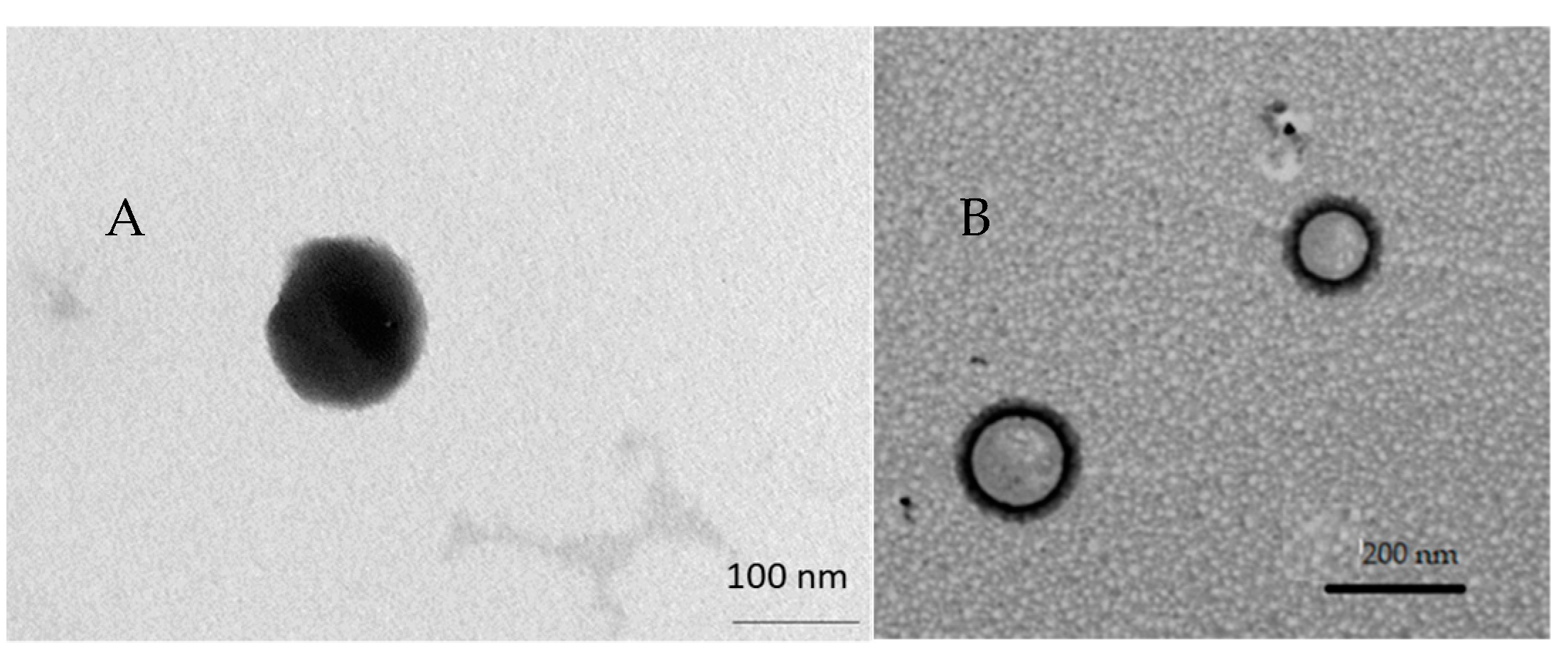
2.4.2. Morphological Characterization
Morphological characterization was achieved by using the transmission electron microscopy (TEM) CM12 from Philips with an accelerating voltage of 80 kV. The aqueous dispersion was diluted 10-fold in deionized water, and 10 μL was applied to a 150-mesh carbon film-covered copper grid. To obtain a thin film, excess sample was eliminated from the grid with a filter paper. After that, 5 μL of phosphotungstic acid solution (1% w/v in water) was dropped onto the grid as a staining medium and the excess solution was removed with a filter paper. Samples were dried for 3 min, after which they were examined with the electron microscope and photographed at an accelerating voltage of 80 kV.
2.5. Determination of Encapsulation Efficiency and Loading Capacity
The amount of TQ or FITC encapsulated in coated or uncoated LP was determined by the dialysis bag method. The membrane (cut-off 3.5 kD) was filled with 2 mL of sample and kept for 1 h under magnetic stirring at 100 rpm and 25 °C, in 1L of deionized water, to remove free TQ. After that, the samples were diluted 1:50 with MeOH HPLC grade, sonicated for 30 min, and analyzed by HPLC-DAD. The EE% has been calculated as follows:
2.6. Storage Stability
LP-TQ and LP-TQ-HA storage stability studies were carried out for two months at +4 °C. Particle size, PdI, zeta-potential, and EE% were evaluated by DLS, ELS, and HPLC-DAD analyses. LP-FITC and LP-FITC-HA storage stability was monitored for 6 h.
2.7. In Vitro Release Study
Regenerated cellulose membranes with a molecular weight cut off 3.5–5 kD were selected to allow TQ diffusion into the acceptor medium, consisting of 200 mL PBS and Tween 80 0.5 % w/v. The dialysis bag was pre-hydrated for 30 min with deionized water, then filled with 2 mL of sample. The bag was doused into an acceptor medium, and the system was incubated at 37 °C under magnetic stirring for 6 h. At predetermined time intervals, 1 mL of the medium was withdrawn and replaced with the same volume of fresh release medium maintained at 37 °C to preserve sink conditions. TQ released at each time interval was quantified using HPLC-DAD.
2.8. Cell Culture Studies
2.8.1. Human Corneal Epithelial Cells (HCE-2)
Human corneal epithelial cells (HCE-2) were cultured in serum-free medium supplemented with 0.05 mg/mL of BPE, 5 ng/mL of epidermal growth factor, 500 ng/mL of hydrocortisone, and 0.005 mg/mL of insulin. The cells were incubated at 37 °C in a humidified incubator containing 5% CO2 air and then plated on pre-treated 75 cm2 flasks with a mixture of 0.01 mg/mL of fibronectin, 0.03 mg/mL of bovine type I collagen, and 0.01 mg/mL of bovine serum albumin. The medium was changed twice a week. Once confluence was reached, the cells were split with a 1:3 ratio in other previously treated flasks.
2.8.2. Human Conjunctival Epithelial Cells (HConEC)
Human conjunctival epithelial cells (HConEC) provided by Innoprot were isolated from the human conjunctiva. The cells were cultured in a culture medium consisting of the basal medium with the addition of the conjunctival epithelial cell growth supplement (CEpiCGS), penicillin/streptomycin (P/S), and fetal bovine serum, at 37 °C in a humidified incubator containing 5% CO2 air. The cells were plated in 75 cm2 flasks previously treated for 1 h with a 1:10 poly-lysine solution in PBS. The culture medium was changed twice a week. Once confluence was reached, the cells were split with a 1:3 ratio in other previously treated flasks.
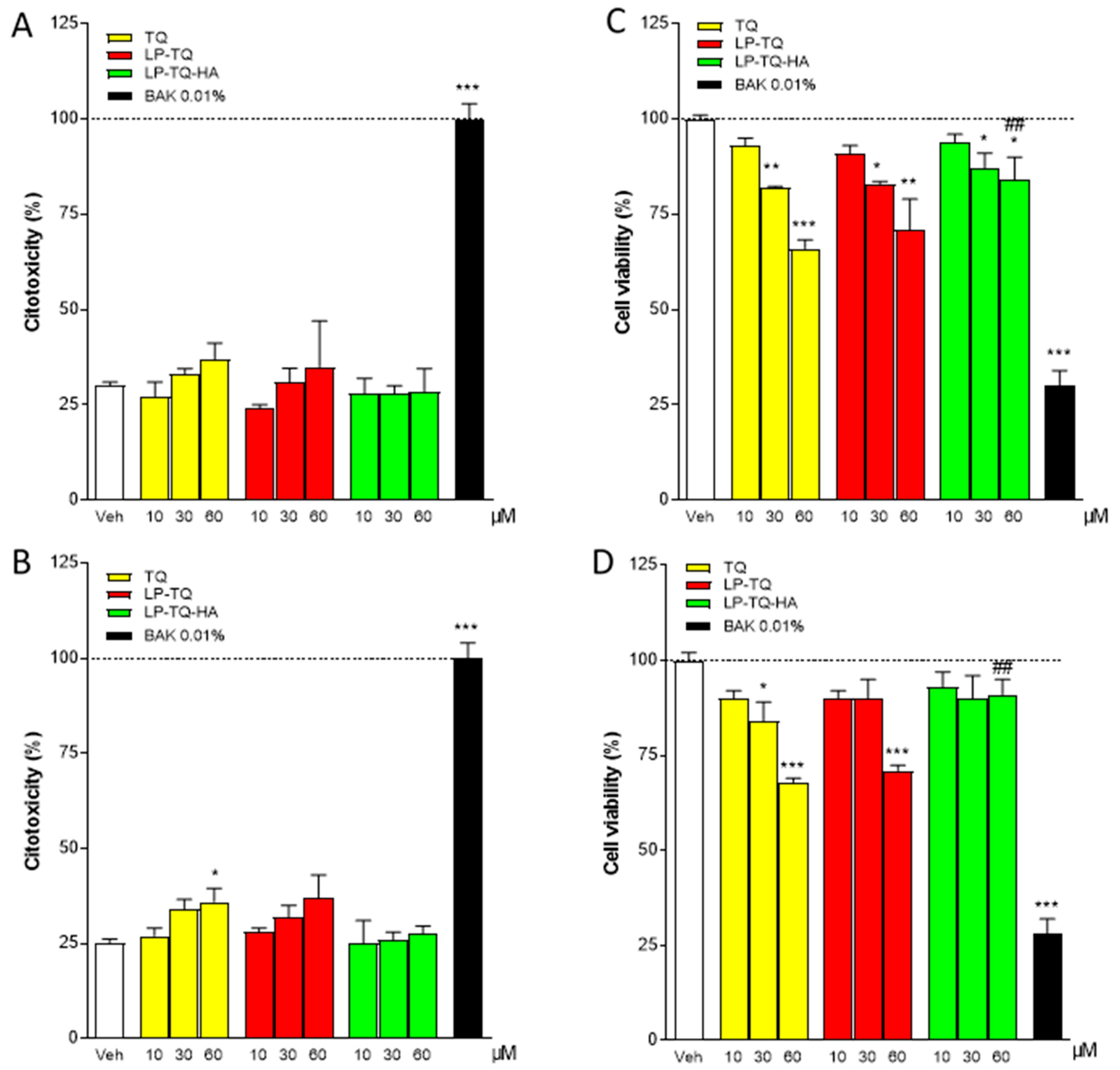
2.8.3. Analysis of In Vitro Cytotoxicity
Human corneal and conjunctival epithelial cells were suspended and plated in 24-well plates (approximately 4 × 10⁴ cells/cm2). Once they reached about 70–80% confluence, the medium was removed, and the cells were exposed to TQ, LP-TQ, and LP-TQ-HA suitably diluted in PBS. PBS was used as a positive control, and 0.01% BAK was used as a negative control for maximal toxicity. Corneal and conjunctival epithelial cells were incubated with TQ solution, TQ formulated in LP-TQ liposomes, or LP-TQ-HA hyaluronic acid-coated liposomes in different concentrations (10, 30 and 60 µM) and for two different periods of time (15 min and 1 h).
MTT Assay
The viability of conjunctival and corneal epithelial cells exposed for two different periods (15 min and 1 h) and in different concentrations of free or formulated TQ was evaluated by MTT assay. Cells were plated in 24-well plates and kept in culture. TQ, LP-TQ, and LP-TQ-HA were incubated in different concentrations for different periods in the vehicle. Part of the medium from each well was withdrawn and stored for the LDH assay. Cells were incubated with MTT at a concentration of 1 mg/mL [
25]. After removing the MTT-containing solution, dimethyl sulfoxide (DMSO) was added to the wells to dissolve the formation, and the absorbance of MTT was read at 550 and 690 nm. The vehicle was used as a positive control. Cell viability was expressed as a percentage of cells incubated only in the vehicle at the corresponding exposure time.
LDH Assay
Damage in human corneal and conjunctival epithelial cells was quantitatively assessed by measuring the amount of LDH released by the damaged cells into the extracellular fluid, 15 min and 1 h after drug exposure, using the LDH kit, as previously described [
26]. The LDH level corresponding to complete cell death was determined for each experiment by analyzing sister cultures exposed to 0.01% BAK. Background LDH release was determined in drug-unexposed control cultures and subtracted from all experimental values. The resulting values correlated linearly with the degree of cell loss estimated upon observation of cultures in phase contrast optics.
2.8.4. Cellular Uptake Studies
For the evaluation of the intracellular content of FITC, the corneal epithelial cells (HCE-2) and the conjunctival cells (HConEC) were exposed for 1 h to LP-FITC or LP-FITC-HA containing 1 mg/mL of FITC, or to a saturated salution of fluorescent probe diluted in PBS. A qualitative assessment of FITC uptake at the cellular level was performed by culturing human corneal and conjunctival cells on histological slides, treated with free FITC or formulated on liposomes for 1 h and fixed in 4% formaldehyde in phosphate buffer 0.1 mol/L, pH 7.4, for 10 min; the cells were then stained with DAPI shielded fluorine (Sigma, Milan, Italy) to visualize the nucleus and were subsequently observed by fluorescence microscopy (Labophot-2 Nikon, Tokyo, Japan). Ten photomicrographs were randomly taken for each sample. Cell uptake was studied by fluorescence microscopy using FITC-labeled liposomes, with a high pressure mercury vapor lamp, 20× objective, NA = 0.75 (OLYMPUS BX3-CBH/U-MCZ). Filter set: excitation 365 nm emission 400 nm high pass DAPI, excitation 494 nm emission 518 nm.
4. Conclusions
In this study, the potential of liposomes as a drug vehicle for the ophthalmic delivery of TQ was investigated. Two liposomal formulations have been designed and optimized. Both liposomes consist of phosphatidylcholine and Plurol Oleique, a liquid lipid which is used to replace cholesterol and ameliorate encapsulation efficiency and which doubles TQ solubility. This formulation was also coated with 0.1% w/v HA. Physical characterization revealed that uncoated and coated liposomes are suitable for ocular administration, with encapsulation efficiency of 70%. The formulations stored at +4 °C were stable for two months, and greater chemical stability was obtained with the HA coating. Liposomes guarantee a prolonged and gradual release, and they reduce the TQ toxicity observed at high dosage, particularly in the case of LP-TQ-HA when tested in both HCEC-2 and HConEC cells.
Finally, the in vitro uptake study, conducted with fluorescent liposomes, showed that both liposomal formulations increased the absorption at the cellular level and, in particular, at nucleus level, when tested in the corneal and conjunctival cells, with the most marked effect for HA-coated liposomes.


 ,
, 


