
“Plurethosome” as Vesicular System for Cutaneous Administration of Mangiferin: Formulative Study and 3D Skin Tissue Evaluation

 ,
,  ,
,  , ,
, ,  , , , ,

Abstract
:1. Introduction
2. Materials and Methods
2.1. Materials
2.2. Vesicular Nanosystem Preparation
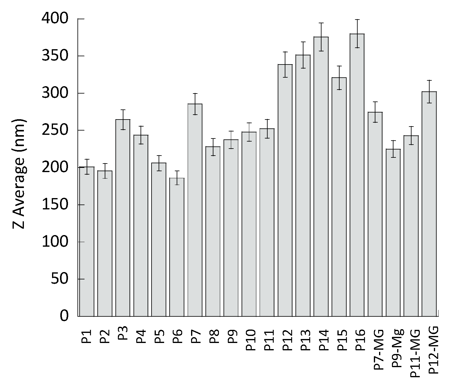
2.3. Photon Correlation Spectroscopy (PCS)
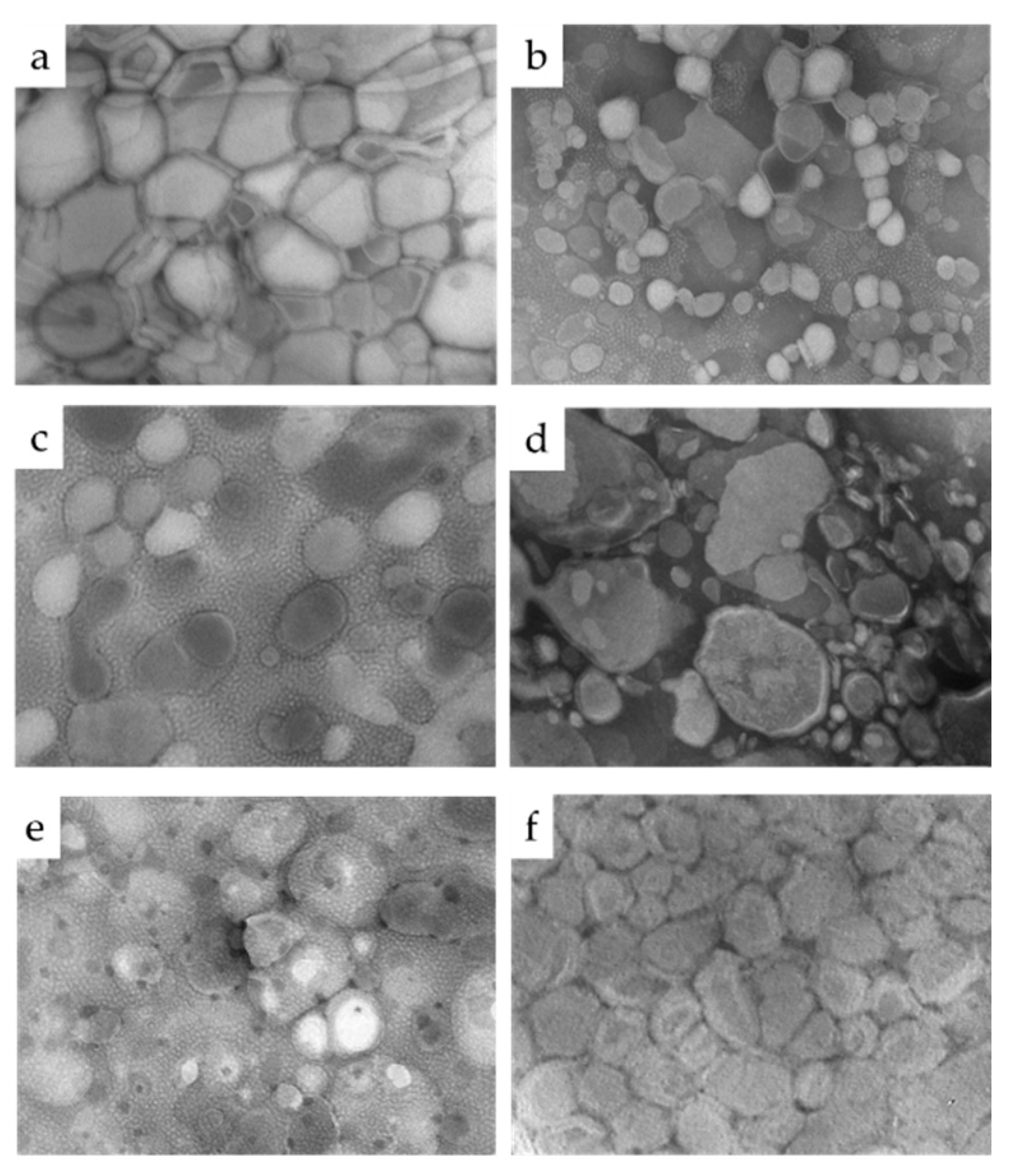
2.4. Transmission Electron Microscopy (TEM)
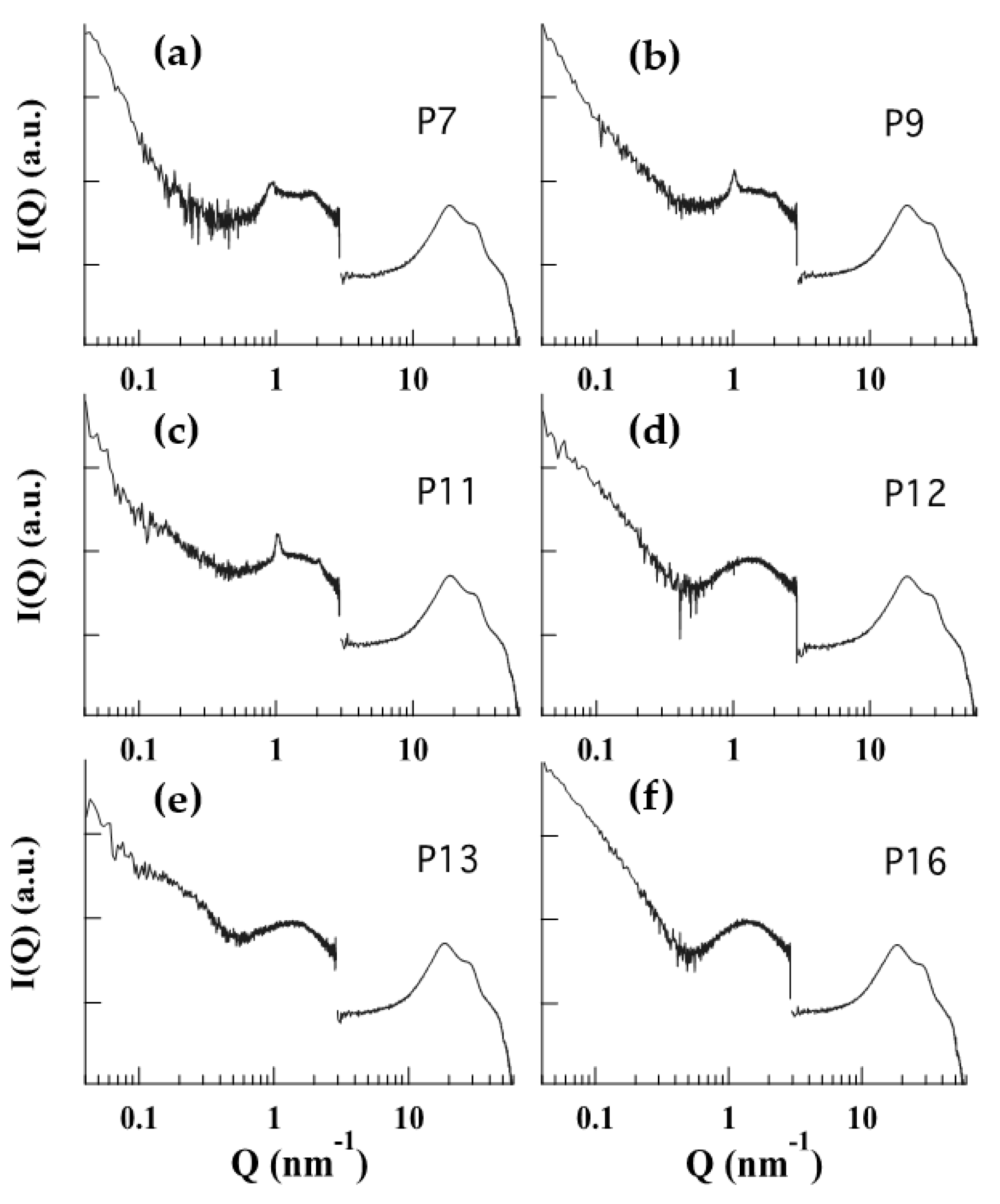
2.5. X-ray Scattering
2.6. Mangiferin (MG) Content of Vesicular Nanosystems
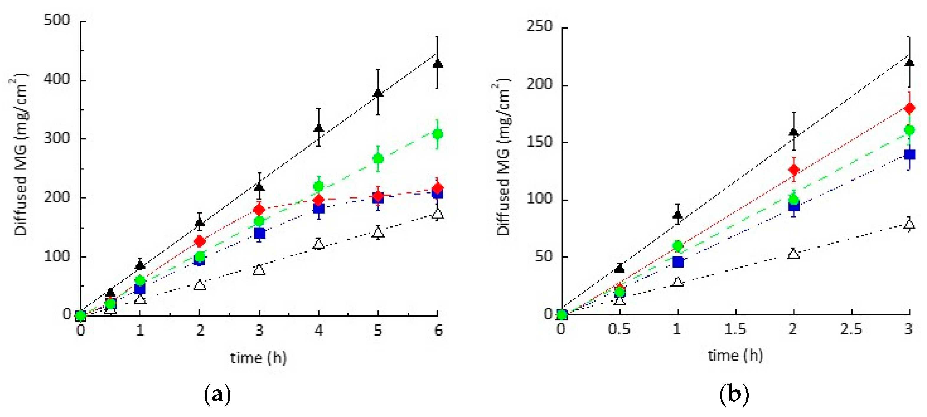
2.7. In Vitro Diffusion Experiments
2.8. HPLC Analysis
2.9. Antioxidant Activity (2,2-diphenyl-1-picrylhydrazyl Assay)
2.10. Rheological Measurements
2.11. Culture and Exposure to Ozone of EpiDerm 3D Skin Models
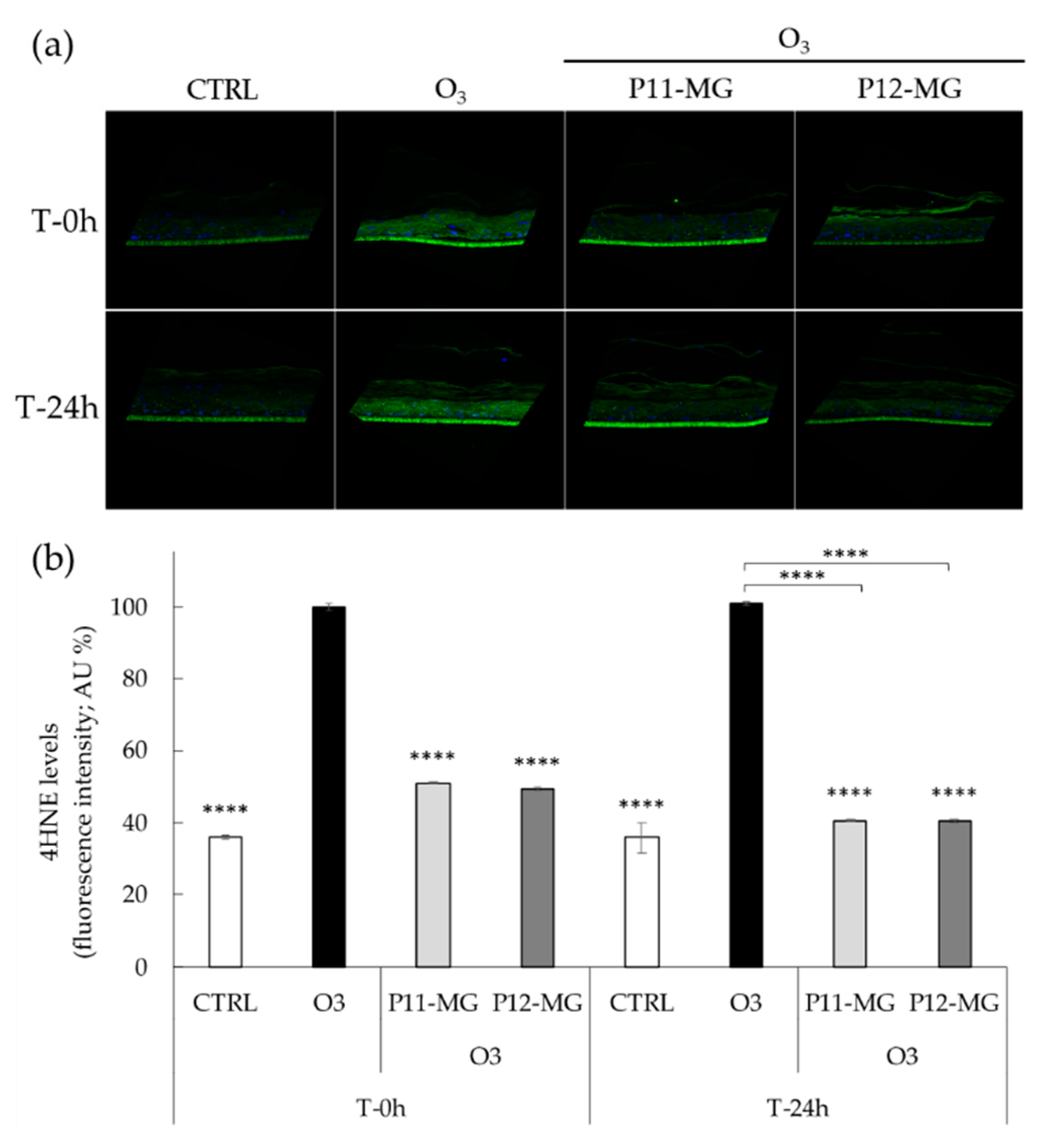
2.12. Immunohistochemistry for 4HNE
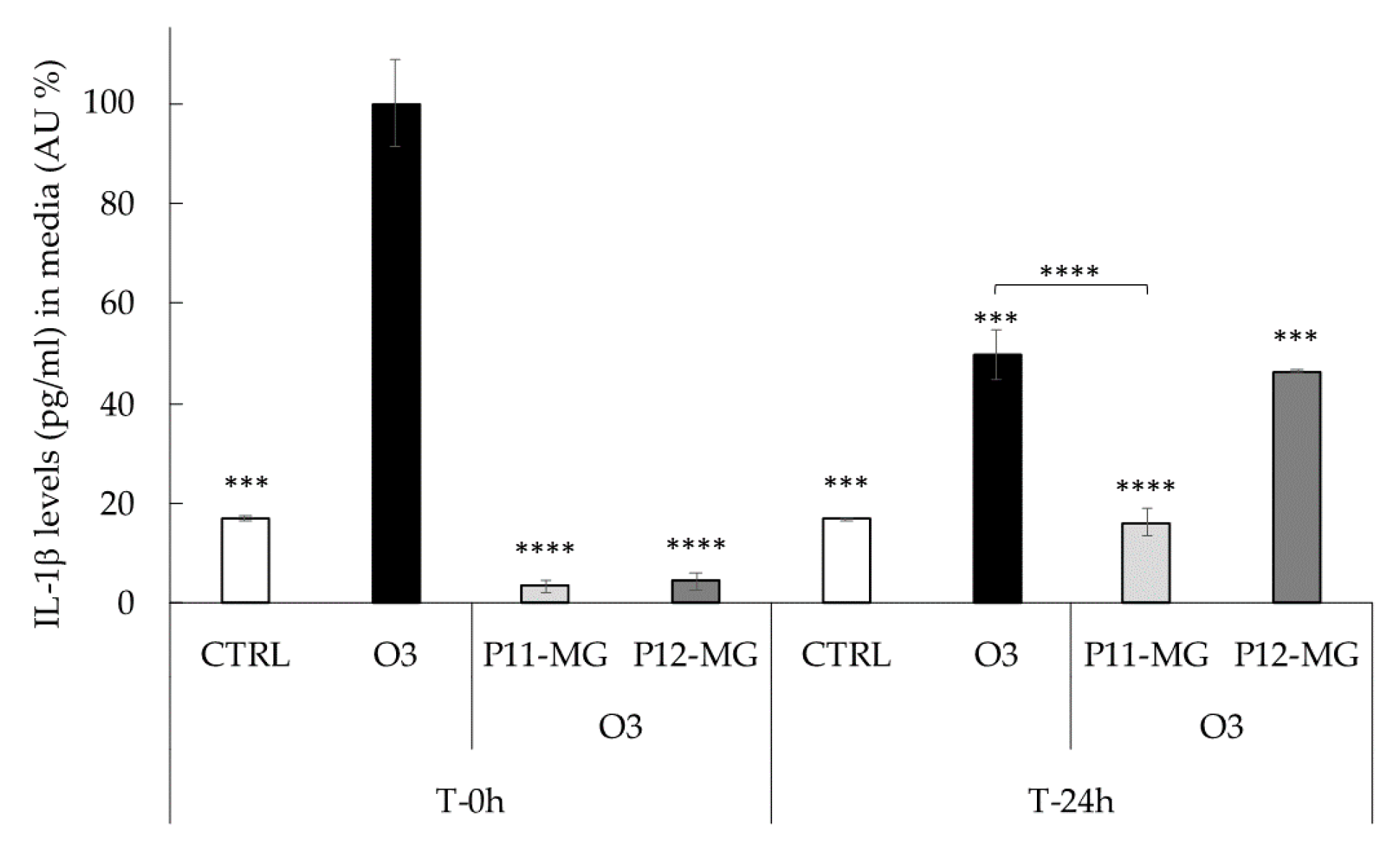
2.13. Detection of IL-1β Using ELISA Assays
3. Results and Discussion
3.1. Preparation of Vesicular Nanosystems
3.2. Characterization of Vesicular Nanosystems
3.3. Preparation and Characterization of MG-Containing Vesicular Nanosystems
3.4. In Vitro MG Diffusion Kinetics
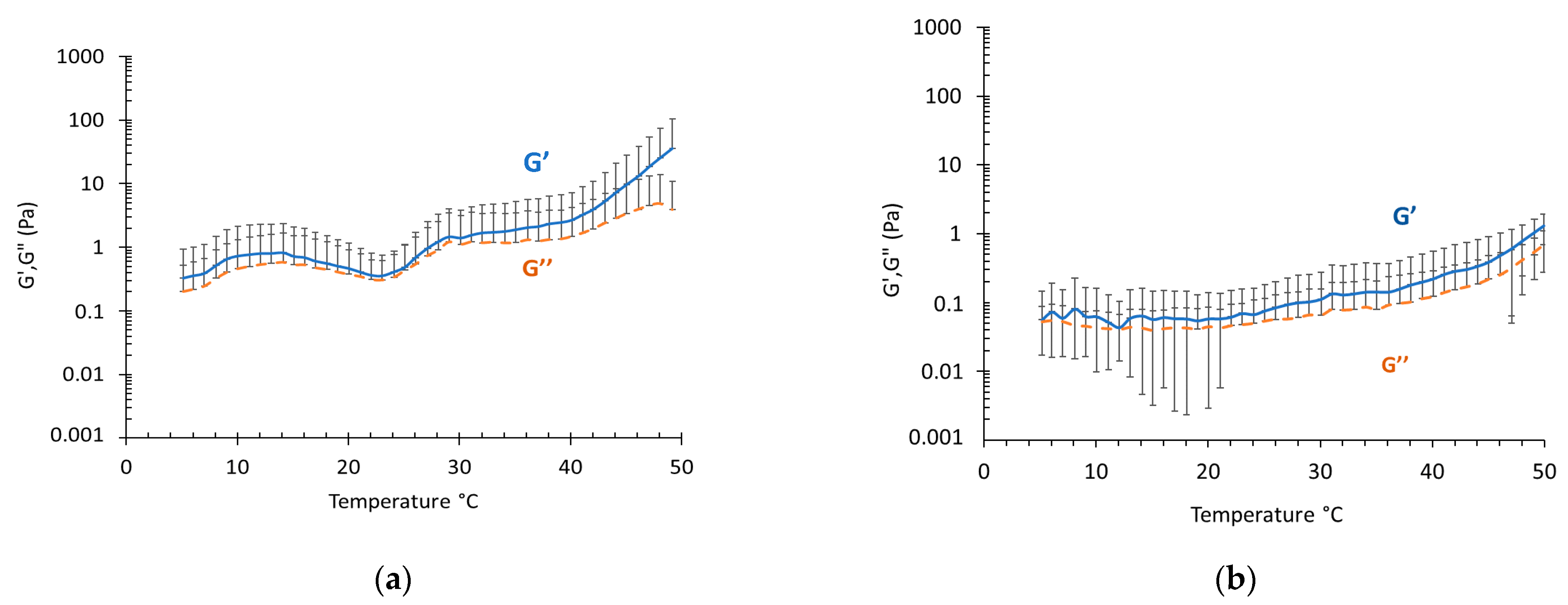
3.5. Rheological Study
3.6. Antioxidant Activity Study
3.7. Effect of Loaded-MG against the O3-Induced ox-Inflammatory Damage in 3D Human Skin Models
4. Conclusions
Supplementary Materials
Author Contributions
Funding
Acknowledgments
Conflicts of Interest
References
- Bălă, G.-P.; Râjnoveanu, R.-M.; Tudorache, E.; Motișan, R.; Oancea, C. Air pollution exposure—The (in)visible risk factor for respiratory diseases. Environ. Sci. Pollut. Res. 2021, 28, 19615–19628. [Google Scholar] [CrossRef] [PubMed]
- Lim, C.C.; Hayes, R.B.; Ahn, J.; Shao, Y.; Silverman, D.T.; Jones, R.R.; Garcia, C.; Bell, M.L.; Thurston, G.D. Long-Term Exposure to Ozone and Cause-Specific Mortality Risk in the United States. Am. J. Respir. Crit. Care Med. 2019, 200, 1022–1031. [Google Scholar] [CrossRef] [PubMed]
- Ferrara, F.; Pambianchi, E.; Woodby, B.; Messano, N.; Therrien, J.-P.; Pecorelli, A.; Canella, R.; Valacchi, G. Evaluating the effect of ozone in UV induced skin damage. Toxicol. Lett. 2021, 338, 40–50. [Google Scholar] [CrossRef]
- Song, H.; Tan, W.; Zhang, X. Ozone Induces Inflammation in Bronchial Epithelial Cells. J. Asthma 2011, 48, 79–83. [Google Scholar] [CrossRef]
- He, C.; Cheng, J.; Zhang, X.; Douthwaite, M.; Pattisson, S.; Hao, Z. Recent Advances in the Catalytic Oxidation of Volatile Organic Compounds: A Review Based on Pollutant Sorts and Sources. Chem. Rev. 2019, 119, 4471–4568. [Google Scholar] [CrossRef]
- Valacchi, G.; Virgili, F.; Cervellati, C.; Pecorelli, A. OxInflammation: From Subclinical Condition to Pathological Biomarker. Front. Physiol. 2018, 9, 858. [Google Scholar] [CrossRef] [Green Version]
- Fuks, K.B.; Woodby, B.; Valacchi, G. Skin damage by tropospheric ozone. Hautarzt 2019. [Google Scholar] [CrossRef] [PubMed]
- Ferrara, F.; Woodby, B.; Pecorelli, A.; Schiavone, M.L.; Pambianchi, E.; Messano, N.; Therrien, J.-P.; Choudhary, H.; Valacchi, G. Additive effect of combined pollutants to UV induced skin OxInflammation damage. Evaluating the protective topical application of a cosmeceutical mixture formulation. Redox Biol. 2020, 34, 101481. [Google Scholar] [CrossRef] [PubMed]
- Kim, K.E.; Cho, D.; Park, H.J. Air pollution and skin diseases: Adverse effects of airborne particulate matter on various skin diseases. Life Sci. 2016, 152, 126–134. [Google Scholar] [CrossRef]
- Krutmann, J.; Liu, W.; Li, L.; Pan, X.; Crawford, M.; Sore, G.; Seite, S. Pollution and skin: From epidemiological and mechanistic studies to clinical implications. J. Dermatol. Sci. 2014, 76, 163–168. [Google Scholar] [CrossRef] [PubMed]
- Telang, M.; Dhulap, S.; Mandhare, A.; Hirwani, R. Therapeutic and cosmetic applications of mangiferin: A patent review. Expert Opin. Ther. Pat. 2013, 23, 1561–1580. [Google Scholar] [CrossRef] [PubMed]
- Matkowski, A.; Kus, P.; Goralska, E.; Wozniak, D. Mangiferin—A Bioactive Xanthonoid, not only from Mango and not just Antioxidant. Mini Rev. Med. Chem. 2013, 13, 439–455. [Google Scholar] [CrossRef] [PubMed]
- Khare, P.; Shanker, K. Mangiferin: A review of sources and interventions for biological activities: Mangiferin. BioFactors 2016, 42, 504–514. [Google Scholar] [CrossRef]
- Khurana, R.K.; Kaur, R.; Lohan, S.; Singh, K.K.; Singh, B. Mangiferin: A promising anticancer bioactive. Pharm. Pat. Anal. 2016, 5, 169–181. [Google Scholar] [CrossRef]
- Jiang, T.; Han, F.; Gao, G.; Liu, M. Mangiferin exert cardioprotective and anti-apoptotic effects in heart failure induced rats. Life Sci. 2020, 249, 117476. [Google Scholar] [CrossRef]
- Zhao, Y.; Wang, W.; Wu, X.; Ma, X.; Qu, R.; Chen, X.; Liu, C.; Liu, Y.; Wang, X.; Yan, P.; et al. Mangiferin antagonizes TNF-α-mediated inflammatory reaction and protects against dermatitis in a mice model. Int. Immunopharmacol. 2017, 45, 174–179. [Google Scholar] [CrossRef]
- Pleguezuelos-Villa, M.; Diez-Sales, O.; Manca, M.L.; Manconi, M.; Sauri, A.R.; Escribano-Ferrer, E.; Nácher, A. Mangiferin glycethosomes as a new potential adjuvant for the treatment of psoriasis. Int. J. Pharm. 2020, 573, 118844. [Google Scholar] [CrossRef]
- Sguizzato, M.; Ferrara, F.; Hallan, S.S.; Baldisserotto, A.; Drechsler, M.; Malatesta, M.; Costanzo, M.; Cortesi, R.; Puglia, C.; Valacchi, G.; et al. Ethosomes and Transethosomes for Mangiferin Transdermal Delivery. Antioxidants 2021, 10, 768. [Google Scholar] [CrossRef]
- Ma, M.; Wang, J.; Guo, F.; Lei, M.; Tan, F.; Li, N. Development of nanovesicular systems for dermal imiquimod delivery: Physicochemical characterization and in vitro/in vivo evaluation. J. Mater. Sci. Mater. Med. 2015, 26, 192. [Google Scholar] [CrossRef]
- Song, C.K.; Balakrishnan, P.; Shim, C.-K.; Chung, S.-J.; Chong, S.; Kim, D.-D. A novel vesicular carrier, transethosome, for enhanced skin delivery of voriconazole: Characterization and in vitro/in vivo evaluation. Colloids Surf. B Biointerfaces 2012, 92, 299–304. [Google Scholar] [CrossRef]
- Song, H.; Wen, J.; Li, H.; Meng, Y.; Zhang, Y.; Zhang, N.; Zheng, W. Enhanced transdermal permeability and drug deposition of rheumatoid arthritis via sinomenine hydrochloride-loaded antioxidant surface transethosome. Int. J. Nanomed. 2019, 14, 3177–3188. [Google Scholar] [CrossRef] [Green Version]
- Abdulbaqi, I.M.; Darwis, Y.; Khan, N.A.K.; Assi, R.A.; Khan, A.A. Ethosomal nanocarriers: The impact of constituents and formulation techniques on ethosomal properties, in vivo studies, and clinical trials. Int. J. Nanomed. 2016, 11, 2279–2304. [Google Scholar] [CrossRef] [PubMed] [Green Version]
- Natsheh, H.; Vettorato, E.; Touitou, E. Ethosomes for Dermal Administration of Natural Active Molecules. Curr. Pharm. Des. 2019, 25, 2338–2348. [Google Scholar] [CrossRef] [PubMed]
- Ascenso, A.; Raposo, S.; Batista, C.; Cardoso, P.; Mendes, T.; Praça, F.G.; Bentley, M.V.L.B.; Simões, S. Development, characterization, and skin delivery studies of related ultradeformable vesicles: Transfersomes, ethosomes, and transethosomes. Int. J. Nanomed. 2015, 10, 5837–5851. [Google Scholar] [CrossRef] [Green Version]
- Costanzo, M.; Esposito, E.; Sguizzato, M.; Lacavalla, M.A.; Drechsler, M.; Valacchi, G.; Zancanaro, C.; Malatesta, M. Formulative Study and Intracellular Fate Evaluation of Ethosomes and Transethosomes for Vitamin D3 Delivery. Int. J. Mol. Sci. 2021, 22, 5341. [Google Scholar] [CrossRef]
- De Luca, C.; Valacchi, G. Surface Lipids as Multifunctional Mediators of Skin Responses to Environmental Stimuli. Mediators Inflamm. 2010, 2010, 321494. [Google Scholar] [CrossRef]
- Grimaudo, M.A.; Pescina, S.; Padula, C.; Santi, P.; Concheiro, A.; Alvarez-Lorenzo, C.; Nicoli, S. Poloxamer 407/TPGS Mixed Micelles as Promising Carriers for Cyclosporine Ocular Delivery. Mol. Pharm. 2018, 15, 571–584. [Google Scholar] [CrossRef]
- Ćirin, D.; Krstonošić, V.; Poša, M. Properties of poloxamer 407 and polysorbate mixed micelles: Influence of polysorbate hydrophobic chain. J. Ind. Eng. Chem. 2017, 47, 194–201. [Google Scholar] [CrossRef]
- Erukova, V.Y.; Krylova, O.O.; Antonenko, Y.N.; Melik-Nubarov, N.S. Effect of ethylene oxide and propylene oxide block copolymers on the permeability of bilayer lipid membranes to small solutes including doxorubicin. Biochim. Biophys. Acta BBA Biomembr. 2000, 1468, 73–86. [Google Scholar] [CrossRef] [Green Version]
- Pecora, R. Dynamic Light Scattering Measurement of Nanometer Particles in Liquids. J. Nanoparticle Res. 2000, 2, 123–131. [Google Scholar] [CrossRef]
- Barbosa, L.R.S.; Ortore, M.G.; Spinozzi, F.; Mariani, P.; Bernstorff, S.; Itri, R. The Importance of Protein-Protein Interactions on the pH-Induced Conformational Changes of Bovine Serum Albumin: A Small-Angle X-Ray Scattering Study. Biophys. J. 2010, 98, 147–157. [Google Scholar] [CrossRef] [PubMed]
- FDA Guidance for Industry: SUPAC-SS: Nonsterile Semisolid Dosage Forms; Scale-Up and Post-Approval Changes: Chemistry, Manufacturing and Controls; In-Vitro Release Testing and In Vivo Bioequivalence Documentation—ECA Academy. Available online: https://www.gmp-compliance.org/guidelines/gmp-guideline/fda-guidance-for-industry-supac-ss-nonsterile-semisolid-dosage-forms-scale-up-and-post-approval-changes-chemistry-manufacturing- (accessed on 11 December 2020).
- Pleguezuelos-Villa, M.; Nácher, A.; Hernández, M.J.; Ofelia Vila Buso, M.A.; Ruiz Sauri, A.; Díez-Sales, O. Mangiferin nanoemulsions in treatment of inflammatory disorders and skin regeneration. Int. J. Pharm. 2019, 564, 299–307. [Google Scholar] [CrossRef] [PubMed]
- Hallan, S.S.; Sguizzato, M.; Mariani, P.; Cortesi, R.; Huang, N.; Simelière, F.; Marchetti, N.; Drechsler, M.; Ruzgas, T.; Esposito, E. Design and Characterization of Ethosomes for Transdermal Delivery of Caffeic Acid. Pharmaceutics 2020, 12, 740. [Google Scholar] [CrossRef]
- Ferrara, F.; Pambianchi, E.; Pecorelli, A.; Woodby, B.; Messano, N.; Therrien, J.-P.; Lila, M.A.; Valacchi, G. Redox regulation of cutaneous inflammasome by ozone exposure. Free Radic. Biol. Med. 2020, 152, 561–570. [Google Scholar] [CrossRef]
- Magnani, N.D.; Muresan, X.M.; Belmonte, G.; Cervellati, F.; Sticozzi, C.; Pecorelli, A.; Miracco, C.; Marchini, T.; Evelson, P.; Valacchi, G. Skin Damage Mechanisms Related to Airborne Particulate Matter Exposure. Toxicol. Sci. 2016, 149, 227–236. [Google Scholar] [CrossRef] [Green Version]
- Romani, A.; Cervellati, C.; Muresan, X.M.; Belmonte, G.; Pecorelli, A.; Cervellati, F.; Benedusi, M.; Evelson, P.; Valacchi, G. Keratinocytes oxidative damage mechanisms related to airbone particle matter exposure. Mech. Ageing Dev. 2018, 172, 86–95. [Google Scholar] [CrossRef]
- Sguizzato, M.; Mariani, P.; Spinozzi, F.; Benedusi, M.; Cervellati, F.; Cortesi, R.; Drechsler, M.; Prieux, R.; Valacchi, G.; Esposito, E. Ethosomes for Coenzyme Q10 Cutaneous Administration: From Design to 3D Skin Tissue Evaluation. Antioxidants 2020, 9, 485. [Google Scholar] [CrossRef]
- Artzner, F.; Geiger, S.; Olivier, A.; Allais, C.; Finet, S.; Agnely, F. Interactions between Poloxamers in Aqueous Solutions: Micellization and Gelation Studied by Differential Scanning Calorimetry, Small Angle X-ray Scattering, and Rheology. Langmuir 2007, 23, 5085–5092. [Google Scholar] [CrossRef]
- Adhikari, U.; Goliaei, A.; Tsereteli, L.; Berkowitz, M.L. Properties of Poloxamer Molecules and Poloxamer Micelles Dissolved in Water and Next to Lipid Bilayers: Results from Computer Simulations. J. Phys. Chem. B 2016, 120, 5823–5830. [Google Scholar] [CrossRef] [PubMed]
- Chandaroy, P.; Sen, A.; Hui, S.W. Temperature-controlled content release from liposomes encapsulating Pluronic F127. J. Control. Release 2001, 76, 27–37. [Google Scholar] [CrossRef]
- Kostarelos, K.; Luckham, P.F.; Tadros, T.F. Addition of (Tri-)Block Copolymers to Phospholipid Vesicles: A Study of the Molecular Morphology and Structure by Using Hydrophobic Dye Molecules as Bilayer Probes. J. Colloid Interface Sci. 1997, 191, 341–348. [Google Scholar] [CrossRef] [PubMed] [Green Version]
- Younus, M.; Hawley, A.; Boyd, B.J.; Rizwan, S.B. Bulk and dispersed aqueous behaviour of an endogenous lipid, selachyl alcohol: Effect of Tween 80 and Pluronic F127 on nanostructure. Colloids Surf. B Biointerfaces 2018, 169, 135–142. [Google Scholar] [CrossRef] [PubMed]
- Zhang, B.; Chen, J.; Lu, Y.; Qi, J.; Wu, W. Liposomes interiorly thickened with thermosensitive nanogels as novel drug delivery systems. Int. J. Pharm. 2013, 455, 276–284. [Google Scholar] [CrossRef] [PubMed]
- Ahmed, S.; Corvis, Y.; Gahoual, R.; Euan, A.; Lai-Kuen, R.; Couillaud, B.M.; Seguin, J.; Alhareth, K.; Mignet, N. Conception of nanosized hybrid liposome/poloxamer particles to thicken the interior core of liposomes and delay hydrophilic drug delivery. Int. J. Pharm. 2019, 567, 118488. [Google Scholar] [CrossRef] [PubMed]
- Sguizzato, M.; Valacchi, G.; Pecorelli, A.; Boldrini, P.; Simelière, F.; Huang, N.; Cortesi, R.; Esposito, E. Gallic acid loaded poloxamer gel as new adjuvant strategy for melanoma: A preliminary study. Colloids Surf. B Biointerfaces 2020, 185, 110613. [Google Scholar] [CrossRef] [PubMed]
- Sguizzato, M.; Mariani, P.; Ferrara, F.; Drechsler, M.; Hallan, S.S.; Huang, N.; Simelière, F.; Khunti, N.; Cortesi, R.; Marchetti, N.; et al. Nanoparticulate Gels for Cutaneous Administration of Caffeic Acid. Nanomaterials 2020, 10, 961. [Google Scholar] [CrossRef]
- Hallan, S.S.; Sguizzato, M.; Drechsler, M.; Mariani, P.; Montesi, L.; Cortesi, R.; Björklund, S.; Ruzgas, T.; Esposito, E. The Potential of Caffeic Acid Lipid Nanoparticulate Systems for Skin Application: In Vitro Assays to Assess Delivery and Antioxidant Effect. Nanomaterials 2021, 11, 171. [Google Scholar] [CrossRef]
- Baloglu, E.; Karavana, S.Y.; Senyigit, Z.A.; Guneri, T. Rheological and mechanical properties of poloxamer mixtures as a mucoadhesive gel base. Pharm. Dev. Technol. 2011, 16, 627–636. [Google Scholar] [CrossRef]
- Fakhari, A.; Corcoran, M.; Schwarz, A. Thermogelling properties of purified poloxamer 407. Heliyon 2017, 3, e00390. [Google Scholar] [CrossRef]
- Pecorelli, A.; Woodby, B.; Prieux, R.; Valacchi, G. Involvement of 4-hydroxy-2-nonenal in pollution-induced skin damage. BioFactors 2019, 45, 536–547. [Google Scholar] [CrossRef] [Green Version]
- Küchler, S.; Strüver, K.; Friess, W. Reconstructed skin models as emerging tools for drug absorption studies. Expert Opin. Drug Metab. Toxicol. 2013, 9, 1255–1263. [Google Scholar] [CrossRef] [PubMed]


| Formulation Code | PC 1 % w/w | T80 2 % w/w | F127 3 % w/w | Ethanol % w/w | Water % w/w | MG 4 % w/w |
|---|---|---|---|---|---|---|
| P1 | 0.6 | - | - | 19.4 | 80.0 | - |
| P2 | 0.6 | - | 0.3 | 19.1 | 80.0 | - |
| P3 | 0.6 | - | 0.6 | 18.8 | 80.0 | |
| P4 | 0.6 | - | 1.2 | 18.2 | 80.0 | - |
| P5 | 0.9 | - | - | 29.1 | 70.0 | - |
| P6 | 0.9 | 0.3 | - | 28.8 | 70.0 | - |
| P7 | 1.8 | - | - | 18.2 | 80.0 | - |
| P8 | 1.8 | - | 0.3 | 17.9 | 80.0 | - |
| P9 | 1.8 | - | 0.6 | 17.6 | 80.0 | - |
| P10 | 1.8 | - | 0.9 | 17.3 | 80.0 | - |
| P11 | 1.8 | - | 1.2 | 17.0 | 80.0 | - |
| P12 | 1.8 | 0.2 | - | 18.0 | 80.0 | - |
| P13 | 1.8 | 0.2 | 1.2 | 16.8 | 80.0 | - |
| P14 | 1.8 | - | 1.6 | 16.6 | 80.0 | - |
| P15 | 2.7 | - | - | 27.3 | 70.0 | - |
| P16 | 2.7 | 0.3 | - | 27.0 | 70.0 | - |
| P7-MG | 1.8 | - | - | 18.12 | 80.0 | 0.08 |
| P9-MG | 1.8 | - | 0.6 | 17.52 | 80.0 | 0.08 |
| P11-MG | 1.8 | - | 1.2 | 16.9 | 80.0 | 0.08 |
| P12-MG | 1.8 | 0.2 | - | 17.5 | 80.0 | 0.08 |
| Formulation | Time | Z Average | Dispersity | Macroscopic |
|---|---|---|---|---|
| Code | (Days) | (nm) ± s.d. | Index ± s.d. | Appearance |
| P1 | 1 | 201.2 ± 1 | 0.06 ± 0.01 | translucent |
| 90 | 206.0 ± 2 | 0.08 ± 0.03 | translucent | |
| P2 | 1 | 195.5 ± 1 | 0.14 ± 0.02 | translucent |
| 90 | 200.0 ± 10 | 0.14 ± 0.03 | translucent | |
| P3 | 1 | 264.4 ± 14 | 0.15 ± 0.02 | translucent |
| 90 | n.d. | n.d. | phase separation | |
| P4 | 1 | 243.7 ± 35 | 0.16 ± 0.01 | milky |
| 90 | n.d. | n.d. | phase separation | |
| P5 | 1 | 206.3 ± 33 | 0.15 ± 0.04 | milky |
| 90 | 225.4 ± 25 | 0.16 ± 0.02 | milky | |
| P6 | 1 | 186.2 ± 20 | 0.13 ± 0.05 | translucent |
| 90 | 190.5 ± 22 | 0.14 ± 0.05 | translucent | |
| P7 | 1 | 285.5 ± 30 | 0.22 ± 0.07 | milky |
| 90 | n.d. | n.d. | phase separation | |
| P8 | 1 | 227.7 ± 30 | 0.18 ± 0.05 | milky |
| 90 | n.d. | n.d. | phase separation | |
| P9 | 1 | 237.3 ± 25 | 0.22 ± 0.09 | milky |
| 90 | 260.2 ± 15 | 0.23 ± 0.07 | milky | |
| P10 | 1 | 247.8 ± 35 | 0.18 ± 0.05 | milky |
| 90 | 270.2 ± 15 | 0.19 ± 0.06 | milky | |
| P11 | 1 | 252.3 ± 25 | 0.18 ± 0.04 | milky |
| 90 | 260.2 ± 17 | 0.19 ± 0.08 | milky | |
| P12 | 1 | 338.5 ± 20 | 0.18 ± 0.07 | quite translucent |
| 90 | 360.5 ± 15 | 0.20 ± 0.08 | quite translucent | |
| P13 | 1 | 351.2 ± 18 | 0.17 ± 0.06 | translucent |
| 90 | 382.7 ± 15 | 0.19 ± 0.05 | translucent | |
| P14 | 1 | 375.6 ± 12 | 0.21 ± 0.09 | milky |
| 90 | n.d. | n.d. | phase separation | |
| P15 | 1 | 320.7 ± 12 | 0.24 ± 0.07 | milky |
| 90 | n.d. | n.d. | phase separation | |
| P16 | 1 | 550.5 ± 80 | 0.25 ± 0.05 | quite translucent |
| 90 | 525.4 ± 20 | 0.26 ±0.07 | quite translucent | |
| P7-MG | 1 | 274.5 ± 20 | 0.21 ± 0.05 | yellowish |
| 90 | n.d. | n.d. | phase separation | |
| P9-MG | 1 | 225.2 ± 15 | 0.20 ± 0.04 | yellowish |
| 90 | 240.2 ± 11 | 0.22 ± 0.07 | yellowish * | |
| P11-MG | 1 | 243.7 ± 25 | 0.18 ± 0.05 | yellowish |
| 90 | 248.7 ± 16 | 0.17 ± 0.09 | yellowish | |
| P12-MG | 1 | 332.5 ± 12 | 0.20 ± 0.04 | yellowish |
| 90 | 334.0 ± 25 | 0.20 ± 0.08 | yellowish |
| Figure | Structure | Lamellar Repeat Distance (nm) |
|---|---|---|
| P7 | multilamellar | 6.76 |
| P9 | multilamellar | 6.19 |
| P11 | multilamellar | 6.13 |
| P12 | lamellar | - |
| P13 | lamellar | - |
| P16 | lamellar | - |
| Formulation | EC a (%) | Recovery b (%) | DPPH IC50 (µg/mL) |
|---|---|---|---|
| Sol-MG c | - | - | 14.06 ± 0.53 |
| P7-MG | 78 ± 2 | 98 ± 1 | n.d. |
| P9-MG | 87 ± 3 | 97 ± 3 | n.d. |
| P11-MG | 88 ± 4 | 99 ± 1 | 16.10 ± 0.23 |
| P12-MG | 71 ± 2 | 98 ± 2 | 20.89 ± 0.08 |
| Parameter | P7-MG | P9-MG | P11-MG | P12-MG | Sol-MG |
|---|---|---|---|---|---|
| F 1 ± s.d. (μg/cm2/h) | 26.44 ± 3.4 | 61.84 ± 5.2 | 47.36 ± 4.7 | 53.40 ± 5.4 | 73.58 ± 10.0 |
| MG (mg/mL) | 0.8 | 0.8 | 0.8 | 0.8 | 0.8 |
| D 2 ± s.d. (cm/h) × 10−3 | 33.05 ± 4.2 | 77.30 ± 6.5 | 59.20 ± 5.8 | 66.75 ± 6.7 | 91.97 ± 12.5 |
| Rm 3 ± s.d. (μg/cm2) | 15 ± 4 | 9 ± 3 | 10 ± 3 | 11 ± 4 | 10 ± 2 |
| Rv 4 ± s.d. (μg/mL) | 610 ± 30 | 575 ± 45 | 580 ± 28 | 480 ± 32 | 360 ± 27 |
| Q6 5 ± s.d. (μg/mL) | 175 ± 15 | 216 ± 18 | 210 ± 19 | 309 ± 28 | 430 ± 40 |
| Rm/Q6 | 0.08 | 0.04 | 0.05 | 0.03 | 0.02 |
| Rv/Q6 | 3.48 | 2.66 | 2.76 | 1.55 | 0.83 |
Publisher’s Note: MDPI stays neutral with regard to jurisdictional claims in published maps and institutional affiliations. |
© 2021 by the authors. Licensee MDPI, Basel, Switzerland. This article is an open access article distributed under the terms and conditions of the Creative Commons Attribution (CC BY) license (https://creativecommons.org/licenses/by/4.0/).
Share and Cite
Sguizzato, M.; Ferrara, F.; Mariani, P.; Pepe, A.; Cortesi, R.; Huang, N.; Simelière, F.; Boldrini, P.; Baldisserotto, A.; Valacchi, G.; et al. “Plurethosome” as Vesicular System for Cutaneous Administration of Mangiferin: Formulative Study and 3D Skin Tissue Evaluation. Pharmaceutics 2021, 13, 1124. https://doi.org/10.3390/pharmaceutics13081124
Sguizzato M, Ferrara F, Mariani P, Pepe A, Cortesi R, Huang N, Simelière F, Boldrini P, Baldisserotto A, Valacchi G, et al. “Plurethosome” as Vesicular System for Cutaneous Administration of Mangiferin: Formulative Study and 3D Skin Tissue Evaluation. Pharmaceutics. 2021; 13(8):1124. https://doi.org/10.3390/pharmaceutics13081124
Chicago/Turabian StyleSguizzato, Maddalena, Francesca Ferrara, Paolo Mariani, Alessia Pepe, Rita Cortesi, Nicolas Huang, Fanny Simelière, Paola Boldrini, Anna Baldisserotto, Giuseppe Valacchi, and et al. 2021. "“Plurethosome” as Vesicular System for Cutaneous Administration of Mangiferin: Formulative Study and 3D Skin Tissue Evaluation" Pharmaceutics 13, no. 8: 1124. https://doi.org/10.3390/pharmaceutics13081124
APA StyleSguizzato, M., Ferrara, F., Mariani, P., Pepe, A., Cortesi, R., Huang, N., Simelière, F., Boldrini, P., Baldisserotto, A., Valacchi, G., & Esposito, E. (2021). “Plurethosome” as Vesicular System for Cutaneous Administration of Mangiferin: Formulative Study and 3D Skin Tissue Evaluation. Pharmaceutics, 13(8), 1124. https://doi.org/10.3390/pharmaceutics13081124