Article

Polylactide Nanocapsules Attenuate Adverse Cardiac Cellular Effects of Lyso-7, a Pan-PPAR Agonist/Anti-Inflammatory New Thiazolidinedione

by
Giani M. Garcia
1,2,
Jérôme Roy
1,
Ivan R. Pitta
3,
Dulcinéia S. P. Abdalla
4,
Andrea Grabe-Guimarães
2,
Vanessa C. F. Mosqueira
2 and
Sylvain Richard
1,*
1
PhyMedExp, Inserm U1046, CNRS UMR 9214, Université de Montpellier, 34270 Montpellier, France
2
Department of Pharmacy, School of Pharmacy, Federal University of Ouro Preto, Ouro Preto 35400-000, Brazil
3
Center of Health Sciences, Federal University of Pernambuco, Recife 50670-420, Brazil
4
Department of Clinical and Toxicological Analyses, Faculty of Pharmaceutical Sciences, University of São Paulo, São Paulo 05508-000, Brazil
*
Author to whom correspondence should be addressed.
Pharmaceutics 2021, 13(9), 1521; https://doi.org/10.3390/pharmaceutics13091521
Submission received: 23 July 2021 / Revised: 12 September 2021 / Accepted: 14 September 2021 / Published: 20 September 2021
(This article belongs to the Special Issue Polymeric Nanocapsules in Drug Delivery)

Abstract

:
Lyso-7 is a novel synthetic thiazolidinedione, which is a receptor (pan) agonist of PPAR α,β/δ,γ with anti-inflammatory activity. We investigated the cardiotoxicity of free Lyso-7 in vitro (4.5–450 nM), and Lyso-7 loaded in polylactic acid nanocapsules (NC) in vivo (Lyso-7-NC, 1.6 mg/kg). In previous work, we characterized Lyso-7-NC. We administered intravenously Lyso-7, Lyso-7-NC, control, and blank-NC once a day for seven days in mice. We assessed cell contraction and intracellular Ca2+ transients on single mice cardiomyocytes enzymatically isolated. Lyso-7 reduced cell contraction and accelerated relaxation while lowering diastolic Ca2+ and reducing Ca2+ transient amplitude. Lyso-7 also promoted abnormal ectopic diastolic Ca2+ events, which isoproterenol dramatically enhanced. Incorporation of Lyso-7 in NC attenuated drug effects on cell contraction and prevented its impact on relaxation, diastolic Ca2+, Ca2+ transient amplitude, Ca2+ transient decay kinetics, and promotion of diastolic Ca2+ events. Acute effects of Lyso-7 on cardiomyocytes in vitro at high concentrations (450 nM) were globally similar to those observed after repeated administration in vivo. In conclusion, we show evidence for off-target effects of Lyso-7, seen during acute exposure of cardiomyocytes to high concentrations and after repeated treatment in mice. Nano-encapsulation of Lyso-7 in polymeric NC attenuated the unwanted effects, particularly ectopic Ca2+ events known to support life-threatening arrhythmias favored by stress or exercise.

1. Introduction

Peroxisome proliferator-activated receptors (PPARs) are members of the nuclear receptor family and function as ligand inducible transcription factors to regulate genes involved in lipid and glucose metabolism [1,2]. Thiazolidinediones (TZDs) are PPARγ agonists widely used as antihyperglycemic agents to improve the liver, muscles, and adipose tissue insulin sensitivity and treat type 2 diabetes mellitus [3]. The TZDs have additional anti-inflammatory and atheroprotective effects, yet the activation of PPARα may also be involved [4]. Despite reported favorable effects on cardiovascular risk factors, ischemic heart diseases and cardiovascular events may increase in diabetic patients [5,6]. TZDs also increase the risk of developing heart failure (HF) [7,8,9]. Treatment with TZD, such as rosiglitazone or pioglitazone, increase stroke risk in patients [10].
Lyso-7 is a new indole-thiazolidine compound (Figure 1A) recently synthesized [11]. It is a promising hybrid molecule acting as a pan partial agonist of PPARγ, PPARα, and PPARβ/δ but also acting as a COX-2 inhibitor [11,12]. Lyso-7 has a potent in vivo beneficial effect on inflammation and microcirculatory damage and the development of atherosclerotic lesions [13]. Lyso-7 exhibits anti-inflammatory effects via inhibition of expression of adhesion proteins, abolishing the adhesion of neutrophils to endothelial cells, and effects on blocking the enhancement of intracellular Ca2+ levels in neutrophils [14].
Polymeric nanocapsules (NC) (Figure 1A) are oily core nanostructures surrounded by a polymeric wall [15,16]). They can encapsulate different lipophilic molecules at high payloads and are biodegradable. They have demonstrated outstanding potential to reduce the cardiotoxicity of several drug candidates such as artemether, halofantrine, and lychnopholide, as reported by our group [17,18,19,20]. These properties may reflect NC’s ability to control drug release in the blood, reducing the free fraction producing potentially toxic adverse effects. Lyso-7 is a lipophilic compound (clog·P = 5.6). After encapsulation in NC, its physicochemical properties, release kinetics, and intravenous pharmacokinetics have been characterized [21]. NC significantly improves the Lyso-7 biopharmaceutical profile. Lyso-7 NC is suitable for oral or intravenous administration due to its nanometrical size dispersion. After intravenous administration in mice, compared with free Lyso-7 administered in solution, NC retained Lyso-7 in the plasma compartment, increasing 14-fold the plasma concentration and 3-fold the heart concentration. Upon intravenous administration, the accumulation of Lyso-7 in the heart was unrelated to blood flow. No apparent cardiotoxicity of Lyso-7 was observed in vivo when the NC formulation was administered intravenously at a dose of 1.6 mg/kg [21].
Due to the TZD class of drugs’ clinical restrictions, our study now aimed to assess the cardiac cellular effects of intravenous repeated administrations of Lyso-7 (free form) and Lyso-7 encapsulated in polylactic acid NC in an experimental rodent model (mice). We investigated the effect of Lyso-7 on the contraction and Ca2+ handling in single left ventricular (LV) myocytes in vitro and after treatment in vivo. Primary objectives were to: (i) detect off-target effects potentially leading to unexpected harmful side effects, and (ii) determine if the use of NC provides cardiac protection.

2. Materials and Methods

2.1. Materials

Lyso-7, [(5Z)-5-[5-bromo-1H-indol-3-yl)methylene]-3-(4-chlorobenzyl)-thiazolidine-2,4-dione] (CAS Number 1505484-42-3) (CAS Registry Number 1505484-42-3), Mw 447.73 g/mol, was synthesized, purified and characterized by Prof Ivan da Rocha Pitta as reported [11]. PLA polymer, a poly-rac-lactide Mn 75,000–120,000 g/mol, poloxamer 188, polysorbate 80, polyethylene glycol 300 (PEG 300), glucose, ethanol and methanol (HPLC grade) were purchased from Sigma-Aldrich (Cotia, Brazil). Symplicity® System (Millipore, Bedford, MA, USA) produced Milli-Q water to prepare all solutions throughout the experiments. Soy lecithin with 75% of phosphatidylcholine (Epikuron® 170) was a generous gift from Lucas Meyer (Champlan, France). Sasol Olefins & Surfactants GmbH (Hamburg, Germany) provided Miglyol 810 N. Tedia (Rio de Janeiro, Brazil) provided ethyl acetate, acetone, N,N-dimethylacetamide (DMA), acetone and dimethylsulfoxide (DMSO) (analytical grade).

2.2. Preparation of Lyso-7 Solution and Lyso-7-Loaded Nanocapsules

Lyso-7 was dissolved in absolute ethanol further diluted in buffers used in each experiment with ethanol concentration no higher than 2% v/v. We used this solution without Lyso-7 as a control in the acute experiments performed in vitro with isolated cardiomyocytes. We prepared the free Lyso-7 solution for intravenous (iv) treatment of mice as follows: we dissolved Lyso-7 in 20 μL of DMSO, 20 μL of polysobate 80, 380 μL of DMA, and 580 μL of PEG 300. This solution was mixed for 10 min and then appropriately diluted in isotonic glucose to obtain 0.5 mg/mL of free Lyso-7 solution used in vivo. We used the same solution without Lyso-7 as a control in vivo.
We used the nanoprecipitation method to make monodispersed nanocapsules loading Lyso-7 (Lyso-7-NC) or without Lyso-7 (blank-NC) as previously described [21]. We prepared Lyso-7-NC with 0.6% wt/v of PLA, 0.75% v/v of Miglyol 810N), and 0.75% wt/v of soybean lecithin dissolved in 10 mL of acetone. We added this organic solution into 0.75% wt/v of Poloxamer 188 solution (20 mL) via a syringe. We maintained dispersion under magnetic stirring for 10 min, and then all organic solvents and part of the water were evaporated under reduced pressure (Heidolph Rotary Evaporator, Schwabach, Germany) to render 5 mL of the aqueous suspension of NC.
The mean hydrodynamic diameters were 296 ± 4.2 nm and 265 ± 1.7 nm, respectively, with dispersion indexes less than 0.3 indicating the monodispersed population of particles in size. NC encapsulated Lyso-7 with high efficiency (83%) at a 0.5 mg/mL Lyso-7 concentration, attributed to Lyso-7 high lipophilicity. We have already characterized these NC in detail [21]. We used blank-NC as controls vs. Lyso-7-NC. All the formulations of Lyso-7, the 0.5 mg/mL iv solution (Lyso-7) and the 0.5 mg/mL Lyso-7 NC (Lyso-7-NC), as well as blank-NC, were passed through a 0.8 μm sterile filter before appropriate dilution in isotonic glucose to allow intravenous injection in mice of the doses established.

2.3. Animals and Cardiomyocytes

We used 7-week-old male C57BL/6J mice (Janvier Labs, Le Genest-Saint-Isle, France) bred and housed (4 mice/cage) under pathogen-free conditions (22 ± 2 °C; 12-h day/12-h night cycle) with standard rodent chow diet, ad libitum access to water, and behavioral needs (wood bedding material, cardboard tunnel, nesting material, and wooden gnawing sticks). For repeated-dose intravenous administration (via tail vein), mice were blindly randomized to the four different groups: vehicle (intravenous solution diluted in isotonic glucose), Lyso-7 (1.6 mg/kg/day), blank-NC (equivalent dose of NC excipients related to 1.6 mg/kg/day), and Lyso-7-NC (1.6 mg/kg/day). We administered mice vehicle and blank-NC as control groups for Lyso-7 and Lyso-7-NC, respectively, for comparisons. Repeated doses of both Lyso-7 and Lyso-7-NC for seven days produced no death in mice. We isolated myocytes freshly from mice of all experimental groups after the seven days of repeated administrations, as shown in Figure 1B.
We assessed the acute effects of Lyso-7 in vitro following the application of 4.5 nM, 45 nM, and 450 nM of Lyso-7 for 15 min on single cardiomyocytes isolated from untreated animals. The hearts were excised after euthanasia of mice by cervical dislocation to ensure rapid death without injecting any substance interfering with cellular physiology. Whole hearts were submitted to the liberase action using a Langendorf perfusion system to obtain individual LV myocytes as described [18,20,22,23,24]. We used only quiescent cardiomyocytes with precise edges. Otherwise, we investigated cells randomly. We carried out the experiments 1 to 4 h after cells enzymatic isolation. Figure 1B shows the general schema of animal experiments and protocols.

2.4. Measurements of Contraction and Intracellular Ca2+ in Single Cardiomyocytes

We field-stimulated myocytes with 1-ms current pulses delivered at 1 Hz to assess cell sarcomere length (SL) shortening, an index of contraction, and intracellular Ca2+ transients recorded simultaneously using an IonOptix system (IonOptix LCC. Milton, MA, USA) with a Zeiss microscope (Carl Zeiss GmbH, Oberkochen, Germany) (40× oil-immersion objective, 0.36 μm/pixel) as described [18,20,22,23,24]. Briefly, cells were bathed in a solution containing (in mM) 117 NaCl, 5.7 KCl, 11 glucose, 1.7 MgCl2, 1.8 CaCl2, 4.4 NaHCO3, 1.5 KH2PO4, and 21 HEPES (pH 7.4); all chemicals bought from Sigma-Aldrich, Saint-Quentin-Fallavier, France. We loaded cells with the dual-emission ratiometric Ca2+ indicator Indo-1AM (2 µM, Invitrogen, Grand Island, NY, USA) to monitor cytosolic Ca2+ determined by the ratio of 405 nm/480 nm fluorescence (a.u.: arbitrary units) [18,20,22,23,24]. We paced cells for 30 s periods followed by a 30-s rest period to investigate spontaneous Ca2+ diastolic events and contractions. In some experiments, we mimicked stress conditions by exposing cells to the β-adrenergic agonist isoproterenol (ISO, 10 nM) for 5 min before the experiments. Data were analyzed using Ionwizard® Software (version 7.4, IonOptix®, Westwood, LA, USA).

2.5. Sparks Confocal

We visualized Ca2+ sparks in quiescent myocytes incubated with the Ca2+ indicator Fluo-4AM (4 μM) (Molecular Probes Inc., Eugene, OR, USA), visualized by confocal imaging with a Zeiss LSM510 microscope (Carl Zeiss Inc., Oberkochen, Germany) equipped with 63×/1.2 N.A. water immersion objective, at 25 °C in line-scan mode (1.5 ms/line, 512 pixels × 3000 lines) as described [18,22,23]. The dye was excited at 488 nm, and the fluorescence emission was collected through a 505-nm long-pass filter. Myocytes were field-stimulated at 1 Hz with 1-ms current pulses delivered via two platinum electrodes, one on each side of the perfusion chamber. During the rest period that followed stimulation, myocytes were repetitively scanned along the entire length of the cell at 1.5-ms intervals, for a maximum of 6 s. We reduced the laser intensity to 5% maximum to decrease cell damage and dye bleaching. Ca2+ sparks were determined by averaging the intensity of each sequential scan line and plotting the mean intensity as a function of time. We used the SparkMaster plugin for ImageJ software (Wayne Rasband, National Institutes of Health, Bethesda, MD, USA) to detect and analyze Ca2+ sparks.

2.6. Statistical Analysis

We performed statistical analyses using GraphPad Prism® Software (Version 6.0, GraphPad Software Inc., San Diego, CA, USA). We expressed all data as mean ± SEM. We used D’Agostino and Pearson omnibus and Shapiro–Wilk normality tests, then a One-way ANOVA test for multiple comparisons. We performed post hoc tests where F was significant (p < 0.05) and no variance inhomogeneity. We used a Tukey post-test to compare all pairs of columns. We used a t-test for Ca2+ waves and Ca2+ sparks. p < 0.05 indicated a significant difference. N indicates the number of mice, and n indicates the number of independent cardiomyocytes isolated from these mice.

3. Results

3.1. Effect of In Vivo Repeated Dose Administration of Lyso-7 on Single Myocytes

3.1.1. Differential Effects of Lyso-7 and Lyso-7-NC on Cellular Contraction and Ca2+

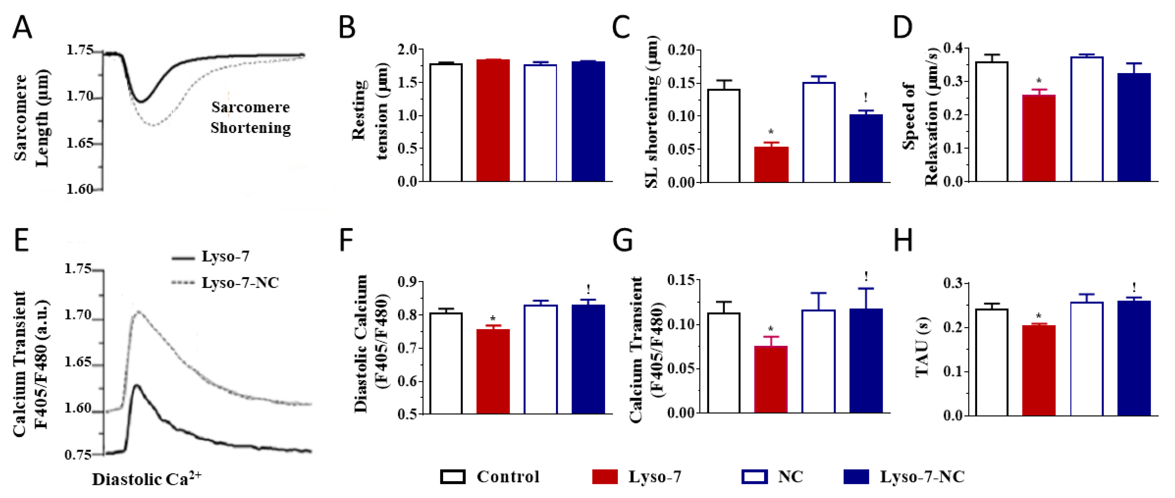
We investigated the effects of the repeated administration of Lyso-7 (free form) on contraction and Ca2+ transient evoked by electrical stimulation of LV myocytes at 1 Hz. No mice died under treatment with the formulations. First, it is worth noting that blank-NC did not affect any parameter relative to the control group (Figure 2, white bars). In contrast, Lyso-7 decreased cell contraction (−64%, SL shortening) and accelerated relaxation (25%) during systolic activity while not affecting resting SL (Figure 2A–D). In parallel, Lyso-7 lowered diastolic Ca2+ (−5%), reduced the Ca2+ transient amplitude (−35%) and accelerated the Ca2+ transient decay kinetics (16%) (Figure 2E–H). However, when Lyso-7 was incorporated in NC, its effect on contraction was less pronounced, by a factor of two (−33%, Lyso-7-NC vs. NC; Figure 2C, ! p < 0.05), and the impact of Lyso-7 on cell relaxation was prevented (Figure 2D) as well as its effect on the diastolic Ca2+, Ca2+ transient amplitude and Ca2+ transient decay kinetics (Figure 2F–H, ! p < 0.05).
We next evaluated the effects of Lyso-7 under the challenge of a maximally active concentration of the β-adrenergic receptor agonist isoproterenol (ISO,10 nM) to mimic the effects of physical exercise and stress [25]. We exposed myocytes to ISO for 5 min before recordings. As expected, in all experimental groups (compare averaged data in Figure 3 with corresponding values in Figure 2), acute exposure of cells to ISO enhanced cell contraction, accelerated relaxation, and, in parallel, increased Ca2+ transient amplitude and accelerated Ca2+ transient decay kinetics in line with enhanced reuptake of Ca2+ in the sarcoplasmic reticulum (SR) via the SERCA2a pump [26]. For example, in the control group, ISO increased cell contraction by 43%, accelerated relaxation by 29%, increased Ca2+ transient amplitude by 81%, and accelerated Ca2+ transient decay kinetics by 60%. Overall, these effects were consistent with those reported in the literature [25]. However, ISO had a two-fold more positive inotropic effect on contraction, compensating for the decreasing impact of Lyso-7 as found in basal conditions while also enhancing the lusitropic impact (Figure 3B,C). In the same line, free Lyso-7 still lowered diastolic Ca2+ and accelerated the Ca2+ transient decay kinetics (Figure 3D,F), similarly to the effects observed in the absence of ISO (Figure 2F,H) except for the Ca2+ transient where the decreasing impact of ISO was partly compensated (compare Figure 2G with Figure 3E). The different effects of Lyso-7 in the presence of ISO were absent or attenuated by nano-encapsulation (Lyso-7-NC formulation). In basal conditions and with ISO, these data showed that Lyso-7 impacts cellular contraction and intracellular Ca2+, but its effects could be mostly prevented or significantly attenuated (e.g., contraction) by encapsulation in NC.

3.1.2. Differential Effects of Lyso-7 and Lyso-7-NC on Ectopic Ca2+ Events at Rest

The repeated administration of Lyso-7 promoted ectopic diastolic Ca2+ events, not observed in the control and NC groups, during resting periods following a train of stimulations (Figure 4A,C). When we incorporated Lyso-7 in NC (Lyso-7-NC group), we detected no significant abnormal activity. We also observed no irregular Ca2+ transient during pacing in any of the four groups. We next tested the effect of an ISO challenge on the promotion of spontaneous Ca2+ events. First of all, ISO (10 nM), as such, favored their occurrence (Figure 4B,D) in line with reports by others [27,28,29]. Nevertheless, Lyso-7 severely enhanced the number of cells exhibiting ectopic Ca2+ events (80% vs. 10% in the absence of ISO), which was abolished in the Lyso-7-NC group (Figure 4D). Therefore, Lyso-7 induced aberrant diastolic Ca2+ events, severely increased by ISO but controlled by NC.

3.2. Acute Effects of Free Lyso-7 on Single Cardiomyocytes In Vitro

3.2.1. Lyso-7 Impairs Contraction and Ca2+ Transient

In separate experiments, we acutely exposed cardiomyocytes from untreated animals for 15 min to Lyso-7 at 4.5 nM, 45 nM, and 450 nM, respectively (Figure 5). The highest concentration corresponded to 200 µg/mL, i.e., a concentration almost one hundred times higher than the blood concentration as determined in studies performed in mice in vivo (1.86 µg/mL; [21]). Free Lyso-7 increased contraction, but only at 450 nM (Figure 5A,C). Lyso-7 also accelerated cellular relaxation dose-dependently (+30% at 450 nM), an effect detected at 45 nM (17%) (Figure 5D). Lyso-7 did not affect resting SL (Figure 5B). In parallel, Lyso-7 at 450 nM lowered diastolic Ca2+ (−29%) (Figure 5E,F) and reduced the amplitude of the Ca2+ transient (−45%) (Figure 5E,G). In contrast, Lyso-7 did not affect the decay of the Ca2+ transient (Figure 5H).
Acute exposure to ISO increased SL shortening (i.e., augmented contraction) approximately twice, except at 450 nM (+25%) due to the intrinsic positive effect of Lyso-7 at this concentration, and enhanced relaxation (+30%) in control myocytes (compare values in Figure 6C,D and Figure 5C,D). In parallel, ISO increased the amplitude of the Ca2+ transient (+80%), the diastolic Ca2+ (+12%), and accelerated the Ca2+ transient decay (+40%) (compare values in Figure 6F–H and Figure 5F–H). Under these experimental conditions (presence of ISO), we observed no effect of Lyso-7 on cell contraction and relaxation, even at 450 nM (Figure 6A,C). In particular, there was no additive inotropic effect. Similarly, Lyso-7 did not affect the Ca2+ transient amplitude, diastolic Ca2+, and Ca2+ transient decay in the presence of ISO at 4.5 nM and 45 nM. However, at 450 nM, Lyso-7 still reduced both the resting Ca2+ (−37% vs. control) and the Ca2+ transient amplitude (−47%; Figure 6F,G) and reminded the effects of Lyso-7 in basal conditions (absence of ISO) (Figure 5F,G). In contrast, Lyso-7 did not affect the decay of the Ca2+ transient (Figure 6H), as seen in the absence of ISO (Figure 5H).

3.2.2. Lyso-7 Promotes Abnormal Ca2+ Events at Rest

We detected no abnormal activity, either during pacing or rest, in control cells for low concentrations of Lyso-7. However, Lyso-7 at 450 nM promoted diastolic ectopic Ca2+ events in nearly 20% of cells (Figure 7A) though they were small and infrequent. The presence of abnormal macroscopic Ca2+ events at rest often results from a ryanodine receptor 2 (RyR2) leakage, generating microscopic Ca2+ events called Ca2+ sparks [27]. Consistent with this possibility, Lyso-7 at 450 nM increased the onset of Ca2+ sparks (+125%; Figure 7C,D). We next tested the effect of Lyso-7 in cells subjected to an ISO challenge. Once again, ISO (10 nM), as such, promoted the occurrence of ectopic Ca2+ waves (in 30% of cells, Figure 7A) and Ca2+ sparks (approximately two-fold, Figure 7C,D). Lyso-7 had no further impact at 4.5 nM and 45 nM in the presence of ISO, but it was worth noting that Lyso-7 severely enhanced the number of cells exhibiting ectopic Ca2+ events (80% vs. 10% in the absence of ISO). In the presence of ISO, all cells exhibited spontaneous diastolic Ca2+ events at 450 nM (Figure 7A). Lyso-7 also further increased Ca2+ sparks frequency (+28%; Figure 7C,D). Therefore, Lyso-7 promoted unwanted spontaneous Ca2+ events, both microscopically and macroscopically, particularly during the β-adrenergic challenge of ISO in line with this pathway’s additive or synergic effect.

4. Discussion

The main result of our study is that the nano-encapsulation of Lyso-7 in PLA-NC protects cardiac cells from the deleterious off-target effects of this drug. Indeed, Lyso-7 free impacted several parameters of the cellular excitation-contraction coupling and, in particular, promoted abnormal spontaneous firing of undesirable ectopic diastolic Ca2+ events known to increase the arrhythmogenic risk in vivo. Nano-encapsulation of Lyso-7 abolished or significantly attenuated the unwanted effects.
A significant result of our study was evidence for some off-target effects of Lyso-7, seen during acute exposure of cardiomyocytes to high concentrations or after repeated treatment in vivo. Indeed, most of the adverse effects, namely on contraction, intracellular Ca2+ handling, and pro-arrhythmogenic Ca2+ events, were retrieved after repeated intravenous administration of Lyso-7 at 1.6 mg/kg/day in mice for seven days. Of note, a counterintuitively divergent effect appeared on cell contraction (i.e., a decrease instead of an increase following acute exposure), suggesting different mechanisms or that cell physiology has been impacted during the repeated administration. Nevertheless, Lyso-7 had many similar effects and profoundly modified the Ca2+ homeostasis of the myocytes when administered acutely and directly on the cardiomyocytes or repeatedly to mice. Lyso-7 lowered the resting Ca2+, decreased the amplitude of the Ca2+ transient, and accelerated its decay kinetics. Finally, Lyso-7 promoted abnormal firing of Ca2+ events during diastolic periods in both experimental conditions.
Ca2+ sparks reflect discrete, elementary Ca2+ signaling events arising from abnormally leaky RyR2 channels [27,28]. They participate critically in initiating and propagating pro-arrhythmogenic spontaneous Ca2+ waves. From a mechanistic point of view, the processes involved here are unclear. RyR2 leakage may participate in depleting SR Ca2+ content, therefore explaining the decrease of Ca2+ transient amplitude. However, since this depletion should increase intracellular Ca2+, it was counterintuitive to find that Lyso-7 decreases diastolic Ca2+. The Ca2+ levels are controlled mainly by the balance between uptake of Ca2+ via SERCA2a and Ca2+ release of the RyR2 receptors. In case of imbalance, abnormal diastolic Ca2+ discharge activates the electrogenic Na+/Ca2+ exchanger (NCX), producing delayed afterdepolarization and potentially ectopic Ca2+ transients [26,27,30]. Therefore, Lyso-7 may enhance the Na+/Ca2+ exchanger (NCX) activity, essential for removing intracellular Ca2+ during relaxation [31], to account for the reduced diastolic Ca2+.
The β-adrenergic challenge did not change much the overall effect of Lyso-7 on the basic Ca2+ handling parameters, but it dramatically enhanced its impact on the firing of macroscopic Ca2+ events as well as Ca2+ sparks at rest. Low RyR2 leakage is insufficient per se to produce arrhythmogenic diastolic Ca2+ release, particularly with enhanced NCX-mediated Ca2+ extrusion, because of the depletion in the SR Ca2+ content [32]. However, under β-adrenergic stimulation, increased SR-Ca2+ content, consistent with Ca2+ transient decay acceleration found here, is known to enhance RyR2 open probability and the associated arrhythmia risks [32] in line with our findings on Ca2+ waves and Ca2+ sparks (Figure 4 and Figure 7). Overall, this result points out a genuine cumulative pro-arrhythmogenic risk of Lyso-7 under stress or physical exercise.
The effects of Lyso-7 were complex, probably involving several mechanisms. In contrast with its impact on Ca2+ handling, Lyso-7 had divergent acute and long-term effects on cell contraction (increase vs. decrease, respectively). The amount of Ca2+ released depends on the amount of Ca2+ stored in the SR [33]. The positive inotropic effect of Lyso-7 on contraction was uncorrelated with the concomitant reduction in the Ca2+ transient amplitude, which was paradoxical. Since this latter parameter is a crucial determinant of contraction, another mechanism must be responsible for the positive inotropy. An increase in the sensitivity of the contractile proteins to Ca2+ is a likely candidate. We did not investigate the underlying mechanism because that was not the main objective of our study. ISO could not prevent, or compensate for, the impact of Lyso-7 on Ca2+ handling parameters such as the decrease in diastolic Ca2+ and Ca2+ transient amplitude. Overall, our data suggest that Lyso-7 has multiple molecular off-targets, direct targeting of contractile protein activity being a likely possibility in addition to Ca2+ handling. Moreover, the divergent effect on contraction, particularly the decrease resulting from repeated exposure of cells to Lyso-7 in vivo, may reflect a long-term impact of the molecule on this parameter. This possibility warrants further thorough investigation on the effect of more prolonged, repeated administration to investigate whether morphological and functional changes occur during chronic exposure at the whole organ level and whether NC ensures long-term cardioprotection.
The most important result of our study was the safety provided by the administration of Lyso-7 in the NC dosage form for seven days. Indeed, Lyso-7 in NC was devoid of cardiotoxicity for most parameters investigated at the cellular level. Lyso-7-NC had only little effect on Ca2+ handling, spontaneous Ca2+ events, and contraction of cardiomyocytes. The overall result was somewhat unexpected because the administration of Lyso-7 encapsulated in polymeric NC increases Lyso-7 body exposure by 14-fold and heart concentration by 3.5-fold, respectively, according to the pharmacokinetics and biodistribution studies [21]. The lipophilic character of free Lyso-7 is also likely to direct the molecule preferentially to the cell membrane, contributing to the increase in the whole heart. Cell membranes would then act as a storage site, a reservoir, gradually releasing the active principle towards its targets over time. The prolonged release of Lyso-7 from the NC in vivo may significantly reduce the fraction of the free molecule and minimize the interaction with the cardiomyocytes and the off-target effects. This is consistent with the absence of the impact of free Lyso-7 when applied at low concentrations (<45 nM). In vitro, the time required to release 50% Lyso-7 from NC is 420 min compared to 30 min dissolution of free Lyso-7, indicating that NC is a sustained release device for this molecule [21]. From our previous pharmacokinetic data, the intravenous dose of 1.6 mg/kg is large enough to induce the effects on COX-1, COX-2, PPAR-α, PPAR-β/δ or PPAR-γ [12,13].

5. Conclusions

The association of Lyso-7 with PLA-NC is promising to provide a potential therapeutic option devoid of significant adverse cardiac effects in line with previous findings from our group showing that NC can attenuate the toxicity of artemether and lychnopholide both at the heart and cardiomyocytes levels [18,19,20]. The NCs were efficient to prevent aberrant Ca2+ events known to support life-threatening Ca2+-dependent ventricular arrhythmias potentially favored during stress or exercise. The interest of this concept can extend to all TZD to treat type 2 diabetes because of risks of increased HF and mortality incidence [34] and all the potential uses of Lyso-7. Although more investigation is warranted, our data may pave the way for NC in future applications.

Author Contributions

Conceptualization, methodology, I.R.P., D.S.P.A., V.C.F.M., G.M.G., S.R.; validation, V.C.F.M., S.R.; investigation and resources, G.M.G., J.R., V.C.F.M., S.R.; formal analysis and data curation, G.M.G., J.R., A.G.-G.; writing—original draft preparation, G.M.G., V.C.F.M., S.R.; writing—review and editing, I.R.P., D.S.P.A., G.M.G., J.R., A.G.-G., V.C.F.M., S.R.; funding acquisition and supervision, V.C.F.M., S.R. All authors have read and agreed to the published version of the manuscript.

Funding

A bilateral research collaborative grant from CAPES/COFECUB between Brazil and France (#768-13; V.C.F.M., A.G.-G., and S.R), NANOBIOMG-Network (#00007-14, APQ-02864-16); FAPEMIG, Minas Gerais, Brazil) supported this work. G.M.G. received a CAPES/DS-Brazil scholarship and CAPES/Embrapa 133-Ed.15/2014 postdoctoral fellowship. IR Pitta, D.S.P. Abdalla, and V.C.F. Mosqueira are research fellows of CNPq, Brazil.

Institutional Review Board Statement

Experimental procedures conformed to European Parliament Directive 2010/63/E.U. and the 22 September 2010 Council on animal protection. They were approved by French Ministère de la Recherche et de l’Enseignement Supérieur (N° 02571.01).

Informed Consent Statement

Not applicable.

Data Availability Statement

The data presented in this study are available in the research article.

Conflicts of Interest

The authors declare no conflict of interest.

References

  1. Michalik, L.; Auwerx, J.; Berger, J.P.; Chatterjee, V.K.; Glass, C.K.; Gonzalez, F.J.; Grimaldi, P.A.; Kadowaki, T.; Lazar, M.A.; O’Rahilly, S.; et al. International Union of Pharmacology. LXI. Peroxisome Proliferator-Activated Receptors. Pharmacol. Rev. 2006, 58, 726–741. [Google Scholar] [CrossRef]
  2. Tan, C.K.; Zhuang, Y.; Wahli, W. Synthetic and natural Peroxisome Proliferator-Activated Receptor (PPAR) agonists as candidates for the therapy of the metabolic syndrome. Expert Opin. Ther. Targets 2017, 21, 333–348. [Google Scholar] [CrossRef]
  3. Hamblin, M.; Chang, L.; Fan, Y.; Zhang, J.; Chen, Y.E. PPARs and the Cardiovascular System. Antioxid. Redox Signal. 2009, 11, 1415–1452. [Google Scholar] [CrossRef]
  4. Orasanu, G.; Ziouzenkova, O.; Devchand, P.R.; Nehra, V.; Hamdy, O.; Horton, E.S.; Plutzky, J. The Peroxisome Proliferator-Activated Receptor-γ Agonist Pioglitazone Represses Inflammation in a Peroxisome Proliferator-Activated Receptor-α–Dependent Manner In Vitro and In Vivo in Mice. J. Am. Coll. Cardiol. 2008, 52, 869–881. [Google Scholar] [CrossRef] [Green Version]
  5. Diamant, M.; Heine, R.J. Thiazolidinediones in Type 2 Diabetes Mellitus. Drugs 2003, 63, 1373–1405. [Google Scholar] [CrossRef] [PubMed]
  6. Lincoff, A.M.; Wolski, K.; Nicholls, S.; Nissen, S.E. Pioglitazone and Risk of Cardiovascular Events in Patients with Type 2 Diabetes Mellitus. JAMA 2007, 298, 1180–1188. [Google Scholar] [CrossRef]
  7. DeLea, T.E.; Edelsberg, J.S.; Hagiwara, M.; Oster, G.; Phillips, L.S. Use of thiazolidinediones and risk of heart failure in people with type 2 diabetes: A retrospective cohort study. Diabetes Care 2003, 26, 2983–2989. [Google Scholar] [CrossRef] [Green Version]
  8. Cheng, A.Y.; Fantus, I.G. Thiazolidinedione-Induced Congestive Heart Failure. Ann. Pharmacother. 2004, 38, 817–820. [Google Scholar] [CrossRef] [PubMed]
  9. Hernandez, A.V.; Usmani, A.; Rajamanickam, A.; Moheet, A. Thiazolidinediones and Risk of Heart Failure in Patients with or at High Risk of Type 2 Diabetes Mellitus. Am. J. Cardiovasc. Drugs 2011, 11, 115–128. [Google Scholar] [CrossRef] [PubMed]
  10. Graham, D.J.; Ouellet-Hellstrom, R.; MaCurdy, T.E.; Ali, F.; Sholley, C.; Worrall, C.; Kelman, J.A. Risk of Acute Myocardial Infarction, Stroke, Heart Failure, and Death in Elderly Medicare Patients Treated with Rosiglitazone or Pioglitazone. JAMA 2010, 304, 411–418. [Google Scholar] [CrossRef] [Green Version]
  11. Santin, J.R.; Uchôa, F.D.; Lima, M.D.C.A.; Rabello, M.; Machado, I.D.; Hernandes, M.Z.; Amato, A.A.; Milton, F.A.; Webb, P.; Neves, F.D.A.R.; et al. Chemical synthesis, docking studies and biological effects of a pan peroxisome proliferator-activated receptor agonist and cyclooxygenase inhibitor. Eur. J. Pharm. Sci. 2013, 48, 689–697. [Google Scholar] [CrossRef]
  12. Santin, J.R.; Machado, I.D.; Rodrigues, S.F.P.; Teixeira, S.; Muscará, M.N.; Galdino, S.L.; Pitta, I.D.R.; Farsky, S.H.P. Role of an Indole-Thiazolidine Molecule PPAR Pan-Agonist and COX Inhibitor on Inflammation and Microcirculatory Damage in Acute Gastric Lesions. PLoS ONE 2013, 8, e76894. [Google Scholar] [CrossRef] [PubMed] [Green Version]
  13. César, F.A.; Rudnicki, M.; Heras, B.D.L.; Boscá, L.; Lima, M.C.; Pitta, I.R.; Abdalla, D.S. New indole-thiazolidine attenuates atherosclerosis in LDLr−/− mice. Vasc. Pharmacol. 2015, 71, 174–180. [Google Scholar] [CrossRef]
  14. Santin, J.R.; Machado, I.D.; Drewes, C.C.; Kupa, L.D.V.K.; Soares, R.M.; Cavalcanti, D.M.; Pitta, I.D.R.; Farsky, S.H. Role of an indole-thiazolidiene PPAR pan ligand on actions elicited by G-protein coupled receptor activated neutrophils. Biomed. Pharmacother. 2018, 105, 947–955. [Google Scholar] [CrossRef]
  15. Mosqueira, V.C.; Legrand, P.; Pinto-Alphandary, H.; Puisieux, F.; Barratt, G. Poly(d,l-Lactide) Nanocapsules Prepared by a Solvent Displacement Process: Influence of the Composition on Physicochemical and Structural Properties. J. Pharm. Sci. 2000, 89, 614–626. [Google Scholar] [CrossRef]
  16. Degobert, G.; Aydin, D. Lyophilization of Nanocapsules: Instability Sources, Formulation and Process Parameters. Pharmaceutics 2021, 13, 1112. [Google Scholar] [CrossRef] [PubMed]
  17. Leite, E.A.; Grabe-Guimarães, A.; Guimarães, H.N.; Machado-Coelho, G.L.L.; Barratt, G.; Mosqueira, V.C. Cardiotoxicity reduction induced by halofantrine entrapped in nanocapsule devices. Life Sci. 2007, 80, 1327–1334. [Google Scholar] [CrossRef] [Green Version]
  18. Branquinho, R.T.; Roy, J.; Farah, C.; Garcia, G.M.; Aimond, F.; Le Guennec, J.-Y.; Saúde-Guimarães, D.; Grabe-Guimaraes, A.; Mosqueira, V.; Lana, M.; et al. Biodegradable Polymeric Nanocapsules Prevent Cardiotoxicity of Anti-Trypanosomal Lychnopholide. Sci. Rep. 2017, 7, srep44998. [Google Scholar] [CrossRef] [Green Version]
  19. Souza, A.C.M.; Mosqueira, V.C.F.; Silveira, A.P.A.; Antunes, L.R.; Richard, S.; Guimarães, H.N.; Grabe-Guimarães, A. Reduced cardiotoxicity and increased oral efficacy of artemether polymeric nanocapsules in Plasmodium berghei-infected mice. Parasitology 2017, 145, 1075–1083. [Google Scholar] [CrossRef] [PubMed]
  20. Souza, A.C.M.; Grabe-Guimarães, A.; Cruz, J.D.S.; Santos-Miranda, A.; Farah, C.; Oliveira, L.T.; Lucas, A.; Aimond, F.; Sicard, P.; Mosqueira, V.C.F.; et al. Mechanisms of artemether toxicity on single cardiomyocytes and protective effect of nanoencapsulation. Br. J. Pharmacol. 2020. [Google Scholar] [CrossRef]
  21. Garcia, G.M.; Oliveira, L.T.; Pitta, I.D.R.; De Lima, M.D.C.A.; Vilela, J.M.C.; Andrade, M.S.; Abdalla, D.S.P.; Mosqueira, V.C.F. Improved nonclinical pharmacokinetics and biodistribution of a new PPAR pan-agonist and COX inhibitor in nanocapsule formulation. J. Control. Release 2015, 209, 207–218. [Google Scholar] [CrossRef] [PubMed] [Green Version]
  22. Thireau, J.; Karam, S.; Fauconnier, J.; Roberge, S.; Cassan, C.; Cazorla, O.; Aimond, F.; Lacampagne, A.; Babuty, M.; Richard, S. Functional evidence for an active role of B-type natriuretic peptide in cardiac remodelling and pro-arrhythmogenicity. Cardiovasc. Res. 2012, 95, 59–68. [Google Scholar] [CrossRef] [Green Version]
  23. Thireau, J.; Karam, S.; Roberge, S.; Roussel, J.; Aimond, F.; Cassan, C.; Gac, A.; Babuty, D.; Le Guennec, J.-Y.; Lacampagne, A.; et al. β-Adrenergic blockade combined with subcutaneous B-type natriuretic peptide: A promising approach to reduce ventricular arrhythmia in heart failure? Heart 2014, 100, 833–841. [Google Scholar] [CrossRef] [Green Version]
  24. Zalvidea, S.; Andre, L.; Loyer, X.; Cassan, C.; Sainte-Marie, Y.; Thireau, J.; Sjaastad, I.; Heymes, C.; Pasquié, J.-L.; Cazorla, O.; et al. ACE Inhibition Prevents Diastolic Ca2+ Overload and Loss of Myofilament Ca2+ Sensitivity after Myocardial Infarction. Curr. Mol. Med. 2012, 12, 206–217. [Google Scholar] [CrossRef] [PubMed] [Green Version]
  25. Collins, H.E.; Rodrigo, G.C. Inotropic Response of Cardiac Ventricular Myocytes to β-Adrenergic Stimulation with Isoproterenol Exhibits Diurnal Variation. Circ. Res. 2010, 106, 1244–1252. [Google Scholar] [CrossRef] [Green Version]
  26. Eisner, D.A.; Caldwell, J.L.; Trafford, A.W.; Hutchings, D. The Control of Diastolic Calcium in the Heart. Circ. Res. 2020, 126, 395–412. [Google Scholar] [CrossRef] [PubMed]
  27. Cheng, H.; Lederer, W.J. Calcium Sparks. Physiol. Rev. 2008, 88, 1491–1545. [Google Scholar] [CrossRef] [Green Version]
  28. Fernández-Velasco, M.; Rueda, A.; Rizzi, N.; Benitah, J.-P.; Colombi, B.; Napolitano, C.; Priori, S.G.; Richard, S.; Gómez, A.M. Increased Ca 2+ Sensitivity of the Ryanodine Receptor Mutant RyR2 R4496C Underlies Catecholaminergic Polymorphic Ventricular Tachycardia. Circ. Res. 2009, 104, 201–209. [Google Scholar] [CrossRef] [Green Version]
  29. Santiago, D.J.; Ríos, E.; Shannon, T.R. Isoproterenol Increases the Fraction of Spark-Dependent RyR-Mediated Leak in Ventricular Myocytes. Biophys. J. 2013, 104, 976–985. [Google Scholar] [CrossRef] [Green Version]
  30. Mechmann, S.; Pott, L. Identification of Na-Ca exchange current in single cardiac myocytes. Nature 1986, 319, 597–599. [Google Scholar] [CrossRef]
  31. Shattock, M.J.; Ottolia, M.; Bers, D.; Blaustein, M.P.; Boguslavskyi, A.; Bossuyt, J.; Bridge, J.H.B.; Chen-Izu, Y.; Clancy, C.E.; Edwards, A.; et al. Na+/Ca2+exchange and Na+/K+-ATPase in the heart. J. Physiol. 2015, 593, 1361–1382. [Google Scholar] [CrossRef] [PubMed] [Green Version]
  32. Venetucci, L.A.; Trafford, A.W.; Eisner, D.A. Increasing Ryanodine Receptor Open Probability Alone Does Not Produce Arrhythmogenic Calcium Waves. Circ. Res. 2007, 100, 105–111. [Google Scholar] [CrossRef] [PubMed] [Green Version]
  33. Fabiato, A.; Fabiato, F. Calcium and cardiac excitation-contraction coupling. Annu. Rev. Physiol. 1979, 41. [Google Scholar] [CrossRef] [PubMed]
  34. Nissen, S.E.; Wolski, K. Effect of Rosiglitazone on the Risk of Myocardial Infarction and Death from Cardiovascular Causes. N. Engl. J. Med. 2007, 356, 2457–2471. [Google Scholar] [CrossRef] [PubMed] [Green Version]
Figure 1. Chemical structure of Lyso-7 and schematic representation of polymeric nanocapsules (A). Presentation of the experimental protocol used to treat mice and isolate the cardiomyocytes to test cardiotoxicity of the formulations (B) directly in vitro (acute effects) or after in vivo treatment followed by cardiomyocyte isolation.
Pharmaceutics 13 01521 g001
Figure 2. Effect of in vivo administration of Lyso-7 and Lyso-7-NC (1.6 mg/kg, once a day for seven days) on contraction (AD) and Ca2+ transient (EH) in freshly isolated cardiomyocytes. Typical recordings of the effects of Lyso-7 (free form) and Lyso-7-NC on sarcomere length (SL) shortening (A) and Ca2+ transient (E), both under field stimulation at 1 Hz. (BD): averaged data of resting SL, SL shortening and relaxation, respectively, and (FH): averaged data of diastolic Ca2+, Ca2+ transient and decay of Ca2+ transient (Tau), respectively, all for the four experimental groups (Control, Lyso-7, NC, and Lyso-7-NC). ANOVA followed by Tukey post-test (p < 0.05). * Lyso-7 vs. Control; ! Lyso-7-NC vs. Lyso-7. n = 4; n = 12–18.
Pharmaceutics 13 01521 g002
Figure 3. Effect of in vivo administration of Lyso-7 and Lyso-7-NC (1.6 mg/kg, once a day for seven days) on contraction (AD) and Ca2+ transient (EH) of freshly isolated cardiomyocytes exposed in vitro to Isoproterenol (ISO, 10 nM). (AC): averaged data of resting sarcomere length (SL), SL shortening and relaxation, respectively, and (DF): averaged data of diastolic Ca2+, Ca2+ transient and decay of Ca2+ transient (Tau), respectively, all for the four experimental groups (Control, Lyso-7, NC, and Lyso-7-NC). ANOVA followed by Tukey post-test (p < 0.05). * Lyso-7 vs. Control; ! Lyso-7-NC vs. Lyso-7. n = 4; n = 12–18.
Pharmaceutics 13 01521 g003
Figure 4. Effect of in vivo administration of Lyso-7 and Lyso-7-NC (1.6 mg/kg, once a day for seven days) on cardiomyocytes’ ectopic diastolic Ca2+ waves. (A) Representative recordings of Ca2+ waves during resting periods after a train of field stimulation at 1 Hz in cardiomyocytes from the four experimental groups (Control, Lyso-7, NC, and Lyso-7-NC); (B) Representative recordings of Ca2+ waves during resting periods after a train of field stimulation at 1 Hz in cardiomyocytes from the four the same experimental groups treated in vitro with Isoproterenol (ISO, 10 nM); (C,D) percentage of cardiomyocytes developing at least one spontaneous Ca2+ wave in absence and presence of ISO (10 nM) in vitro, respectively, for the four experimental groups (Control, Lyso-7, NC, and Lyso-7-NC). We used a t-test. p < 0.05,* Lyso-7 vs. control; ! Lyso-7-NC vs. Lyso-7. a.u.: arbitrary units. n = 4; n = 12–18.
Pharmaceutics 13 01521 g004
Figure 5. Acute effect of Lyso-7 on contraction (AD) and Ca2+ transient (EH) in cardiomyocytes of untreated mice. Typical recordings of Lyso-7 (450 nM) effects on sarcomere length (SL) shortening (A) and Ca2+ transient (E), both under field stimulation at 1 Hz. (BD) averaged data of resting SL, SL shortening and relaxation, respectively, and (FH): averaged data of diastolic Ca2+, Ca2+ transient and decay of Ca2+ transient (Tau), respectively, for Control, and increasing concentration of Lyso-7 (4.5, 45, and 450 nM). ANOVA followed by Tukey post-test (p < 0.05). * Lyso-7 vs. Control. Control refers to the absence of Lyso-7. n = 12–18.
Pharmaceutics 13 01521 g005
Figure 6. Acute in vitro effect of Lyso-7 on contraction (AD) and Ca2+ transient (EH) of cardiomyocytes under IsoproTable 10. nM). Typical recordings of Lyso-7 (450 nM) effects on (A) sarcomere length (SL) shortening and (E) Ca2+ transient, both under field stimulation at 1 Hz. (BD) averaged data of resting SL, SL shortening and relaxation, respectively, and (FH) averaged data of diastolic Ca2+, Ca2+ transient and decay of Ca2+ transient (Tau), respectively, for Control, and increasing concentration of Lyso-7 (4.5, 45, and 450 nM). ANOVA followed by Tukey post-test (p < 0.05). * Lyso-7 vs. Control. Control refers to the absence of Lyso-7. n = 12–18.
Pharmaceutics 13 01521 g006
Figure 7. Acute in vitro effect of Lyso-7 on cardiomyocytes’ abnormal spontaneous diastolic Ca2+ events under IsoproTable 10. nM) challenge. (A) percentage of cardiomyocytes developing at least one spontaneous Ca2+ wave in the absence (left panel) and presence (right panel) of Isoproterenol (ISO, 10 nM) during resting periods after a train of field stimulation at 1 Hz in cardiomyocytes. (B) Representative recordings of Ca2+ waves under ISO. n = 12–18 cells. (C) Typical line-scan confocal images of Ca2+ sparks from Fluo-4-AM loaded cardiomyocytes; n = 7–10. (D) Mean of frequency of Ca2+ sparks measured in the different conditions indicated. C = Control refers to the absence of Lyso-7 (450 nM). We used a t-test. p < 0.05, * Lyso-7 vs. Control in the absence of ISO (left part of the panel); # Lyso-7 vs. Control in the presence of ISO (right part of the panel); ! with ISO vs. without ISO.
Pharmaceutics 13 01521 g007
Publisher’s Note: MDPI stays neutral with regard to jurisdictional claims in published maps and institutional affiliations.