Brief Report

Chlorotoxin and Lung Cancer: A Targeting Perspective for Drug Delivery

by
Archana Shrestha
1,†,
Behnaz Lahooti
2,†,
Constantinos M. Mikelis
2,3 and
George Mattheolabakis
1,*
1
School of Basic Pharmaceutical and Toxicological Sciences, College of Pharmacy, University of Louisiana at Monroe, Monroe, LA 71209, USA
2
Department of Pharmaceutical Sciences, School of Pharmacy, Texas Tech University Health Sciences Center, Amarillo, TX 79106, USA
3
Laboratory of Molecular Pharmacology, Department of Pharmacy, University of Patras, 26504 Patras, Greece
*
Author to whom correspondence should be addressed.
These authors contributed equally to this work.
Pharmaceutics 2022, 14(12), 2613; https://doi.org/10.3390/pharmaceutics14122613
Submission received: 21 October 2022 / Revised: 11 November 2022 / Accepted: 22 November 2022 / Published: 26 November 2022
(This article belongs to the Section Drug Targeting and Design)

Abstract

:
In the generational evolution of nano-based drug delivery carriers, active targeting has been a major milestone for improved and selective drug accumulation in tissues and cell types beyond the existing passive targeting capabilities. Among the various active targeting moieties, chlorotoxin, a peptide extracted from scorpions, demonstrated promising tumor cell accumulation and selection. With lung cancer being among the leading diagnoses of cancer-related deaths in both men and women, novel therapeutic methodologies utilizing nanotechnology for drug delivery emerged. Given chlorotoxin’s promising biological activity, we explore its potential against lung cancer and its utilization for active targeting against this cancer’s tumor cells. Our analysis indicates that despite the extensive chlorotoxin’s research against glioblastoma, lung cancer research with the molecule has been limited, despite some promising early results.

1. Introduction

The advent of nanotechnology presented innovative solutions for drug delivery to the existing traditional methodologies. Numerous new nanosized delivery carriers provided novel perspectives and opportunities to overcome traditional drug delivery limitations. At the same time, a stream of pioneering variations on these nanocarrier designs expanded their original capabilities [1]. For example, liposomes evolved from basic lipid bilayers to long-circulating liposomes and immunoliposomes [2], while similar “evolutionary steps” occurred in virtually every other drug delivery nanocarrier type [2,3,4,5,6].
Active targeting methodologies were introduced for the selective accumulation of drugs to a subset of cells in vivo, in addition to any passive drug accumulation to the desired tissues that may take place [1]. This straightforward but challenging objective relies on molecules that have an affinity towards specific proteins. Certain proteins/receptors are overexpressed on diseased vs. normal cells, and using molecules to identify and bind to these proteins can accommodate the specific delivery of compounds to these cells [7]. Antibodies, antibody fragments, aptamers, sugars, and peptides have prominently been used for their ability to identify, bind, and promote endocytosis of drug carriers attached to them preferentially to specific cells [7,8]. Active targeting found applicability to various drug carrier types, including liposomes (i.e., immunoliposomes) [2], polymeric nanoparticles [9], dendrimers [10], and inorganic nanoparticles [11], among others. Briefly, active targeting has been used to target cells of the immune system [12,13], endo-/epithelial cells [14,15], and, most prominently, cancer cells [7].
Among the different molecule types used for active targeting, peptides have attracted extensive interest [16,17]. By using peptides conjugated with drugs or carriers for targeting receptors overexpressed in diseased cells, active targeting against these cells could be achieved [18]. Receptor-specific ligands based on peptides present advantages compared to antibodies or small molecules. These include: (a) decreased immunogenicity; (b) controlled chemistry capable of producing a large variety of distinct potential structures; and (c) the possibility of reduced peptide production cost, depending on the length and complexity of the peptide [19]. Representative of the peptide structural variety for active targeting is that peptides as short as three amino acids in length have been used for active targeting of drug delivery carriers [20], while much larger peptides have also been evaluated [19].
Animal venoms are complex mixtures of bioactive proteins and peptides, among other compounds, naturally designed (or naturally selected) to have substantial effects at low concentrations on a potential recipient [21,22]. In this article, we focus on an animal venom-derived peptide known as chlorotoxin (CTX), which is increasingly recognized for its capacity to preferentially attach to cancer cells and is used for active targeting. Although we provide a brief overview of how chlorotoxin came to prominence for differentiating glioma cancer cells vs. normal cells, we focus on its use in lung cancer and describe the up-to-date literature on this cancer type. With lung cancer being the second most frequently observed cancer in new cases and the leading cause of cancer-related deaths in the USA, active targeting methodologies can potentially enhance the therapeutic abilities of drugs against the disease [23]. CTX’s demonstrated potential for active targeting should be of significant value against this cancer type. Here, we show that early research on CTX presented a favorable behavior for active targeting against cancer cells, including lung cancer. However, we identified a rather contrasting finding, where there is limited literature on CTX’s activity against lung cancer cells decades after the molecule’s discovery and its recognition for active targeting. As the reasons for CTX’s limited applicability in lung cancer are not apparent, we attempt to build a comprehensive understanding of its relatively limited application for active targeting against this disease.

2. Materials and Methods

2.1. Materials

DMEM and F12K media were purchased from GibcoTM (Life technologies, Carlsbad, CA, USA). Bovine Serum Albumin (BSA) was obtained by R&D Systems (Minneapolis, MN, USA). CellTiter-Fluor cell viability Assay was obtained from Promega (WI, USA). Other chemicals were of analytical grade, obtained from Fisher, VWR or Sigma. Recombinant Chlorotoxin was obtained from Alfa chemistry (#ACM163515353; Ronkokoma, NY, USA).

2.2. Cell Culture

A549 cells were cultured in DMEM/F12K media, with 10% fetal bovine serum and 1% penicillin/streptomycin. H1975 cells were cultured in RPMI media supplemented with 10% fetal bovine serum and 1% penicillin/streptomycin. Both cell lines were maintained at 37 °C with 5% CO2 supply in humidified conditions.

2.3. Cell Viability

Briefly, 1 × 10⁴ cells of A549 or H1975 were seeded in the wells of a 96-well, black, optical bottom plate with complete media. Following overnight incubation at 37 °C, the cells were treated with varying concentrations of CTX in DMSO diluted into the media, and incubated for 24, 48, and 72 h. Each concentration was evaluated in sextuplicates. An equal volume of DMSO in complete media served as the control. Following incubation, live cells were detected using the CellTiter-Fluor cell viability Assay, following manufacturer’s instructions, and predicted IC50 values were determined using GraphPad Prism (Version 8, GraphPad Software LLC, San Diego, CA, USA).

2.4. Quantitative Phase Imaging Microscopy

We seeded 25,000 cells/well for A549 and 30,000 cells/well for H1975 in 24-well glass bottom plates (Cellvis Cat#P24-1.5H-N), overnight at 37 °C. Subsequently, varying concentrations of CTX in DMSO diluted in media were added to wells in triplicates, and the cells were under constant observation for 24 h using Livecyte (Phasefocus, Sheffield, UK) for determining doubling time and quantifying morphological characteristics. Control group was an equal volume of DMSO in media.

2.5. Statstical Analysis

We performed one-way ANOVA followed by post hoc Tukey’s test to determine the significance of difference between the treated vs. untreated groups unless otherwise specified. We present the mean values ± standard errors; p values < 0.05 were considered statistically significant.

3. Discussion

3.1. Chlorotoxin Selectively Binds to Cancer Cells—Receptor Targeting

CTX is a neurotoxin with 36 amino acids and four disulfide bonds between the cysteine residues at 2 to 19, 5 to 28, 16 to 33, and 20 to 35 (Figure 1) [24]. The molecule was isolated from the Leiurus quinquestriatus scorpion venom [25] and attracted attention for its capacity to differentiate between glioma cancer cells and normal cells, favored by CTX’s apparent/hypothesized capacity to cross the blood-brain barrier [26]. The list of potential cancer cell targets was subsequently expanded further.
Two conjugates of CTX, one with 131I [27], a radioactive iodine, and the other with Cy5.5 [28], a NIR fluorescent molecule, were synthesized, and they presented the potential for identification and visual discrimination of cancerous vs. normal tissue. The studies revealed that CTX’s tumor cell discriminating properties in vivo could improve pre-operative tumor identification and identify and mark tumor areas with a visually detectable dye during surgical procedures. Though the focus was on glioma, the technology also presented high potential against medulloblastoma, prostate cancer, intestinal cancers, and sarcoma. Hence, this approach would permit improved tumor detection and surgical precision, achieving higher precision in removing tumor foci and, thus, minimizing the damage to normal tissue [28]. Furthermore, these applications illustrated CTX’s potential for drug delivery. Simply stated, since CTX can carry and deliver a dye molecule to the tumor cells, it can potentially deliver other types of molecules, such as drugs.
CTX binds to small-conductance epithelial chloride channels, inducing cellular internalization [29]. Representatively, as glioma cells reduce in size, through reduction of the cytoplasm volume, this requires secretion of K+ and Cl ions, among others [29]. The result is an increase of chloride channel abundance to support the process in the glioma cells, while these chloride channels are absent or with a relatively lowered presence on normal cells. Thus, CTX’s chloride channel targeting favors the molecule’s attachment on glioma cells. Further analysis indicated that CTX also selectively binds to matrix metalloproteinase- (MMP)-2 [26]. This receptor is of particular interest as it participates in the degradation of the extracellular matrix and associates with increased metastatic potential for different tumor cells [30,31]. MMP-2 upregulation has been detected in melanoma [32], breast [33,34], ovarian [35,36], pancreatic [37,38], prostate [39,40], and lung cancers [31,41], among others, and associates with metastasis or poor prognosis.
Finally, Kesavan et al. [42] reported that CTX also binds to Annexin A2. They utilized the 131I-CTX complex, which attached to PANC-1 cells in an annexin A2-expression dependency. Further analysis in HUVECs revealed the binding of CTX to Annexin A2. In another study, McGonigle et al. [43] reported that CTX targeted Neuropilin-1, a tumor, and endothelial cell endocytic receptor. This plurality of potential tumor cell-overexpressed receptor targets for CTX presents the current ambiguity in our understanding of its function, though the conclusion that CTX preferentially targets cancer over normal cells remains.

3.2. Chlorotoxin and Lung Cancer: Promising but Limited Research

With the elevated lung cancer occurrence and mortality rate, it is evident that novel methodologies for improved therapeutics are necessary. Active targeting against lung cancer cells, similar to other cancers, presents potential advantages for improved pharmacokinetics, reduced side effects, and improved therapeutic outcomes. As described above, CTX demonstrated a promising capacity to differentiate between tumor and normal cells, which prompted its evaluation for active targeting against lung cancer cells, as presented below.
Lyons et al. [44] used a biotin conjugate of a synthetic, biologically active CTX to evaluate its capacity to attach to different cancer types. Among the cancer types tested, the authors evaluated lung carcinomas and reported that the molecule demonstrated a consistent positive cancer tissue staining (i.e., small cell lung carcinomas), while presenting a negative staining to normal tissues, including lung tissue.
In the study using the Cy5.5-CTX conjugate mentioned above, Veiseh et al. [28] reported that they were able to detect lung metastasis in an animal model of prostate cancer following intravenous (i.v.) administration of the conjugate using transgenic mice that expressed SV40T gene at the prostate epithelium. Gribbin et al. [45] reported that a phase I clinical trial of the 131I-CTX molecule, also termed 131I-TM-601, demonstrated tumor-specific uptake, following i.v. administration and observation with a gamma camera or SPECT imaging. Among the patients with different cancer types that presented a positive tumor uptake by the CTX-modified molecule, there were also patients with non-small cell lung cancer. Contrastingly, in patients with small cell lung cancer, tumor uptake of the molecule was not detected. Kesavan et al. [42] reported a strong binding affinity of CTX with A549 cells, which was later corroborated by Wiranowska et al. [46], who demonstrated that TM-601 was taken up by A549 lung cancer cells in vitro. Thus, these studies corroborate a strong potential for CTX to be used for lung cancer active targeting.
Not surprisingly, CTX was evaluated for active drug delivery. In an approach that resembles the 131I-CTX and Cy5.5-CTX conjugates, Graf et al. [47] developed a Pt(IV) complex with CTX. Platinum (Pt)-based compounds are at the forefront of lung cancer treatment [48]. Pt is the central metal atom in the Pt(II) coordinated complexes in drugs such as cisplatin, oxaliplatin, carboplatin, and others [49], while Pt(IV) drugs have been produced, as Pt(IV) is reduced to the active Pt(II) in situ in the cells [50]. The researchers determined that the CTX-Pt (i.e., Pt(IV)) presented relatively decreased cytotoxicity in A549 lung cancer cells compared to cisplatin (i.e., Pt(II)). The authors reported that this was also previously observed with other ligand-Pt(IV) conjugates, such as with cell-penetrating peptides, attributed to the Pt(IV)’s inherent reduced toxicity compared to cisplatin (i.e., Pt(II)). Nonetheless, by including additional cancer cells in their study, the authors concluded that the CTX conjugate with Pt(IV) had higher toxicity vs. Pt(IV) alone, attributed to an active targeting effect as the effect was cell type-dependent, i.e., receptor expression-mediated [47].
More recently, Yang et al. [51] reported the development of a magnetic nanochain linked to CTX and conjugated with curcumin. The researchers indicated that the CTX-conjugate targeted A549 cells in vitro, and the tumors were detectable using magnetic resonance imaging in vivo. Finally, daily i.v. administration of the conjugate to the animals inhibited early tumor growth.
With a focus on nanotechnology, but not specifically on lung cancer, Qin et al. [52] developed a CTX-modified liposomal formulation of doxorubicin. The authors evaluated the nanocarrier’s capacity to target metastatic breast tumors that develop in the lungs, using mice bearing highly metastatic 4T1 cell tumors. Compared to non-modified liposomes, the CTX-modified liposomes had a more robust uptake by the 4T1 cells in vitro and presented a stronger accumulation in the tumor area in vivo. Furthermore, the CTX-modified liposomes inhibited both primary tumor growth and lung metastases development more potently than the unmodified liposomes. Briefly, no metastatic lung nodules were detected in the CTX-modified liposomes treated group, while the unmodified liposomes treated group presented a 4/6 incidence rate of metastasis in the lungs.
Although these studies demonstrated a promising potential of CTX for lung cancer treatment, the research on the topic has not expanded. This becomes more intriguing by recognizing that: (a) identification of CTX’s binding to chloride channels took place in 1993 [53]; (b) CTX active targeting against tumor methodologies developed as early as 1998 [27]; and (c) CTX active targeting against lung cancer were reported as early as 2002 [44]. These dates indicate a more than two decades old field. For comparison, three other peptides, Arg-Gly-Asp (RGD), HER-2, and TAT peptides with similarly ~2–3 decades since their discovery [54,55,56], have been extensively studied for active targeting against lung cancer. For example, limited research (~10 articles) has been produced for CTX and lung cancer vs. hundreds of articles for the other three peptides. Although this analysis may not be comprehensive, it is apparent that chlorotoxin utilization in lung cancer has been limited. In contrast, a search on CTX and glioma presents a different perspective, with >110 articles on the topic, and relatively similar numbers of papers for the other three aforementioned targeting moieties on the same cancer.
It seems that early research on CTX and its tumor-targeting capacity was dominated by glioma-focused studies, which could have contributed to the high CTX-based publications on this disease. Nonetheless, the skewed result of limited CTX’s research abundance against lung cancer does not necessarily indicate a limited activity of the molecule against this cancer type or any other cancer, and the presented research above could indicate the contrary.

3.3. Identification and Comparison of CTX’s Targeting Capacity

The limited but promising data on CTX and lung cancer draws a favorable picture of the molecule’s potential against this disease, but does not fully address the CTX’s importance for active targeting against lung cancer. A comparison between CTX and other targeting moieties, or a comparison of targeted receptor expressions, could potentially assist in further evaluating the molecule’s potential for targeting lung cancer cells. Unfortunately, we did not identify literature comparing CTX and other targeting moieties directly. Furthermore, expression levels of different receptors do not lead to an ideal comparison. The reason is that, at least in the studies we evaluated during the preparation of this manuscript, the researchers describe a relative receptor expression between tumor and normal tissue/cells and not an absolute quantification of receptor abundance per cell or gram of tissue or similar. An interesting indicator for absolute quantification would be the analysis of RNA-seq data, where normalized FPKM values for genes are provided, and, by extension, the expression could be obtained for different genes based on quantitative values. Unfortunately, as RNA levels to protein expression correlation does not necessarily take place, the FPKM values do not ensure that they correlate to the corresponding protein expressions or even more, to membrane-bound receptors [57,58]. Furthermore, the tissue expression of specific proteins may not necessarily correspond to tumor-(or tumor cell-) specific protein expression, as the baseline expression of a receptor in normal cells may vary by cell type, organ, and other complex physiological or gene regulatory mechanisms [57,58]. In vitro cell lines can be viable alternative models but do not necessarily reflect the in vivo tumor microenvironment [59].
Another perspective is the binding constant of the targeting moiety to the targeted receptors. For example, two potential binding sites were identified for CTX against glioma cells, a high-affinity site with a dissociation constant (Kd) = 4.2 nM and a low-affinity site with a Kd = 660 nM. However, it was assumed that the targets are glioma-specific chloride ion channels or a receptor that modulates chloride channel’s activity [27]. In another study, de-amidated CTX, which is assumed to be generated in situ at the tumors following CTX administration, demonstrated binding with Neuropilin-1 with a Kd value of ~240 nM [43]. Deshane et al. [60] evaluated CTX’s binding to MMP-2 using an enzyme-linked immunosorbent-based assay and identified 50% CTX binding was achieved at a concentration of 115 nM. For comparison, two FDA-approved antibodies, Cetuximab, a monoclonal antibody against EGFR for treatment of various cancers, has a Kd of 0.38 nM [61], and Trastuzumab (i.e., herceptin), a recombinant antibody against HER2, has a Kd of ~5–9 nM [62]. In another case, folic acid, a molecule frequently used for active targeting against lung cancer, has a Kd value of ~0.4 nM [63,64]. Table 1 presents the Kd values for some representative molecules used in drug delivery for active targeting. Thus, CTX presents a relatively comparable, though potentially higher, Kd value compared to other targeting moieties used for active targeting in drug delivery. In fact, CTX’s binding capacity, as defined by its Kd values, should be characterized as medium strength affinity [65], similar to other peptides presented in Table 1.
Although this information may indicate how CTX has the potential to target specific receptors, the actual tumor-type dependent abundance of receptors on tumor cells may produce variations to the molecule’s attachment capacity. A review by Dardevet et al. [30] reported that CTX was successfully used for dying lung cancer tissues vs. normal lungs, indicating successful selectivity. Furthermore, Kesavan et al.’s [42] comparison of CTX’s binding between A549 lung cancer cells and U87-MG glioblastoma cells demonstrated that the molecule should have comparable behavior in the two cancer types. Briefly, the authors quantified the binding of TM-601 (i.e., CTX) by measuring the radioactivity of technetium-99m-labeled TM-601 bound to glioblastoma cells (i.e., U87-MG), NSCLC cells (i.e., A549) and pancreatic cancer cells (i.e., Panc-1). The authors reported that A549 and U87-MG cells demonstrated comparable IC50 values (18.2 nM vs. 10.7 nM) while Panc-1 cells presented higher IC50 value (106 nM). Kesavan et al.’s results may indicate that CTX’s attachment capacity to lung cancer cells may be comparable to glioblastoma cells. However, it is challenging to generalize and reach the same hypothetical conclusion for all lung cancer types, as the study took place with a single lung cancer cell line.
Similar to other peptides, CTX has limitations that may deter potential research on the molecule. For example, peptides have been limited by chemical and physical instability, short half-life, and challenges associated with cell membrane permeability or endosomal escape [73,74]. Furthermore, the CTX’s relatively long 36-aminoacid chain with four disulfide bonds and a 3D structure is not easily produced using traditional peptide synthesis. These factors can potentially become prohibitive for active research, which may explain any hesitation from the research community to rapidly expand in the lung cancer field. Yet, these limitations do not justify CTX’s limited utilization in lung cancer compared to glioblastoma, or in one cancer type vs. another.

3.4. CTX Activity on Lung Cancer Cells

The limited literature of CTX’s activity on lung cancer does not only hinder our understanding on the molecule’s potential as an active targeting agent, but also does not provide significant information on the activity of CTX directly to the lung cancer cells. For example, in an interesting study by Ayed et al. [75], the authors reported the effect of CTX’s C-terminal amidation on tumor cell proliferation/cytotoxicity using a variety of cell lines, including U87MG glioblastoma cells, MCF-7 breast cancer cells, PC3 prostate cancer cells, and A549 lung cancer cells. The overall conclusion of their study was that CTX did not demonstrate significant toxicity or inhibition of proliferation in these cell lines at 10 μM concentration up to 48 h.
CTX’s promising potential as an active targeting moiety against lung cancer prompted us to evaluate the molecule against lung cancer cells. Therefore, we evaluated CTX’s activity against two lung cancer cell lines, A549 and H1975. First, we evaluated the cytotoxicity of the molecule in the two cell lines in a range of concentrations. Our analysis indicated that CTX did not demonstrate a significant cytotoxicity, with predicted IC50 values above its reported Kd binding concentrations (Figure 2). Similarly, we performed morphometric analyses via Quantitative Phase Imaging (QPI) microscopy, using a Livecyte microscope (Phase Focus, Sheffield, UK) [76], to evaluate the effect of various CTX concentrations on lung cancer cells for approximately 24 h (Figure 3). CTX did not significantly alter the doubling time for the two cell lines and did not cause an identifiable trend in the change of speed and instantaneous velocity for both cell lines, despite the statistical analysis indicating significant differences between concentrations. In fact, the statistical significances can be attributed to the large number of cells being evaluated simultaneously by the microscope, which allows for increased accuracy in the detected properties per study group. Nonetheless, the increasing CTX concentration did not demonstrate a precise behavior in the parameters mentioned above. Finally, increasing CTX’s concentration did not present any identifiable trend regarding the cells’ sphericity and perimeter as well. These results indicate that in the studied concentrations, CTX did not induce appreciable morphometric changes in the cells [76]. Thus, we did not identify an impact on the lung cancer cells’ activity by CTX (for the tested parameters), at least in concentrations up to 32 μM, which are higher than the molecule’s suggested Kd value.

4. Conclusions

CTX appears to have no appreciable effect directly on cancer cells, which can contribute to its classification as purely a targeting moiety. With the existing body of literature, there is no reason to assume that CTX does not work for active targeting against lung cancer cells. Furthermore, the existing limited research indicates the opposite, and that CTX can be a useful targeting moiety against the disease. Thus, the molecule’s limited utilization against lung cancer may only be limited by the lack of active research focus on the topic until now. Nonetheless, further work is necessary to ascertain its potential for active targeting against this disease and the potential benefits that it can provide, similar to the benefits that the molecule provided against glioblastoma.

Author Contributions

A.S. and B.L. performed experiments; G.M. and C.M.M. supervised the experiments; C.M.M. reviewed and edited the manuscript; G.M. contributed to the conception of the article, wrote and revised the final manuscript. All authors have read and agreed to the published version of the manuscript.

Funding

This work was supported for G.M. by the Research Competitiveness Subprogram (RCS) of the Louisiana Board of Regents through the Board of Regents Support Fund (LEQSF(2021-24)-RD-A-23) and for C.M.M. in part by the National Institutes of Health Grant (NCI) R15CA231339 and Texas Tech University Health Sciences Center (TTUHSC) School of Pharmacy Office of the Sciences grant. The core TTUHSC equipment used was obtained through the Cancer Prevention Research Institute of Texas (CPRIT) Grant RP200572. The funders had no role in study design, decision to write, or preparation of the manuscript.

Institutional Review Board Statement

Not Applicable.

Informed Consent Statement

Not Applicable.

Data Availability Statement

Data available on request.

Conflicts of Interest

The authors declare that the research was conducted without any commercial or financial relationships that could be construed as a potential conflict of interest.

References

  1. Mattheolabakis, G.; Rigas, B.; Constantinides, P.P. Nanodelivery strategies in cancer chemotherapy: Biological rationale and pharmaceutical perspectives. Nanomedicine 2012, 7, 1577–1590. [Google Scholar] [CrossRef] [PubMed]
  2. Torchilin, V.P. Recent advances with liposomes as pharmaceutical carriers. Nat. Rev. Drug Discov. 2005, 4, 145–160. [Google Scholar] [CrossRef] [PubMed]
  3. Suk, J.S.; Xu, Q.; Kim, N.; Hanes, J.; Ensign, L.M. PEGylation as a strategy for improving nanoparticle-based drug and gene delivery. Adv. Drug. Deliv. Rev. 2016, 99, 28–51. [Google Scholar] [CrossRef] [PubMed] [Green Version]
  4. Karakoti, A.S.; Das, S.; Thevuthasan, S.; Seal, S. PEGylated inorganic nanoparticles. Angew. Chem. Int. Ed. Engl. 2011, 50, 1980–1994. [Google Scholar] [CrossRef]
  5. Thakur, S.; Kesharwani, P.; Tekade, R.K.; Jain, N.K. Impact of pegylation on biopharmaceutical properties of dendrimers. Polymer 2015, 59, 67–92. [Google Scholar] [CrossRef]
  6. Harris, J.M.; Chess, R.B. Effect of pegylation on pharmaceuticals. Nat. Rev. Drug Discov. 2003, 2, 214–221. [Google Scholar] [CrossRef]
  7. Bazak, R.; Houri, M.; El Achy, S.; Kamel, S.; Refaat, T. Cancer active targeting by nanoparticles: A comprehensive review of literature. J. Cancer Res. Clin. Oncol. 2015, 141, 769–784. [Google Scholar] [CrossRef] [Green Version]
  8. Irache, J.M.; Salman, H.H.; Gamazo, C.; Espuelas, S. Mannose-targeted systems for the delivery of therapeutics. Expert Opin. Drug. Deliv. 2008, 5, 703–724. [Google Scholar] [CrossRef]
  9. Mukherjee, B.; Satapathy, B.S.; Mondal, L.; Dey, N.S.; Maji, R. Potentials and challenges of active targeting at the tumor cells by engineered polymeric nanoparticles. Curr. Pharm. Biotechnol. 2013, 14, 1250–1263. [Google Scholar] [CrossRef]
  10. Madaan, K.; Kumar, S.; Poonia, N.; Lather, V.; Pandita, D. Dendrimers in drug delivery and targeting: Drug-dendrimer interactions and toxicity issues. J. Pharm. Bioallied Sci. 2014, 6, 139–150. [Google Scholar] [CrossRef]
  11. Montaseri, H.; Kruger, C.A.; Abrahamse, H. Inorganic Nanoparticles Applied for Active Targeted Photodynamic Therapy of Breast Cancer. Pharmaceutics 2021, 13, 296. [Google Scholar] [CrossRef] [PubMed]
  12. Gaspar, N.; Zambito, G.; Lowik, C.; Mezzanotte, L. Active Nano-targeting of Macrophages. Curr. Pharm. Des. 2019, 25, 1951–1961. [Google Scholar] [CrossRef] [PubMed]
  13. Caminschi, I.; Maraskovsky, E.; Heath, W.R. Targeting Dendritic Cells in vivo for Cancer Therapy. Front. Immunol. 2012, 3, 13. [Google Scholar] [CrossRef] [PubMed] [Green Version]
  14. Simone, E.; Ding, B.S.; Muzykantov, V. Targeted delivery of therapeutics to endothelium. Cell Tissue Res. 2009, 335, 283–300. [Google Scholar] [CrossRef] [Green Version]
  15. Kan, S.; Hariyadi, D.M.; Grainge, C.; Knight, D.A.; Bartlett, N.W.; Liang, M. Airway epithelial-targeted nanoparticles for asthma therapy. Am. J. Physiol. Lung Cell Mol. Physiol. 2020, 318, L500–L509. [Google Scholar] [CrossRef]
  16. Lau, J.L.; Dunn, M.K. Therapeutic peptides: Historical perspectives, current development trends, and future directions. Bioorg. Med. Chem. 2018, 26, 2700–2707. [Google Scholar] [CrossRef]
  17. London, N.; Raveh, B.; Schueler-Furman, O. Modeling peptide-protein interactions. Methods Mol. Biol. 2012, 857, 375–398. [Google Scholar] [CrossRef]
  18. Scodeller, P.; Asciutto, E.K. Targeting Tumors Using Peptides. Molecules 2020, 25, 808. [Google Scholar] [CrossRef] [Green Version]
  19. Liu, M.; Fang, X.; Yang, Y.; Wang, C. Peptide-Enabled Targeted Delivery Systems for Therapeutic Applications. Front. Bioeng. Biotechnol. 2021, 9, 701504. [Google Scholar] [CrossRef]
  20. Calandro, P.; Iovenitti, G.; Zamperini, C.; Candita, F.; Dreassi, E.; Chiariello, M.; Angelucci, A.; Schenone, S.; Botta, M.; Mancini, A. Plasmin-Binding Tripeptide-Decorated Liposomes Loading Pyrazolo[3,4-d]pyrimidines for Targeting Hepatocellular Carcinoma. ACS Med. Chem. Lett. 2018, 9, 646–651. [Google Scholar] [CrossRef]
  21. Bordon, K.C.F.; Cologna, C.T.; Fornari-Baldo, E.C.; Pinheiro-Junior, E.L.; Cerni, F.A.; Amorim, F.G.; Anjolette, F.A.P.; Cordeiro, F.A.; Wiezel, G.A.; Cardoso, I.A.; et al. From Animal Poisons and Venoms to Medicines: Achievements, Challenges and Perspectives in Drug Discovery. Front. Pharmacol. 2020, 11, 1132. [Google Scholar] [CrossRef] [PubMed]
  22. Herzig, V.; Cristofori-Armstrong, B.; Israel, M.R.; Nixon, S.A.; Vetter, I.; King, G.F. Animal toxins—Nature’s evolutionary-refined toolkit for basic research and drug discovery. Biochem. Pharmacol. 2020, 181, 114096. [Google Scholar] [CrossRef] [PubMed]
  23. Siegel, R.L.; Miller, K.D.; Fuchs, H.E.; Jemal, A. Cancer Statistics, 2021. CA Cancer. J. Clin. 2021, 71, 7–33. [Google Scholar] [CrossRef]
  24. Gregory, A.J.; Voit-Ostricki, L.; Lovas, S.; Watts, C.R. Effects of Selective Substitution of Cysteine Residues on the Conformational Properties of Chlorotoxin Explored by Molecular Dynamics Simulations. Int. J. Mol. Sci. 2019, 20, 1261. [Google Scholar] [CrossRef] [PubMed] [Green Version]
  25. Dardevet, L.; Rani, D.; Aziz, T.A.; Bazin, I.; Sabatier, J.M.; Fadl, M.; Brambilla, E.; De Waard, M. Chlorotoxin: A helpful natural scorpion peptide to diagnose glioma and fight tumor invasion. Toxins 2015, 7, 1079–1101. [Google Scholar] [CrossRef]
  26. Cohen, G.; Burks, S.R.; Frank, J.A. Chlorotoxin-A Multimodal Imaging Platform for Targeting Glioma Tumors. Toxins 2018, 10, 496. [Google Scholar] [CrossRef] [Green Version]
  27. Soroceanu, L.; Gillespie, Y.; Khazaeli, M.B.; Sontheimer, H. Use of Chlorotoxin for Targeting of Primary Brain Tumors. Cancer Res. 1998, 58, 4871–4879. [Google Scholar]
  28. Veiseh, M.; Gabikian, P.; Bahrami, S.B.; Veiseh, O.; Zhang, M.; Hackman, R.C.; Ravanpay, A.C.; Stroud, M.R.; Kusuma, Y.; Hansen, S.J.; et al. Tumor paint: A chlorotoxin:Cy5.5 bioconjugate for intraoperative visualization of cancer foci. Cancer Res. 2007, 67, 6882–6888. [Google Scholar] [CrossRef] [Green Version]
  29. McFerrin, M.B.; Sontheimer, H. A role for ion channels in glioma cell invasion. Neuron Glia. Biol. 2006, 2, 39–49. [Google Scholar] [CrossRef] [Green Version]
  30. Webb, A.H.; Gao, B.T.; Goldsmith, Z.K.; Irvine, A.S.; Saleh, N.; Lee, R.P.; Lendermon, J.B.; Bheemreddy, R.; Zhang, Q.; Brennan, R.C.; et al. Inhibition of MMP-2 and MMP-9 decreases cellular migration, and angiogenesis in in vitro models of retinoblastoma. BMC Cancer 2017, 17, 434. [Google Scholar] [CrossRef] [Green Version]
  31. Merchant, N.; Nagaraju, G.P.; Rajitha, B.; Lammata, S.; Jella, K.K.; Buchwald, Z.S.; Lakka, S.S.; Ali, A.N. Matrix metalloproteinases: Their functional role in lung cancer. Carcinogenesis 2017, 38, 766–780. [Google Scholar] [CrossRef] [PubMed]
  32. Hofmann, U.B.; Westphal, J.R.; Waas, E.T.; Zendman, A.J.; Cornelissen, I.M.; Ruiter, D.J.; van Muijen, G.N. Matrix metalloproteinases in human melanoma cell lines and xenografts: Increased expression of activated matrix metalloproteinase-2 (MMP-2) correlates with melanoma progression. Br. J. Cancer 1999, 81, 774–782. [Google Scholar] [CrossRef] [PubMed] [Green Version]
  33. Ren, F.; Tang, R.; Zhang, X.; Madushi, W.M.; Luo, D.; Dang, Y.; Li, Z.; Wei, K.; Chen, G. Overexpression of MMP Family Members Functions as Prognostic Biomarker for Breast Cancer Patients: A Systematic Review and Meta-Analysis. PLoS ONE 2015, 10, e0135544. [Google Scholar] [CrossRef] [Green Version]
  34. Baum, O.; Hlushchuk, R.; Forster, A.; Greiner, R.; Clezardin, P.; Zhao, Y.; Djonov, V.; Gruber, G. Increased invasive potential and up-regulation of MMP-2 in MDA-MB-231 breast cancer cells expressing the beta3 integrin subunit. Int. J. Oncol. 2007, 30, 325–332. [Google Scholar] [PubMed]
  35. Kenny, H.A.; Lengyel, E. MMP-2 functions as an early response protein in ovarian cancer metastasis. Cell Cycle 2009, 8, 683–688. [Google Scholar] [CrossRef] [PubMed] [Green Version]
  36. Perigny, M.; Bairati, I.; Harvey, I.; Beauchemin, M.; Harel, F.; Plante, M.; Tetu, B. Role of immunohistochemical overexpression of matrix metalloproteinases MMP-2 and MMP-11 in the prognosis of death by ovarian cancer. Am. J. Clin. Pathol. 2008, 129, 226–231. [Google Scholar] [CrossRef] [PubMed]
  37. Ellenrieder, V.; Alber, B.; Lacher, U.; Hendler, S.F.; Menke, A.; Boeck, W.; Wagner, M.; Wilda, M.; Friess, H.; Buchler, M.; et al. Role of MT-MMPs and MMP-2 in pancreatic cancer progression. Int. J. Cancer 2000, 85, 14–20. [Google Scholar] [CrossRef]
  38. Juuti, A.; Lundin, J.; Nordling, S.; Louhimo, J.; Haglund, C. Epithelial MMP-2 expression correlates with worse prognosis in pancreatic cancer. Oncology 2006, 71, 61–68. [Google Scholar] [CrossRef]
  39. Xie, T.; Dong, B.; Yan, Y.; Hu, G.; Xu, Y. Association between MMP-2 expression and prostate cancer: A meta-analysis. Biomed. Rep. 2016, 4, 241–245. [Google Scholar] [CrossRef] [Green Version]
  40. Ross, J.S.; Kaur, P.; Sheehan, C.E.; Fisher, H.A.; Kaufman, R.A., Jr.; Kallakury, B.V. Prognostic significance of matrix metalloproteinase 2 and tissue inhibitor of metalloproteinase 2 expression in prostate cancer. Mod. Pathol. 2003, 16, 198–205. [Google Scholar] [CrossRef] [Green Version]
  41. Han, L.; Sheng, B.; Zeng, Q.; Yao, W.; Jiang, Q. Correlation between MMP2 expression in lung cancer tissues and clinical parameters: A retrospective clinical analysis. BMC Pulm. Med. 2020, 20, 283. [Google Scholar] [CrossRef]
  42. Kesavan, K.; Ratliff, J.; Johnson, E.W.; Dahlberg, W.; Asara, J.M.; Misra, P.; Frangioni, J.V.; Jacoby, D.B. Annexin A2 is a molecular target for TM601, a peptide with tumor-targeting and anti-angiogenic effects. J. Biol. Chem. 2010, 285, 4366–4374. [Google Scholar] [CrossRef] [Green Version]
  43. McGonigle, S.; Majumder, U.; Kolber-Simonds, D.; Wu, J.; Hart, A.; Noland, T.; TenDyke, K.; Custar, D.; Li, D.; Du, H.; et al. Neuropilin-1 drives tumor-specific uptake of chlorotoxin. Cell Commun. Signal 2019, 17, 67. [Google Scholar] [CrossRef] [Green Version]
  44. Lyons, S.A.; O’Neal, J.; Sontheimer, H. Chlorotoxin, a scorpion-derived peptide, specifically binds to gliomas and tumors of neuroectodermal origin. Glia 2002, 39, 162–173. [Google Scholar] [CrossRef]
  45. Gribbin, T.E.; Senzer, N.; Raizer, J.J.; Shen, S.; Nabors, L.B.; Wiranowska, M.; Fiveash, J.B. A phase I evaluation of intravenous (IV) 131I-chlorotoxin delivery to solid peripheral and intracranial tumors. J. Clin. Oncol. 2009, 27, e14507. [Google Scholar] [CrossRef]
  46. Wiranowska, M.; Colina, L.O.; Johnson, J.O. Clathrin-mediated entry and cellular localization of chlorotoxin in human glioma. Cancer Cell Int. 2011, 11, 27. [Google Scholar] [CrossRef] [PubMed] [Green Version]
  47. Graf, N.; Mokhtari, T.E.; Papayannopoulos, I.A.; Lippard, S.J. Platinum(IV)-chlorotoxin (CTX) conjugates for targeting cancer cells. J. Inorg. Biochem. 2012, 110, 58–63. [Google Scholar] [CrossRef] [PubMed] [Green Version]
  48. Gandhi, L.; Rodriguez-Abreu, D.; Gadgeel, S.; Esteban, E.; Felip, E.; De Angelis, F.; Domine, M.; Clingan, P.; Hochmair, M.J.; Powell, S.F.; et al. Pembrolizumab plus Chemotherapy in Metastatic Non-Small-Cell Lung Cancer. N. Engl. J. Med. 2018, 378, 2078–2092. [Google Scholar] [CrossRef]
  49. Dilruba, S.; Kalayda, G.V. Platinum-based drugs: Past, present and future. Cancer Chemother. Pharmacol. 2016, 77, 1103–1124. [Google Scholar] [CrossRef]
  50. Mattheolabakis, G.; Ling, D.; Ahmad, G.; Amiji, M. Enhanced Anti-Tumor Efficacy of Lipid-Modified Platinum Derivatives in Combination with Survivin Silencing siRNA in Resistant Non-Small Cell Lung Cancer. Pharm. Res. 2016, 33, 2943–2953. [Google Scholar] [CrossRef] [Green Version]
  51. Yang, Y.; Huang, Z.; Pu, X.; Yin, G.; Wang, L.; Gao, F. Fabrication of magnetic nanochains linked with CTX and curcumin for dual modal imaging detection and limitation of early tumour. Cell Prolif. 2018, 51, e12486. [Google Scholar] [CrossRef]
  52. Qin, C.; He, B.; Dai, W.; Zhang, H.; Wang, X.; Wang, J.; Zhang, X.; Wang, G.; Yin, L.; Zhang, Q. Inhibition of metastatic tumor growth and metastasis via targeting metastatic breast cancer by chlorotoxin-modified liposomes. Mol. Pharm. 2014, 11, 3233–3241. [Google Scholar] [CrossRef] [PubMed]
  53. Maertens, C.; Wei, L.; Tytgat, J.; Droogmans, G.; Nilius, B. Chlorotoxin does not inhibit volume-regulated, calcium-activated and cyclic AMP-activated chloride channels. Br. J. Pharmacol. 2000, 129, 791–801. [Google Scholar] [CrossRef] [Green Version]
  54. Pierschbacher, M.D.; Ruoslahti, E. Cell attachment activity of fibronectin can be duplicated by small synthetic fragments of the molecule. Nature 1984, 309, 30–33. [Google Scholar] [CrossRef] [PubMed]
  55. Sawyers, C.L. Herceptin: A First Assault on Oncogenes that Launched a Revolution. Cell 2019, 179, 8–12. [Google Scholar] [CrossRef] [Green Version]
  56. Young Kim, H.; Young Yum, S.; Jang, G.; Ahn, D.R. Discovery of a non-cationic cell penetrating peptide derived from membrane-interacting human proteins and its potential as a protein delivery carrier. Sci. Rep. 2015, 5, 11719. [Google Scholar] [CrossRef] [Green Version]
  57. Gry, M.; Rimini, R.; Stromberg, S.; Asplund, A.; Ponten, F.; Uhlen, M.; Nilsson, P. Correlations between RNA and protein expression profiles in 23 human cell lines. BMC Genom. 2009, 10, 365. [Google Scholar] [CrossRef] [Green Version]
  58. Koussounadis, A.; Langdon, S.P.; Um, I.H.; Harrison, D.J.; Smith, V.A. Relationship between differentially expressed mRNA and mRNA-protein correlations in a xenograft model system. Sci. Rep. 2015, 5, 10775. [Google Scholar] [CrossRef] [Green Version]
  59. Carlson, M.W.; Iyer, V.R.; Marcotte, E.M. Quantitative gene expression assessment identifies appropriate cell line models for individual cervical cancer pathways. BMC Genom. 2007, 8, 117. [Google Scholar] [CrossRef] [PubMed] [Green Version]
  60. Deshane, J.; Garner, C.C.; Sontheimer, H. Chlorotoxin inhibits glioma cell invasion via matrix metalloproteinase-2. J. Biol. Chem. 2003, 278, 4135–4144. [Google Scholar] [CrossRef] [Green Version]
  61. Patel, D.; Lahiji, A.; Patel, S.; Franklin, M.; Jimenez, X.; Hicklin, D.J.; Kang, X. Monoclonal antibody cetuximab binds to and down-regulates constitutively activated epidermal growth factor receptor vIII on the cell surface. Anticancer Res. 2007, 27, 3355–3366. [Google Scholar]
  62. Costantini, D.L.; Chan, C.; Cai, Z.; Vallis, K.A.; Reilly, R.M. (111)In-labeled trastuzumab (Herceptin) modified with nuclear localization sequences (NLS): An Auger electron-emitting radiotherapeutic agent for HER2/neu-amplified breast cancer. J. Nucl. Med. 2007, 48, 1357–1368. [Google Scholar] [CrossRef]
  63. Yi, Y.S. Folate Receptor-Targeted Diagnostics and Therapeutics for Inflammatory Diseases. Immune Netw. 2016, 16, 337–343. [Google Scholar] [CrossRef] [Green Version]
  64. Doucette, M.M.; Stevens, V.L. Folate receptor function is regulated in response to different cellular growth rates in cultured mammalian cells. J. Nutr. 2001, 131, 2819–2825. [Google Scholar] [CrossRef] [PubMed] [Green Version]
  65. Erijman, A.; Rosenthal, E.; Shifman, J.M. How structure defines affinity in protein-protein interactions. PLoS ONE 2014, 9, e110085. [Google Scholar] [CrossRef] [PubMed] [Green Version]
  66. Li, Z.; Zhao, R.; Wu, X.; Sun, Y.; Yao, M.; Li, J.; Xu, Y.; Gu, J. Identification and characterization of a novel peptide ligand of epidermal growth factor receptor for targeted delivery of therapeutics. FASEB J. 2005, 19, 1978–1985. [Google Scholar] [CrossRef] [PubMed]
  67. Zhou, J.; Joshi, B.P.; Duan, X.; Pant, A.; Qiu, Z.; Kuick, R.; Owens, S.R.; Wang, T.D. EGFR Overexpressed in Colonic Neoplasia Can be Detected on Wide-Field Endoscopic Imaging. Clin. Transl. Gastroenterol. 2015, 6, e101. [Google Scholar] [CrossRef]
  68. Toporkiewicz, M.; Meissner, J.; Matusewicz, L.; Czogalla, A.; Sikorski, A.F. Toward a magic or imaginary bullet? Ligands for drug targeting to cancer cells: Principles, hopes, and challenges. Int. J. Nanomed. 2015, 10, 1399–1414. [Google Scholar] [CrossRef] [Green Version]
  69. Kumar Kulabhusan, P.; Hussain, B.; Yuce, M. Current Perspectives on Aptamers as Diagnostic Tools and Therapeutic Agents. Pharmaceutics 2020, 12, 646. [Google Scholar] [CrossRef]
  70. Mirian, M.; Khanahmad, H.; Darzi, L.; Salehi, M.; Sadeghi-Aliabadi, H. Oligonucleotide aptamers: Potential novel molecules against viral hepatitis. Res. Pharm. Sci. 2017, 12, 88–98. [Google Scholar] [CrossRef] [Green Version]
  71. Goddard, Z.R.; Marin, M.J.; Russell, D.A.; Searcey, M. Active targeting of gold nanoparticles as cancer therapeutics. Chem. Soc. Rev. 2020, 49, 8774–8789. [Google Scholar] [CrossRef]
  72. Fuchs, H.; Gessner, R. Iodination significantly influences the binding of human transferrin to the transferrin receptor. Biochim. Biophys. Acta 2002, 1570, 19–26. [Google Scholar] [CrossRef] [PubMed]
  73. Fosgerau, K.; Hoffmann, T. Peptide therapeutics: Current status and future directions. Drug Discov. Today 2015, 20, 122–128. [Google Scholar] [CrossRef] [PubMed] [Green Version]
  74. Erazo-Oliveras, A.; Muthukrishnan, N.; Baker, R.; Wang, T.Y.; Pellois, J.P. Improving the endosomal escape of cell-penetrating peptides and their cargos: Strategies and challenges. Pharmaceuticals 2012, 5, 1177–1209. [Google Scholar] [CrossRef]
  75. Ayed, A.S.; Omran, M.A.A.A.; Nabil, Z.I.; Strong, P.N.; Newton, K.A.; Abdel-Rahman, M.A. C-Terminal Amidation of Chlorotoxin Does Not Affect Tumour Cell Proliferation and Has No Effect on Toxin Cytotoxicity. Int. J. Pept. Res. Ther. 2021, 27, 659–667. [Google Scholar] [CrossRef]
  76. Lodeserto, P.; Rossi, M.; Blasi, P.; Farruggia, G.; Orienti, I. Nanospermidine in Combination with Nanofenretinide Induces Cell Death in Neuroblastoma Cell Lines. Pharmaceutics 2022, 14, 1215. [Google Scholar] [CrossRef] [PubMed]
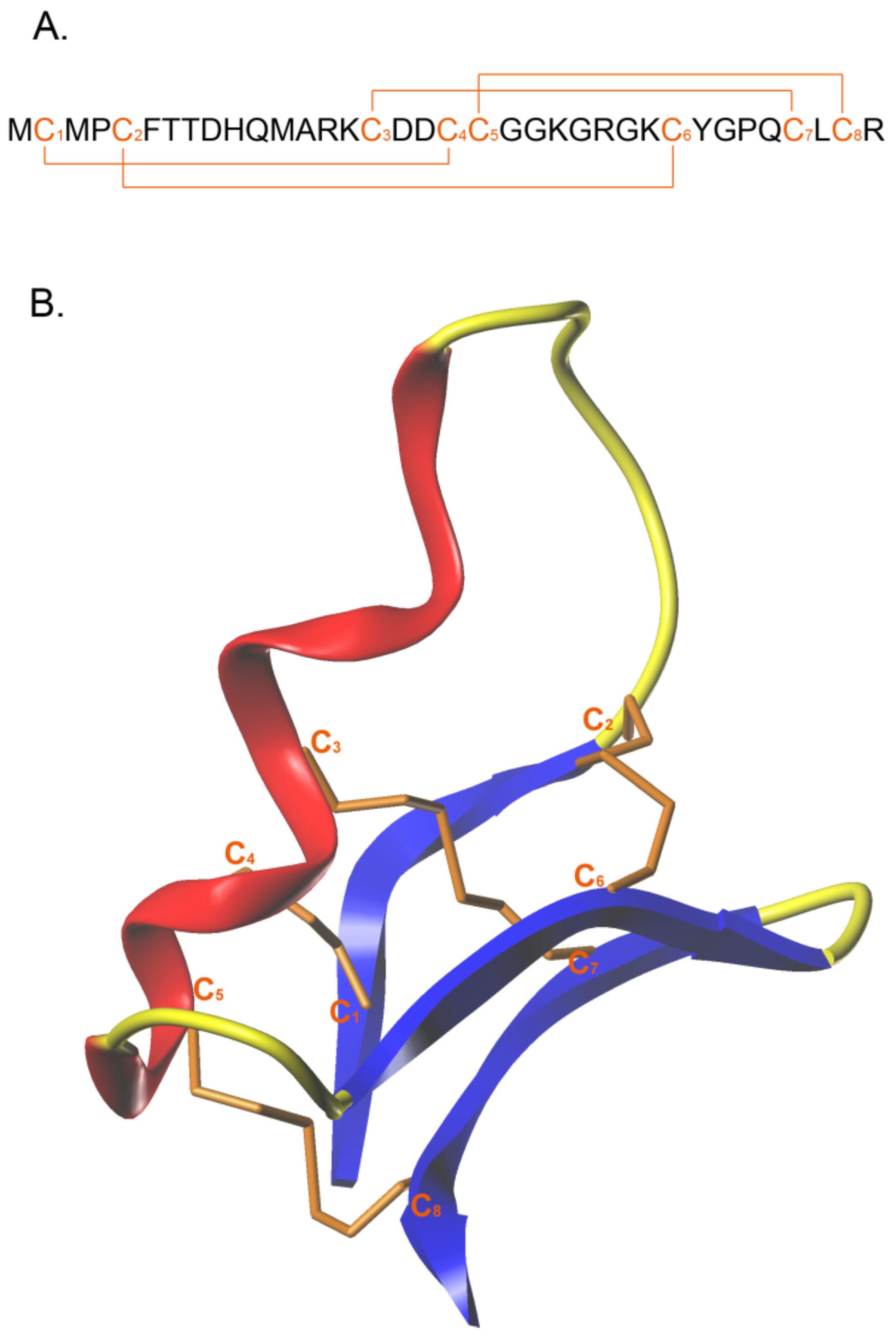
Figure 1. Chlorotoxin’s amino acid sequence (A) and structure (B) with the disulfide bonds presented. Reprinted with permission from Ref. [25], 2015, MDPI.
Pharmaceutics 14 02613 g001
Figure 2. Cytotoxicity analysis of CTX at varying concentrations in A549 (left) and H1975 (right) cells at 24, 48, and 72 h. Different amounts of CTX were dissolved in an equal volume of DMSO, prior to dilution into complete media for each well to the targeted desired concentration, in sextuplicates. An equal volume of DMSO in complete media served as the control. All values are presented as averages with SEM. N/A: not available, i.e., IC50 value is unpredictable. The A549 and H1975 cell lines were obtained from ATCC.
Pharmaceutics 14 02613 g002
Figure 3. Cell doubling time, track speed, instantaneous velocity, sphericity, and perimeter were monitored for approximately 24 h using Livecyte QPI microscopy. A549 (left) and H1975 (right) cells were cultured in 24-well plates, and CTX at varying concentrations was introduced (similar to cytotoxicity analysis above). All samples were in triplicate wells. Cell doubling time data are the average of three wells per treatment group, with standard error of the mean. Colored graphs indicate value distribution based on values from all individually measured cells. The statistical analysis is one-way ANOVA, followed by Tukey’s post-hoc test. All statistical analyses reported are in comparison to the control-DMSO group. *: p < 0.05; ***: p < 0.001.
Pharmaceutics 14 02613 g003
Table 1. Representative active targeting moieties and their respective dissociation constant values. The list includes peptides, antibodies, and aptamers used for active targeting.
Targeting MoietyMolecule TypeTargetKd (nM)Ref.
GE11PeptideEGFR22.28[66]
QRHKPREPeptideEGFR50[67]
RGDPeptideIntegrins ανβ3 and ανβ53.2–100[68]
TrastuzumabAntibodyHER25–9[62]
BevacizumabAntibodyVEGF0.5[68]
NOX-E36AptamerMCP-11.32[69]
NU172AptamerThrombin0.3–0.5[69]
AB3AptamerOFA/iLRP101[69]
AO-01AptamerMatrix-binding domain180[70]
Hyaluronic acidPoly saccharideCD440.3[71]
TransferrinGlycoproteinTransferrin Receptor0.23[72]
Publisher’s Note: MDPI stays neutral with regard to jurisdictional claims in published maps and institutional affiliations.