Article

The Ca2+ Channel Blocker Verapamil Inhibits the In Vitro Activation and Function of T Lymphocytes: A 2022 Reappraisal

by
José Ignacio Veytia-Bucheli
1,2,
Den Alejandro Alvarado-Velázquez
1,3,
Lourival Domingos Possani
1,
Roberto González-Amaro
4 and
Yvonne Rosenstein
1,*
1
Departamento de Medicina Molecular y Bioprocesos, Instituto de Biotecnología, Universidad Nacional Autónoma de México, Av. Universidad 2001, Cuernavaca 62210, Mexico
2
Laboratoire de Chimie Bio-Organique, Département de Chimie, Faculté des Sciences, Université de Namur, Rue de Bruxelles 615, 5000 Namur, Belgium
3
Posgrado en Ciencias, Instituto de Investigación en Ciencias Básicas y Aplicadas, Universidad Autónoma del Estado de Morelos, Av. Universidad 1001, Cuernavaca 62209, Mexico
4
Centro de Investigación en Ciencias de la Salud y Biomedicina, Universidad Autónoma de San Luis Potosí, Av. Sierra Leona 550, San Luis Potosí 78210, Mexico
*
Author to whom correspondence should be addressed.
Pharmaceutics 2022, 14(7), 1478; https://doi.org/10.3390/pharmaceutics14071478
Submission received: 15 May 2022 / Revised: 12 July 2022 / Accepted: 12 July 2022 / Published: 15 July 2022
(This article belongs to the Section Clinical Pharmaceutics)

Abstract

:
Ca2+ channel blockers (CCBs) are commonly used to treat different cardiovascular conditions. These drugs disrupt the intracellular Ca2+ signaling network, inhibiting numerous cellular functions in different cells, including T lymphocytes. We explored the effect of the CCB verapamil on normal human peripheral blood T cell activation, proliferation, and cytokine production. Cells were activated by ligating CD3 or CD3/CD28 in the presence or absence of verapamil, and the expression of activation-induced cell surface molecules (CD25, CD40L, CD69, PD-1, and OX40), cell proliferation, and cytokine release were assessed by flow cytometry. Verapamil exerted a dose-dependent inhibitory effect on the expression of all the activation-induced cell surface molecules tested. In addition, verapamil diminished T cell proliferation induced in response to CD3/CD28 stimulation. Likewise, the production of Th1/Th17 and Th2 cytokines was also reduced by verapamil. Our data substantiate a potent in vitro suppressive effect of verapamil on T lymphocytes, a fact that might be relevant in patients receiving CCBs.

1. Introduction

Ion channels allow the flux of ions across cell membranes and control critical physiological processes such as muscle contraction, nerve impulse, hormonal secretion, gene expression, proliferation, apoptosis, development, and cell migration [1]. Ca2+ ions are universal second messengers that drive these cellular responses by binding to numerous Ca2+-sensitive effector proteins that transduce the information encoded in the spatiotemporal dynamics of Ca2+ signaling.
Alterations in Ca2+ signaling are the cause of numerous diseases [2,3]. Ca2+ channel blockers (CCBs) such as amlodipine, diltiazem, nifedipine, and verapamil are widely used to treat hypertension, cardiac arrhythmias, angina pectoris, and the prophylaxis of migraine headaches. Blocking the voltage-gated L-type (slowly inactivating) Ca2+ channels in vascular smooth muscle results in vasodilation and decreased peripheral vascular resistance. In cardiac muscle cells, the inhibition of L-type Ca2+ channels produces negative chronotropic, dromotropic, and inotropic effects [4,5].
Although CCBs are considered safe and adverse reactions are uncommon, different conditions are associated with CCB therapy, including increased risk for viral infections, hyporeactivity in delayed hypersensitivity skin tests, low response to vaccination, and reduced in vitro lymphocyte proliferation [6,7,8]. Accordingly, CCB therapy has been associated with a diminished rate of graft rejection of solid organ transplants [9,10,11,12,13] as well as an increased risk of cancer in elderly patients [9,14].
The levels of intracellular Ca2+ have a central role in T cell function. The engagement of the T cell receptor (TCR) with its cognate antigen presented by antigen-presenting cells triggers the mobilization of Ca2+ from intracellular stores, followed by a sustained elevation dependent on extracellular Ca2+. Lasting Ca2+ fluxes are necessary to recruit to and retain in the nucleus isoforms of the nuclear factor of activated T cells (NFAT), and other transcription factors to control the clonal expansion, cell survival, differentiation, and production of effector molecules [2].
In humans, Ca2+ release-activated Ca2+ channel protein 1 (ORAI1) is the predominant contributor to the TCR-induced Ca2+ influx. However, additional Ca2+ permeable channels have been proposed to mediate Ca2+ influx in T cells, including certain voltage-gated Ca2+ (Cav) channels, transient receptor potential (TRP) channels, and purinergic ionotropic (P2X) receptors (reviewed in [1,15,16]). Additionally, the K+ voltage-gated channel subfamily A member 3 (Kv1.3) and the intermediate conductance Ca2+-activated K+ channel protein 4 (KCa3.1) regulate the membrane potential through the efflux of K+ cations to the extracellular space and generate the electrochemical potential that drives the entry of Ca2+ through the plasma membrane during T cell activation [17,18]. The blockade of the ion channels involved in the Ca2+ signaling network results in impaired T cell activation and function [2].
As the specificity of CCBs for L-type Ca2+ channels is poor and unrelated channels are often affected by these drugs, this study aimed to further explore, in vitro, the putative immunosuppressive effect of verapamil, a widely used CCB, a phenomenon that might be relevant in vivo.

2. Materials and Methods

2.1. Blood Samples and T Cell Purification

This procedure was approved by the Bioethics Committee of the Instituto de Biotecnología. Buffy coats from anonymized healthy donors were obtained from the Centro Estatal de la Transfusión Sanguínea (Cuernavaca, Mexico). Peripheral blood mononuclear cells (PBMCs) were separated through Ficoll-Paque PLUS (GE Healthcare Bio-Sciences AB, Uppsala, Sweden) density gradient centrifugation. Cells were resuspended in RPMI-1640 medium (HyClone, GE Healthcare Life Sciences, Logan, UT, USA) supplemented with 2% heat-inactivated fetal calf serum (By Productos, Guadalajara, Mexico) and incubated in 100 mm tissue-culture treated polystyrene dishes (8 × 10⁷ cells/dish) at 37 °C in 5% CO2 overnight to promote adhesion of monocytes to the plastic surface and cell arrest. Non-adherent cells were recovered to purify T cells by magnetic cell sorting (negative selection) with the Pan T Cell Isolation Kit (Miltenyi Biotec GmbH, Bergisch Gladbach, Germany). Briefly, non-T cells were labeled with a monoclonal antibody cocktail (biotin-conjugated anti-CD14, -CD15, -CD16, -CD19, -CD34, -CD36, -CD56, -CD123, and -CD235a). Subsequently, the preparation was incubated with anti-biotin secondary antibodies conjugated with magnetic MicroBeads and transferred to a LD Column (Miltenyi Biotec GmbH) placed on a MidiMACS Separator (Miltenyi Biotec GmbH) permanent magnet. T cells were recovered by elution. Purity (CD3+ cells) was always > 95%, as determined by flow cytometry with PE anti-human CD3d antibody (1 μg/mL, clone 7D6, Caltag Laboratories, Burlingame, CA, USA).

2.2. T Cell Stimulation Assays

T cells (1 × 10⁶ cells/mL) in RPMI-1640 medium supplemented with 10% heat-inactivated fetal calf serum were stimulated in 48-well polystyrene cell culture plates (5 × 10⁵ cells/well). T cells were stimulated with anti-human CD3e mAb (clone OKT3, IgG2a, home purified) in the presence or absence of anti-human CD28 mAb (clone CD28.2, IgG1, BioLegend, San Diego, CA, USA). The anti-CD3e antibody was immobilized on the wells’ surface (100 μL/well of a 9.5 μg/mL antibody solution in phosphate-buffered saline (PBS)) by incubating the plates for 2 h at 37 °C. Wells were washed three times with PBS to remove unbound antibody, after which the cells were seeded, and soluble anti-CD28 antibody (2 μg/mL) was added to the cells and cross-linked with goat anti-mouse IgG1 antibodies (2 μg/mL) (SouthernBiotech, Birmingham, AL, USA). Where indicated, verapamil hydrochloride (Kener, Toluca, Mexico) was added at the indicated concentrations five minutes before the onset of stimulation. Plates were incubated at 37 °C in 5% CO2 for the indicated times.

2.3. Cytokine Production Profile

The supernatants from T cells activated as indicated above for 24 or 72 h were collected, and cytokines were measured using the LEGENDplex Human Th Cytokine Panel 13-plex (BioLegend) and flow cytometry. Samples were acquired in a BD FACSCanto II (BD Biosciences, San Jose, CA, USA) flow cytometer with the BD FACSDiva (version 6.1.3, BD Biosciences) software and analyzed with the LEGENDplex Data Analysis Software (BioLegend).

2.4. Flow Cytometry

For cell viability determination, cells were washed with PBS and incubated with 500 μL of a 1:100,000 dilution of the Fixable Viability Dye eFluor 780 (Life Technologies, Carlsbad, CA, USA) in PBS at 4 °C for 30 min in the dark. To evaluate cell proliferation, 2 × 10⁷ cells were labeled with 1 mL of 10 µM CFSE (Life Technologies, Eugene, OR, USA) in RMPI at 37 °C for 15 min in the dark and then washed with RPMI-1640 supplemented with 10% heat-inactivated fetal calf serum. Then, cell proliferation was assessed after 120 h of stimulation. For fluorescent antibody staining, cells were washed with PBS. Fc receptors were blocked with 100 μL of 10% human serum in FACS solution (PBS supplemented with 0.5% bovine serum albumin and 0.1% sodium azide) at 4 °C for 30 min, and cells were stained in a final volume of 150 μL with a master mix containing the following fluorescent antibodies: APC anti-human CD25 (1 μg/mL, clone BC96, BioLegend), PE anti-human CD69 (0.25 μg/mL, clone FN50, BioLegend), FITC anti-human CD40L/CD154 (1 μg/mL, clone 24–31, BioLegend), Pacific Blue anti-human PD-1/CD279 (1.25 μg/mL, clone EH12.2H7, BioLegend), and PerCP/Cy5.5 anti-human OX40/CD134 (2.5 μg/mL, clone Ber-ACT35, BioLegend). Cells were incubated for 30 min at 4 °C in the dark, washed with FACS solution to remove unbound antibodies, fixed with 2% paraformaldehyde in PBS, and stored at 4 °C until analysis. Samples were acquired on a BD FACSCanto II (BD Biosciences) flow cytometer with the BD FACSDiva (BD Biosciences) software and analyzed using the FlowJo (version 8.7, FlowJo, LLC, Ashland, OR, USA) software. The expression level of the different molecules was expressed as mean fluorescence intensity (MFI) or percentage of positive cells.

2.5. Statistical Analysis

GraphPad Prism 8.0.2 (GraphPad Software, San Diego, CA, USA) software was used for data analysis and graph creation. Data are shown as arithmetic mean + standard deviation of the mean (SD). The Shapiro–Wilk test was used to determine the normality of the data. For comparisons between two groups, paired Student’s t-test (parametric data) and Wilcoxon matched-pairs test (non-parametric data) were used. For comparisons among multiple groups, repeated-measures ANOVA followed by Tukey’s Multiple Comparison post hoc test (parametric data) and Friedman test followed by Dunn’s post hoc test (non-parametric data) were used. Statistical significance was set at p < 0.05.

3. Results

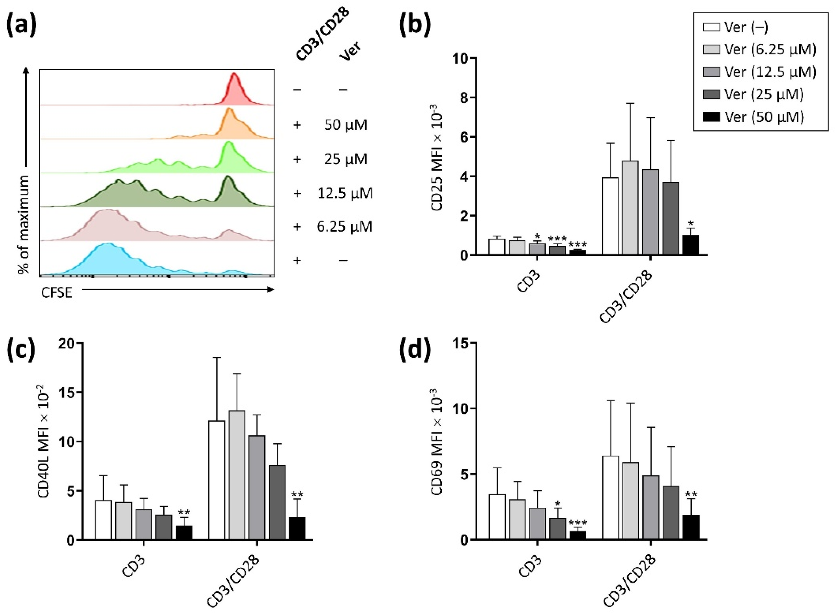
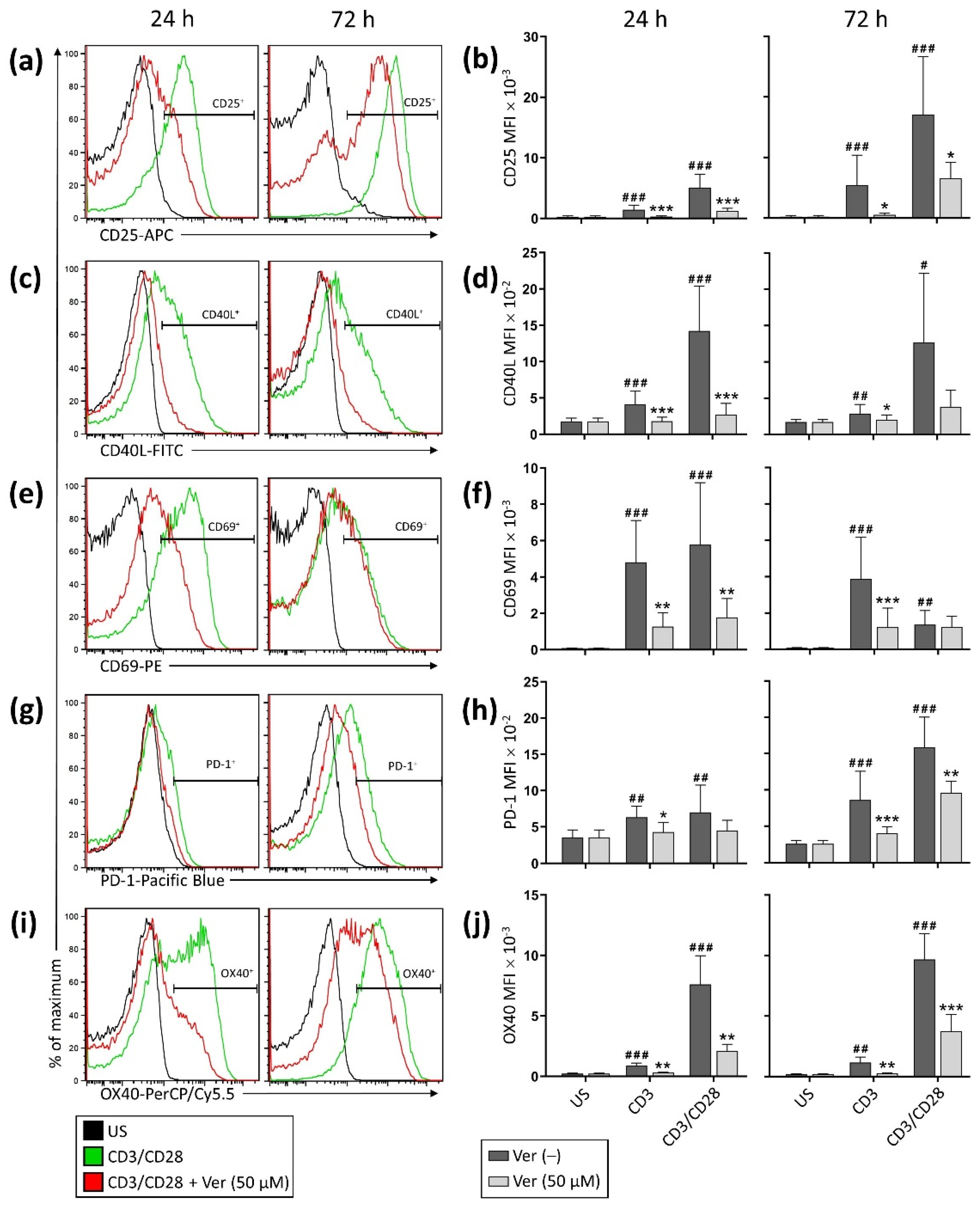
3.1. Verapamil Inhibits T Cell Activation and Proliferation

We first evaluated the possible effect of verapamil on T cell proliferation. As shown in Figure 1a, verapamil exerted a dose-dependent inhibitory effect on the T lymphocyte proliferation in response to CD3 stimulation and CD28 costimulation, with almost complete inhibition at 50 μM. Furthermore, verapamil induced a dose-dependent reduction in the expression level of the activation-induced cell surface molecules CD25, CD40L, and CD69 (Figure 1b–d).
Additional experiments showed that along with inhibiting the expression of CD25, CD40L, and CD69, verapamil inhibited the expression of other activation-induced cell surface molecules, namely PD-1 and OX40 (Figure 2). These results were observed following an incubation period of 24 or 72 h (data shown consider the MFI). Comparable results were observed when the data were expressed as the percentage of lymphocytes expressing the activation-induced cell surface molecules (Figure S1).
Interestingly, when cells were stimulated through CD3/CD28 as compared to only CD3, the inhibitory effect of verapamil was less profound, consistent with CD28 recruiting additional signaling pathways that bypass the TCR-activated Ca2+-dependent events inhibited by verapamil, partially rescuing T cell function [19].
Cell viability was not affected in the presence of verapamil at the end of the 24 h activation period (Figure S2). However, after 72 h verapamil significantly decreased cell viability, particularly in unstimulated cells, reflecting the importance of the Ca2+ signaling network in the survival of quiescent and activated T cells.

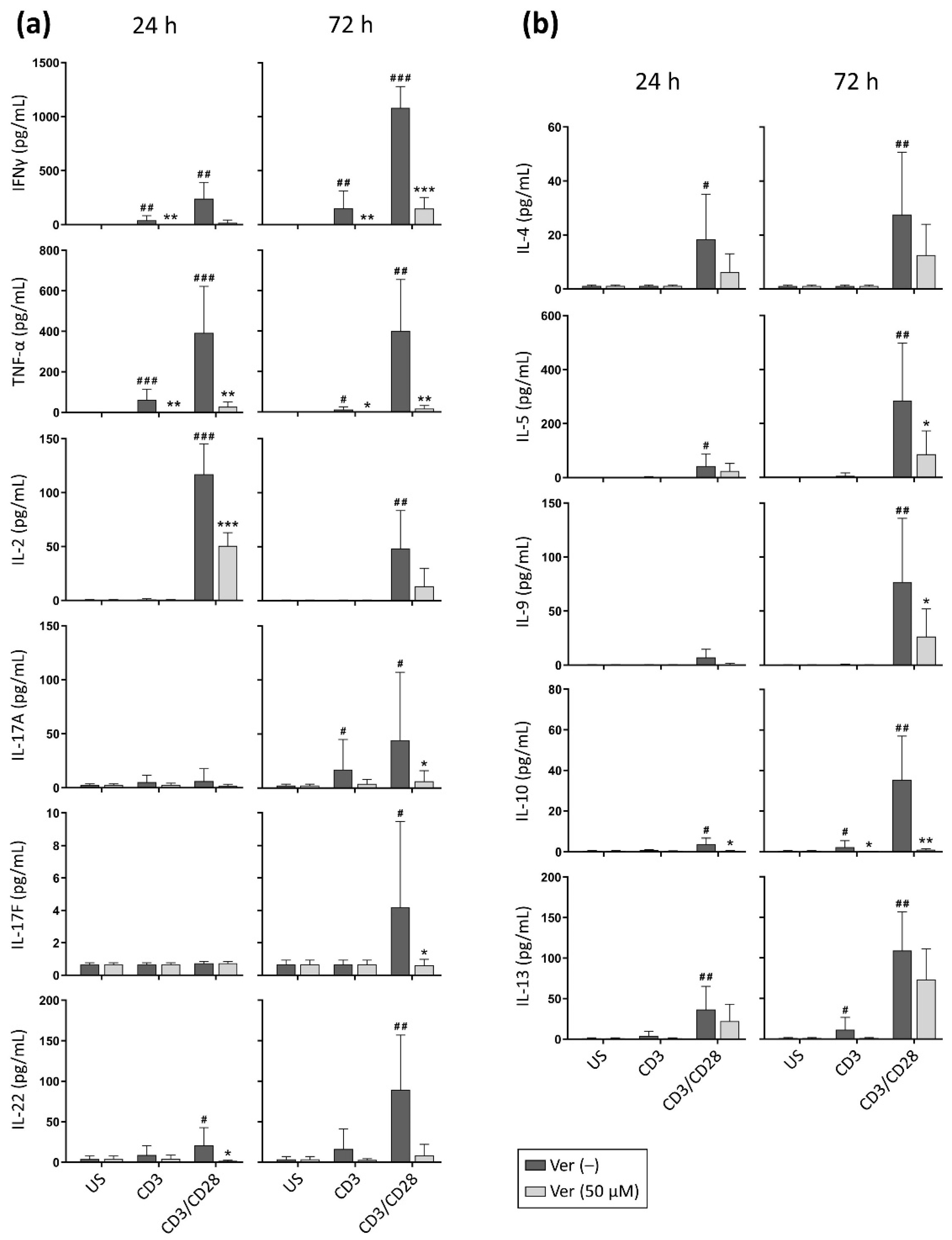
3.2. Verapamil Impairs the Production of Cytokines

T lymphocytes produce a variety of cytokines that play a central role in the final configuration of an immune response [20]. Since the intracellular Ca2+ concentration has a central role in the recruitment of several transcription factors (e.g., NFAT isoforms), promoting the expression of different cytokine genes, we evaluated whether verapamil altered cytokine production following T cell stimulation. As expected, T lymphocytes stimulated through CD3 produced small amounts of the cytokines tested (Figure 3); however, a significant decrease in IFNγ, TNF-α, and IL-10 in the media could be detected when verapamil was present.
When engaging CD3 and CD28, the cells released considerable quantities of the cytokines of the multiplex detection kit, except IL-6 or IL-21, which were not detected under our experimental conditions. In the presence of verapamil, a noticeable and significant decrease in IFNγ, TNF-α, IL-2, -5, -9, -10, -17A, -17F, and -22 concentration was detected in the media (Figure 3). IL-4 and -13 were also reduced in the presence of verapamil, although this was not statistically significant due to interindividual variation.
As T cell viability is not compromised after 24 h of culture in the presence of verapamil (Figure S2), and T cell division begins 40–50 h after stimulation [21], the mere inhibitory effect of verapamil on T cell activation could explain the decrease in cytokines in the media at the 24 h time-point. However, the reduction detected at the 72 h time-point reflects the combination of the inhibitory effect of verapamil on T cell activation, the reduced proliferation (Figure 1), and reduced cell viability (Figure S2). Our experiments do not allow us to elucidate whether the inhibitory effect of verapamil on cytokine production could be due, at least in part, to the blockade of lymphocyte proliferation. However, consistent with our results, a study evaluating the effects of verapamil analogues on the growth of Mycobacterium tuberculosis [22] reports that concentrations of verapamil as low as 6 μM inhibited the expansion of M. tuberculosis-specific T cells as well as IFNγ production by proliferating cells.
Overall, in vitro data (Figure 3) indicate that verapamil impairs the production of Th1/Th17 (IFNγ, TNF-α, IL-2, -17A, -17F, and -22) and Th2 (IL-4, -5, -9, -10, and -13) cytokines by stimulated T lymphocytes. Similar to the effect on the expression of activation-induced cell surface molecules, verapamil impaired cytokine production to a lesser extent when cells were costimulated, as the CD3/CD28 stimulus was more refractory to the inhibitory effects of verapamil, partially rescuing T cell function.

4. Discussion

Ca2+ signaling is tightly controlled during human T cell activation. While ORAI1 channels are the main Ca2+ entry pathway for a sustained increase in cytoplasmic Ca2+ concentrations, other ion channels such as Kv1.3, KCa3.1, Cav, TRP channels, and P2X receptors have been proposed to modulate Ca2+ influx in T cells as well [1,15,16]. Ca2+ binds to ubiquitous Ca2+ sensors, including calmodulin (CaM), regulating protein kinases/phosphatases that activate transcription factors such as NFAT, cAMP-responsive element-binding protein (CREB), myocyte-specific enhancer factor 2 (MEF2), and nuclear factor-κB (NF-κB) isoforms. These transcription factors modulate the transcription of genes involved in cell proliferation, differentiation, cytokine production, survival, and cell death, thus placing the control of Ca2+ fluxes at the center of the T cell decision-making process [16,23].
Verapamil is a CCB commonly used to treat cardiovascular disorders. By inhibiting Ca2+ influx through L-type Ca2+ channels, CCBs promote cardiac and vascular smooth muscle cell relaxation [4,5]. However, these drugs are promiscuous ion channel blockers and have pleiotropic effects, as their targets are widely distributed [24]. As T cells are increasingly used for immunotherapy, one of the current challenges is understanding the role of the different molecules that participate in T cell activation and differentiation. The main goal of this investigation was to expand the available information regarding the immunosuppressive effect of verapamil on human T lymphocytes.
T cell activation requires the coordinated signals from the TCR and co-receptor molecules such as CD28 to transit to an effector cell phenotype. As the expression of CD25, CD40L, CD69, PD-1, and OX40 is Ca2+-dependent [25,26], we evaluated the effect of verapamil on the expression of these activation-induced cell surface molecules in CD3/CD28-stimulated T cells. These molecules regulate essential processes during the T cell response, such as the expression of co-stimulatory molecules, cytokine secretion, antibody isotype switching, the acquisition of an effector/memory phenotype, the regulation of the T cell migration pattern, cell survival, proliferation, promotion of tolerance, and return to immune homeostasis [27,28,29,30,31].
Under our experimental conditions, verapamil profoundly affected T cell activation. We showed that the addition of verapamil to purified human peripheral blood T lymphocytes at the onset of T cell activation resulted in a pronounced inhibition of CD25, CD40L, CD69, PD-1, and OX40 expression, likely resulting from the lack of activation of Ca2+-dependent transcription factors to stimulate gene transcription [25,32]. The recruitment and prolonged presence of Ca2+-dependent transcription factors in the nucleus is also indispensable for the transcription of cytokine genes [25,26], prompting us to evaluate the effect of verapamil on cytokine production. Accordingly, and consistent with previous reports [33,34,35,36,37,38,39,40,41,42,43], T cells also produced fewer Th1/Th17 and Th2 cytokines when in the presence of verapamil.
Interestingly, the verapamil-induced inhibition was more pronounced when only the TCR was engaged, as the simultaneous ligation of CD28 and the TCR partially bypassed this inhibition, likely due to the activation of alternative signaling pathways that do not entirely rely on sustained Ca2+ fluxes.
These results are consistent with previous descriptions of the immunomodulatory effect of verapamil on human T cells, whereby verapamil was reported to inhibit Ca2+ entry, ATP production, CD25 expression, the accumulation of inositol phosphates, the cell cycle progression, and the uptake of precursor molecules for the synthesis of proteins, RNA and DNA. Verapamil was also shown to impair the generation and function of cytotoxic T cells and the remodeling of T cell cytoskeleton, chemotaxis, motility, and transmigration [33,34,36,37,38,39,40,41,44,45,46,47,48,49,50,51,52,53,54,55,56]. Together with these reports, our data suggest that verapamil likely exerts a downregulatory effect on the different T cell subsets and could act as a broad-spectrum immunosuppressive molecule.
It is worth mentioning that the data we report herein were obtained within verapamil concentrations of ~10–50 µM. While the ORAI1 channels are resistant to verapamil [24], this range tallies with the concentrations required to inhibit other ion channels that modulate Ca2+ influx in human T cells such as KCa3.1 (IC50 = 28 μM [17]), Kv1.3 (IC50 = 8 μM [57]), Cav (IC50 ~10 μM for L-type [58], and ~20 μM for T-type channels [59,60]), as well as the P-glycoprotein (≥2 μM [42]). Human T cells also express significant levels of P-glycoprotein, an ATP-dependent transporter that participates in the non-specific transmembrane transport of metabolites, xenobiotics, and endogenous peptides/proteins. Inhibiting P-glycoprotein activity by monoclonal antibodies or small molecules such as verapamil results in a profound suppression of cytokine release from activated PBMCs [42], adding a deficient transmembrane cytokine transport to the verapamil’s overall T cell immunomodulatory effect. The possibility that these concentrations of verapamil affect additional molecular targets should not be excluded.
As a lipophilic molecule, verapamil is widely distributed from plasma to body tissues. In a rat animal model, following a single intraperitoneal injection of verapamil (30 mg/kg), peak plasma concentrations were around 1.5 μM, while 15 to 85 times higher amounts were found in organs such as lungs, liver, kidneys, and heart [61]. In humans, wide variations in the disposition of verapamil and drug accumulation can occur between individuals during a multiple-dose oral regime. Considering that with standard doses, peak plasma concentrations of 0.1–2.0 μM are reached [62], concentrations within the immunosuppressive range could be expected in tissues undergoing an immune response.
Verapamil is marketed as a racemic mixture of equal proportions of the S(–)- and R(+)-verapamil enantiomers. Although the S(–) enantiomer is more potent at inhibiting Ca2+ uptake in human mitogen-stimulated lymphocytes, both enantiomers have been reported to be equally potent suppressors of T cell function [39], suggesting that verapamil’s immunosuppressive effects may be, at least partially, independent of the inhibition of transmembrane Ca2+ fluxes.
Although obtained in vitro, with total T cells, our results reinforce previous data suggesting that verapamil would be expected to act on different T cell subsets, disabling T cell responses. Furthermore, verapamil and other CCBs are known to compromise other cells important for the generation of an immune response such as monocytes/macrophages [63], neutrophils [63,64,65,66], NK cells [33,39,46,67,68], B cells [69,70,71], and endothelial cells [53,54,72]. Overall, our data underpin a potent suppressive effect of verapamil on T lymphocytes, a phenomenon that might be relevant for patients receiving CCBs. In some cases (e.g., patients requiring immunosuppressive therapy), this phenomenon might be considered a desirable effect. In contrast, in others (e.g., patients with arterial hypertension without other diseases), this effect should be avoided or, at least, taken into account.

Supplementary Materials

The following supporting information can be downloaded at: https://www.mdpi.com/article/10.3390/pharmaceutics14071478/s1. Figure S1: Verapamil decreases the percentage of cells expressing the activation-induced cell surface molecules; Figure S2: Verapamil compromises T cell viability during long culture periods.

Author Contributions

Conceptualization, J.I.V.-B., R.G.-A. and Y.R.; methodology, J.I.V.-B. and D.A.A.-V.; software, not applicable; validation, J.I.V.-B. and D.A.A.-V.; formal analysis, J.I.V.-B.; investigation, J.I.V.-B. and D.A.A.-V.; resources, Y.R., R.G.-A., and L.D.P.; data curation, not applicable; writing—original draft preparation, J.I.V.-B. and Y.R.; writing—review and editing, Y.R., L.D.P. and R.G.-A.; visualization, J.I.V.-B., R.G.-A. and Y.R.; supervision, Y.R., R.G.-A. and L.D.P.; project administration, Y.R. and R.G.-A.; funding acquisition, Y.R. All authors have read and agreed to the published version of the manuscript.

Funding

This research was partially funded by CONACYT (grant number A1-S-15601), and DGAPA/UNAM (grant number IN212519) to Y.R. The APC was funded by Y.R., R.G.-A. and L.D.P.

Institutional Review Board Statement

The study was conducted in accordance with the Declaration of Helsinki and approved by the Bioethics Committee of the Instituto de Biotecnología (protocol code 220990, 01/15/2014).

Informed Consent Statement

Informed consent was waived because buffy coats from healthy donors were anonymous, with no possibility of linking them to the person from whom they were collected.

Data Availability Statement

The data presented in this study are available within this article.

Acknowledgments

The authors are indebted to Ángel Cárdenas-Hernández for the generous gift of the verapamil hydrochloride used in this work. The authors thank Erika Melchy for technical support, and the members of the Rosenstein and Possani laboratories for their valuable comments on this work. J.I.V.-B. and D.A.A.-V. were recipients of a graduate student fellowship from CONACYT (fellowship holder numbers 289448 and 894574).

Conflicts of Interest

The authors declare no conflict of interest. The funders had no role in the design of the study; in the collection, analyses, or interpretation of data; in the writing of the manuscript, or in the decision to publish the results.

References

  1. Feske, S.; Wulff, H.; Skolnik, E.Y. Ion Channels in Innate and Adaptive Immunity. Annu. Rev. Immunol. 2015, 33, 291–353. [Google Scholar] [CrossRef] [Green Version]
  2. Feske, S. Calcium Signalling in Lymphocyte Activation and Disease. Nat. Rev. Immunol. 2007, 7, 690–702. [Google Scholar] [CrossRef]
  3. Brini, M.; Calì, T.; Ottolini, D.; Carafoli, E. Neuronal Calcium Signaling: Function and Dysfunction. Cell. Mol. Life Sci. 2014, 71, 2787–2814. [Google Scholar] [CrossRef]
  4. Elliott, W.J.; Ram, C.V.S. Calcium Channel Blockers. J. Clin. Hypertens. 2011, 13, 687–689. [Google Scholar] [CrossRef]
  5. Abernethy, D.R.; Schwartz, J.B. Calcium-Antagonist Drugs. N. Engl. J. Med. 1999, 341, 1447–1457. [Google Scholar] [CrossRef]
  6. Derenne, F.; Vanhaeverbeek, M.; Brohée, D. Nifedipine-Induced Hyporeactivity in Delayed Hypersensitivity Skin Tests. Int. J. Immunopharmacol. 1987, 9, 741–744. [Google Scholar] [CrossRef]
  7. Tietz, N.W.; Thompson, J. Possible Concentration-Dependent Suppression of Immune Response by Verapamil. Arch. Fam. Med. 1995, 4, 368–369. [Google Scholar] [CrossRef]
  8. McFadden, J.; Bacon, K.; Camp, R. Topically Applied Verapamil Hydrochloride Inhibits Tuberculin-Induced Delayed-Type Hypersensitivity Reactions in Human Skin. J. Investig. Dermatol. 1992, 99, 784–786. [Google Scholar] [CrossRef] [Green Version]
  9. Moslen, M.T.; Balakumaran, A. Calcium Channel Blockers and Cancer: Due to Loss of Immune Surveillance? Am. J. Hypertens. 1996, 9, 1050–1051. [Google Scholar] [CrossRef] [Green Version]
  10. Suthanthiran, M.; Haschemeyer, R.H.; Riggio, R.R.; Adubor, C.; Friedman, G.S.; Cheigh, J.S.; Wang, J.C.; Fotino, M.; Stubenbord, W.T.; Saal, S.D. Excellent Outcome with a Calcium Channel Blocker-Supplemented Immunosuppressive Regimen in Cadaveric Renal Transplantation. A Potential Strategy to Avoid Antibody Induction Protocols. Transplantation 1993, 55, 1008–1013. [Google Scholar] [CrossRef]
  11. Dawidson, I.; Rooth, P.; Lu, C.; Sagalowsky, A.; Diller, K.; Palmer, B.; Peters, P.; Risser, R.; Sandor, Z.; Seney, F. Verapamil Improves the Outcome after Cadaver Renal Transplantation. J. Am. Soc. Nephrol. 1991, 2, 983–990. [Google Scholar] [CrossRef] [PubMed]
  12. Chitwood, K.K.; Heim-Duthoy, K.L. Immunosuppressive Properties of Calcium Channel Blockers. Pharmacotherapy 1993, 13, 447–454. [Google Scholar] [CrossRef] [PubMed]
  13. Palmer, B.F.; Dawidson, I.; Sagalowsky, A.; Sandor, Z.; Lu, C.Y. Improved Outcome of Cadaveric Renal Transplantation Due to Calcium Channel Blockers. Transplantation 1991, 52, 640–645. [Google Scholar] [CrossRef] [PubMed]
  14. Pahor, M.; Guralnik, J.M.; Salive, M.E.; Corti, M.C.; Carbonin, P.; Havlik, R.J. Do Calcium Channel Blockers Increase the Risk of Cancer? Am. J. Hypertens. 1996, 9, 695–699. [Google Scholar] [CrossRef] [Green Version]
  15. Fenninger, F.; Jefferies, W.A. What’s Bred in the Bone: Calcium Channels in Lymphocytes. J. Immunol. 2019, 202, 1021–1030. [Google Scholar] [CrossRef] [Green Version]
  16. Trebak, M.; Kinet, J.-P. Calcium Signalling in T Cells. Nat. Rev. Immunol. 2019, 19, 154–169. [Google Scholar] [CrossRef]
  17. Chandy, K.G.; Wulff, H.; Beeton, C.; Pennington, M.; Gutman, G.A.; Cahalan, M.D. K+ Channels as Targets for Specific Immunomodulation. Trends Pharmacol. Sci. 2004, 25, 280–289. [Google Scholar] [CrossRef] [Green Version]
  18. Beeton, C.; Barbaria, J.; Giraud, P.; Devaux, J.; Benoliel, A.-M.; Gola, M.; Sabatier, J.M.; Bernard, D.; Crest, M.; Beraud, E. Selective Blocking of Voltage-Gated K+ Channels Improves Experimental Autoimmune Encephalomyelitis and Inhibits T Cell Activation. J. Immunol. 2001, 166, 936–944. [Google Scholar] [CrossRef] [Green Version]
  19. Riha, P.; Rudd, C.E. CD28 Co-Signaling in the Adaptive Immune Response. Self Nonself 2010, 1, 231–240. [Google Scholar] [CrossRef] [Green Version]
  20. Cantrell, D. Signaling in Lymphocyte Activation. Cold Spring Harb. Perspect. Biol. 2015, 7, a018788. [Google Scholar] [CrossRef] [Green Version]
  21. Li, Y.; Kurlander, R.J. Comparison of Anti-CD3 and Anti-CD28-Coated Beads with Soluble Anti-CD3 for Expanding Human T Cells: Differing Impact on CD8 T Cell Phenotype and Responsiveness to Restimulation. J. Transl. Med. 2010, 8, 104. [Google Scholar] [CrossRef] [PubMed] [Green Version]
  22. Abate, G.; Ruminiski, P.G.; Kumar, M.; Singh, K.; Hamzabegovic, F.; Hoft, D.F.; Eickhoff, C.S.; Selimovic, A.; Campbell, M.; Chibale, K. New Verapamil Analogs Inhibit Intracellular Mycobacteria without Affecting the Functions of Mycobacterium-Specific T Cells. Antimicrob. Agents Chemother. 2015, 60, 1216–1225. [Google Scholar] [CrossRef] [PubMed] [Green Version]
  23. Srikanth, S.; Gwack, Y. Orai1-NFAT Signalling Pathway Triggered by T Cell Receptor Stimulation. Mol. Cells 2013, 35, 182–194. [Google Scholar] [CrossRef] [Green Version]
  24. Luik, R.M.; Lewis, R.S. New Insights into the Molecular Mechanisms of Store-Operated Ca2+ Signaling in T Cells. Trends Mol. Med. 2007, 13, 103–107. [Google Scholar] [CrossRef]
  25. Vaeth, M.; Kahlfuss, S.; Feske, S. CRAC Channels and Calcium Signaling in T Cell-Mediated Immunity. Trends Immunol. 2020, 41, 878–901. [Google Scholar] [CrossRef]
  26. Jash, A.; Sahoo, A.; Kim, G.-C.; Chae, C.-S.; Hwang, J.-S.; Kim, J.-E.; Im, S.-H. Nuclear Factor of Activated T Cells 1 (NFAT1)-Induced Permissive Chromatin Modification Facilitates Nuclear Factor-ΚB (NF-ΚB)-Mediated Interleukin-9 (IL-9) Transactivation. J. Biol. Chem. 2012, 287, 15445–15457. [Google Scholar] [CrossRef] [PubMed] [Green Version]
  27. Karnell, J.L.; Rieder, S.A.; Ettinger, R.; Kolbeck, R. Targeting the CD40-CD40L Pathway in Autoimmune Diseases: Humoral Immunity and Beyond. Adv. Drug Deliv. Rev. 2019, 141, 92–103. [Google Scholar] [CrossRef] [PubMed]
  28. Cibrián, D.; Sánchez-Madrid, F. CD69: From Activation Marker to Metabolic Gatekeeper. Eur. J. Immunol. 2017, 47, 946–953. [Google Scholar] [CrossRef]
  29. Létourneau, S.; Krieg, C.; Pantaleo, G.; Boyman, O. IL-2- and CD25-Dependent Immunoregulatory Mechanisms in the Homeostasis of T-Cell Subsets. J. Allergy Clin. Immunol. 2009, 123, 758–762. [Google Scholar] [CrossRef]
  30. Webb, G.J.; Hirschfield, G.M.; Lane, P.J.L. OX40, OX40L and Autoimmunity: A Comprehensive Review. Clin. Rev. Allergy Immunol. 2016, 50, 312–332. [Google Scholar] [CrossRef]
  31. Sharpe, A.H.; Pauken, K.E. The Diverse Functions of the PD1 Inhibitory Pathway. Nat. Rev. Immunol. 2018, 18, 153–167. [Google Scholar] [CrossRef] [PubMed]
  32. González-Amaro, R.; Cortés, J.R.; Sánchez-Madrid, F.; Martín, P. Is CD69 an Effective Brake to Control Inflammatory Diseases? Trends Mol. Med. 2013, 19, 625–632. [Google Scholar] [CrossRef] [PubMed] [Green Version]
  33. Weir, M.R.; Peppler, R.; Gomolka, D.; Handwerger, B.S. Additive Inhibition of Afferent and Efferent Immunological Responses of Human Peripheral Blood Mononuclear Cells by Verapamil and Cyclosporine. Transplantation 1991, 51, 851–857. [Google Scholar] [CrossRef] [PubMed]
  34. Walz, G.; Zanker, B.; Wieder, K.; Hadro, E.; Moscovitch-Lopatin, M.; Smith, B.R.; Strom, T.B. Similar Effects of Cyclosporine and Verapamil on Lymphokine, Interleukin 2 Receptor, and Proto-Oncogene Expression. Transplantation 1989, 47, 331–334. [Google Scholar] [CrossRef]
  35. Matsumori, A.; Nishio, R.; Nose, Y. Calcium Channel Blockers Differentially Modulate Cytokine Production by Peripheral Blood Mononuclear Cells. Circ. J. 2010, 74, 567–571. [Google Scholar] [CrossRef] [Green Version]
  36. Weir, M.R. Therapeutic Benefits of Calcium Channel Blockers in Cyclosporine-Treated Organ Transplant Recipients: Blood Pressure Control and Immunosuppression. Am. J. Med. 1991, 90, 32S–36S. [Google Scholar] [CrossRef]
  37. Bruserud, O. In Vitro Effects of R-Verapamil on the Cytokine Environment and T-Lymphocyte Proliferation When Human T-Lymphocyte Activation Takes Place in the Presence of Acute Myelogenous Leukemia Blasts. Cancer Chemother. Pharmacol. 1996, 39, 71–78. [Google Scholar] [CrossRef]
  38. Walz, G.; Zanker, B.; Barth, C.; Wieder, K.J.; Clark, S.C.; Strom, T.B. Transcriptional Modulation of Human IL-6 Gene Expression by Verapamil. J. Immunol. 1990, 144, 4242–4248. [Google Scholar]
  39. Weir, M.R.; Peppler, R.; Gomolka, D.; Handwerger, B.S. Evidence That the Antiproliferative Effect of Verapamil on Afferent and Efferent Immune Responses Is Independent of Calcium Channel Inhibition. Transplantation 1992, 54, 681–685. [Google Scholar] [CrossRef]
  40. Zanker, B.; Marx, S.; Strom, T.B.; Köhler, H. The Immunosuppressive Effects of Verapamil upon Mitogen Activated and Allo-Antigen Inducible Human Cytotoxic T-Lymphocytes. Int. J. Immunopharmacol. 1994, 16, 507–517. [Google Scholar] [CrossRef]
  41. Bruserud, O. Effect of Verapamil on T-Lymphocyte Activation in Vitro. Scand. J. Immunol. 1985, 21, 73–79. [Google Scholar] [CrossRef] [PubMed]
  42. Drach, J.; Gsur, A.; Hamilton, G.; Zhao, S.; Angerler, J.; Fiegl, M.; Zojer, N.; Raderer, M.; Haberl, I.; Andreeff, M.; et al. Involvement of P-Glycoprotein in the Transmembrane Transport of Interleukin-2 (IL-2), IL-4, and Interferon-Gamma in Normal Human T Lymphocytes. Blood 1996, 88, 1747–1754. [Google Scholar] [CrossRef] [PubMed] [Green Version]
  43. Kooijman, R.; Devos, S.; Hooghe-Peters, E. Inhibition of in Vitro Cytokine Production by Human Peripheral Blood Mononuclear Cells Treated with Xenobiotics: Implications for the Prediction of General Toxicity and Immunotoxicity. Toxicol. Vitr. 2010, 24, 1782–1789. [Google Scholar] [CrossRef] [PubMed]
  44. Kennes, B.; Hubert, C.; Brohee, D.; Neve, P. Early Biochemical Events Associated with Lymphocyte Activation in Ageing. I. Evidence That Ca2+ Dependent Processes Induced by PHA Are Impaired. Immunology 1981, 42, 119–126. [Google Scholar]
  45. Bruserud, O.; Hamann, W.; Patel, S.; Pawelec, G. CD4+ TCR Alpha Beta+ T-Cell Clones Derived Shortly after Allogeneic Bone Marrow Transplantation: Theophyllamine and Verapamil Inhibit Proliferation of Functionally Heterogeneous T-Cells. Int. J. Immunopharmacol. 1992, 14, 783–789. [Google Scholar] [CrossRef]
  46. Witkowski, J.M.; Siebert, J.; Lukaszuk, K.; Trawicka, L. Comparison of Effect of a Panel of Membrane Channel Blockers on the Proliferative, Cytotoxic and Cytoadherence Abilities of Human Peripheral Blood Lymphocytes. Immunopharmacology 1993, 26, 53–63. [Google Scholar] [CrossRef]
  47. Marx, M.; Weber, M.; Merkel, F.; Meyer zum Büschenfelde, K.H.; Köhler, H. Additive Effects of Calcium Antagonists on Cyclosporin A-Induced Inhibition of T-Cell Proliferation. Nephrol. Dial. Transplant. 1990, 5, 1038–1044. [Google Scholar] [CrossRef]
  48. Nakabayashi, H.; Komada, H.; Yoshida, T.; Takanari, H.; Izutsu, K. Lymphocyte Calmodulin and Its Participation in the Stimulation of T Lymphocytes by Mitogenic Lectins. Biol. Cell 1992, 75, 55–59. [Google Scholar] [CrossRef]
  49. Birx, D.L.; Berger, M.; Fleisher, T.A. The Interference of T Cell Activation by Calcium Channel Blocking Agents. J. Immunol. 1984, 133, 2904–2909. [Google Scholar]
  50. Weir, M.R.; Peppler, R.; Gomolka, D.; Handwerger, B.S. Calcium Channel Blockers Inhibit Cellular Uptake of Thymidine, Uridine and Leucine: The Incorporation of These Molecules into DNA, RNA and Protein in the Presence of Calcium Channel Blockers Is Not a Valid Measure of Lymphocyte Activation. Immunopharmacology 1993, 25, 75–82. [Google Scholar] [CrossRef]
  51. Maisel, A.S.; Murray, D.; Polizzi, S.; Motulsky, H.J.; Brodde, O.E.; van Tits, L.J.; Michel, M.C. Does Verapamil Act as an Immunomodulatory Drug in Vivo? Immunopharmacology 1991, 22, 85–91. [Google Scholar] [CrossRef]
  52. Bacon, K.B.; Westwick, J.; Camp, R.D.R. Potent and Specific Inhibition of IL-8-, IL-1α- and IL-1β-Induced in Vitro Human Lymphocyte Migration by Calcium Channel Antagonists. Biochem. Biophys. Res. Commun. 1989, 165, 349–354. [Google Scholar] [CrossRef]
  53. Blaheta, R.A.; Hailer, N.P.; Brude, N.; Wittig, B.; Leckel, K.; Oppermann, E.; Bachmann, M.; Harder, S.; Cinatl, J.; Scholz, M.; et al. In Vitro Analysis of Verapamil-Induced Immunosuppression: Potent Inhibition of T Cell Motility and Lymphocytic Transmigration through Allogeneic Endothelial Cells. Transplantation 2000, 69, 588–597. [Google Scholar] [CrossRef] [PubMed]
  54. Yamaguchi, M.; Suwa, H.; Miyasaka, M.; Kumada, K. Selective Inhibition of Vascular Cell Adhesion Molecule-1 Expression by Verapamil in Human Vascular Endothelial Cells. Transplantation 1997, 63, 759–764. [Google Scholar] [CrossRef] [PubMed]
  55. Chandy, K.G.; DeCoursey, T.E.; Cahalan, M.D.; McLaughlin, C.; Gupta, S. Voltage-Gated Potassium Channels Are Required for Human T Lymphocyte Activation. J. Exp. Med. 1984, 160, 369–385. [Google Scholar] [CrossRef] [Green Version]
  56. DeCoursey, T.E.; Chandy, K.G.; Gupta, S.; Cahalan, M.D. Voltage-Dependent Ion Channels in T-Lymphocytes. J. Neuroimmunol. 1985, 10, 71–95. [Google Scholar] [CrossRef]
  57. Rauer, H.; Grissmer, S. Evidence for an Internal Phenylalkylamine Action on the Voltage-Gated Potassium Channel Kv1.3. Mol. Pharmacol. 1996, 50, 1625–1634. [Google Scholar]
  58. Johnson, B.D.; Hockerman, G.H.; Scheuer, T.; Catterall, W.A. Distinct Effects of Mutations in Transmembrane Segment IVS6 on Block of L-Type Calcium Channels by Structurally Similar Phenylalkylamines. Mol. Pharmacol. 1996, 50, 1388–1400. [Google Scholar]
  59. Freeze, B.S.; McNulty, M.M.; Hanck, D.A. State-Dependent Verapamil Block of the Cloned Human Ca(v)3.1 T-Type Ca(2+) Channel. Mol. Pharmacol. 2006, 70, 718–726. [Google Scholar] [CrossRef] [Green Version]
  60. Bergson, P.; Lipkind, G.; Lee, S.P.; Duban, M.-E.; Hanck, D.A. Verapamil Block of T-Type Calcium Channels. Mol. Pharmacol. 2011, 79, 411–419. [Google Scholar] [CrossRef]
  61. Hamann, S.R.; Todd, G.D.; McAllister, R.G. The Pharmacology of Verapamil. V. Tissue Distribution of Verapamil and Norverapamil in Rat and Dog. Pharmacology 1983, 27, 1–8. [Google Scholar] [CrossRef] [PubMed]
  62. Hamann, S.R.; Blouin, R.A.; McAllister, R.G. Clinical Pharmacokinetics of Verapamil. Clin. Pharmacokinet. 1984, 9, 26–41. [Google Scholar] [CrossRef] [PubMed]
  63. Levy, R.; Dana, R.; Gold, B.; Alkan, M.; Schlaeffer, F. Influence of Calcium Channel Blockers on Polymorphonuclear and Monocyte Bactericidal and Fungicidal Activity. Isr. J. Med. Sci. 1991, 27, 301–306. [Google Scholar]
  64. Kazanjian, P.H.; Pennington, J.E. Influence of Drugs That Block Calcium Channels on the Microbicidal Function of Human Neutrophils. J. Infect. Dis. 1985, 151, 15–22. [Google Scholar] [CrossRef] [PubMed]
  65. Marino, F.; Cattaneo, S.; Cosentino, M.; Rasini, E.; Di Grazia, L.; Fietta, A.M.; Lecchini, S.; Frigo, G. Diazepam Stimulates Migration and Phagocytosis of Human Neutrophils: Possible Contribution of Peripheral-Type Benzodiazepine Receptors and Intracellular Calcium. Pharmacology 2001, 63, 42–49. [Google Scholar] [CrossRef] [PubMed]
  66. Ishizaki, T.; Yano, E.; Evans, P.H. Cellular Mechanisms of Reactive Oxygen Metabolite Generation from Human Polymorphonuclear Leukocytes Induced by Crocidolite Asbestos. Environ. Res. 1997, 75, 135–140. [Google Scholar] [CrossRef] [PubMed]
  67. Huwyler, T.; Hirt, A.; Felix, D.; Morell, A. Effect of Cations and Cation Channel Blockers on Human Natural Killer Cells. Int. J. Immunopharmacol. 1985, 7, 573–576. [Google Scholar] [CrossRef]
  68. Markham, P.N.; Ellis, T.M.; Tambur, A.R.; Gebel, H.M. Differential Sensitivity of Resting and IL-2 Activated NK Cells to R-Verapamil. Transplantation 1996, 62, 1883–1888. [Google Scholar] [CrossRef]
  69. Degiannis, D.; Hornung, N.; Luke-Gustites, D.; Raskova, J.; Raska, K. IL-4 Receptor Expression by SAC-Activated B-Lymphocytes: Its Role in B-Cell Proliferation and the Effect of Cyclosporine (CsA), Prednisolone and Verapamil. Int. J. Immunopharmacol. 1993, 15, 829–832. [Google Scholar] [CrossRef]
  70. Brent, L.H.; Butler, J.L.; Woods, W.T.; Bubien, J.K. Transmembrane Ion Conductance in Human B Lymphocyte Activation. J. Immunol. 1990, 145, 2381–2389. [Google Scholar]
  71. Dugas, B.; Vazquez, A.; Delfraissy, J.-F.; Gérard, J.-P.; Rannou, M.-T.; Galanaud, P. Human B Cell Activation: Selective Sensitivity of the Early Stages to Calcium Channel-Blocking Drugs. Eur. J. Immunol. 1986, 16, 162–167. [Google Scholar] [CrossRef] [PubMed]
  72. Blaheta, R.A.; Nelson, K.; Oppermann, E.; Leckel, K.; Harder, S.; Cinatl, J.; Weber, S.; Shipkova, M.; Encke, A.; Markus, B.H. Mycophenolate Mofetil Decreases Endothelial Prostaglandin E2 in Response to Allogeneic T Cells or Cytokines. Transplantation 2000, 69, 1977–1981. [Google Scholar] [CrossRef] [PubMed]
Figure 1. Verapamil inhibits T cell proliferation and activation in a dose-dependent manner. Purified T cells were stimulated (CD3 alone or CD3/CD28) in the absence or presence of different concentrations of verapamil (6.25–50 μM). (a) Cell proliferation was evaluated by CFSE dilution after 120 h of culture. Cells were stained after 24 h of culture for (b) CD25, (c) CD40L, and (d) CD69. Expression level of the activation-induced cell surface molecules is expressed as MFI. Data from 4 donors are shown as mean + SD. The significance of the pairwise comparisons between cells cultured without and with verapamil is indicated with asterisks (* p < 0.05, ** p < 0.01, *** p < 0.001).
Pharmaceutics 14 01478 g001
Figure 2. Verapamil decreases the expression of T cell activation-induced cell surface molecules. Purified T cells were stimulated (CD3 alone or CD3/CD28) in the absence or presence of verapamil (50 μM). Cells were stained for (a) CD25, (c) CD40L, (e) CD69, (g) PD-1, and (i) OX40 after 24 and 72 h of culture. A histogram of a representative donor for each activation-induced cell surface molecule is shown. (b,d,f,h,j) Expression level of the activation-induced cell surface molecules is expressed as MFI. Data from 4–12 donors are shown as mean + SD. The significance of the pairwise comparisons between unstimulated and stimulated (CD3 alone or CD3/CD28) cells is indicated with hash signs (# p < 0.05, ## p < 0.01, ### p < 0.001). The significance of the pairwise comparisons between cells cultured without and with verapamil is indicated with asterisks (* p < 0.05, ** p < 0.01, *** p < 0.001).
Pharmaceutics 14 01478 g002
Figure 3. Verapamil decreases the production of Th1/Th17 and Th2 cytokines by T cells. Purified T cells were stimulated (CD3 alone or CD3/CD28) in the absence or presence of verapamil (50 μM). After 24 and 72 h of culture, cell supernatants were collected, and (a) Th1/Th17 (IFNγ, TNF-α, IL-2, -17A, -17F, and -22) and (b) Th2 (IL-4, -5, -9, -10, and -13) cytokines were quantified with a multiplex assay. Data from 6–12 donors are shown as mean + SD. The significance of the pairwise comparisons between unstimulated and stimulated (CD3 alone or CD3/CD28) cells is indicated with hash signs (# p < 0.05, ## p < 0.01, ### p < 0.001). The significance of the pairwise comparisons between cells cultured without and with verapamil is indicated with asterisks (* p < 0.05, ** p < 0.01, *** p < 0.001).
Pharmaceutics 14 01478 g003
Publisher’s Note: MDPI stays neutral with regard to jurisdictional claims in published maps and institutional affiliations.

