Article

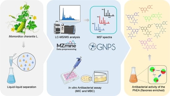
Antibacterial Activity from Momordica charantia L. Leaves and Flavones Enriched Phase

by
Abraão de Jesus B. Muribeca
1,†,
Paulo Wender P. Gomes
2,3,†,
Steven Souza Paes
1,
Ana Paula Alves da Costa
4,
Paulo Weslem Portal Gomes
5,
Jéssica de Souza Viana
1,
José Diogo E. Reis
1,
Sônia das Graças Santa R. Pamplona
1,
Consuelo Silva
1,6,
Anelize Bauermeister
7,
Lourivaldo da Silva Santos
1 and
Milton Nascimento da Silva
1,*
1
Institute of Exact and Natural Sciences, Federal University of Pará, Augusto Corrêa, 01, Guamá, Belém 66075-110, PA, Brazil
2
Collaborative Mass Spectrometry Innovation Center, University of California San Diego, La Jolla, San Diego, CA 92093, USA
3
Skaggs School of Pharmacy and Pharmaceutical Sciences, University of California, La Jolla, San Diego, CA 92093, USA
4
Department of Natural Science, Campus XIX, State University of Pará, Rodovia PA 154, Km 28, Cajú, Salvaterra 66860-000, PA, Brazil
5
Institute of Biology, University of Campinas, Monteiro Lobato, 255, Barão Geraldo, Campinas 13083-862, SP, Brazil
6
Pharmaceutical Science Post-Graduation Program, Faculty of Pharmacy, Federal University of Pará, Belém 66075-110, PA, Brazil
7
Institute of Biomedical Sciences, University of São Paulo, São Paulo 05508-900, SP, Brazil
*
Author to whom correspondence should be addressed.
These authors contributed equally to this work.
Pharmaceutics 2022, 14(9), 1796; https://doi.org/10.3390/pharmaceutics14091796
Submission received: 9 July 2022 / Revised: 1 August 2022 / Accepted: 4 August 2022 / Published: 26 August 2022

Abstract

:
Momordica charantia L. (Cucurbitaceae) is a plant known in Brazil as “melão de São Caetano”, which has been related to many therapeutic applications in folk medicine. Herein, we describe antibacterial activities and related metabolites for an extract and fractions obtained from the leaves of that species. An ethanolic extract and its three fractions were used to perform in vitro antibacterial assays. In addition, liquid chromatography coupled to mass spectrometry and the molecular networking approach were used for the metabolite annotation process. Overall, 25 compounds were annotated in the ethanolic extract from M. charantia leaves, including flavones, terpenes, organic acids, and inositol pyrophosphate derivatives. The ethanolic extract exhibited low activity against Proteus mirabilis (MIC 312.5 µg·mL−1) and Klebsiella pneumoniae (MIC 625 µg·mL−1). The ethyl acetate phase showed interesting antibacterial activity (MIC 156.2 µg·mL−1) against Klebsiella pneumoniae, and it was well justified by the high content of glycosylated flavones. Therefore, based on the ethyl acetate phase antibacterial result, we suggest that M. charantia leaves could be considered as an alternative antibacterial source against K. pneumoniae and can serve as a pillar for future studies as well as pharmacological application against the bacteria.

Graphical Abstract

1. Introduction

Clinical infections caused by resistant bacteria have become a major public health concern worldwide, and approximately 700,000 deaths per year are caused by this type of bacteria [1]. It is estimated that by 2050, there will be more than 10 million deaths/year credited to ‘superbugs’, with the expectation of the highest rate being reported in developing countries [2]. The demand for new antimicrobial agents has been growing at the same time as the discovery and advancement of multi-resistant bacteria. Therefore, researchers have been focusing a great effort on the search for new therapeutic alternatives against multidrug-resistant bacteria [3]. In this sense, plant derivatives appear as potential sources of new drugs that act in different ways to deactivate or block the growth of such pathogens [4].
The Cucurbitaceae family includes several species widely distributed throughout the tropical and subtropical regions. There are antimicrobial activities associated with crude extracts and isolated metabolites of species from this family [5]. Among the species, Momordica charantia L., popularly known as “melão de São Caetano”, is considered an invasive plant in Brazil and can be found in different places in the country. It is frequently grown in orchards and coffee plantations, or even on fences and debris in abandoned land [6], and is composed of compound classes such as terpenoids, saponins, and flavonoids [5,7]. This species can be highlighted due to its antimicrobial [5], nutraceutical and inflammatory properties [8], as well as healing of gastric ulcers [9], rheumatism [10], and so on. In developing countries, M. charantia has been employed in folk medicine for several other pharmacological purposes, such as to treat toothache, diarrhea, furuncle, cancer, hypertension, obesity, bacterial and viral infections, diabetes, pneumonia, and even AIDS [11,12,13,14,15,16].
Although the chemical characterization of such plants can lead to new pharmaceuticals, it remains challenging due to the presence of several components with different physical – chemical properties, including many in relatively small quantities [17]. Nuclear magnetic resonance (NMR) techniques provide valuable structural information used for structure characterization and therefore, provide unequivocal identification. Liquid chromatography coupled to mass spectrometry (LC-MS) plays a crucial role as it is compatible with chromatographic techniques that allow the separation of compounds in the sample, leading to a deeper investigation of the whole chemical content due to the high sensitivity of MS that can detect metabolites of picomole to femtomole levels in some cases [18]. Furthermore, MS allows the detection of metabolites as ions in the form of mass to charge ratio (m/z) [19], and also allows the fragmentation of the ions at the gas phase by collision-induced dissociation (CID), providing MS/MS spectra that contain valuable information to contribute to annotation of the chemical structure [20]. It is worth mentioning that the acquisition of scan spectra by mass spectrometry is fast, and a single chromatographic run can provide thousands of MS spectra. Therefore, methods that allow a fast and easy analysis of such data are welcome in this field.
In this context, molecular networking, a tool from the Global Natural Products Social Molecular Networking (GNPS) [21] infrastructure, allows the visualization of MS/MS data by organizing it by spectral similarity. This is a very effective strategy, especially because establishing a similarity relationship provides organization into molecular families (usually related chemical structures), and, therefore, finds related metabolites in the dataset even in small amounts. GNPS also contains a spectral library of known compounds, and the molecular networking allows automatic searches in the spectral library, which contributes to speed up the dereplication of known compounds, a phytochemical approach that has been widely used in recent decades, including the modest contribution of these authors [22,23,24,25,26], and which is also unknown by recognition of the analogs into the molecular families.
Based on the antimicrobial potential already reported for M. charantia, this study describes the evaluation of in vitro antibacterial activity for the ethanolic extract and fractions from the leaves of M. charantia. In addition, liquid chromatography coupled to high-resolution mass spectrometry (LC-HRMS) analyses allowed the chemical characterization of the samples.

2. Materials and Methods

2.1. Botanical Material and Extraction

Leaves of M. charantia were collected in the municipality of Soure (0°23′50′′ S and 49°27′02′′ W), Marajó Island, PA, Brazil. The botanical material was incorporated (voucher: MFS009218) in the Prof. Dr. Marlene Freitas da Silva (MFS) Herbarium of the State University of Pará. Permission to access the Brazilian genetic patrimony was provided by SISGEN (A695619).
The leaves were washed with water from the Direct-Q5 system (Millipore, Darmstadt, Germany) and decontaminated with sodium hypochlorite solution (NaOCl, 0.1%) acquired from Dinâmica (Jaraguá do Sul, SC, Brazil). The samples were dried in an air circulation oven (Quimis, Brazil) at 45 °C until constant weight. The dry material was crushed in ball mills up to a granulometry of 60 to 100 μm, obtaining 146.54 g. The mass was subjected to extraction with 1 L of ethanol (Tedia, Fairfield, OH, USA) at room temperature for 24 h (2×), and 35.30 g of crude extract was obtained after the solvent evaporation process. Thus, 1 g of ethanolic extract (EE) was mixed with a hydroalcoholic solution, consisting of 60 mL of ultra-pure water, 20 mL of ethanol, and 1 mL of hydrochloric acid (Dinâmica, Indaiatuba, SP, Brazil). The resulting solution was subjected to liquid – liquid partition (LLP) to obtain the hexane (PhHex), ethyl acetate (PhEA), and hydroalcoholic (PhWOH) phases, respectively.

2.2. Liquid Chromatography-High Resolution Mass Spectrometry Analysis

The analyses were performed on a Xevo G2-S QqTOF mass spectrometer (Waters Corp., Milford, MA, USA) equipped with a LockSpray source. The instrument was calibrated with a mass of reference (leucine-enkephalin) utilized for accurate mass measurements. MassLynx 4.1 software was used for system control and data acquisition. The samples were analyzed in a BEH C18 column (Waters Corp.; 50 mm; 2.1 mm; 1.7 μm particle size) using ultra-pure water (solvent A) and acetonitrile (solvent B), both containing 0.1% formic acid. The column temperature was maintained at 40 °C. Linear gradient elution was performed with a flow of 300 μL/min and 5 – 95% of solvent B in 20 min. The injection volume of the samples was 5 μL. The mass spectra data were recorded in a negative ionization mode (ESI) for a mass range from m/z 50 to 1200. The source temperature was set to 120 °C with a cone gas flow of 50 L/h. The desolvation gas flow was set to 600 L/h at a temperature of 150 °C. The capillary was set at 3.0 kV with cone voltage at 40 V. The settings of the data-dependent acquisition (DDA) experiments were: centroid format, number of ions selected 5 (Top5 experiment), the normalized collision energy (NCE) was set to 10, 20, 30, 40 and 50, scan rate of 0.5 sec, charge states of +1 and +2, tolerance window of ±0.2 Da and peak extract window of 2 Da, tolerance of deisotope ± 3 Da, extraction tolerance of deisotope 6 Da.

2.3. Mass Spectrum Data Treatment

The raw files of the EE and PhEA acquired in the Xevo G2-S QTof mass spectrometer (Waters Corp., Milford, MA, USA) were converted into mzML format using the software MS Convert of the ProteoWizard package [27] and were processed with the software MZmine 2.53 version [28]. The limit for the detection of ions in negative mode at the MS1 level was set at 1.0 × 10³ and MS2 at 5.0 × 101. Chromatograms were constructed using ADAP with a minimum group number of 3 and a minimum group intensity limit of 1.0 × 10³, a min highest intensity of 3.0 × 10³, and an m/z tolerance of 10.0 ppm. The local minimum search algorithm was used to deconvolve the chromatogram, with an m/z tolerance of 0.5 for the pairing of MS2 and 0.2 min for RT. Isotopes were detected using a peak window with a tolerance of 10.0 ppm, an RT tolerance of 0.5 min, a maximum charge of 1. For peak alignment, the tolerance of m/z 10 ppm was used, scores for m/z of 75 and 25 for RT with a tolerance of 0.2 min. The resulting list was filtered to remove duplicates and lines with no associated MS2 spectrum. Then, gap filling was used to fill in the gaps in the peak list. The resultant files were exported using FBMN-GNPS.

2.4. Molecular Networks

The molecular network was built from the mgf and CSV files exported from MZmine. We used metadata to organize metabolite information according to the online workflow (available online: https://ccms-ucsd.github.io/GNPSDocumentation/, accessed on 30 January 2022) available on the GNPS website (available online: http://gnps.ucsd.edu, accessed on 8 July 2022) [21]. The tolerance of m/z for the precursor ion was adjusted to 0.02 Da and for fragment ion to 0.02 Da. Minimum cosine score above 0.5 and the minimum number of fragment ions were fixed on 4. The spectra on the network were then searched in the GNPS spectral libraries. The database spectra were filtered with a minimum cosine score above 0.6 and a minimum of 4 fragment ions correspondence. The job is available online at the link: https://gnps.ucsd.edu/ProteoSAFe/status.jsp?task=4890e08934bb4334b8738a346c10e7c4, accessed on 8 July 2022. The results were visualized and organized using Cytoscape version 3.8.2 (Seattle, WA, USA) [29]. Lastly, the correlation between PhEA and bioactivity score of the metabolites was obtained from NPAnalyst [30].

2.5. In Vitro Antibacterial Assay

Three strains of bacteria were used to evaluate the potential of the extracts: Staphylococcus aureus ATCC 25923, and Klebsiella pneumoniae ATCC 700603, provided by Instituto Evandro Chagas, Pará State (Brazil) and the bacteria Proteus mirabilis LACEN 8/7 (human isolated) provided by the Central Laboratory of Pará State collection. These microorganisms were selected for being common pathogens that can infect humans, animals or plants. The pure cultures were maintained by routine sub-culturing at one-week intervals in BHI broth (Brain Heart Infusion, Kasvi, Spain), incubated at 37 °C, and spiked for 24 h for their metabolic activation. The minimum inhibitory concentration (MIC) and minimum bactericidal concentration (MBC) were conducted using a method approved by the Clinical and Laboratory Standards Institute in 96-well microtitration plates [31].

2.6. Determination of Minimum Inhibitory Concentration (MIC) and Minimum Bactericidal Concentration (MBC)

The antimicrobial susceptibility test was conducted using a method approved by the Clinical and Laboratory Standards Institute [31]. The tests were carried out with a stock solution of 5 mg·mL−1 of the crude extract/phases using the successive dilution method to obtain the concentrations from 2500 to 78.1 µg·mL−1. Ciprofloxacin (Medley, Brazil) and vancomycin (1 mg·mL−1 each) were used as positive controls and BHI (Brain Heart Infusion) culture medium was used as negative control.
A total of 5 mg of extract/phases was dissolved in 100 μL of DMSO (Neon, Brazil) contained in Eppendorf tubes. Then, 900 μL of sterile BHI was added and stirred for better homogenization. In a 96-well microtitration plate (KASVI, Brazil), 100 μL of BHI broth was added to each well. Then, 100 μL of solution containing the samples was added to the first well of each column.
In each well, 5 μL of the bacterial suspension (104 Colony Forming Unit CFU/mL as required by CLSI) was inoculated and adjusted to 0.5 McFarland Standard scale and then the plates were incubated at 37 °C for 24 h. The results were read by adding 10 μL of TTC (2,3,5-triphenyltetrazolic chloride, NEON, Brazil). To prepare the dye, 0.2 g of TTC was added to the penicillin type flask containing 10 mL of sterile distilled water. The final solution showed a translucent color, and when in contact with environments where there are microorganisms presents a red color.
The type of activity presented in each concentration (bacteriostatic or bactericidal) was checked. In the cavities where there was no red color caused by the reaction of TTC with the bacteria, 5 μL of the volume contained in the wells was (re) inoculated in a Petri dish containing BHI agar culture medium and incubated at 37 °C for 24 h. Wells where bacteria grew indicated a bacteriostatic effect at that concentration and wells without bacteria indicated a bactericidal effect.

3. Results and Discussion

3.1. Antibacterial Activity

The EE and the PhEA of M. charantia showed antimicrobial activity and, therefore, they were selected to be analyzed by LC-MS/MS. Both positive controls of vancomycin and ciprofloxacin had a MIC of 7.8 µg·mL−1. The EE demonstrated a good bactericidal effect against Klebsiella pneumoniae and Proteus mirabilis. The PhEA stands out for presenting the best result with a MIC of 156.2 µg·mL−1 for the K. pneumoniae, suggesting that this presents bioactive substances, possibly the glycosylated flavones, responsible for such activity (Table 1). We highlighted the bactericidal effect of Klebsiella Pneumonie, and Proteus Mirabilis, both Gram-negative strains. The literature [32] reports that the cell wall of Gram-negative bacteria acts as a barrier to a number of substances, including antibiotics. However, recently it was confirmed that quercetin derivatives have strong antibacterial action against Gram-negative bacteria [33], and the ethyl acetate phase (PhEA, see Table 1) described in this study showed high content of quercetin (16) derivatives, i.e., quercetin-O-sambubioside (4), quercetin-O-glucoside (6), quercetin-O-glucosyl-6′′-acetate (9), and quercetin-O-acetylpentoside (13). MIC values < 100 µg·mL−1 are considered significant antimicrobials; moderate inhibitors present MIC in the range of 100 to 625 µg·mL−1; and inhibitors with MIC > 625 µg·mL−1 are considered weak [34]. In this sense, the ethyl acetate phase showed moderate activity (Table 1) against K. pneumoniae (MIC of 156.2 µg·mL−1), while the ethanolic extract showed moderate activity against P. mirabilis (312.5 µg·mL−1) and weak activity against K. pneumoniae and S. aureus (625 µg·mL−1). We emphasize that the MIC value against K. pneumoniae is in the range moderate–significant, which characterize PhEA, a source of candidate inhibitors of important hospital bacteria.
In previous reports, antimicrobial activity for leaves, fruits and seeds was reported against some clinically important bacteria [20]. The extract of the leaves showed the main results of being a potent inhibitor for Staphylococcus aureus, moderate for Staphylococcus epidermidis and weak for Candida albicans. Studies with extracts of the seed showed interesting activities against Escherichia coli, Salmonella typhi, and Staphylococcus aureus, but less activity against Pseudomonas aeruginosa [35].

3.2. Identification of Chemical Constituents

The total ion profiles of the EE and PhEA were recorded from 50 to 1200 Da in 20 min (Figure 1). The molecular formulas, main fragment ions, and putative names are shown in Table 2. A total of 32 major metabolites were detected in the EE. Of these, 25 compounds were annotated in level 2 and 3 of identification according to MSI [36] based on HRMS and MS/MS data and the literature; most of these compounds have well characterized fragmentation (MS/MS) profiles [37].

3.3. Molecular Networking (GNPS Annotation)

The molecular networking created with EE and PhEA showed 224 parent ions after removing the blank. Seven compounds (6, 7, 8, 10, 12, 23 and 32) including isomers were annotated based on MS2 data available in the GNPS spectral libraries. A family of flavones was reported, and the structure of the compounds is shown in Figure 2.
The peak 6 [M − H] of m/z 463.0869 with the main fragments m/z 301, 271, and 179 was annotated as quercetin-O-glucoside. The loss of the sugar unit [(M − H) − H2O] explains the fragment of m/z 301. However, the position of the hydroxyl groups in ring B, as well as the glycosyl moiety, cannot be confirmed with only MS/MS data. The loss of H2O following CO [(M − H) − H2O − CO] justifies the m/z 271 [39]; lastly, the loss of C7H6O2 on the B ring by retrocyclisation [39] explains the fragment of m/z 179. The peak 7 [M − H] of m/z 579.1355 was annotated as kaempferol O-glucoside-O-pentoside (product ions: m/z 463, 399, 327, 285, 151, 109). The losses of C5H8O3 [(M − H) − C5H8O3] and C7H16O5 [(M − H) − C5H8O3] confirm the presence of two sugar units in the molecule, however, as discussed before, the positions of the sugar moieties cannot be certainly confirmed. Furthermore, characteristic fission from sugars (0,2 X1 mechanism) [40] suggests the m/z 327, the ion of m/z 285, occurs by losses of two sugar units, and the loss of C8H6O2 referred to the C ring [(M − H) − C8H6O2] explains the ion of m/z 151. The base structure coumarin is identified by loss of C9H4O4 to the B ring [(M − H) − C9H4O4] characterizing the fragment of m/z 109. The peak 8 [M − H] of m/z 593.15 was annotated as luteolin-O-rutinoside and this molecule showed main fragments of m/z 547, 447, 357, 327, 285. The loss of C2H6O is very common in glycosylated flavones [41], which explains the fragment of m/z 547. In addition, from m/z 593 to 447 loss of C6H10O4 [(M − H) − 146] occurs and from m/z 447 to 285 loss of another sugar unit occurs. Lastly, m/z 285 is confirmed as the aglycone peak. The other fragments are very well discussed in the literature [40]; losses of C3H6O3 [(M – H) − 146 − 90] and CH2O [(M − H) − 146 − 90 − 30] suggested the ions of m/z 357 and 327, respectively. The peak 10 [(M − H)] of m/z 447.0926 was annotated as kaempferol-O-glucoside and the fragments of m/z 327, 284, 255, and 227 are very well discussed in the literature [39,42]. In summary, fission of kind 0,2 X1 occurs in the glycoside to the ion m/z 327, then, the aglycone (m/z 284) is confirmed by radical cleavage [3Y0 − H] followed by a loss of CH2O [3Y0 – CH2O] and CO [3Y0 − CH2O − CO] to the fragments of m/z 255 and 227. The peak 12 [M − H] of m/z 477.1035 was annotated as isorhamnetin-O-glucoside. The loss of C2H6O in the glucoside [41] explains the ion m/z 431 [(M − H) − 46] and the 0,2 X1 mechanism confirms the loss of C4H8O4 [(M − H) − 120] to the ion of m/z 357. The loss of glucoside occurs as m/z 315 [(M − H) − C6H10O5] followed by loss of radical CH3 from m/z 315 [(M − H) − C6H10O5 − CH3]•−. Aglycone corresponds to the ion of m/z 285; ion of m/z 271 was formed by a loss of CO2 [(M − H) − C6H10O5 − CO2] and retro-Diels-Alder (RDA) from m/z 151 [43,44].
The peak 32 [M − H] of m/z 571.2882 was characterized as 1-hexadecanoyl-sn-glycero-3-phospho-(1′-myo-inositol), an important inositol pyrophosphates derivative present in plants as a signaling metabolite [45,46,47]. While information about this new class of molecules in plants is still scarce, the enzymes responsible for their synthesis have recently been elucidated [48]. In this sense, 32 is being reported for the first time in the genus. Despite the report of some sulfur-derived compounds in the M. charantia sample [49], they were not observed in this work.
A molecular family of glycosylated terpene derivatives (Figure 3) was detected in the EE. The ion of m/z 385.1856 is referent from peak 3, identified as hydroxy-2,4,4-trimethyl-3-(3-oxobutyl)-2-cyclohexen-1-one glucoside, and the pathway of fragmentation has been shown in previous reports [50]. The fragmentation of the ion m/z 385 generated an ion at m/z 223, which refers to a neutral loss of a glycoside [(M − H) − 162]. A loss of H2O indicates the ion m/z 205 followed by losses C2H2O and C3H8 suggesting the ions of m/z 163 [(M − H) − 42] and 119 [(M − H) − 42 − 44], respectively. Lastly, m/z 113 is explained by ring fission and loss of C7H10O [(M − H) − 110]. This compound is included in the monoterpenes class and derivatives already reported in the literature from the Cucurbitaceae family [51].
According to Figure 2, the compounds 3, 22, 25, and 27 showed the presence of glycoside. In this sense, following the chemosystematic from the genus, for example, the compound 25 was characterized as momordicoside L isomer, a cucurbitane-type triterpenoid already reported for the species [52]. According to the literature [52], the most intense product ion of m/z 471 corresponds to the aglycone after the loss of glycoside [(M − H) − Glc], and in addition a loss of C3H6O (propan-2-one) in C-5 characterized the ion of m/z 575 [(M − H) − 58] following loss of C2H2 to the fragment of m/z 549. Lastly, the ion of m/z 343 occurs by loss of C8H16O [(M − H) − Glc − 128] from the aglycone. Furthermore, this highlights that the cucurbitane is related to the genus [53,54] and confirms the chemosystematic possibility that this study has found an isomer of momordicoside L. The peak 22 showed a match in the GNPS library and it was annotated as hederagenin base-2H + 1O, O-AcetylHex. Peak 27 was suggested as a triterpene glycoside derivative, and we believe it has the same base structure of momordicoside L based on the molecular network. Furthermore, compound 27 has a difference of 4 Da concerning momordicoside L, suggesting two unsaturated bonds. Finally, the compound 23 [M − H] of m/z 721.4172 was annotated as aederagenin-O-acetyl-hex.

3.4. Bioactivity and Structure

Previous studies reported that flavonoids have antimicrobial activity [55,56]. These stand out even more because of their antibacterial properties, especially against strains of Gram-negative bacteria, which are responsible for serious opportunistic infections and are resistant to common therapies. In this sense, the study of plants with high flavonoid content should be highlighted [57]. Thus, our study focused on the ethyl acetate phase (MIC 156.2 µg·mL−1) against K. pneumoniae, which stands out for its ability to develop enzymatic resistance mechanisms and is considered to be largely responsible for several infectious diseases [58]. The PhEA proved to be rich in flavones, for instance quercetin-O-glucoside (6) and luteolin-O-rutinoside (8) (Figure 4), which confirms the correlation of the observed antimicrobial activity, as well the EE.
Our chemical prospecting data summarize chemical constituents that have in common a structural core of flavones, well known for a variety of activities [25]. Furthermore, isolated flavonols such as quercetin and kaempferol already show promising results as antimicrobials [59] as well against K. pneumoniae (MIC > 256 µg·mL−1) [60] and are the base structures of the main protagonists of the PhEA in this study. We emphasize carefully that the nominal results have a much more expressive value than many works given the same biological answer; perhaps the answer may be even more significant if the studies aim at obtaining isolated compounds. However, we emphasize that the rapid annotation provided using the LC-MS/MS technique does not require isolation, but shows an understanding of the active extension of the extract, directing more objective studies to that specific class. In addition, the literature [61] treats enriched phases as the main drivers for more specific studies, not to reach the main compounds responsible for the activity but to exclude those who may be acting as deterrents of the activity. In this sense, we are not being categorical in pointing only to these substances as active, but we are presenting a more interference-free sample.

4. Conclusions

This study showed the extract and phase ethyl acetate from M. Charantia leaves as an antibacterial agent. A total of 32 major compounds were detected, and, of these, 25 were annotated based on mass spectrometry data. Compounds including flavones, terpenes, organic acids, and inositol pyrophosphate derivatives are reported for the first time for the genus Momordica. The ethanolic extract exhibited low activity against Proteus mirabilis and Klebsiella pneumoniae. However, the phase ethyl acetate enriched with flavones showed interesting antibacterial inhibition against K. pneumoniae. Hence, we show that the leaves are a renewable antibacterial source and can serve as a pillar for future studies.

Author Contributions

Original design: A.d.J.B.M., P.W.P.G. (Paulo Wender P. Gomes), S.S.P. and A.P.A.d.C. designed this study; A.d.J.B.M. and J.D.E.R. conducted plant extractions and phase separations; S.S.P., A.P.A.d.C. and J.d.S.V. conducted the biological experiments; P.W.P.G. (Paulo Weslem P. Gomes) conducted the botanical identification. Conceptualization, data curation, and formal analysis: A.d.J.B.M., P.W.P.G. (Paulo Wender P. Gomes) and S.S.P. writing—review, visualization and editing: P.W.P.G. (Paulo Wender P. Gomes), S.d.G.S.R.P. C.S., A.B., L.d.S.S. and M.N.d.S. Advisor, funding acquisition and resources: L.d.S.S. and M.N.d.S. Project administration: M.N.d.S. All authors have read and agreed to the published version of the manuscript.

Funding

Coordenação de Aperfeiçoamento de Pessoal de Nível Superior (CAPES), Process: 88882.445389/2019-01, Modality: Doctoral Scholarship. Pró-reitoria de Pesquisa—Universidade Federal do Pará (UFPA) and Universidade do Estado do Pará (UEPA) for the financial support.

Institutional Review Board Statement

Not applicable.

Informed Consent Statement

Not applicable.

Data Availability Statement

All supporting data used in this study are available from the authors.

Acknowledgments

The authors would like to express their deepest gratitude to Waters Technologies do Brasil LTDA for the technical support for LC-MS/MS analysis presented here.

Conflicts of Interest

We declare no current or potential conflict of interest related to this article.

References

  1. Who No Time to Wait: Securing the Future from Drug-Resistant Infections. Available online: https://www.who.int/antimicrobial-resistance/interagency-coordination-group/final-report/en/ (accessed on 31 December 2020).
  2. O’Neill, J. Tackling Drug-Resistant Infections Globally: Final Report and Recommendations; The Government of the United Kingdom: London, UK, 2016. [Google Scholar]
  3. Ali Mirza, S.; Afzaal, M.; Begum, S.; Arooj, T.; Almas, M.; Ahmed, S.; Younus, M. Chapter 11-Uptake Mechanism of Antibiotics in Plants. In Antibiotics and Antimicrobial Resistance Genes in the Environment; Hashmi, M.Z., Ed.; Elsevier: Amsterdam, The Netherlands, 2020; Volume 1, pp. 183–188. ISBN 9780128188828. [Google Scholar]
  4. Michelin, D.C.; Moreschi, P.E.; Lima, A.C.; Nascimento, G.G.F.; Paganelli, M.O.; Chaud, M.V. Avaliação da atividade antimicrobiana de extratos vegetais. Rev. Bras. Farmacogn. 2005, 15, 316–320. [Google Scholar] [CrossRef]
  5. Guarniz, W.A.S.; Canuto, K.M.; Ribeiro, P.R.V.; Dodou, H.V.; Magalhaes, K.N.; Sá, K.; do Nascimento, P.G.G.; Silva, K.L.; Sales, G.W.P.; Monteiro, M.P.; et al. Momordica Charantia L. Variety from Northeastern Brazil: Analysis of Antimicrobial Activity and Phytochemical Components. Pharmacogn. J. 2019, 11, 1312–1324. [Google Scholar] [CrossRef]
  6. Lorenzi, H. Plantas Daninhas Do Brasil: Terrestres, Aquáticas, Parasitas e Tóxicas. 3\textordfeminine Edição. Inst. Plant. Bras. 2000, 309, 640. [Google Scholar]
  7. Jia, S.; Shen, M.; Zhang, F.; Xie, J. Recent Advances in Momordica Charantia: Functional Components and Biological Activities. Int. J. Mol. Sci. 2017, 18, 2555. [Google Scholar] [CrossRef]
  8. Bortolotti, M.; Mercatelli, D.; Polito, L. Momordica Charantia, a Nutraceutical Approach for Inflammatory Related Diseases. Front. Pharmacol. 2019, 10, 486. [Google Scholar] [CrossRef]
  9. Gürdal, B.; Kültür, Ş. An Ethnobotanical Study of Medicinal Plants in Marmaris (Muğla, Turkey). J. Ethnopharmacol. 2013, 146, 113–126. [Google Scholar] [CrossRef] [PubMed]
  10. Polito, L.; Bortolotti, M.; Maiello, S.; Battelli, M.; Bolognesi, A. Plants Producing Ribosome-Inactivating Proteins in Traditional Medicine. Molecules 2016, 21, 1560. [Google Scholar] [CrossRef]
  11. Chen, J.; Tian, R.; Qiu, M.; Lu, L.; Zheng, Y.; Zhang, Z. Trinorcucurbitane and Cucurbitane Triterpenoids from the Roots of Momordica Charantia. Phytochemistry 2008, 69, 1043–1048. [Google Scholar] [CrossRef]
  12. Polito, L.; Djemil, A.; Bortolotti, M. Plant Toxin-Based Immunotoxins for Cancer Therapy: A Short Overview. Biomedicines 2016, 4, 12. [Google Scholar] [CrossRef]
  13. Grover, J.K.; Yadav, S.P. Pharmacological Actions and Potential Uses of Momordica Charantia: A Review. J. Ethnopharmacol. 2004, 93, 123–132. [Google Scholar] [CrossRef]
  14. Raman, A.; Lau, C. Anti-Diabetic Properties and Phytochemistry of Momordica Charantia L. (Cucurbitaceae). Phytomedicine 1996, 2, 349–362. [Google Scholar] [CrossRef]
  15. Bailey, C.J.; Day, C.; Leatherdale, B.A. Traditional Treatments for Diabetes from Asia and the West Indies. Pract. Diabetes Int. 1986, 3, 190–192. [Google Scholar] [CrossRef]
  16. Dans, A.M.L.; Villarruz, M.V.C.; Jimeno, C.A.; Javelosa, M.A.U.; Chua, J.; Bautista, R.; Velez, G.G.B. The Effect of Momordica Charantia Capsule Preparation on Glycemic Control in Type 2 Diabetes Mellitus Needs Further Studies. J. Clin. Epidemiol. 2007, 60, 554–559. [Google Scholar] [CrossRef] [PubMed]
  17. Hussein, R.A.; El-Anssary, A.A. Plants Secondary Metabolites: The Key Drivers of the Pharmacological Actions of Medicinal Plants. In Herbal Medicine; IntechOpen: London, UK, 2019. [Google Scholar]
  18. Lei, Z.; Huhman, D.V.; Sumner, L.W. Mass Spectrometry Strategies in Metabolomics. J. Biol. Chem. 2011, 286, 25435–25442. [Google Scholar] [CrossRef] [PubMed]
  19. Kim, H.W.; Choi, S.Y.; Jang, H.S.; Ryu, B.; Sung, S.H.; Yang, H. Exploring Novel Secondary Metabolites from Natural Products Using Pre-Processed Mass Spectral Data. Sci. Rep. 2019, 9, 17430. [Google Scholar] [CrossRef]
  20. Graça, G.; Cai, Y.; Lau, C.-H.E.; Vorkas, P.A.; Lewis, M.R.; Want, E.J.; Herrington, D.; Ebbels, T.M.D. Automated Annotation of Untargeted All-Ion Fragmentation LC—MS Metabolomics Data with MetaboAnnotatoR. Anal. Chem. 2022, 94, 3446–3455. [Google Scholar] [CrossRef]
  21. Wang, M.; Carver, J.J.; Phelan, V.V.; Sanchez, L.M.; Garg, N.; Peng, Y.; Nguyen, D.D.; Watrous, J.; Kapono, C.A.; Luzzatto-Knaan, T.; et al. Sharing and Community Curation of Mass Spectrometry Data with Global Natural Products Social Molecular Networking. Nat. Biotechnol. 2016, 34, 828–837. [Google Scholar] [CrossRef]
  22. Gomes, P.W.P.; Barretto, H.; Reis, J.D.E.; Muribeca, A.; Veloso, A.; Albuquerque, C.; Teixeira, A.; Braamcamp, W.; Pamplona, S.; Silva, C.; et al. Chemical Composition of Leaves, Stem, and Roots of Peperomia Pellucida (L.) Kunth. Molecules 2022, 27, 1847. [Google Scholar] [CrossRef]
  23. Gomes, P.; Quirós-Guerrero, L.; Muribeca, A.; Reis, J.; Pamplona, S.; Lima, A.H.; Trindade, M.; Silva, C.; Souza, J.N.S.; Boutin, J.; et al. Constituents of Chamaecrista Diphylla (L.) Greene Leaves with Potent Antioxidant Capacity: A Feature-Based Molecular Network Dereplication Approach. Pharmaceutics 2021, 13, 681. [Google Scholar] [CrossRef] [PubMed]
  24. Gomes, P.W.P.; Pamplona, T.C.D.L.; Navegantes-Lima, K.C.; Quadros, L.B.G.; Oliveira, A.L.B.; Santos, S.M.; e Silva, C.Y.Y.; Silva, M.J.C.; Souza, J.N.S.; Quirós-Guerrero, L.M.; et al. Chemical Composition and Antibacterial Action of Stryphnodendron Pulcherrimum Bark Extract, “Barbatimão” Species: Evaluation of Its Use as a Topical Agent. Arab. J. Chem. 2021, 14, 103183. [Google Scholar] [CrossRef]
  25. Gomes, P.; Quirós-Guerrero, L.; Silva, C.; Pamplona, S.; Boutin, J.A.; Eberlin, M.; Wolfender, J.-L.; Silva, M. Feature-Based Molecular Network-Guided Dereplication of Natural Bioactive Products from Leaves of Stryphnodendron Pulcherrimum (Willd.) Hochr. Metabolites 2021, 11, 281. [Google Scholar] [CrossRef] [PubMed]
  26. Santiago, J.C.C.; Albuquerque, C.A.B.; Muribeca, A.d.J.B.; Sá, P.R.C.; Pamplona, S.d.G.S.R.; Silva, C.Y.Y.e.; Ribera, P.C.; Fontes-Júnior, E.d.A.; da Silva, M.N. Margaritaria Nobilis L.f. (Phyllanthaceae): Ethnopharmacology and Application of Computational Tools in the Annotation of Bioactive Molecules. Metabolites 2022, 12, 681. [Google Scholar] [CrossRef] [PubMed]
  27. Holman, J.D.; Tabb, D.L.; Mallick, P. Employing ProteoWizard to Convert Raw Mass Spectrometry Data. Curr. Protoc. Bioinform. 2014, 46, 1–9. [Google Scholar] [CrossRef] [PubMed]
  28. Pluskal, T.; Castillo, S.; Villar-Briones, A.; Oresic, M. MZmine 2: Modular Framework for Processing, Visualizing, and Analyzing Mass Spectrometry-Based Molecular Profile Data. BMC Bioinform. 2010, 11, 395. [Google Scholar] [CrossRef] [PubMed]
  29. Shannon, P.; Markiel, A.; Ozier, O.; Baliga, N.S.; Wang, J.T.; Ramage, D.; Amin, N.; Schwikowski, B.; Ideker, T. Cytoscape: A Software Environment for Integrated Models of Biomolecular Interaction Networks. Genome Res. 2003, 13, 2498–2504. [Google Scholar] [CrossRef]
  30. Lee, S.; van Santen, J.A.; Farzaneh, N.; Liu, D.Y.; Pye, C.R.; Baumeister, T.U.H.; Wong, W.R.; Linington, R.G. NP Analyst: An Open Online Platform for Compound Activity Mapping. ACS Cent. Sci. 2022, 8, 223–234. [Google Scholar] [CrossRef] [PubMed]
  31. CLSI M100-S11. Performance Standards for Antimicrobial Susceptibility Testing. Clin. Microbiol. Newsl. 2001, 23, 49. [Google Scholar] [CrossRef]
  32. Tortora, G.J.; Funke, B.R.; Case, C.L.; Weber, D.; Bair, W. Microbiology, 13th ed.; Pearson: London, UK, 2018; ISBN 9780134605180. [Google Scholar]
  33. Osonga, F.J.; Akgul, A.; Miller, R.M.; Eshun, G.B.; Yazgan, I.; Akgul, A.; Sadik, O.A. Antimicrobial Activity of a New Class of Phosphorylated and Modified Flavonoids. ACS Omega 2019, 4, 12865–12871. [Google Scholar] [CrossRef]
  34. Kuete, V. Potential of Cameroonian Plants and Derived Products against Microbial Infections: A Review. Planta Med. 2010, 76, 1479–1491. [Google Scholar] [CrossRef]
  35. Jabeen, U.; Khanum, A. Isolation and Characterization of Potential Food Preservative Peptide from Momordica Charantia L. Arab. J. Chem. 2017, 10, S3982–S3989. [Google Scholar] [CrossRef]
  36. Sumner, L.W.; Amberg, A.; Barrett, D.; Beale, M.H.; Beger, R.; Daykin, C.A.; Fan, T.W.-M.; Fiehn, O.; Goodacre, R.; Griffin, J.L.; et al. Proposed Minimum Reporting Standards for Chemical Analysis. Metabolomics 2007, 3, 211–221. [Google Scholar] [CrossRef] [PubMed] [Green Version]
  37. Amaral, J.G.; Bauermeister, A.; Pilon, A.C.; Gouvea, D.R.; Sakamoto, H.T.; Gobbo-Neto, L.; Lopes, J.L.C.; Lopes, N.P. Fragmentation Pathway and Structural Characterization of New Glycosylated Phenolic Derivatives from Eremanthus Glomerulatus Less (Asteraceae) by Electrospray Ionization Tandem Mass Spectrometry. J. Mass Spectrom. 2017, 52, 783–787. [Google Scholar] [CrossRef] [PubMed]
  38. Bittremieux, W.; Avalon, N.E.; Thomas, S.P.; Kakhkhorov, S.A.; Aksenov, A.A.; Gomes, P.W.P.; Aceves, C.M.; Rodríguez, A.M.C.; Gauglitz, J.M.; Gerwick, W.H.; et al. Open Access Repository-Scale Propagated Nearest Neighbor Suspect Spectral Library for Untargeted Metabolomics. bioRxiv 2022. [Google Scholar] [CrossRef]
  39. Li, A.; Hou, X.; Wei, Y. Fast Screening of Flavonoids from Switchgrass and Mikania Micrantha by Liquid Chromatography Hybrid-Ion Trap Time-of-Flight Mass Spectrometry. Anal. Methods 2018, 10, 109–122. [Google Scholar] [CrossRef]
  40. Colombo, R.; Yariwake, J.H.; McCullagh, M. Study of C- and O-Glycosylflavones in Sugarcane Extracts Using Liquid Chromatography: Exact Mass Measurement Mass Spectrometry. J. Braz. Chem. Soc. 2008, 19, 483–490. [Google Scholar] [CrossRef]
  41. March, R.; Brodbelt, J. Analysis of Flavonoids: Tandem Mass Spectrometry, Computational Methods, and NMR. J. Mass Spectrom. 2008, 43, 1581–1617. [Google Scholar] [CrossRef]
  42. Kumar, S.; Singh, A.; Kumar, B. Identification and Characterization of Phenolics and Terpenoids from Ethanolic Extracts of Phyllanthus Species by HPLC-ESI-QTOF-MS/MS. J. Pharm. Anal. 2017, 7, 214–222. [Google Scholar] [CrossRef]
  43. Du, L.-Y.; Zhao, M.; Xu, J.; Qian, D.-W.; Jiang, S.; Shang, E.-X.; Guo, J.-M.; Duan, J.-A. Analysis of the Metabolites of Isorhamnetin 3-O-Glucoside Produced by Human Intestinal Flora in Vitro by Applying Ultraperformance Liquid Chromatography/Quadrupole Time-of-Flight Mass Spectrometry. J. Agric. Food Chem. 2014, 62, 2489–2495. [Google Scholar] [CrossRef]
  44. Chen, Y.; Yu, H.; Wu, H.; Pan, Y.; Wang, K.; Jin, Y.; Zhang, C. Characterization and Quantification by LC-MS/MS of the Chemical Components of the Heating Products of the Flavonoids Extract in Pollen Typhae for Transformation Rule Exploration. Molecules 2015, 20, 18352–18366. [Google Scholar] [CrossRef]
  45. Nagy, R.; Grob, H.; Weder, B.; Green, P.; Klein, M.; Frelet-Barrand, A.; Schjoerring, J.K.; Brearley, C.; Martinoia, E. The Arabidopsis ATP-Binding Cassette Protein AtMRP5/AtABCC5 Is a High Affinity Inositol Hexakisphosphate Transporter Involved in Guard Cell Signaling and Phytate Storage. J. Biol. Chem. 2009, 284, 33614–33622. [Google Scholar] [CrossRef]
  46. Desai, M.; Rangarajan, P.; Donahue, J.L.; Williams, S.P.; Land, E.S.; Mandal, M.K.; Phillippy, B.Q.; Perera, I.Y.; Raboy, V.; Gillaspy, G.E. Two Inositol Hexakisphosphate Kinases Drive Inositol Pyrophosphate Synthesis in Plants. Plant J. 2014, 80, 642–653. [Google Scholar] [CrossRef] [PubMed]
  47. Raboy, V. Seed Total Phosphate and Phytic Acid. In Molecular Genetic Approaches to Maize Improvement; Kriz, A.L., Larkins, B.A., Eds.; Springer: Berlin/Heidelberg, Germany, 2009; pp. 41–53. ISBN 9783540689225. [Google Scholar]
  48. Freed, C.; Adepoju, O.; Gillaspy, G. Can Inositol Pyrophosphates Inform Strategies for Developing Low Phytate Crops? Plants 2020, 9, 115. [Google Scholar] [CrossRef]
  49. Chan, L.Y.; Wang, C.K.L.; Major, J.M.; Greenwood, K.P.; Lewis, R.J.; Craik, D.J.; Daly, N.L. Isolation and Characterization of Peptides from Momordica Cochinchinensis Seeds. J. Nat. Prod. 2009, 72, 1453–1458. [Google Scholar] [CrossRef] [PubMed]
  50. Li, Z.; Tu, Z.; Wang, H.; Zhang, L. Ultrasound-Assisted Extraction Optimization of α-Glucosidase Inhibitors from Ceratophyllum Demersum L. and Identification of Phytochemical Profiling by HPLC-QTOF-MS/MS. Molecules 2020, 25, 4507. [Google Scholar] [CrossRef]
  51. Kai, H.; Baba, M.; Okuyama, T. Two New Megastigmanes from the Leaves of Cucumis Sativus. Chem. Pharm. Bull. 2007, 55, 133–136. [Google Scholar] [CrossRef] [PubMed]
  52. Ma, J.; Krynitsky, A.J.; Grundel, E.; Rader, J.I. Quantitative Determination of Cucurbitane-Type Triterpenes and Triterpene Glycosides in Dietary Supplements Containing Bitter Melon (Momordica Charantia) by HPLC-MS/MS. J. AOAC Int. 2012, 95, 1597–1608. [Google Scholar] [CrossRef] [PubMed]
  53. Ferreira, M.-J.U.; Duarte, N.; Reis, M.; Madureira, A.M.; Molnár, J. Euphorbia and Momordica Metabolites for Overcoming Multidrug Resistance. Phytochem. Rev. 2014, 13, 915–935. [Google Scholar] [CrossRef]
  54. Ramalhete, C.; Mansoor, T.A.; Mulhovo, S.; Molnár, J.; Ferreira, M.-J.U. Cucurbitane-Type Triterpenoids from the African Plant Momordica Balsamina. J. Nat. Prod. 2009, 72, 2009–2013. [Google Scholar] [CrossRef]
  55. Dixon, R.A.; Howles, P.A.; Lamb, C.; He, X.Z.; Reddy, J.T. Prospects for the Metabolic Engineering of Bioactive Flavonoids and Related Phenylpropanoid Compounds. Adv. Exp. Med. Biol. 1998, 439, 55–66. [Google Scholar] [CrossRef]
  56. Alcaráz, L.E.; Blanco, S.E.; Puig, O.N.; Tomás, F.; Ferretti, F.H. Antibacterial Activity of Flavonoids Against Methicillin-Resistant Staphylococcus Aureus Strains. J. Theor. Biol. 2000, 205, 231–240. [Google Scholar] [CrossRef]
  57. Mirzoeva, O.K.; Grishanin, R.N.; Calder, P.C. Antimicrobial Action of Propolis and Some of Its Components: The Effects on Growth, Membrane Potential and Motility of Bacteria. Microbiol. Res. 1997, 152, 239–246. [Google Scholar] [CrossRef]
  58. Kołpa, M.; Wałaszek, M.; Gniadek, A.; Wolak, Z.; Dobroś, W. Incidence, Microbiological Profile and Risk Factors of Healthcare-Associated Infections in Intensive Care Units: A 10 Year Observation in a Provincial Hospital in Southern Poland. Int. J. Environ. Res. Public Health 2018, 15, 112. [Google Scholar] [CrossRef] [PubMed]
  59. Hodek, P. Flavonoids. In Metabolism of Drugs and Other Xenobiotics; Wiley-VCH Verlag GmbH & Co. KGaA: Weinheim, Germany, 2012; pp. 543–582. ISBN 9783527630905. [Google Scholar]
  60. Lin, R.-D.; Chin, Y.-P.; Lee, M.-H. Antimicrobial Activity of Antibiotics in Combination with Natural Flavonoids against Clinical Extended-Spectrum Beta-Lactamase (ESBL)-Producing Klebsiella Pneumoniae. Phytother. Res. 2005, 19, 612–617. [Google Scholar] [CrossRef] [PubMed]
  61. Wang, E.; Li, Y.; Maguy, B.L.; Lou, Z.; Wang, H.; Zhao, W.; Chen, X. Separation and Enrichment of Phenolics Improved the Antibiofilm and Antibacterial Activity of the Fractions from Citrus Medica L. Var. Sarcodactylis in Vitro and in Tofu. Food Chem. 2019, 294, 533–538. [Google Scholar] [CrossRef] [PubMed]
Figure 1. Total ion chromatogram (TIC) in the negative ionization mode of the ethanolic extract (EE), and ethyl acetate phase (PhEA).
Pharmaceutics 14 01796 g001
Figure 2. Feature-based molecular networking (GNPS) based on MS/MS data (ESI) from GNPS library [21] and suspect list [38]. The arrows indicate the nodes that have a match in the spectral library and their respective structure. Node text indicates the parent ion, node color reports the extract of the plant (orange: ethanolic extract from leaves; blue: phase ethyl acetate from leaves).
Pharmaceutics 14 01796 g002
Figure 3. Representation of hydroxy-2,4,4-trimethyl-3-(3-oxobutyl)-2-cyclohexen-1-one glucoside (3) clusters in ethanolic extract using MS/MS in negative ionization mode.
Pharmaceutics 14 01796 g003
Figure 4. Community network. PhEA illustrated as square white node. Two examples of predicted bioactive flavones (6,8). The color of metabolite nodes is defined by cluster score (0.5–0.75). The size corresponds to the activity score (normalized data).
Pharmaceutics 14 01796 g004
Table 1. Bacteria growth behavior in the presence of the extract and phases at different concentrations.
Concentration
(µg·mL−1)
EEPhHexPhEAPhWOHEEPhHexPhEAPhWOHEEPhHexPhEAPhWOH
Klebsiella pneumoniaeProteus mirabilisStaphylococcus aureus
2500=+=+=++++
1250=+=+=++++++
625=+=+=++++++
312.5++=+=+++++++
156.2++=+++++++++
78.1++++++++++++
39.0++++++++++++
Note: NP: natural product; EE: ethanolic extract; PhHex: hexane phase; PhEA: ethyl acetate phase; PhWOH: hydroalcoholic phase; = bactericidal effect; − bacteriostatic effect; + not active.
Table 2. The identified or tentatively identified compounds of ethanolic extract and ethyl acetate phase from Momordica charantia by LC-HRMS.
PeakRt (Min)Molecular Formula[M − H] (m/z)Main Product
Ions (MS/MS)
Annotated CompoundEEPhEA
CalculatedAccurateError (ppm)
10.42C13H24O13387.1139387.11292.6341, 278, 179melibiosex-
21.63C12H14O8285.0610285.06022.8153dihydrobenzoic acid pentosexx
33.23C20H32O10a 431.1917a 431.19101.6385.1856 [M − H], 223, 205, 163, 119, 113hydroxy-2,4,4-trimethyl-3-(3-oxobutyl)-2-cyclohexen-1-one glucosidexx
43.71C26H28O16595.1299595.13061.2445, 300, 272, 251, 191, 178quercetin-O-sambubiosidexx
53.97C27H30O16609.1456609.14580.3463, 301rutinxx
64.08C21H20O12463.0877463.08691.7301, 271, 179quercetin-O-glucosidexx
74.14C26H28O15579.1350579.13480.3463, 399, 327, 285, 151, 109kaempferol-O-glucoside-O-pentosidex-
84.39C27H30O15593.1506593.15110.8547, 447, 357, 327, 285luteolin-O-rutinosidexx
94.37C23H22O13505.0982505.09800.4300, 271, 255, 243, 178, 151quercetin-O-glucosyl-6′′-acetatexx
104.53C21H20O11447.0927447.09260.2327, 284, 255, 227kaempferol-O-glucosidexx
114.65C7H6O3137.0239137.02288.0934-hydroxybenzoic acidx-
124.67C22H22O12477.1033477.10350.4431, 357, 315, 300, 285, 271, 151isorhamnetin-O-glucosidex-
134.87C23H22O12489.1038489.10331.0285, 255, 227quercetin-O-acetylpentosidexx
144.98C20H34O9a 417.2125a 417.21132.9371.2052 [M − H], 209, 161,icariside B6xx
155.66C18H32O7359.2070359.20612.5343, 305, 287, 239, 227, 209, 197, 171unknownx-
165.77C15H10O7301.0348301.03412.3273, 245, 193, 179, 151, 121quercetinxx
177.14C18H32O5327.2171327.21622.7291, 229, 171trihydroxy octadecadienoic acid isomerxx
187.24C18H32O5327.2171327.21603.3291, 229, 171trihydroxy octadecadienoic acid isomerxx
197.64C18H34O5329.2328329.23202.4211, 171trihydroxy octadecenoic acidxx
208.38C37H60O11679.4057a 679.40530.6633.3994 [M − H], 285momordicoside L isomerx-
218.58C18H28O4307.1909307.19032.0289, 267, 235, 209, 185unknownx-
229.06C39H60O13a 735.3956a 735.39630.9689.3918 [M − H], 667, 599, 527, 339hederagenin base-2H + 1O, O-AcetylHexx-
239.74C39H62O12721.4163721.41561.0675 [M − H], 633, 513, 275, 193hederagenin-O-AcetylHexx-
249.18C18H26O4305.1753305.17462.3287, 249, 207unknownx-
259.37C37H60O11a 679.4057a 679.40600.4633.4015 [M − H], 575, 549, 471, 343momordicoside L isomerx-
269.92C46H56O6703.3999703.40610.6659, 633, 597, 482, 350unknownx-
2710.0C30H60O16a 675.3803a 675.373510629.3677 [M − H], 569, 467, 447, 339, 297triterpene glycosides derivativexx
2810.4C37H60O11a 679.4057a 679.40520.7633.4084 [M − H], 530, 339, 291, 137momordicoside L isomerx-
2911.51C18H29O3293.2117293.21092.7275, 235, 183, 121unknownx-
3011.56C28H62O21733.3705733.37293.3689, 554, 412, 364, 259, 175unknownx-
3112.0C36H54O10645.3639645.36390.0601, 559, 513, 407, 339, 243, 168, 127unknownx-
3214.9C32H44O9571.2907571.28824.4525, 481, 391, 325, 315, 255, 241, 1531-Hexadecanoyl-sn-glycero-3-phospho-(1′-myo-inositol) isomerx-
Note: a [M + HCOOH − H]; EE: ethanolic extract; PhEA: phase ethyl acetate; x: presence; -: absent.
Publisher’s Note: MDPI stays neutral with regard to jurisdictional claims in published maps and institutional affiliations.

