1. Introduction
Cancer ranks as a leading cause of mortality worldwide, but efficient therapeutic modalities against cancer have remained a challenge over past decades [
1,
2]. Although traditional first-line cancer treatment approaches, including chemotherapy, radiotherapy, and surgery, have made great progress in clinical practices, they are still associated with some adverse effects, such as multidrug resistance, immune system impairment, or systemic toxicity [
3,
4,
5]. Recently, the fast development of molecular imaging technology has ushered in new frontiers in the field of anti-cancer therapies. Minimally invasive or noninvasive imaging-guided therapeutic modalities, such as microwave therapy, phototherapy, and acoustic therapy, are attracting researchers’ attention and showing promising potential for cancer treatment due to their precisely spatiotemporal controllability, special tumor-killing activity, and elevated therapeutic outcomes [
6,
7,
8].
Recently, sonodynamic therapy (SDT), a noninvasive treatment modality through combining sonosensitizers with low-intensity ultrasound (US), is rapidly developing to be a promising approach for oncotherapy. Thanks to the merits of deep tissue-penetrating capability (>10 cm), low cost, and non-invasiveness, SDT has been demonstrated to be more effective than photodynamic therapy (PDT), which suffers from the main limitation in the low-light penetration of tissue depth [
9,
10,
11]. The predominant mechanism of SDT is considered to be the production of cytotoxic reactive oxygen species (ROS) from sonosensitizers irradiated by US, such as cytotoxic singlet oxygen (
1O
2), leading to damage to intracellular DNA and apoptosis of cancer cells [
12,
13]. So far, numerous sonosensitizers, including organic and inorganic materials, have been developed [
14]. Among them, chlorin e6 (Ce6), an FDA-approved organic sensitizer of porphyrin derivatives [
15], shows some unique advantages over other sonosensitizers, including effective activation by US and fluorescence imaging capability, which makes it possible to realize imaging-guided SDT for precise tumor therapy [
16,
17,
18,
19]. Unfortunately, SDT still has some problems. For example, the hypoxic tumor microenvironment (TME) may restrict the therapeutic efficacy of SDT due to the continuous O
2 consumption during SDT [
20,
21]. In addition, the short diffusion distance of ROS also makes it difficult to kill large-scale tumor cells [
22,
23]. All these disadvantages mentioned above show that there is still a challenge for the optimal therapeutic outcome by only SDT.
Unlike SDT, photothermal therapy (PTT) is another promising oxygen-independent anti-tumor strategy, which employs photothermal agents to transduce light energy into hyperthermia to kill tumors [
24,
25]. The application of PTT to tumor treatment is highly effective; however, the limited tissue penetration of PTT usually results in incomplete ablation for some large tumors, which greatly decreases its anti-tumor efficacy [
26]. Thus, SDT or PTT, as a single therapeutic modality, cannot achieve a satisfactory outcome. Previous studies have reported that hyperthermia-induced enhancement of blood perfusion during PTT could alleviate hypoxia and promote SDT-mediated ROS generation [
27,
28]. Documents have also demonstrated that the combined use of PTT and SDT could overcome the inherent limitations of the single PTT or SDT modality and achieve significantly improved anti-tumor effects [
29,
30]. To date, many photothermal agents with strong near-infrared (NIR) absorption, such as Au, Pt, and Pd metal nanoparticles (NPs), have been developed for PTT treatment of tumors [
31,
32,
33]. Nevertheless, most of them face the dilemma of potential tissue toxicity and low photothermal conversion efficiency.
Melanin is a natural polyphenol substance existing in biological tissues, including the hair and skin of mammals, the ink of cuttlefish, black sesame, bacteria, etc. [
34,
35,
36,
37,
38]. Also, melanin is an endogenous chromophore with photoacoustic (PA) imaging capability, exhibiting high sensitivity and spatial resolution in in vivo deep-seated tissues [
39]. Generally, there are two main ways to obtain melanin NPs. One is chemical synthesis, and the other is biological extraction. The former is environmentally unfriendly and has poor biocompatibility, and the latter is procedurally complex, low efficiency, and expensive. Herein, we explored a biosynthetic route to obtain melanin NPs via genetically engineered bacteria and fabricated the SDT/PTT dual-functional liposomes (designated as MC@Lip) by encapsulating the biosynthetic melanin NPs into a hydrophilic core and Ce6 into the hydrophobic lipid shell. The genetic circuit, which harbors a tyrosinase gene named Tyr1 under the control of a TlpA promoter inhibited by the temperature-sensitive repressor protein (TlpA36), was introduced into E.coli. The genetically engineered bacteria can express tyrosinase in a temperature-induced manner, catalyzing Tyrosine substrate to convert into melanin. The relatively slow biosynthetic procedure makes it possible to fabricate different particle sizes of melanin NPs by controlling the incubation time of bacteria with tyrosine substrate. The biosynthetic melanin NPs with 8 nm particle size and Ce6 sonosensitizer were further encapsulated into liposomes via the filming-rehydration method and obtained the MC@Lip. After these resulting MC@Lip were further intravenously administrated into the tumor-bearing mice, they would target the tumor site by the enhanced permeability and retention (EPR) effect. Upon the MC@Lip becoming enriched in the tumor, they would be received with acoustic and laser irradiation, activating the combined treatments of SDT and PTT and greatly improving the anti-tumor efficacy (
Figure 1).
2. Materials and Methods
2.1. Materials
1,2-distearoyl-sn-glycero-3-phosphoethanolamine-N-[methoxy(polyethylene glycol)-2000] (DSPE-PEG2000), 1,2-dipalmitoyl-sn-glycero-3-phosphatidylcholine (DPPC), and cholesterol were bought from Avanti Polar Lipids Inc (Alabaster, AL, USA). Calcein-AM, propidium iodide, and the CCK-8 (Cell Counting Kit-8) assay were purchased from Dojindo Laboratories (Kumamoto, Japan). 2,7-Dichloro-fluorescein diacetate (DCFH-DA) was purchased from Beyotime Biotechnology (Shanghai, China). Chlorin e6 (Ce6) and Singlet Oxygen Sensor Green (SOSG) were bought from Meilun Biotechnology Co., Ltd. (Dalian, China). 2,2,6,6-Tetramethylpiperidine (TEMP), 4, 6-diamidino-2-phenylindole (DAPI), and L-tyrosine were purchased from Sigma Aldrich (St. Louis, MO, USA). 1.3-diphenylisobenzofuran (DPBF) was bought from Solarbio Biotechnology Co., Ltd. (Beijing, China). Copper (II) Sulphate Pentahydrate was purchased from Titan Scientific Co., Ltd. (Shanghai, China). The sodium dodecyl sulfate-polyacrylamide gel electrophoresis (SDS-PAGE) Gel Fast Preparation Kit was bought from Epizyme Biomedical Technology Co., Ltd. (Shanghai, China).
2.2. Synthesis of Biosynthetic Melanin
The tyrosinase gene named Tyr1 from Bacillus megaterium was synthesized by Kingsley Genetics and cloned into the pJC vector under the control of the pTlpA promoter by a Seamless Cloning Kit (D7010M, Beyotime Biotechnology, Shanghai, China), followed by transformation into E. coli BL21. Tyrosinase expression was induced at OD 0.6 in a shaking incubator at 25 °C, 30 °C, or 37 °C for different times, followed by analysis with SDS-PAGE. The gel was visualized by staining with Coomassie brilliant blue solution after electrophoresis. Melanin was subsequently synthesized by supplementing it with 0.04 mg mL−1 CuSO4, as well as 0.8 mg mL−1 L-tyrosine, when the genetically engineered bacteria at OD 0.6 in a shaking incubator at 37 °C. The bacterial cells were firstly removed by centrifugation (7000 rpm, 8 min), and the black supernatant containing melanin nanoparticles was further passed through a Millipore 0.22 μm polyethersulfone membrane to remove cell debris. Afterwards, the black supernatant was washed with deionizing water several times by using a 10 K ultrafiltration membrane with centrifugation (7000 rpm, 5 min), and the final melanin solution was harvested and lyophilized for further use.
2.3. Characterization of Biosynthetic Melanin
The morphological characterization of melanin nanoparticles was examined by TEM at an acceleration voltage of 100 kV (JEM-1200EX). Dynamic light scattering (DLS) measurement (Zeta Sizer, Malvern, PA, USA) was applied to estimate the size distribution of melanin nanoparticles. UV-Vis-NIR absorbance spectra of biosynthetic melanin were determined at 25, 50, 100, 200, 400, or 800 μg mL−1 concentrations. The linear relationship between the concentration of biosynthetic melanin and the absorbance in the UV-Vis-NIR region was plotted at the wavelength 808 nm. Fourier-transform infrared spectroscopy (FTIR) was applied to identify the functional groups of biosynthetic melanin. Briefly, 1 mg melanin powder was mixed with 160 mg anhydrous potassium bromide (KBr; Specac, Orpington, UK), which was compressed into a translucent pellet with a hydraulic press (HY-12). The transmitted FTIR measurements (INVENIO R+RAMII, Bruker, Karlsruhe, Germany) were performed in an air background.
2.4. Fabrication of MC@Lip
Biosynthetic melanin and Ce6-loaded liposomes (termed MC@Lip) were fabricated through the thin-film hydration method. Briefly, 0.8 mg Ce6 in 1 mL methanol was added into the 1 mL CHCl3-containing lipid mixture (DPPC, cholesterol, and DSPE-PEG-2000, weight ratio at 3:1:1). Then, this solution was transferred into a round-bottom tube. The solvent was removed with nitrogen gas flow to form a uniform thin film, followed by vacuum drying for 2 h. Afterwards, the film was hydrated in 2 mL of melanin solution (8 mg mL−1) at 37 °C. The MC@Lip was harvested via mini-extruders under an extrusion method with the 200 nm and 100 nm membranes separately. The homogenized liposomes were further purified via a centrifugal filter (MWCO = 50 kDa) at 4000× g for 10 min. After that, the MC@Lip was re-dispersed with deionized water, and the final liposomes were attained and stored at 4 °C in the dark for further use. The unloaded, Ce6-loaded, or melanin-loaded liposomes were also prepared by this similar process.
2.5. Characterization of MC@Lip
The structure and morphology of MC@Lip were examined by transmission electron microscopy (TEM, JEM-1200EX, Tokyo, Japan). The photo absorption spectra and fluorescence spectra of Ce6 and biosynthetic melanin were recorded using a microplate spectrophotometer (Multiskan GO, Thermo Scientific, Waltham, MA, USA) and multi-mode microplate reader (Bio Tek Synergy4, Winooski, VT, USA), respectively. The hydrodynamic particle size distribution and zeta potential were measured by using a Malvern Zetasizer Nano S90 instrument (Malvern, Worcestershire, UK). The particle sizes of MC@Lip dispersed in phosphate-buffered saline (PBS), fetal bovine serum (FBS), or water were recorded after 0, 1, 3, 5, or 7 days. The standard concentration curves of Ce6 and biosynthetic melanin solutions at a wavelength of 404 nm and 808 nm were measured to evaluate the amount of Ce6 and biosynthetic melanin encapsulated into MC@Lip. The supernatant was collected for absorbance tests at 404 nm and 808 nm to determine the mass of free Ce6 and melanin, respectively. The encapsulation efficiency of Ce6 and melanin were calculated by equation as follows: encapsulation efficiency (%) = (Weight of Total Ce6 or melanin inputs-Weight of Free Ce6 or melanin)/Total Weight of Ce6 or melanin inputs × 100%.
2.6. In Vitro 1O2 Generation from MC@Lip by US Irradiation
For the quantitative analysis of 1O2 generation, MC@Lip was suspended in PBS (Ce6 at the concentration of 12 μg mL−1), and DPBF (50 µM) was added. Then, the mixture was irradiated by US (1.0 MHz, 1.7 W cm−2, 50% duty cycle) for 5 min in the dark. Finally, the absorbance intensity at the wavelength of 398 nm in each group was measured via a multi-mode microplate reader. The DPBF consumption was calculated by the following equation: the consumption of DPBF (%) = (the before irradiation absorbance of DPBF—the post irradiation absorbance of DPBF)/the before irradiation absorbance of DPBF × 100%. Additionally, the MC@Lip solution was added into the SOSG (5 µM), and then the mixture was treated under US irradiation (1.0 MHz, 1.7 W cm−2, 50% duty cycle) at various time durations (0 s, 30 s, 60 s, 90 s, or 120 s) to evaluate the 1O2 generation. A multifunctional microplate reader (λex =488 nm, λem = 525 nm) was used to detect the fluorescence (FL) intensity of ROS generation. The 1O2 generation from MC@Lip under US irradiation was also detected by TEMP. Typically, MC@Lip (Ce6 at the concentration of 12 μg mL−1) with the TEMP (50 mM) was irradiated with US (1.0 MHz, 1.7 W cm−2, 50% duty cycle) for 1 min. After that, the A300-10/12 electron spin resonance (ESR) spectrometer (Bruker, Karlsruhe, Germany) was applied to immediately detect the 1O2 generation. The melanin + TEMP as the control group was also tested.
2.7. In Vitro Photothermal Effect of MC@Lip
The photothermal effect of MC@Lip in vitro was evaluated after 808 nm NIR laser irradiation of MC@Lip with different concentrations of melanin. The distance between the laser and sample was fixed at 10 cm. Additionally, the 808 nm NIR laser with various power densities was applied for laser irradiation. During this test, water, blank lip, free Ce6, and melanin were set as controls. An infrared thermal imaging camera (FLIR E4) with an accuracy of 0.1 °C was used to record the temperature variations and infrared radiation (IR) thermal images.
2.8. Cytotoxicity of MC@Lip
Dulbecco’s Modified Eagle Medium (DMEM) added with 1% penicillin/streptomycin and 10% FBS was used to culture the murine breast cancer 4T1 cell line and normal bEnd.3 cell line at 37 °C under a 5% CO2 atmosphere. Cells in the exponential growth phase were further applied for the experiments. To evaluate the cytotoxicity of MC@Lip, 4T1 and bEnd.3 cells were seeded into the 96-well plate with a density of 1 × 10⁴ per well overnight. After cell adhesion, the cell medium was replaced with a new medium containing MC@Lip at various melanin concentrations (0, 25, 50, 100, 200, 400, and 600 μg mL−1). After 24 h co-incubation, the standard CCK-8 assay based on the wavelength of 450 nm absorbance via a multi-mode microplate reader was used to test cell viabilities.
2.9. Cell Uptake of MC@Lip and 1O2 Generation
4T1 cells were incubated with MC@Lip (melanin at the concentration of 200 μg mL−1) or melanin solution at 200 μg mL−1 for 4 h, and then cells were washed twice and imaged by an inverted fluorescent microscope (Thermo Fisher Invitrogen EVOSM5000, Taunton, MA, USA). Intracellular 1O2 production upon exposure to US irradiation was detected by a ROS assay kit (DCFH-DA). 4T1 cells were seeded into the 96-well culture plates with a density of 1 × 10⁴ cells per well and divided into four groups: the group without any treatment (Control), only US irradiation group (US), MC@Lip group (MC@Lip), and MC@Lip under US irradiation group (MC@Lip + US). After incubation with MC@Lip (Ce6 at the concentration 12 μg mL−1 in DMEM) at 37 °C for 4 h, DCFH-DA was used to replace the culture media, and the cells were exposed to (1.0 MHz, 1.7 W cm−2, 50% duty cycle) for 1 min. Next, 30 min later, the cells were gently washed with PBS three times and then imaged by inverted fluorescent microscope to evaluate the production of 1O2.
2.10. In Vitro Tumor Cell-Killing Experiments
4T1 cells were seeded in 96-well culture plates with a density of 1 × 10⁴ cells per well and then divided into six groups: the group without any treatment (Control), MC@Lip group (MC@Lip), US combined with NIR irradiation group (US + NIR), MC@Lip combined with US irradiation group (MC@Lip + US), MC@Lip combined with NIR irradiation group (MC@Lip + NIR), and MC@Lip combined with US and NIR irradiation group (MC@Lip + US + NIR). For the control group, cells were cultured with serum-free culture medium. For the MC@Lip group, cells were cultured with MC@Lip solution (melanin concentration of 200 μg mL−1) for 4 h without any irradiation. For the US + NIR group, cells were treated by US irradiation (1.0 MHz, 1.7 W cm−2, 50% duty cycle, 1 min) and to NIR irradiation (808 nm and 1 W cm−2, 5 min) without MC@Lip. For the MC@Lip + US group, cells were cultured with MC@Lip solution for 4 h with only US irradiation (1.0 MHz, 1.7 W cm−2, 50% duty cycle, 1 min). For the MC@Lip + NIR group, cells were cultured with MC@Lip solution for 4 h with only NIR irradiation (808 nm and 1 W cm−2, 5 min). For the MC@Lip + US + NIR group, cells were cultured with MC@Lip solution for 4 h with irradiation of US (1.0 MHz, 1.7 W cm−2, 50% duty cycle, 1 min) and NIR (808 nm, 1 W cm−2, 5 min). After 7–8 h, the standard CCK-8 assay was applied to test cell viabilities. Meanwhile, after the above treatments, cells of different groups were also stained with Calcein-AM and PI solutions, showing living cells fluoresced green and dead cells fluoresced red, respectively, in the inverted fluorescent microscope.
2.11. In Vivo Fluorescence and PA Imaging
All animals (female BALB/c mice: 16~18 g, 4–6 weeks) were bought from Zhuhai Bai shi tong Biotechnology Co. Ltd. All in vivo experiments were carried out under the approved protocols of the Shenzhen Institutes of Advanced Technology and the Animal Care and Use Committee of the Chinese Academy of Sciences. As for the establishment of the subcutaneous xenograft mice tumor model, 4T1 cells with a density of 1 × 10⁶ were subcutaneously administrated into the right flank of each female BALB/c mouse. After seven days, the tumors volumes were allowed to reach ~80 mm3 before the experiments. For the PA imaging of MC@Lip in vivo, 4T1 tumor-bearing mice (n = 3) were intravenously administrated with MC@Lip dispersion at the melanin dose of 60 mg kg−1 (200 μL). PA imaging at tumors was acquired at different time points (0 h, 2 h, 4 h, 8 h, 12 h, and 24 h) after injection by using a Vevo LAZR X system. Both PA and ultrasound (UA) signals were acquired with the laser wavelength set at 700 nm. Moreover, quantitative analysis of PA signals at the tumors was performed correspondingly. For in vivo fluorescence imaging of MC@Lip, 4T1 tumor-bearing mice (n = 3) were intravenously injected with MC@Lip dispersion at the Ce6 dose of 3.8 mg kg−1 (200 μL). Fluorescence images in the tumor regions were recorded at different times post injection (0 h, 2 h, 4 h, 8 h, 12 h, and 24 h) by using a fluorescence system (SPECTRUM, Caliper, Hopkinton, MA, USA). After 24 h, the major organs and tumor site of mice were dissected to obtain fluorescence images ex vivo. Moreover, the relative fluorescence intensities of the major organs and tumor site were measured correspondingly. In addition, tumors were dissected for ultrathin sections and stained with DAPI for fluorescence microscopy detection of Ce6.
2.12. In Vivo Anti-Tumor Efficacy of MC@Lip
The 4T1 tumor-bearing mice were divided into six groups at random (n = 5), including the control group, MC@Lip group, US + NIR group, MC@Lip + US group, MC@Lip + NIR group, and MC@Lip + US + NIR group. Mice in the control and US + NIR groups were intravenously injected with PBS. The mice in the MC@Lip, MC@Lip + US, MC@Lip + NIR, and MC@Lip + US + NIR groups were administered with 200 μL MC@Lip dispersion at the Ce6 dose of 3.8 mg kg−1 and melanin dose of 60 mg kg−1. As for the MC@Lip + US group, US irradiation (1.0 MHz, 1.7 W cm−2, 50% duty cycle, 5 min) was conducted at 4 h post injection. As for the MC@Lip + NIR group, NIR irradiation (808 nm, 1.0 W cm−2, 10 min) was conducted at 4 h post injection. In the US and NIR irradiation groups (US + NIR and MC@Lip + US + NIR), US irradiation (1.0 MHz, 1.7 W cm−2, 50% duty cycle, 5 min) was conducted and then exposure to NIR irradiation (808 nm, 1.0 W cm−2, 10 min) at 4 h post injection. The temperature changes and IR thermal images of the tumor region were recorded by FLIR E4. The body weight and tumor size of mice were recorded and calculated every other day after treatments. The tumor volume (V) was measured by the following formula: V = length × width2/2. The changes of tumor volume and body weight were normalized using the relative tumor volumes named V/V0 (V0 represented the initial tumor volume before the treatment) and relative body weight W/W0 (W0 represented the initial body weight before the treatment), respectively, for 20 days. The survival of mice was monitored for 60 days for evaluation of the anti-tumor efficacy. When the tumor volume of mice reached more than 2000 mm3, mice were then sacrificed, and the tumor sites were sectioned into slices for staining with hematoxylin and eosin (H&E), Ki-67, and the TdT-mediated dUTP Nick-End Labeling (TUNEL) assay. To sum up, the subcutaneous xenograft mice tumor model was initially established by subcutaneously administrating 4T1 cells into the right flank of female BALB/c mouse. Seven days later, after 4 h post injection of MC@Lip, the 4T1 tumor-bearing mice were treated with 5 min ultrasound firstly and then 10 min NIR irradiation to achieve MC@Lip-mediated SDT and PTT.
2.13. Blood and Histological Analysis
To analyze the in vivo biosafety, healthy BALB/c mice (n = 3) were intravenously administrated with 200 μL of MC@Lip dispersion at the Ce6 dose of 3.8 mg kg−1 and melanin dose of 60 mg kg−1. The other three healthy mice were employed as the control group without any treatment. After 14 days, they were sacrificed. The blood samples were used for biochemistry analysis and major organs, including the heart, liver, lung, spleen, and kidney, were harvested for H&E staining.
2.14. Statistical Analysis
GraphPad Prism 6 software was used to conduct statistical analyses. The data are shown as the mean value ± standard deviation (SD). Significance was evaluated via Student’s t-test or one-way ANOVA with Tukey’s post hoc test. The asterisks (* p < 0.05, ** p < 0.01, *** p < 0.001) represent statistical significance, while n.s. is considered no significance.
4. Discussion
Considering that tyrosinase is the crucial enzyme for the formation of melanin in the biosynthesis pathway, the tyr1 gene was synthesized and inserted into a heat-inducible gene circuit under the control of the pTlpA promoter. When the temperature is below 37 °C, the tyr1 gene would not be transcribed due to the repression of TlpA36 protein, which is constitutively expressed in the bacteria. Upon these bacteria being exposed to 37 °C, the TlpA36 repression protein would be deactivated and lose its binding with the pTlpA promoter, leading to the transcription and expression of the tyr1 gene. To test this, we transformed this plasmid with the heat-inducible gene circuit into the E. coli BL21 bacteria and incubated them at the different temperatures of 25 °C, 30 °C, or 37 °C. The SDS-PAGE result indicated that the tyr1 gene can express in a heat-inducible manner (
Figure 2A). Next, we further examined the bacterium-mediated biosynthesis capability of melanin. The change of suspension color of different temperature-cultivated bacteria demonstrated that only the 37 °C cultivated bacteria could catalyze L-tyrosine into melanin. Interestingly, we also found the suitable particle size of biosynthetic melanin with 8nm under the 12 h incubation time with L-tyrosine. Additionally, the photo absorption and FTIR spectra of biosynthetic melanin nanoparticles were consistent with the natural melanin nanoparticles previously reported [
34,
35,
36,
37,
38]. Thus, we successfully fabricated the melanin nanoparticles via the genetically engineered bacteria with the controllable particle size. Due to their relatively slower enzymatic reaction process than the chemical method for catalyzing the formation of melanin nanoparticles, it is especially favorable to obtain the melanin nanoparticles with suitable size by this biosynthetic way. We also found that the larger the melanin nanoparticles that were used to encapsulate into liposomes, the bigger the size of the MC@Lip would be, which was confirmed in
Figure 3D. Given that the favorable small size is about 100 nm for tumor penetration via the EPR effect, we selected the 8 nm biosynthetic melanin nanoparticles to fabricate the MC@Lip. Together with the above results in
Figure 3, we successfully fabricated MC@Lip with an appropriate particle size and high stability in physiological conditions, which were essential for the anti-cancer therapeutic application.
Since Ce6 and melanin in MC@Lip can function as the sonosensitizer and photothermal agents, respectively, we detected the sonodynamic and photothermal effects of MC@Lip. These data in
Figure 4A–C indicated that
1O
2 could efficiently be generated in the MC@Lip + US group due to the presence of Ce6. Moreover, we further investigated the photothermal effect of MC@Lip under NIR irradiation. We found that the ΔT in the MC@Lip + Laser group was higher than the free melanin and Lip group. Based on this result, liposomal confinement might effectively decrease the laser irradiation energy loss of free melanin in the solvent, which was in accordance with the previous study [
38]. In addition, the suitable concentration and laser power density were optimized as follows: melanin concentration at 200 μg mL
−1 and NIR laser power at 1.00 W cm
−2 (808 nm). Generally, photothermal conversion efficiency (η) is considered as the gold standard for evaluating the photothermal capability of NPs for PTT. The η in this study was quantified to be 42.4%, which was in consistency to the reported melanin nanoparticles [
30,
34]. Collectively, these results provided strong support for MC@Lip to be applied as a promising sonosensitizer and photothermal agent with the efficient
1O
2 production and photothermal conversion capability.
After the successful validation of the efficient
1O
2 production and photothermal performance of MC@Lip, we subsequently examined their capability of PTT and SDT in vitro. The cytotoxicity of MC@Lip to 4T1 breast cancer cells and bEnd.3 normal cells were negligible, showing that MC@Lip was highly biocompatible. The fluorescence displayed in
Figure 5B,C indicates the successful cellular internalization of MC@Lip and confirms the ROS generation of MC@Lip upon being received with US irradiation. Strikingly, the MC@Lip + US + NIR group showed the strongest cytotoxicity to 4T1 cells, with the lowest cell viability, which was further confirmed by the Calcein AM/PI staining assay.
Next, it has been well known that tumor imaging plays an essential role in image-guided therapy. These results in
Figure 6 indicate that MC@Lip possesses good PA and fluorescence imaging performance, making it possible to visualize the aggregation of MC@Lip nanoparticles in the tumor. It is especially important for someone to choose the appropriate time to apply the US and NIR laser irradiation for treating the tumor.
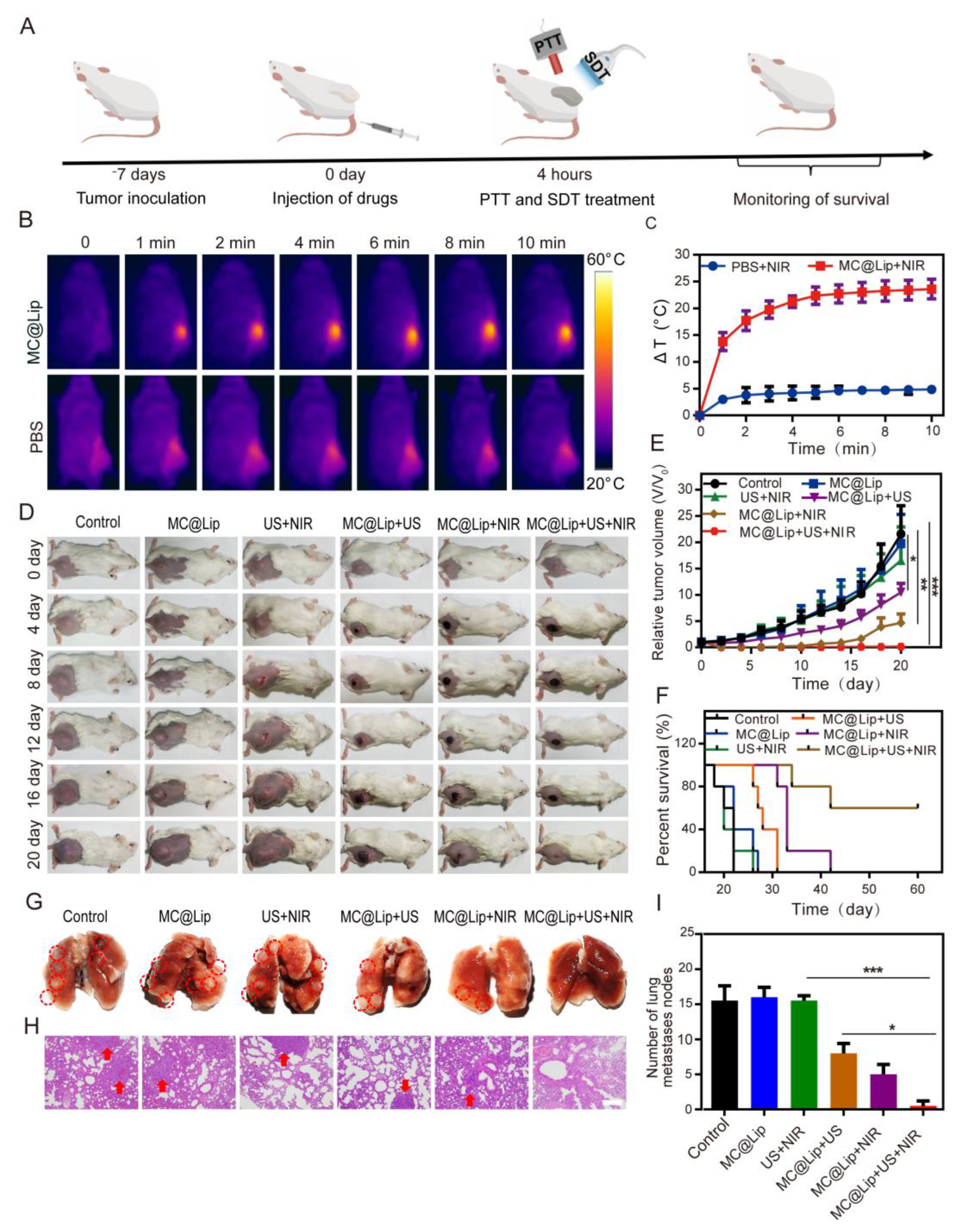
Finally, we further examined the anti-tumor efficacy of MC@Lip-mediated PTT and SDT. This result in
Figure 7B,C confirms that MC@Lip had the desirable PTT potential to ablate the tumor tissue. Collectively, the strongest tumor growth inhibition performance and longest survival time with no noticeable lung metastasis in the MC@Lip + US + NIR group confirmed that MC@Lip-based PTT-SDT harvested the therapeutical efficacy superior to that of monotherapy of PTT or SDT. No histological morphology and negligible difference in the typical markers of liver and kidney function further confirmed that MC@Lip had the favorable biosafety.

 ,
, 


