Article

Anti-Arthritic and Immunomodulatory Potential of Methanolic, n-Hexane, and Ethyl Acetate Fractions of Bark of Acacia modesta on Complete Freund’s Adjuvant-Induced Arthritis in Rats

1
Department of Pharmacology, Faculty of Pharmacy, The University of Lahore, Lahore 54000, Pakistan
2
Department of Pharmacology, Institute of Pharmacy, Faculty of Pharmaceutical and Allied Health Sciences, Lahore College for Women University, Jail Road, Lahore 54000, Pakistan
3
Faculty of Pharmaceutical Sciences, University of Central Punjab (UCP), Lahore 54000, Pakistan
4
Department of Pharmacology, University of Health Sciences, Lahore 54000, Pakistan
5
Department of Chemistry, Government College University Faisalabad, Faisalabad 38000, Pakistan
6
Department of Pharmaceutics, College of Pharmacy, King Saud University, Riyadh 11451, Saudi Arabia
7
Institute of Experimental and Clinical Pharmacology and Toxicology, University of Lübeck, 23562 Lübeck, Germany
*
Authors to whom correspondence should be addressed.
Pharmaceutics 2023, 15(9), 2228; https://doi.org/10.3390/pharmaceutics15092228
Submission received: 3 August 2023 / Revised: 21 August 2023 / Accepted: 22 August 2023 / Published: 29 August 2023

Abstract

:
Rheumatoid arthritis is an autoimmune disorder and topic of interest for researchers due to its increasing frequency and limited treatment. Acacia modesta Wall is known to treat rheumatic disorders in the traditional system of medicinal plants. Traditional medicines are still required for the treatment of this disease due to the large number of side-effects caused by commercial medicines. In the current study, the antiarthritic potential of methanolic extract (AM-metha), n-hexane (AM-hexa) fraction, and ethyl acetate (AM-etha) fraction of the bark of A. modesta against a complete Freund’s adjuvant rat model was evaluated. Evaluation using a digital plethysmometer, macroscopic evaluation, and histopathological evaluation were conducted to determine the paw volume and arthritic scoring. ELISA was performed to assess the PGE2 levels. RT-PCR was used to evaluate the expression levels of MMP2, MMP3, MMP9, NF-κB, IL6, IL1β, TNFα, and VEGF. Biochemical and hematological analyses were also conducted. GC/MS was also carried out to analyze the presence of medicinal compounds. The data revealed a marked reduction in the paw volume, arthritic scoring, and histopathological parameters, indicating the anti-arthritic potential of the plant. Treatment with plant extracts and fractions markedly down-regulated MMP2, MMP3, MMP9, NF-κB, IL6, IL1β, TNFα, and VEGF levels. Similarly, PGE2 levels were also found to be ameliorated in the treatment groups, indicating the immunomodulatory property of plant bark. Plant treatment nearly normalized hematological parameters such as counts of WBCs, RBCs, and platelets, along with Hb content, thereby validating the anti-arthritic activity. GC/MS analysis disclosed the presence of strong anti-inflammatory compounds such as lupeol, oleic acid, and squalene. The study showed that A. modesta possesses anti-arthritic and immunomodulatory potential linked to significant down-regulation of pro-inflammatory and inflammatory biomarkers.

1. Introduction

Inflammation is a natural reaction in the body and occurs due to many endogenous and exogenous factors [1]. Swelling, pain, heat, redness, and loss of function are the signs of inflammation [2]. Rheumatoid arthritis (RA) is a chronic disorder that is responsible for the inflammation of synovial joints and hypertrophy [3]. RA affects about 0.5–1% of the population of the world and it is three times more likely in women than in men [4]. RA severely affects the ankle joints, causing pannus development, eroded bones and cartilage, and inflamed synovial membrane, and causes many changes in the morphology of joints [5]. Despite the fact that inflammation is known to be a biological host defense response, chronic inflammatory responses are considered to be a major cause of RA [2].
The etiology of this disease is still not fully understood, but many immune pathways, including those of immune and non-immune cells, contribute to the release of various proinflammatory cytokines, chemokines, proteases (MMPs), and other matrix-lysing enzymes, resulting in cartilage and bone damage [6]. Higher levels of pro-inflammatory cytokines (e.g., TNFα, IL1, and IL6) caused by an imbalance between pro- and anti-inflammatory cytokines leads to the pathogenesis of RA [7]. TNF-α is involved in the damage of bone and cartilage via the enhancement of other pro-inflammatory cytokines and osteoclast formation [8]. IL6 is another immunomodulator that is actively involved in the pathogenesis of RA [9]. The activation of IL1β in rheumatoid synovium up-regulates the matrix metalloproteinase (MMP) levels, which mediates synovial inflammation, leading to damage of bones and joints [10]. The MMPs are considered to be responsible for the degradation of cartilage in the inflamed joints [11]. PGE2 is another inflammatory mediator that participates in the degradation of bone and cartilage via activation of MMPs [12]. As a mediator of angiogenesis and inflammation, vascular endothelial growth factor (VEGF) also contributes to the pathogenesis of disease [13]. NF-κB is not only a recognized regulator of inflammation in RA, but is also involved in broader aspects of RA pathology such as abnormal apoptosis, producing a Th1 response and stimulation of osteoclast activity [14,15]. Considering the significant role of these mediators in the pathogenesis of RA, the effects of a plant extract and its fractions on expression levels of these mediators were evaluated in the current study.
Many pharmacological agents, such as conventional synthetic disease-modifying antirheumatic drugs (DMARDs), targeted synthetic DMARDs, and biologic DMARDs, are given to treat RA in order to maintain joint activity. Nonsteroidal anti-inflammatory drugs (NSAIDs) and glucocorticoids (GCs) are used as an adjunct and as a symptomatic treatment for inflammation [16]. However, severe adverse effects of these treatments are reported due to their high dose and long-lasting usage [17]. Plants and their constituents are receiving recommendations due to their maximum pharmacological efficacy, minimum adverse reactions, and low costs, especially for inflammatory and immunomodulatory disorders [18]. Globally, herbal medicines are now gaining importance as traditional systems of medicine. Constituents isolated from them have been reported to be active constituents in drug discovery [19].
Acacia modesta Wall is an average sized tree belonging to the family Mimosaceae, and is widely found in rocky ground in India and Pakistan. The tree is locally called Phulai and Palosa [20]. Acacia modesta bark has been traditionally used for the treatment of rheumatic disorders [21,22]. Locally, A. modesta is used for the relief of various bone-, muscle-, and stomach-related disorders [23]. Research studies have shown that A. modesta possesses analgesic, anti-inflammatory, antiplatelet [20], antioxidant [24], anti-hyperglycemic [25], antidiarrheal, antisecretory, antispasmodic [26], antimicrobial [27], antidepressant, and anticoagulant [28] activities.
The current study aimed to determine the antiarthritic potential of AM-metha (methanolic) extract, AM-hexa (n-hexane fraction), and AM-etha (ethyl acetate fraction) of the bark of A. modesta Wall against a complete Freund’s adjuvant (CFA)-induced rat model of arthritis.

2. Materials and Method

2.1. Extract Formation and Fractionation

The bark was taken from the Bheen village, Chakwal district, Punjab province, and was identified by Dr-Zaheer-ud-din Khan, Government College University, Lahore (GC.Herb.Bot.3542). The fine powder (1 kg) was poured into methanol (3 L) in a dark container. After 1 week, the material was filtered through a filter sheet (Whatman number 1). Then, the filtrate was concentrated under reduced pressure using a rotary evaporator (Lab tech EV3111plus) [29]. The percentage yield of methanol extract of A. modesta was calculated to be 10.6%.
The fractionation was undertaken by dissolving methanol extract in 500 mL of distilled water and then the mixture was mixed with 500 mL of n-hexane in a separating funnel. Then, the n-hexane fraction was collected and concentrated under reduced pressure using a rotary evaporator. The n-hexane fraction yield was calculated to be 4%. Later, the same procedure was repeated to obtain the ethyl acetate fraction. The ethyl acetate fraction yield was calculated to be 4.3% [30].

2.2. Animal Housing

Sprague Dawley rats (150–250 g and 6–8 weeks old) of both genders were used for this study. Standard animal diet, humidity (40–60%), 12 h dark/light cycle, and temperature (24–26 °C) were provided to the rats in the animal house of the Faculty of Pharmacy, The University of Lahore. The study was conducted according to national, international, and institutional ethics guidelines regarding animal experiments under the approval number IREC-2019-91, issued by the Institutional Research Ethics Committee, The University of Lahore [31].

2.3. Treatment Design and Induction of Arthritis

For this study, 6 groups were created and each group had 6 rats. CFA at a dose of 0.15 mL (Santa Cruz biotechnology) was injected at day 0 to develop arthritis in the sub-plantar area of the left hind paw in all groups except the normal control. The dosing was started on the 8th day and ended on the 22nd day. After 24 h on day 23, all rats were sacrificed to begin the study [29]. All treatment groups are shown in Table 1.

2.4. Arthritic Score and Paw Volume

A macroscopic scoring method was used to measure the visible signs, such as inflammation, redness, and swelling, in the injected paw at days 8, 13, 18, and 23 of arthritic induction. Score 0 indicated normal, 1 indicated minor, 2 indicated mild, 3 indicated moderate, and 4 indicated severe signs [34]. The paw volume was measured using a water displacement digital plethysmometer (LE 7500, Panlab, Barcelona, Spain) at days 8, 13, 18, and 23 after arthritic induction. The non-injected paw was taken as the negative control. The volume of the non-injected paw was subtracted from the volumes of the injected paw to estimate the increase in paw volume. In addition, % inhibition was also calculated [35].

2.5. Ankle Joint Histopathology

After euthanasia, the ankle joints were separated, fixed, and decalcified. The paraffin blocks were sliced at a thickness of 5 μm and then stained with hematoxylin and eosin (H&E). The histopathological markers of arthritis were evaluated by a blinded histopathologist using 0–4 semi-quantification criteria [36].

2.6. Biochemical and Hematological Biomarkers

Alkaline phosphate (ALP), alanine aminotransferase (ALT), aspartate aminotransferase (AST), creatinine, total bilirubin, and urea levels were measured in serum samples as part of biochemical analysis using a chemistry analyzer and the kit protocol. Platelet, white blood cell (WBC), red blood cell (RBC), and hemoglobin (Hb) levels were measured in blood samples using an automatic hematology analyzer [37].

2.7. PGE2 Protein Levels

The sandwich ELISA method as per the kit protocol (Bioassay technology laboratory, Shanghai, China) was used for measuring PGE2 levels in serum samples. Optical density (OD) was observed at 450 nm [17].

2.8. Determination of MMP2, MMP3, MMP9, NF-κB, IL6, IL1β, TNFα, and VEGF Expression Levels

Total RNA from the blood samples was extracted using the TRIzol method. The samples were processed with chloroform, isopropyl alcohol, and 75% ethanol for RNA extraction. After extraction, the RNA samples were processed to determine their quantity and purity using a nanodrop spectrophotometer [38]. Each sample containing 1000 ng RNA was reverse transcribed according to the kit manufacturer’s protocol (Thermo Scientific, Waltham, MA, USA) to synthesize cDNA [39]. Amplification of cDNA (1µL) was performed by mixing it with forward primer (1 µL), reverse primer (1 µL), nuclease-free water (2 µL), and PCR Master Mix (5 µL) (Thermo Scientific). This PCR mixture was placed in a thermal cycler, which was programed for 39 cycles of denaturation, annealing, and extension. Ensemble Genome Browser online was used to design gene-specific primers of MMP2, MMP3, MMP9, NF-κB, and IL-1β. Primer sequences of IL-6, TNFα, and VEGF were chosen from previous articles, as shown in Table 2. GAPDH served as the housekeeping gene. The obtained PCR product was visualized using gel electrophoresis and a gel documentation imaging system, while ImageJ software 1.47t was used for densitometry [40].

2.9. GC/MS Analysis

GC/MS (GCMS-5975c) analysis was performed to determine the presence of constituents in bark extract and fractions. The analysis was performed using capillary column # HP-5MS (30 m × 250 μm × 0.25 μm) and helium with the flow rate of 1 mL/min was used as an inert gas. The oven temperature was initially programmed at 60 °C for 0 min; the temperature was then decreased at 5 °C/min and increased to 80 °C for 2 min, and then decreased at 10 °C/min and increased to 310 °C for 4 min. The total run time of the solutions was 33 min. The relative voltage for MS was 47 and 200 °C was programmed for the quadrupole analyzer. The mass-to-charge range was set at 30 to 700. The GC/MS analysis was performed using the method published in a previous article with slight changes [35].

2.10. Statistical Analysis

GraphPad Prism 5 was used for statistical analysis. One-way analysis of variance and Tukey’s post hoc test were applied for analysis and comparison of groups. All data are presented as the mean ± standard deviation. Significant differences were considered at a p > 0.001, b p > 0.01, and c p > 0.05 when test groups were compared with the disease group.

3. Results

3.1. A. modesta Attenuated Arthritic Score and Paw Volume

Arthritic score was increased in all groups except the normal control group at day 8. In the normal control group, disease was not developed; because the readings of this group were taken as zero, no bar chart is shown. At days 13, 18, and 23, arthritic score was gradually increased in the arthritic control group, while AM-metha, AM-hexa, AM-etha, and piroxicam treatment groups had a lower arthritic score at different day intervals. At the end of the treatment, all treated groups had a remarkably reduced score when compared with the arthritic control group.
AM-metha, AM-hexa, AM-etha, and piroxicam significantly reduced the paw volume when compared with the arthritic control group. Percentage inhibition of paw volume was also calculated using a previously reported formula. AM-metha (29.47%), AM-hexa (27.74%), AM-etha (28.32%), and piroxicam (28.90%) inhibited paw volume in comparison with the arthritic control group, as shown in Figure 1.

3.2. A. modesta Ameliorated Histopathological Parameters of Arthritis

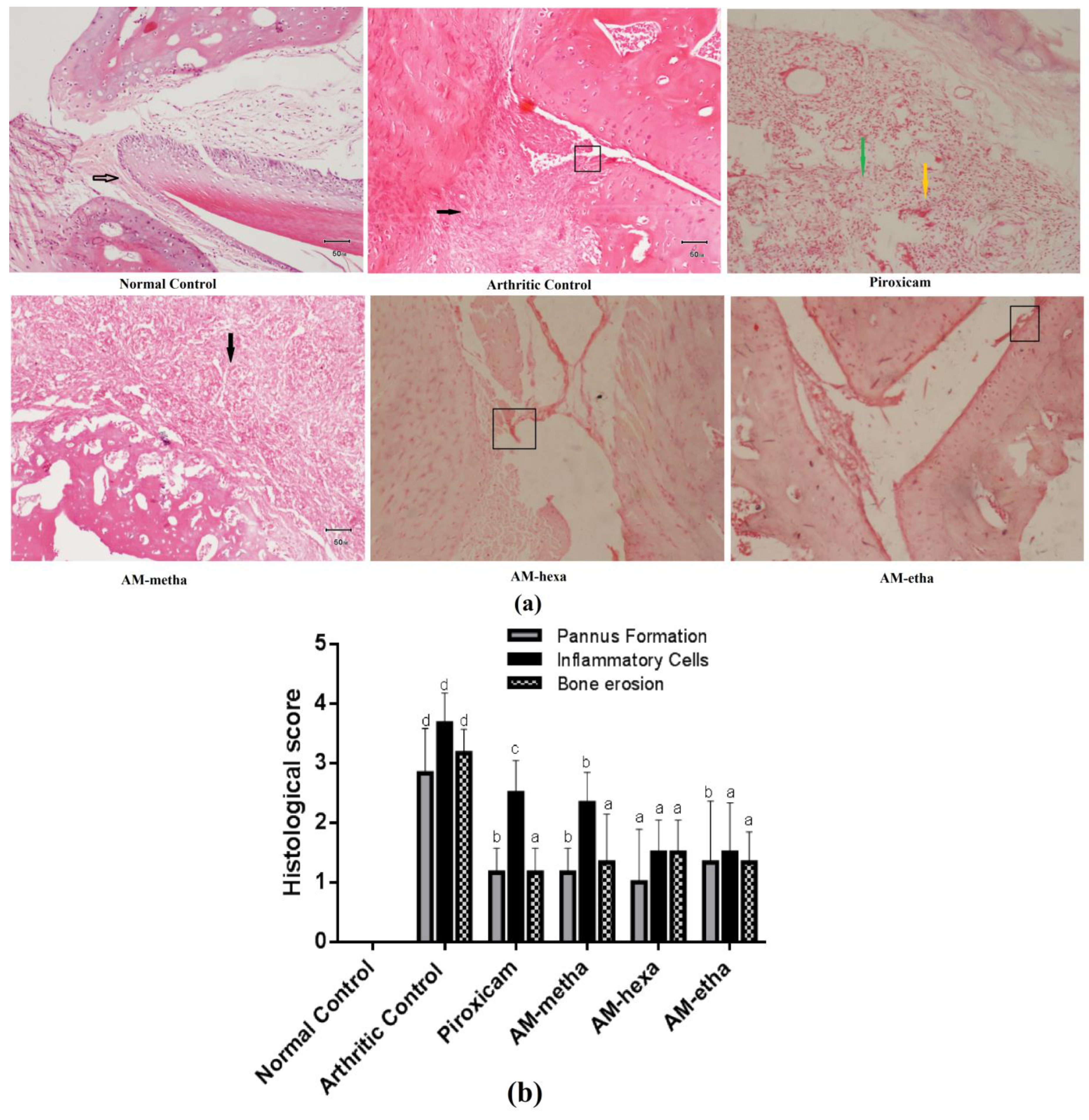
AM-metha extract, AM-hexa fraction, AM-etha fraction, and piroxicam displayed reduction in erosion of bones, inflammatory cell infiltration, and pannus formation in comparison with the arthritic control. The normal control showed normal bone and a normal synovial layer (black arrow); the arthritic control showed marked pannus formation (black arrow) and eroded cartilage (rectangular); piroxicam showed fibrosis formation (green arrow) and moderate infiltration of inflammatory cells (yellow arrow); AM-metha displayed moderate pannus formation (black arrow); AM-hexa showed eroded bone at the level of the joint (rectangular); and AM-etha showed mild erosion of joint cartilage; see Figure 2a,b.

3.3. A. modesta Significantly Reduced PGE2 Levels

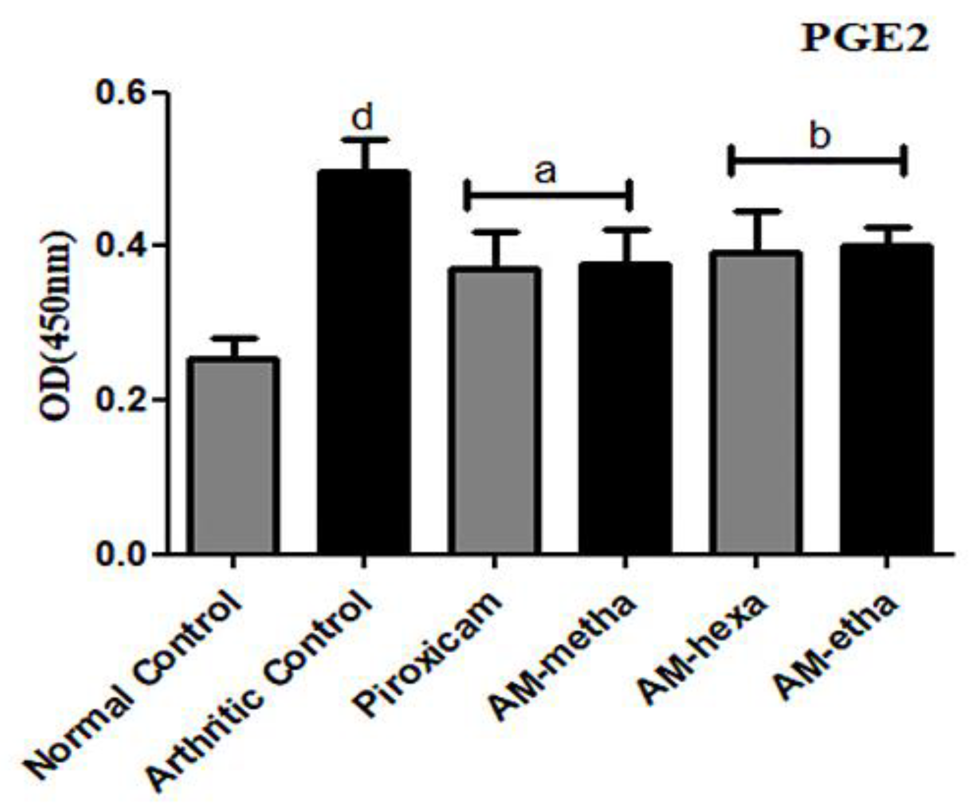
Levels of PGE2 were found to be significantly raised in the arthritic control in comparison with the normal control. The treatment with extract of AM-metha and its fractions, such as AM-hexa and AM-etha, prominently reduced the PGE2 levels when compared with the arthritic control. Piroxicam also significantly decreased the PGE2 levels as compared to the arthritic control, as shown in Figure 3.

3.4. A. modesta Significantly Reduced Matrix Metalloproteinase, Pro-Inflammatory Cytokine, Transcription Factor, and Growth Factor Expression Levels

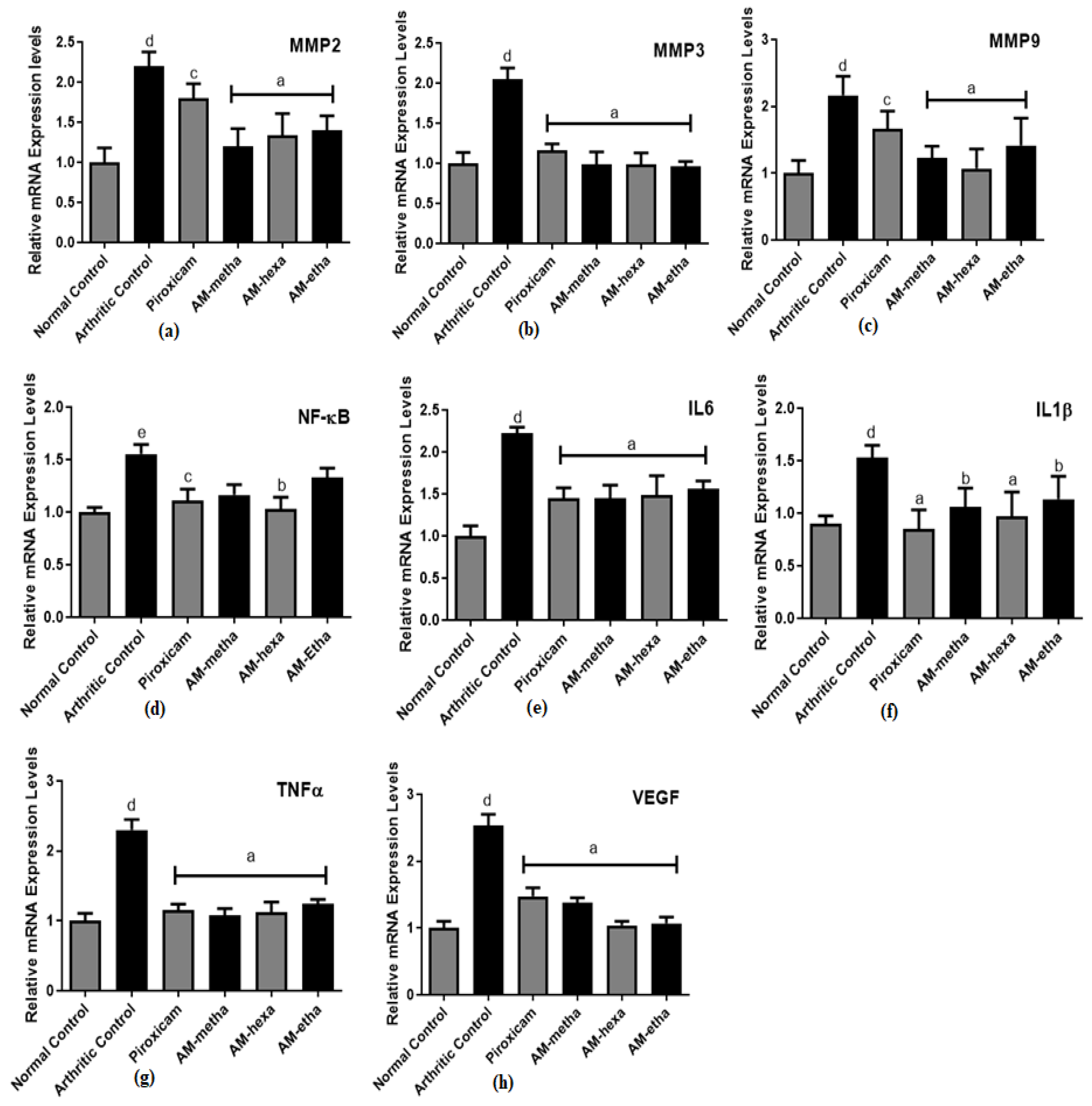
Expression levels of matrix metalloproteinases, such as MMP2, MMP3, and MMP9, were increased in all groups after CFA induction, but after treatment with AM-metha, AM-hexa, and AM-etha, their levels were significantly reduced when compared with the arthritic control. Expression levels of pro-inflammatory cytokines, such as IL6, IL1β, and TNFα, were also found to be higher in the arthritic control as compared with the normal control. Treatment with AM-metha, AM-hexa, and AM-etha reduced their levels significantly when compared with the arthritic control. The transcription factor NF-κB and the growth factor VEGF were significantly increased in arthritic control rats as compared to normal control rats. All treatment groups displayed a significant reduction in VEGF expression levels, while AM-metha and AM-etha showed a non-significant reduction in NF-κB expression levels as compared to the arthritic control group. Piroxicam also showed comparable results (Figure 4).

3.5. A. modesta Improved Hematological Markers and Did Not Display Harm to Liver and Kidneys

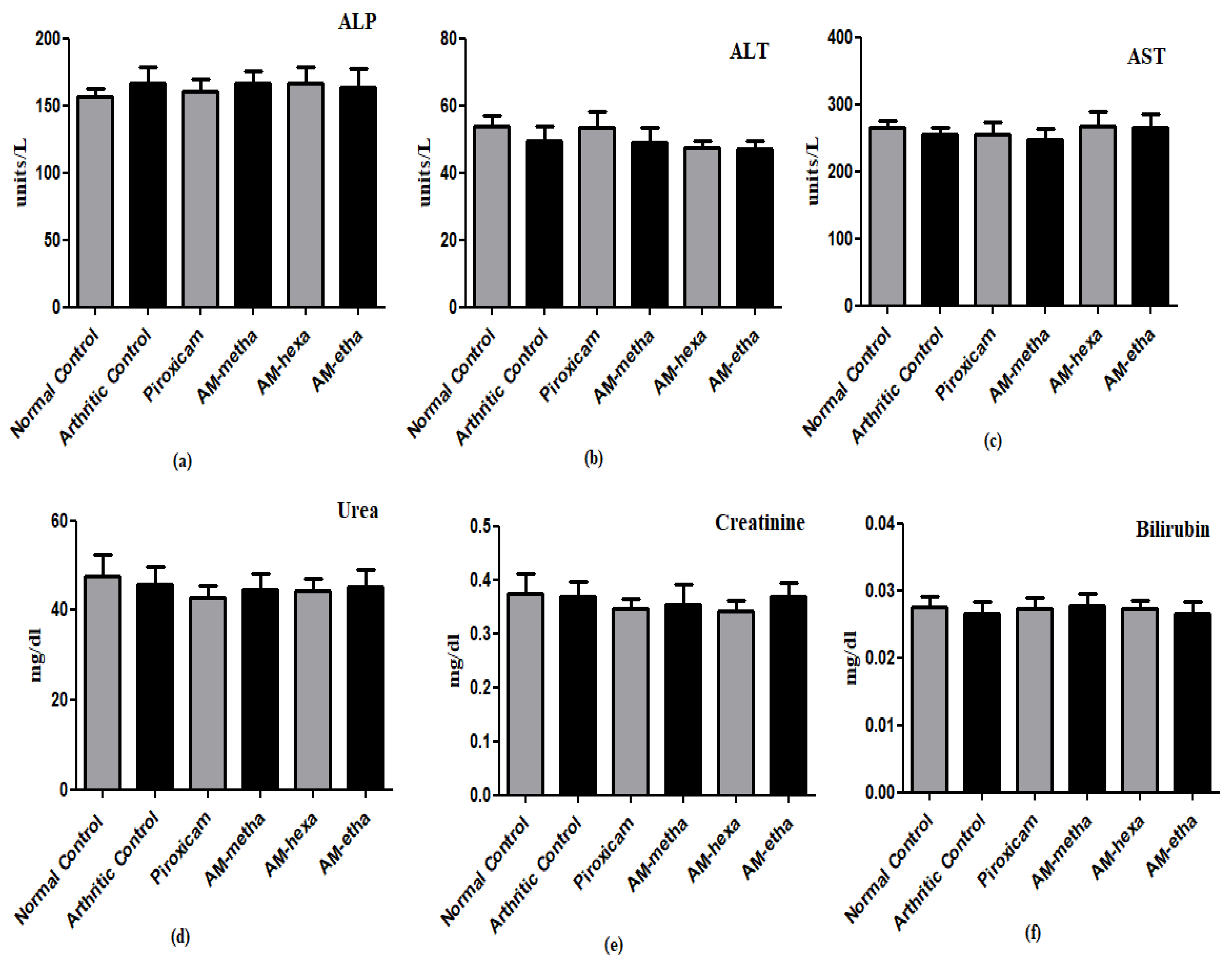
The arthritic control group showed reduced hemoglobin content and RBC counts as compared to the normal control. Treatment with AM-metha, AM-hexa, AM-etha, and piroxicam showed elevated Hb content and RBC counts. A significant elevation in platelet and WBC counts was observed in arthritic control rats as compared to normal control rats, respectively. AM-metha, AM-hexa, and AM-etha groups displayed a reduction in the levels. The attenuation was also seen in piroxicam-treated rats (Table 3). The levels of ALP, total bilirubin, creatinine, urea, AST, and ALT were also evaluated in the current study and a statistically non-significant difference was detected among all groups when they were compared with each other (Figure 5).

3.6. GC/MS Analysis of Extract and Fractions of A. modesta

GC/MS analysis of the methanolic extract, n-hexane fraction, and ethyl acetate fraction of A. modesta was conducted to determine the presence of anti-inflammatory compounds. The bioactive compounds, with their retention time, total percentage, molecular formula, and molecular weight, are shown in Table 4 for methanolic extract, Table 5 for n-hexane fraction, and Table 6 for ethyl acetate fraction.

4. Discussion

Rheumatoid arthritis (RA) is an inflammatory autoimmune disorder subsequently resulting in the formation of pannus, infiltration of inflammatory cells, and synovial hyperplasia, followed by eroded bone and cartilage [37]. In this disease, macrophage and fibroblast-like synoviocytes are the main contributory factors in pannus formation and facilitate bone and joint damage during joint inflammation [6]. The CFA rat model is the model of choice and is commonly used for the evaluation of chronic inflammatory disease due to its similarities with human RA. This model is known to trigger synovial hyperplasia, inflammation, and damage to bone and cartilage by up-regulating the pro-inflammatory cytokines and infiltrating inflammatory cells [5]. A marked inflammation was seen in the hind paw of rats injected with CFA. The vascular exudative phenomenon transports the immune cells to the site of the injected area in the acute phase of inflammation. Then, production of pro-inflammatory cytokines facilitates the formation of pannus, synovial hyperplasia, and damage to bone and cartilage in the chronic phase of inflammation [37]. In the current study, a reduction in paw volume, arthritic score, and histopathological findings was found in plant-treated groups, which showed the anti-arthritic potential of A. modesta.
The pro-inflammatory cytokines, such as TNFα, IL6, and IL1β, play a dominant part in the pathogenesis of RA [36,43]. Cytokine-mediated inflammation is a hallmark of autoimmune diseases. Cytokines such as IL1, 1L6, and TNFα are released from synovial tissues into the systemic circulation and participate in the degradation and inflammation of joints. The levels of 1L6, IL1β, and TNFα are found to be higher in arthritic patients [3,44]. Cytokines work in many areas of inflammatory processes associated with inflammation regulation, autoimmune events, inflammation of synovial membranes, and damage of articular joints [6]. In RA, the activation and differentiation of osteoclast are facilitated by the increased level of pro-inflammatory cytokines. IL1β takes part in the development of osteoclasts by stimulating the RANKL (receptor activator of NF-kappa B ligand), which possesses osteoclastogenic properties. The activation of IL1β in the synovium tissue causes overexpression of MMPs, leading to damage of bone and joints [10]. TNFα also up-regulates the levels of MMPs and PGE2 via fibroblasts and macrophages, leading to synovitis, bone resorption, and cartilage erosion. It also enhances the levels of another pro-inflammatory cytokine, i.e., IL6 [5], which is considered to be a multifunctional cytokine and participates in the activation of osteoclastogenesis, the inflammatory process, synovitis, and the up-regulation of MMPs, leading to bone and cartilage destruction in the affected joints [45]. Attenuation in the gene expression levels of TNFα, IL6, and IL-1β was found in plant-bark-treated groups as compared to the disease group, showing the immunomodulatory property of A. modesta.
MMPs are inflammatory enzymes that are involved in the damage of joints by degrading the components of the extracellular matrix [46]. MMPs such as MMP2, MMP3, and MMP9 are up-regulated in arthritis, leading to damage to bones and cartilage of joints via activation of IL1β, TNFα, and many other cytokines. These cytokines regulate the monitoring of MMPs, mainly through the mitogen-activated protein kinase (MAPK) signal transduction pathway [47]. VEGF, as an inflammatory and angiogenetic mediator, causes the activation of pro-inflammatory cytokines and the development of new vessels by stimulating the immune and endothelial cells. This activation then leads to an inflammatory and angiogenetic event in RA [13]. PGE2 is the key prostaglandin among the various prostaglandins, and is activated by synovial fibroblasts, chondrocytes, and pro-inflammatory cytokines such as IL1β and TNFα [48]. PGE2 is abundantly found in RA patients and acts as an inflammatory and pain marker in RA. It is known to damage bone and cartilage by activating metalloprotease enzymes [12]. NF-ĸB is a transcription factor that has a major involvement in the pathogenesis of rheumatoid arthritis [49]. The activation of NF-κB plays a significant role in the stimulation of pro-inflammatory cytokines and MMPs, and in the inhibition of apoptosis, leading to development of disease [48]. The levels of PGE2, MMP2, MMP3, MMP9, VEGF, and NF-ĸB were down-regulated in extract- and fraction-treated groups when compared with the disease group, validating the immunomodulatory and anti-inflammatory properties of A. modesta, as indicated by the down-regulation of pro-inflammatory cytokines.
The biochemical levels in serum, such as those of ALP, AST, ALT, total bilirubin, urea, and creatinine, were evaluated to determine the safety of plant bark in the liver and kidney [29]. The higher levels of liver enzymes such as ALP, AST, and ALT in the systemic circulation is an indication of hepatic damage [34]. Apart from this, ALP also acts as a biomarker of bone damage and its levels become elevated in RA [50]. Elevated levels of total bilirubin act as a prognostic indicator for acute to chronic liver disease [51]. The higher levels of creatinine and urea in the systemic circulation are a sign of renal damage because they act as a biomarker of renal disease. In the normal state, these compounds are filtered out from the blood through the kidneys, whereas in a pathological state, their levels become elevated in the circulation due to the inability of the kidneys to excrete them properly [29]. Our study found non-significant differences in the levels of ALP, ALT, AST, urea, creatinine, and total bilirubin when compared with each other, which suggests that no harm to kidney or liver was caused by plant bark treatment. The changes in the levels of hematological parameters in CFA-induced rats are linked to the severity of RA. Anemia is common in rheumatoid arthritis, characterized by low levels of RBCs and Hb content [31]. The anemic condition occurs due to the abnormality in the storage of iron in the synovial tissue and reticuloendothelial system. In addition, the inability of bone marrow to generate adequate cells is also a causative factor in the attenuation of RBCs and Hb content. Activation of the immune system against antigen attack leads to high levels of WBCs and platelets in RA [32]. Elevation of WBC count is associated with the activation of the immune system against destructive pathogens, ultimately leading to activation of inflammatory modulators [36]. Platelets play a significant role in inflammation and immunomodulation. Microparticles released from platelets interact with WBCs and produce systemic and joint inflammation in RA [52]. Treatment with plant bark increased the levels of RBCs and Hb content and decreased the levels of WBCs and platelets when compared with the arthritic control, thus the validating anti-arthritic potential of A. modesta.
Although not perfect, chromatography is a widely used method for the identification of constituents in extracts and fractions. The GC-MS analysis of Acacia modesta Wall revealed the presence of previously reported anti-inflammatory and antioxidant phytochemicals, which might be attributed to its antiarthritic property. Lupeol is known to be a powerful anti-inflammatory and antioxidant compound. It reduces the progression of arthritis through down-regulation of the P13K/Akt signaling pathway [53,54,55]. Oleic acid possess anti-inflammatory activity [56]. It produces its effect by targeting proinflammatory cytokines TNF-α and IL-1β [57]. γ-Sitosterol has both anti-inflammatory and antioxidant effects [58,59]. Squalene is reported to be an anti-inflammatory constituent [60]. Hexadecanoic acid, methyl ester, and 5-Hydroxymethyl furfural produce antioxidant effects [61,62]. All these phytochemicals and others found in this study may be credited with the anti-inflammatory activity, which resulted in amelioration of RA.

5. Conclusions

Treatment with Acacia modesta Wall. bark ameliorated rheumatoid arthritis. The data indicate that treatment with A. modesta reduced the paw volume, arthritic score, pannus formation, cartilage and bone erosion, and infiltration of inflammatory cells, thus showing the anti-arthritic potential of the plant. The anti-arthritic potential may be ascribed to down-regulation of MMP2, MMP3, MMP9, IL1β, TNFα, IL6, NF-κB, PGE2, and VEGF levels. Anti-inflammatory constituents such as lupeol, oleic acid, squalene, and various others found in the extract and fractions of plant bark might be credited with the anti-inflammatory activity, resulting in suppression of rheumatoid arthritis. Further studies are required for standardization of the plant extract before its consideration as a viable treatment option alongside conventional medicines.

Author Contributions

K.M.: Conceptualization, Methodology, Formal analysis, Writing—original draft, Data curation, Investigation; A.S.: Conceptualization, Supervision, Project administration, Investigation, methodology, Final approval; M.A.K.: Methodology, Validation, Visualization, Writing—review and editing, Formal analysis; H.H.: Methodology, Validation, Visualization, Writing—review and editing; M.S.: Formal analysis, validation, Writing—review and editing, investigation; A.I.; Formal analysis, Funding acquisition, Visualization, Writing—review and editing; G.A.S.: Data curation; Funding acquisition, Writing—review and editing, validation; A.M.: Formal analysis, Writing—review and editing, data curation, validation; T.A.: Validation, Writing—review and editing, Methodology, Investigation; Z.A.S.: Visualization; Formal analysis, review & editing, investigation; Y.A.B.J.: Funding acquisition, validation, Writing—review and editing, resources, visualization. All authors reviewed and commented on the manuscript. All authors have read and agreed to the published version of the manuscript.

Funding

This research is supported by the Researchers Supporting Project Number (RSP2023R457), King Saud University, Riyadh, Saudi Arabia.

Institutional Review Board Statement

All the experiments were approved under the approval number IREC-2019-91, issued from the Institutional Research Ethics Committee, The University of Lahore.

Informed Consent Statement

Not applicable.

Data Availability Statement

All the data is contained in the manuscript.

Acknowledgments

The authors would like to extend their sincere appreciation to the Researchers Supporting Project, King Saud University, Riyadh, Saudi Arabia. We would like to thank University of Lahore, Lahore–Punjab, Pakistan for providing facilities for experimental procedures.

Conflicts of Interest

The authors declare no conflict of interest.

Abbreviations

PGE2: Prostaglandin E2, MMP2: Matrix metalloproteinase 2, MMP3: Matrix metalloproteinase 3, MMP9: Matrix metalloproteinase 9, NF-κB: Nuclear factor kappa chain enhancer of activated B cells, IL6: Interleukin 6, IL1β: Interleukin 1β, TNFα: Tumor necrosis factor α, VEGF: Vascular endothelial growth factor, GC/MS: Gas Chromatography/Mass spectrometry, ELISA: Enzyme-linked immunosorbent assay.

References

  1. Pezone, A.; Olivieri, F.; Napoli, M.V.; Procopio, A.; Avvedimento, E.V.; Gabrielli, A. Inflammation and DNA damage: Cause, effect or both. Nat. Rev. Rheumatol. 2023, 19, 200–211. [Google Scholar] [CrossRef] [PubMed]
  2. Yi, Y.-S. Role of inflammasomes in inflammatory autoimmune rheumatic diseases. Korean J. Physiol. Pharmacol. 2018, 22, 1–15. [Google Scholar] [CrossRef] [PubMed]
  3. Shrivastava, A.K.; Pandey, A. Inflammation and rheumatoid arthritis. J. Physiol. Biochem. 2012, 69, 335–347. [Google Scholar] [CrossRef] [PubMed]
  4. Huang, Y.; Guo, L.; Chitti, R.; Sreeharsha, N.; Mishra, A.; Gubbiyappa, S.K.; Singh, Y. Wogonin ameliorate complete Freund’s adjuvant induced rheumatoid arthritis via targeting NF-κB/MAPK signaling pathway. Biofactors 2020, 46, 283–291. [Google Scholar] [CrossRef] [PubMed]
  5. Mbiantcha, M.; Almas, J.; Shabana, S.U.; Nida, D.; Aisha, F. Anti-arthritic property of crude extracts of Piptadeniastrum africanum (Mimosaceae) in complete Freund’s adjuvant-induced arthritis in rats. BMC Complement. Altern. Med. 2017, 17, 111. [Google Scholar] [CrossRef]
  6. Fang, Q.; Zhou, C.; Nandakumar, K.S. Molecular and Cellular Pathways Contributing to Joint Damage in Rheumatoid Arthritis. Mediat. Inflamm. 2020, 2020, 3830212. [Google Scholar] [CrossRef]
  7. Sivasakthi, P.; Priya, E.S.; Selvan, P.S. Molecular insights into phytochemicals exhibiting anti-arthritic activity: Systematic review. Inflamm. Res. 2021, 70, 665–685. [Google Scholar] [CrossRef]
  8. Brzustewicz, E.; Bryl, E. The role of cytokines in the pathogenesis of rheumatoid arthritis–Practical and potential application of cytokines as biomarkers and targets of personalized therapy. Cytokine 2015, 76, 527–536. [Google Scholar] [CrossRef]
  9. Ingawale, D.K.; Patel, S.S. Hecogenin exhibits anti-arthritic activity in rats through suppression of pro-inflammatory cytokines in Complete Freund’s adjuvant-induced arthritis. Immunopharmacol. Immunotoxicol. 2017, 40, 59–71. [Google Scholar] [CrossRef]
  10. Ruscitti, P.; Cipriani, P.; Carubbi, F.; Liakouli, V.; Zazzeroni, F.; Di Benedetto, P.; Berardicurti, O.; Alesse, E.; Giacomelli, R. The Role of IL-1β in the Bone Loss during Rheumatic Diseases. Mediat. Inflamm. 2015, 2015, 782382. [Google Scholar] [CrossRef]
  11. Araki, Y.; Mimura, T. Matrix Metalloproteinase Gene Activation Resulting from Disordred Epigenetic Mechanisms in Rheumatoid Arthritis. Int. J. Mol. Sci. 2017, 18, 905. [Google Scholar] [CrossRef]
  12. Yan, Y.; Singh, G.K.; Zhang, F.; Wang, P.; Liu, W.; Zhong, L.; Yang, L. Comparative Study of Normal and Rheumatoid Arthritis Fibroblast-Like Synoviocytes Proliferation under Cyclic Mechanical Stretch: Role of Prostaglandin E2. Connect. Tissue Res. 2011, 53, 246–254. [Google Scholar] [CrossRef]
  13. Lee, Y.H.; Bae, S.-C. Correlation between circulating VEGF levels and disease activity in rheumatoid arthritis: A meta-analysis. Z. Rheumatol. 2018, 77, 240–248. [Google Scholar] [CrossRef]
  14. Liu, T.; Zhang, L.; Joo, D.; Sun, S.C. NF-κB signaling in inflammation. Signal. Transduct. Targeted. Ther. 2017, 2, 17023. [Google Scholar] [CrossRef] [PubMed]
  15. Makarov, S.S. NF-κB in rheumatoid arthritis: A pivotal regulator of inflammation, hyperplasia, and tissue destruction. Arthritis Res. Ther. 2001, 3, 200–206. [Google Scholar] [CrossRef] [PubMed]
  16. Radu, A.-F.; Bungau, S.G. Management of Rheumatoid Arthritis: An Overview. Cells 2021, 10, 2857. [Google Scholar] [CrossRef] [PubMed]
  17. Zhang, Z.; Chinnathambi, A.; Alharbi, S.A.; Bai, L. Copper oxide nanoparticles from Rabdosia rubescens attenuates the complete Freund’s adjuvant (CFA) induced rheumatoid arthritis in rats via suppressing the inflammatory proteins COX-2/PGE2. Arab. J. Chem. 2020, 13, 5639–5650. [Google Scholar] [CrossRef]
  18. Laurindo, L.F.; de Maio, M.C.; Minniti, G.; de Góes Corrêa, N.; Barbalho, S.M.; Quesada, K.; Guiguer, E.L.; Sloan, K.P.; Detregiachi, C.R.; Araújo, A.C.; et al. Effects of medicinal plants and phytochemicals in Nrf2 pathways during inflammatory bowel diseases and related colorectal cancer: A comprehensive review. Metabolites 2023, 13, 243. [Google Scholar] [CrossRef]
  19. Feng, W.; Ao, H.; Peng, C. Gut Microbiota, Short-Chain Fatty Acids, and Herbal Medicines. Front. Pharmacol. 2018, 9, 1354. [Google Scholar] [CrossRef]
  20. Bukhari, I.A.; Khan, R.A.; Gilani, A.H.; Ahmed, S.; Saeed, S.A. Analgesic, anti-inflammatory and anti-platelet activities of the methanolic extract of Acacia modesta leaves. Inflammopharmacology 2010, 18, 187–196. [Google Scholar] [CrossRef]
  21. Kamal, M.; Adnan, M.; Murad, W.; Bibi, H.; Tariq, A.; Rahman, H.; Shinwari, Z.K. Anti-rheumatic potential of Pakistani medicinal plants: A review. Pak. J. Bot. 2016, 48, 399–413. [Google Scholar]
  22. Murad, W.; Azizullah, A.; Adnan, M.; Tariq, A.; Khan, K.U.; Waheed, S.; Ahmad, A. Ethnobotanical assessment of plant resources of Banda Daud Shah, District Karak, Pakistan. J. Ethnobiol. Ethnomed. 2013, 9, 77. [Google Scholar] [CrossRef] [PubMed]
  23. Sher, H.; Aldosari, A.; Ahmad, S. Ethnoecological appraisal of Acacia modesta Wall. common tree of dry ecosystem in Pakistan. Afr. J. Agric. Res. 2012, 7, 5083–5091. [Google Scholar] [CrossRef]
  24. Ur, M.S.R.; Chaudhry, M.A. Evaluation of antioxidant and hepatoprotective effect of Acacia modesta wall against paracetamol induced hepatotoxicity. Br. J. Pharm. Res. 2015, 5, 336–343. [Google Scholar]
  25. Saleem, B.; Islam, M.; Saeed, H.; Imtiaz, F.; Asghar, M.; Saleem, Z.; Mehmood, A.; Naheed, S. Investigations of Acacia modesta Wall. leaves for in vitro anti-diabetic, proliferative and cytotoxic effects. Braz. J. Pharm. Sci. 2018, 54, e17467. [Google Scholar] [CrossRef]
  26. Qazi, N.G.; Khan, A.-U.; Ali, F. Anti-diarrheal, anti-secretory, anti-spasmodic and antiulcer activities of Acacia modesta (Mimosaceae) aerial parts. Trop. J. Pharm. Res. 2017, 16, 2231. [Google Scholar] [CrossRef]
  27. Jawla, S.; Kumar, Y.; Khan, M.S. Antimicrobial and antihyperglycemic activities of Acacia modesta leaves. Pharmacologyonline 2011, 2, 331–343. [Google Scholar]
  28. Latif, S.; Ismail, H.; Khan, M.R.; Rahim, A.A.; Mehboob, R.; Dilshad, E.; Sajid, M.; Haider, S.I.; Anwaar, S.; Majeed, M.N. Pharmacological evaluation of Acacia modesta bark for antipyretic, anti-inflammatory, analgesic, antidepressant and anticoagulant activities in Sprague Dawley rats. Pak. J. Pharm. Sci. 2020, 33, 1015–1023. [Google Scholar]
  29. Shabbir, A.; Batool, S.A.; Basheer, M.I.; Shahzad, M.; Sultana, K.; Tareen, R.B.; Iqbal, J.; Hassan, S.U. Ziziphora clinopodioides ameliorated rheumatoid arthritis and inflammatory paw edema in different models of acute and chronic inflammation. Biomed. Pharmacother. 2018, 97, 1710–1721. [Google Scholar] [CrossRef]
  30. Uttra, A.M.; Hasan, U.H. Anti-arthritic activity of aqueous-methanolic extract and various fractions of Berberis orthobotrys Bien ex Aitch. BMC Complement. Altern. Med. 2017, 17, 371. [Google Scholar] [CrossRef]
  31. Mahnashi, M.H.; Jabbar, Z.; Alamgeer; Irfan, H.M.; Asim, M.H.; Akram, M.; Saif, A.; Alshahrani, M.A.; Alshehri, M.A.; Asiri, S.A. Venlafaxine demonstrated anti-arthritic activity possibly through down regulation of TNF-α, IL-6, IL-1β, and COX-2. Inflammopharmacology 2021, 29, 1413–1425. [Google Scholar] [CrossRef]
  32. Shabbir, A.; Shahzad, M.; Ali, A.; Zia-Ur-Rehman, M. Discovery of New Benzothiazine Derivative as Modulator of Pro- and Anti-inflammatory Cytokines in Rheumatoid Arthritis. Inflammation 2016, 39, 1918–1929. [Google Scholar] [CrossRef] [PubMed]
  33. Riaz, M.; Khan, A.; Zia-Ul-Haq, M.; Dima, L. Mechanism of anti-inflammatory and anti-nociceptive actions of acacia modesta in animal models. Pak. J. Zool. 2015, 47, 1723–1730. [Google Scholar]
  34. Cui, X.; Wang, R.; Bian, P.; Wu, Q.; Seshadri, V.D.D.; Liu, L. Evaluation of antiarthritic activity of nimbolide against Freund’s adjuvant induced arthritis in rats. Artif. Cells Nanomed. Biotechnol. 2019, 47, 3391–3398. [Google Scholar] [CrossRef] [PubMed]
  35. Uroos, M.; Abbas, Z.; Sattar, S.; Umer, N.; Shabbir, A.; Rehman, S.U.; Sharif, A. Nyctanthes arbor-tristis Ameliorated FCA-Induced Experimental Arthritis: A Comparative Study among Different Extracts. Evid.-Based Complement. Altern. Med. 2017, 2017, 4634853. [Google Scholar] [CrossRef]
  36. Tian, Z.; Chinnathambi, A.; Alahmadi, T.A.; Mohan, S.K.; Veeraraghavan, V.P.; Jaganathan, S.K. Anti-arthritic activity of Tin oxide-Chitosan-Polyethylene glycol carvacrol nanoparticles against Freund’s adjuvant induced arthritic rat model via the inhibition of cyclooxygenase-2 and prostaglandin E2. Arab. J. Chem. 2021, 14, 103293. [Google Scholar] [CrossRef]
  37. Das, C.; Bose, A.; Das, D. Ayurvedic Balarista ameliorate anti-arthritic activity in adjuvant induced arthritic rats by inhibiting pro-inflammatory cytokines and oxidative stress. J. Tradit. Complement. Med. 2020, 11, 228–237. [Google Scholar] [CrossRef]
  38. Mobashar, A.; Shabbir, A.; Shahzad, M.; Gobe, G. Preclinical Rodent Models of Arthritis and Acute Inflammation Indicate Immunomodulatory and Anti-Inflammatory Properties of Juglans regia Extracts. Evid.-Based Complement. Altern. Med. 2022, 2022, 1695701. [Google Scholar] [CrossRef]
  39. Sharif, M.; Anjum, I.; Shabbir, A.; Syed, S.K.; Mobeen, I.; Shahid, M.H.; Sarwar, K. Amelioration of Ovalbumin-Induced Allergic Asthma by Juglans regia via Downregulation of Inflammatory Cytokines and Upregulation of Aquaporin-1 and Aquaporin-5 in Mice. J. Trop. Med. 2022, 2022, 6530095. [Google Scholar] [CrossRef]
  40. Mobashar, A.; Shabbir, A.; Shahzad, M.; Hassan, S.U. Evaluation of Immunomodulatory and Antiarthritic Potential of Trigonella gharuensis Extracts. Evid.-Based Complement. Altern. Med. 2020, 2020, 8836080. [Google Scholar] [CrossRef]
  41. Akhtar, T.; Ali, G.; Sheikh, N. Immunosuppressant-Induced Oxidative Stress and Iron: A Paradigm Shift from Systemic to Intrahepatic Abnormalities. Oxid. Med. Cell. Longev. 2020, 2020, 8675275. [Google Scholar] [CrossRef] [PubMed]
  42. Zhu, J.; Su, C.; Chen, Y.; Hao, X.; Jiang, J. Electroacupuncture on ST36 and GB39 Acupoints Inhibits Synovial Angiogenesis via Downregulating HIF-1α/VEGF Expression in a Rat Model of Adjuvant Arthritis. Evid.-Based Complement. Altern. Med. 2019, 2019, 5741931. [Google Scholar] [CrossRef] [PubMed]
  43. Kondo, N.; Kuroda, T.; Kobayashi, D. Cytokine Networks in the Pathogenesis of Rheumatoid Arthritis. Int. J. Mol. Sci. 2021, 22, 10922. [Google Scholar] [CrossRef] [PubMed]
  44. Hu, S.-L.; Chang, A.-C.; Huang, C.-C.; Tsai, C.-H.; Lin, C.-C.; Tang, C.-H. Myostatin Promotes Interleukin-1β Expression in Rheumatoid Arthritis Synovial Fibroblasts through Inhibition of miR-21-5p. Front. Immunol. 2017, 8, 1747. [Google Scholar] [CrossRef]
  45. Hashizume, M.; Mihara, M. The Roles of Interleukin-6 in the Pathogenesis of Rheumatoid Arthritis. Arthritis 2011, 2011, 765624. [Google Scholar] [CrossRef]
  46. Li, R.-L.; Duan, H.-X.; Liang, Q.; Huang, Y.-L.; Wang, L.-Y.; Zhang, Q.; Wu, C.-J.; Liu, S.-Q.; Peng, W. Targeting matrix metalloproteases: A promising strategy for herbal medicines to treat rheumatoid arthritis. Front. Immunol. 2022, 13, 1046810. [Google Scholar] [CrossRef]
  47. Burrage, P.S.; Mix, K.S.; Brinckerhoff, C.E. Matrix Metalloproteinases: Role in Arthritis. Front. Biosci. 2006, 11, 529–543. [Google Scholar] [CrossRef]
  48. Fattahi, M.J.; Mirshafiey, A. Prostaglandins and Rheumatoid Arthritis. Arthritis 2012, 2012, 239310. [Google Scholar] [CrossRef]
  49. Świerkot, J.; Nowak, B.; Czarny, A.; Zaczyńska, E.; Sokolik, R.; Madej, M.; Korman, L.; Sebastian, A.; Wojtala, P.; Lubiński, Ł.; et al. The Activity of JAK/STAT and NF-κB in Patients with Rheumatoid Arthritis. Adv. Clin. Exp. Med. 2016, 25, 709–717. [Google Scholar] [CrossRef]
  50. Hussain, A.; Aslam, B.; Muhammad, F.; Faisal, M.N.; Kousar, S.; Mushtaq, A.; Bari, M.U. Anti-arthritic activity of Ricinus communis L. and Withania somnifera L. extracts in adjuvant-induced arthritic rats via modulating inflammatory mediators and subsiding oxidative stress. Iran. J. Basic. Med. Sci. 2021, 24, 951–961. [Google Scholar] [CrossRef]
  51. López-Velázquez, J.A.; Chávez-Tapia, N.C.; Ponciano-Rodríguez, G.; Sánchez-Valle, V.; Caldwell, S.H.; Uribe, M.; Méndez-Sánchez, N. Bilirubin alone as a biomarker for short-term mortality in acute-on-chronic liver failure: An important prognostic indicator. Ann. Hepatol. 2014, 13, 98–104. [Google Scholar] [CrossRef]
  52. Harifi, G.; Sibilia, J. Pathogenic role of platelets in rheumatoid arthritis and systemic autoimmune diseases: Perspectives and therapeutic aspects. Saudi Med. J. 2016, 37, 354. [Google Scholar] [CrossRef] [PubMed]
  53. Saleem, M. Lupeol, a novel anti-inflammatory and anti-cancer dietary triterpene. Cancer Lett. 2009, 285, 109–115. [Google Scholar] [CrossRef]
  54. Song, T.; Shi, R.; Vijayalakshmi, A.; Lei, B. Protective effect of lupeol on arthritis induced by type II collagen via the suppression of P13K / AKT signaling pathway in Sprague dawley rats. Environ. Toxicol. 2022, 37, 1814–1822. [Google Scholar] [CrossRef] [PubMed]
  55. Wal, A.; Srivastava, R.S.; Wal, P.; Rai, A.; Sharma, S. Lupeol as a magical drug. Pharm. Biol. Eval. 2015, 2, 142–151. [Google Scholar]
  56. Santamarina, A.B.; Pisani, L.P.; Baker, E.J.; Marat, A.D.; Valenzuela, C.A.; Miles, E.A.; Calder, P.C. Anti-inflammatory effects of oleic acid and the anthocyanin keracyanin alone and in combination: Effects on monocyte and macrophage responses and the NF-κB pathway. Food Funct. 2021, 12, 7909–7922. [Google Scholar] [CrossRef]
  57. Medeiros-De-Moraes, I.M.; Gonçalves-De-Albuquerque, C.F.; Kurz, A.R.M.; Oliveira, F.M.d.J.; de Abreu, V.H.P.; Torres, R.C.; Carvalho, V.F.; Estato, V.; Bozza, P.T.; Sperandio, M.; et al. Omega-9 Oleic Acid, the Main Compound of Olive Oil, Mitigates Inflammation during Experimental Sepsis. Oxid. Med. Cell. Longev. 2018, 2018, 6053492. [Google Scholar] [CrossRef]
  58. Azhar, A.S.; Suhaila, H.B.; Imad, H.H. Analysis of bioactive chemical compounds of Euphorbia lathyrus using gas chromatography-mass spectrometry and Fourier-transform infrared spectroscopy. J. Pharmacogn. Phytother. 2016, 8, 109–126. [Google Scholar] [CrossRef]
  59. Singh, S.; Patra, A. Evaluation of adaptogenic potential of Polygonatum cirrhifolium (Wall.) Royle: In vitro, in vivo and in silico studies. S. Afr. J. Bot. 2018, 121, 159–177. [Google Scholar] [CrossRef]
  60. Latief, M.; Muhaimin, M.; Amanda, H.; Prahandika, G.; Tarigan, I.L. Anti-inflammatory activities of squalene compound of methanol extract of Abroma augusta L. J. Teknol. Lab. 2020, 9, 176–185. [Google Scholar] [CrossRef]
  61. Mujeeb, F.; Bajpai, P.; Pathak, N. Phytochemical Evaluation, Antimicrobial Activity, and Determination of Bioactive Components from Leaves of Aegle marmelos. BioMed Res. Int. 2014, 2014, 497606. [Google Scholar] [CrossRef] [PubMed]
  62. Shapla, U.M.; Solayman, M.; Alam, N.; Khalil, M.I.; Gan, S.H. 5-Hydroxymethylfurfural (HMF) levels in honey and other food products: Effects on bees and human health. Chem. Cent. J. 2018, 12, 35. [Google Scholar] [CrossRef] [PubMed]
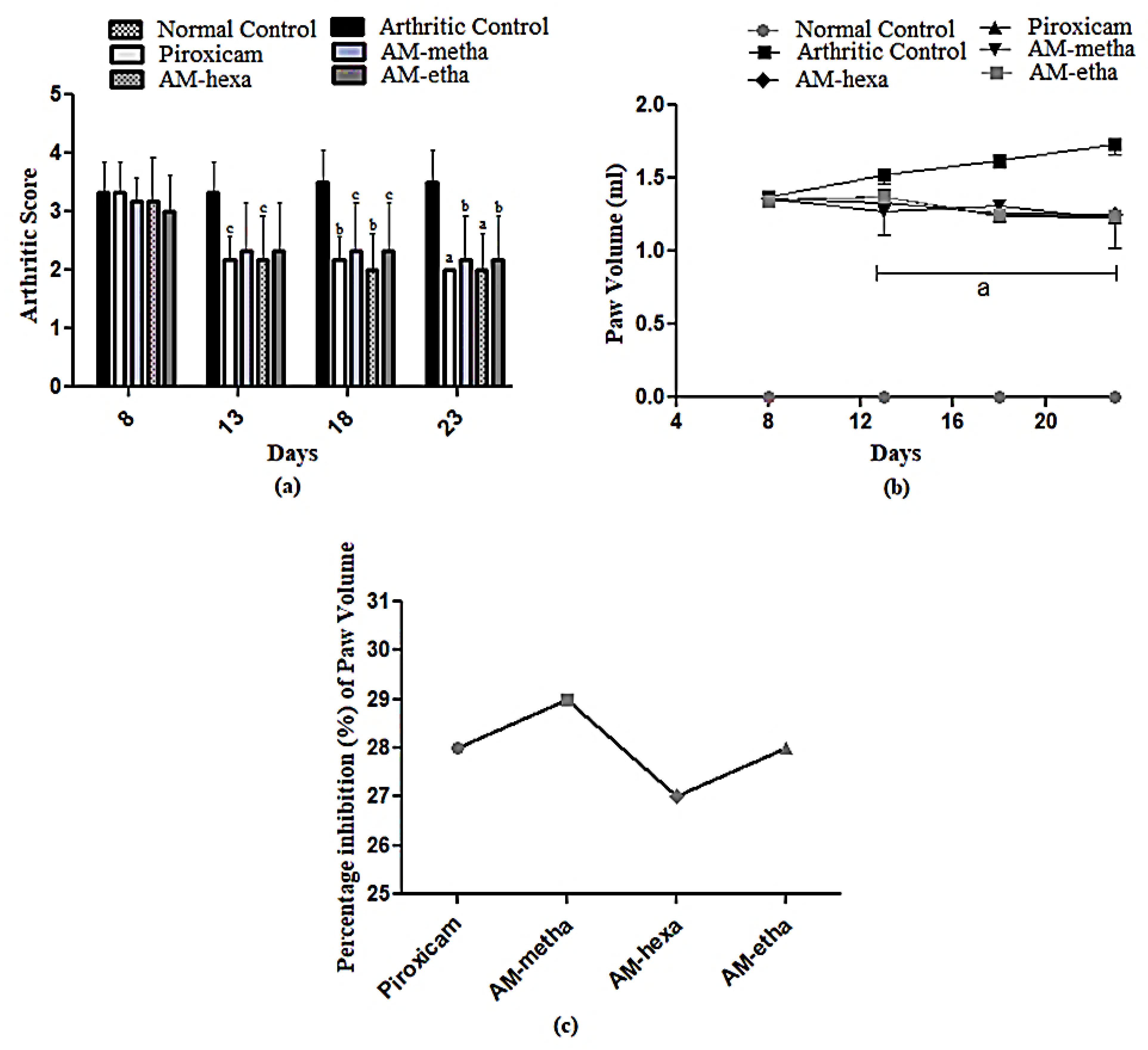
Figure 1. Arthritic index and paw volume. (a) c p < 0.05, b p < 0.01, and a p < 0.001, and (b) a p < 0.001 when other group values were compared with arthritic control; (c) % inhibition when compared with the arthritic control. The highest percentage inhibition was shown by the AM-metha group.
Pharmaceutics 15 02228 g001
Figure 2. (a) Photomicrograph (100×) of histopathological findings (H&E stained). (b) Values are shown as mean ± SD. When treatment group’s values were compared with the arthritic control, then c p < 0.05, b p < 0.01 and a p < 0.001 were considered. d p < 0.001 when arthritic control was compared with normal control.
Pharmaceutics 15 02228 g002
Figure 3. PGE2 protein level. AM-meth, AM-hexa, and AM-etha decreased the levels of PGE2 as compared to the arthritic control group. Here, a p < 0.001 and b p < 0.01 are compared to the arthritic group, whereas d p < 0.001 shows the comparison between the normal control and arthritic groups.
Pharmaceutics 15 02228 g003
Figure 4. AM-metha and AM-hexa prominently reduced the expression levels of (a) MMP2, (b) MMP3, (c) MMP9, (d) NF-κB, (e) IL6, (f) IL1β, (g) TNFα, and (h) VEGF, while AM-metha and AM-etha showed a non-significant reduction in NF-κB expression levels as compared to the arthritic control group. Here, a p < 0.001, b p < 0.01, and c p < 0.05 are compared to the arthritic group, whereas d p < 0.001 and e p < 0.01 shows the comparison between the normal control and arthritic groups.
Pharmaceutics 15 02228 g004
Figure 5. Biochemical markers. In the estimation of (a) ALP, (b) ALT, (c) AST, (d) urea, (e) creatinine, and (f) total bilirubin levels, a non-significant difference was found in all groups when compared with each other.
Pharmaceutics 15 02228 g005
Table 1. Treatment design.
Treatment GroupsDoseRoute of AdministrationReferences
Normal ControlNormal SalineOrally[29]
Arthritic Controli. CFA
ii. Normal saline
i. Injected
ii. Orally
Piroxicam10 mg/kg b.w to arthritic ratsIntraperitonially[32]
AM-metha500 mg/kg b.w to arthritic ratsOrally[33]
AM-hexa500 mg/kg b.w to arthritic ratsOrally
AM-etha500 mg/kg b.w to arthritic ratsOrally
Table 2. Primer list and sequences.
MarkersForward/Reverse PrimersAnnealing Temp (°C)Product SizeReferences
IL65′-GTCAACTCCATCTGCCCTTCAG-3′
5′-GGCAGTGGCTGTCAACAACAT-3′
59270[41]
MMP35′-CCTTTTGATGGGCCTGGAAT-3′
5′-GTGACATCATCTTGTCCATCG-3′
54 107ENSRNOG00000032626
IL1β5′-CCTGCTAGTGTGGATGTTC-3′
5′-GAGGTGCTGAGTTACCAGTT-3′
57390ENSRNOG00000004649
MMP95′-CCACCGAGCTATCCACTCAT-3′
5′-GTCCGGTTTCAGCATGTTTT-3′
56.1159ENSRNOG00000017539
NF-κB5′-CCGAGATAATGACAGCGTGT-3′
5′-CCTTGGGAACGATATGATGG-3′
58.4217ENSRNOG00000023258
TNF-α5′-ACAAGGCTGCCCCGACTAT-3′
5′-CTCCTGGTATGAAGTCCGAAATC-3′
6067[41]
VEGF5′-GTTCAGAGCGGAGAAAGCATT-3′
5′-CTTGCAACGCGAGTCTGTGT-3′
6080[42]
MMP25′-GCAACAAGTATGAGAGCTGC-3′
5′-CGGTCATCATCGTAGTTGGT-3′
5785ENSRNOG00000016695
Table 3. Hematological parameters.
MarkersNormal ControlArthritic ControlPiroxicamAM-MethaAM-HexaAM-Etha
Hemoglobin
(g/dL)
13.52 ± 1.3579.142 ± 0.521 d13.50 ± 0.411 a13.13 ± 0.096 a13.99 ± 0.625 a13.05 ± 0.884 a
RBCs
(106/µL)
7.143 ± 0.6625.417 ± 0.789 e7.188 ± 0.895 b7.750 ± 0.684 a7.503 ± 0.545 b7.625 ± 1.072 a
WBCs
(103/µL)
9.723 ± 0.41617.46 ± 2.350 d13.77 ± 1.256 c14.32 ± 2.062 b14.04 ± 1.192 b13.28 ± 0.710 c
Platelets
(103/µL)
418.7 ± 54.64711.5 ± 42.70 d555.7 ± 27.93 a550.2 ± 33.58 a637.0 ± 32.48 c547.2 ± 37.49 a
Here, a p < 0.001, b p < 0.01 and c p < 0.05 are compared to the arthritic group, whereas d p < 0.001 and e p < 0.01 are compared to the normal control group.
Table 4. Identified components of methanolic extract of A. modesta from mass chromatograms.
Identified CompoundsMolecular FormulaRetention TimeMolecular Weight (g/mol)Total (%Age)
2-Propanone, 1-cyclopentyl-C8H14O8.4801262.294
5-HydroxymethylfurfuralC6H6O311.5021261.582
Phenol,2,4-bis(1,1-dimethylethyl)-C14H22O15.5332062.230
Hexadecanoic acid, methyl esterC17H34O220.1152702.949
Estra-1,3,5(10)-trien-17β-olC18H24O20.4672567.127
3-(2,6,6-Trimethyl-cyclohex-1-enyl)-propionic acid, methyl esterC13H22O220.8762101.943
9,12-Octadecadienoic acid (Z, Z)-, methyl esterC19H34O221.7572942.212
Methyl stearateC19H38O221.8182983.853
6-Octadecenoic acid, methyl ester, (Z)-C19H36O222.0292961.875
9,12-Octadecadienoic acid (Z, Z)-C18H32O222.1112802.663
Kaur-16-eneC20H3222.8572722.440
17-Norkaur-15-ene, 13-methyl-, (8β, 13β)-C20H3223.3402723.198
Podocarp-7-en-3β-ol, 13β-methyl-13-vinyl-C20H32O23.4902881.785
1,6,10,14-Hexadecatetraen-3-ol, 3,7,11,15-tetramethyl-, (E, E)-C20H34O23.6032901.672
Naphthalene, decahydro-1,1,4a-trimethyl-6-methylene-5-(3-methyl-2,4-pentadienyl)-, [4aS-(4aα,5αC20H3223.8902724.854
Phenol,2,2′-methylenebis [6-(1,1-dimethylethyl)-4-methyl-C23H32O224.6513402.562
Androst-5-en-17-ol,4,4-dimethyl-C21H34O24.7643025.426
Estra-1,3,5(10)-trien-17-one,3-hydroxy-2-methoxy-C19H24O325.41930017.137
Podocarp-13-ene-14-glycolic acid, 7-hydroxy-8,13-dimethyl-3-oxo-, -lactoneC21H30O425.6003461.911
BolasteroneC21H32O225.8043161.954
16-Allopregnen-3β-ol-20-oneC21H32O226.2413161.496
CampesterolC28H48O30.7024002.046
StigmasterolC29H48O30.9804124.189
ϒ-SitosterolC29H50O31.5154143.981
LupeolC30H50O32.45042616.618
Table 5. Identified components of n-hexane fraction of A. modesta from mass chromatograms.
Identified CompoundsMolecular FormulaRetention TimeMolecular Weight (g/mol)Total (%Age)
2-Pyrrolidinone, 1-methyl-C5H9NO7.455994.1584
Heptadecane,2,6,10,15-tetramethyl-C21H4415.3142961.121
Phenol,2,4-bis(1,1-dimethylethyl)-C14H22O15.5332061.760
Eicosane,2-methyl-C21H4417.8762961.229
Hexadecanoic acid, methyl esterC17H34O220.1222702.358
1,2-Benzenedicarboxylic acid, butyl 8-methylnonyl esterC22H34O420.5443621.507
7,10-Octadecadienoic acid, methyl esterC19H34O221.7572941.569
10-Octadecenoic acid, methyl esterC19H36O221.8102961.201
Ethyleneglycol dipelargonateC20H38O423.2573422.256
Phenol,2,2′-methylenebis [6-(1,1-dimethylethyl)-4-methyl-C23H32O223.8973401.125
Estra-1,3,5(10)-trien-17-one,3-hydroxy-2-methoxy-C19H24O324.6513007.974
Bis(2-ethylhexyl) phthalateC24H38O425.4043906.118
ϒ-SitosterolC29H50O25.62341441.430
Methylenebis(2,4,6-triisopropylphenylphosphine)C31H50P231.5154842.582
Table 6. Identified components of ethyl acetate fraction of A. modesta from mass chromatograms.
Identified CompoundsMolecular FormulaRetention TimeMolecular Weight (g/mol)Total (%Age)
Phenol,2,4, bis(1,1-dimethylethyl)-C14H22O15.5332061.760
Hexadecanoic acid, methyl esterC17H34O220.1152702.004
n-Hexadecanoic acidC16H32O220.4692562.677
8,11-Octadecadienoic acid, methyl esterC19H34O221.7572942.453
10-Octadecadienoic acid, methyl esterC19H36O221.8102961.574
Oleic acidC18H34O222.1492821.279
9-Octadeccenoic acid,12-hydroxy-, methyl ester, [R-(Z)]-C19H36O323.5053121.472
1-Naphthalenemethanol, Decahydro-5-(5-hydroxy-3-methyl-3-pentenyl)-1,4a-dimethyl-6-methyleC20H34O223.8973061.333
Phenol,2,2′-methylenebis [6-(1,1-dimethylethyl)-4-methyl-C23H32O224.6513404.530
Androst-5-en-17-ol,4,4-dimethyl-C21H34O24.7643021.644
Hexadecanoic acid,1-(hydroxymethyl)-1,2-ethanediyl esterC35H68O525.2695681.008
Estra-1,3,5(10)-trien-17-one,3-hydroxy-2-methoxy-C19H24O325.4043004.534
Bis(2-ethylhexyl) phthalateC24H38O425.63039065.902
SqualeneC30H5O27.6354100.911
StigmasterolC29H48O30.9734121.033
sϒ-SitosterolC29H50O31.5154141.918
LupeolC30H50O32.4354263.696
Disclaimer/Publisher’s Note: The statements, opinions and data contained in all publications are solely those of the individual author(s) and contributor(s) and not of MDPI and/or the editor(s). MDPI and/or the editor(s) disclaim responsibility for any injury to people or property resulting from any ideas, methods, instructions or products referred to in the content.