Review

Targeting DNA Damage Repair and Immune Checkpoint Proteins for Optimizing the Treatment of Endometrial Cancer

1
College of Biological and Pharmaceutical Engineering, West Anhui University, Lu’an 237012, China
2
Anhui Province Key Laboratory of Medical Physics and Technology, Institute of Health and Medical Technology, Hefei Institutes of Physical Science, Chinese Academy of Sciences, Hefei 230031, China
*
Author to whom correspondence should be addressed.
Pharmaceutics 2023, 15(9), 2241; https://doi.org/10.3390/pharmaceutics15092241
Submission received: 5 June 2023 / Revised: 4 August 2023 / Accepted: 12 August 2023 / Published: 30 August 2023
(This article belongs to the Special Issue Recent Advances in Drug Targeting for Cancer Treatment)

Abstract

:
The dependence of cancer cells on the DNA damage response (DDR) pathway for the repair of endogenous- or exogenous-factor-induced DNA damage has been extensively studied in various cancer types, including endometrial cancer (EC). Targeting one or more DNA damage repair protein with small molecules has shown encouraging treatment efficacy in preclinical and clinical models. However, the genes coding for DDR factors are rarely mutated in EC, limiting the utility of DDR inhibitors in this disease. In the current review, we recapitulate the functional role of the DNA repair system in the development and progression of cancer. Importantly, we discuss strategies that target DDR proteins, including PARP, CHK1 and WEE1, as monotherapies or in combination with cytotoxic agents in the treatment of EC and highlight the compounds currently being evaluated for their efficacy in EC in clinic. Recent studies indicate that the application of DNA damage agents in cancer cells leads to the activation of innate and adaptive immune responses; targeting immune checkpoint proteins could overcome the immune suppressive environment in tumors. We further summarize recently revolutionized immunotherapies that have been completed or are now being evaluated for their efficacy in advanced EC and propose future directions for the development of DDR-based cancer therapeutics in the treatment of EC.

1. Introduction

Endometrial cancer (EC) is one of the most aggressive malignancies among gynecological cancers in women, with an estimated 417,000 new diagnoses in 2020 globally, and the incidence of EC is rising in over 20 countries, especially for high-grade EC [1,2,3]. The prevalence of established risk factors for EC varies and has been increasing in past decades, including factors such as early menarche, late menopause, nulliparity, menopausal hormone use, obesity and diabetes [4,5].
Endometrial cancers are broadly classified into two subtypes, with the majority of patients (80%) being diagnosed with type I ECs which show a low-grade, endometrioid histology; they are driven by excessive estrogen exposure and associated with a favorable prognosis. In contrast, type II ECs are high-grade cancers, commonly serous and of a clear-cell morphology; they are estrogen-independent and are associated with a poor prognosis, accounting for 40% of EC-related deaths [6,7]. Clinically, type I ECs are often diagnosed early and successfully treated with surgery and/or radiotherapy, whereas serous type II ECs are often diagnosed late and treated with whole-body chemotherapy [2,8]. Unfortunately, tumor recurrence easily occurs and ultimately leads to a five-year survival rate of under 30% in high-grade or serous ECs, and the treatment options for these types of disease are limited.
Previous molecular profiling has shown that the loss of PTEN and PIK3CA and PIK3R1 mutations coexist in type I EC, leading to the continuous activation of the oncogenic PI3K-AKT pathway [9,10,11,12]. Other commonly mutated genes in type I ECs include KRAS, FGFR2, ARID1A and CTNNB1, along with microsatellite instability, which is found in about one-third of type I ECs [13,14,15]. In contrast, type II ECs are often characterized by alterations in TP53, PIK3CA, PPP2R1A and FBXW7 [16,17,18]. Currently, molecular characteristic studies including whole-exome sequencing and TCGA’s integrated genome sequencing have classified the endometrioid and serous histology of EC into four distinct subtypes. The first is the POLE-mutated (ultramutated) subgroup, and patients of this group exhibit a favorable prognosis. The second is the hypermutated subgroup, which includes microsatellite instability and MLH1-promoter hypermethylation. The third and fourth subgroups are characterized by low copy number alterations and high copy number alterations, respectively [9,19,20]. High-copy-number (serous-like) tumors always exhibit a TP53 gene mutation, and the patients in this group have the worst prognosis. While most low-grade or intermediate-grade endometrioid ECs have favorable prognoses, the majority of high-grade or serous-like ECs always experience recurrence or tumor progression, and these patients ultimately succumb to their disease [19].
Targeting the DNA damage repair system has shown extreme therapeutic potential in a variety of cancer therapies, and the efficacy of this treatment has been linked to the DDR pathway’s ability in cancer cells [21]. However, recent studies have demonstrated that HR activity is competent in the majority of type I ECs, limiting the application of a DDR-based inhibitor in this type of cancer [9,22]. Nonetheless, there have been several studies that have tried to target DNA damage repair proteins, including PARP, which plays an important role in mediating single-strand break (SSB) and double-strand break (DSB) DNA repair in the treatment of EC; the results have demonstrated a modest outcome [23,24]. In contrast, sequencing data have shown that the majority of high-grade or serous ECs are HR-deficient, and some oncogenes such as MYC and CCNE1, which are inducers of genome instability, are amplified in a subset of serous ECs, causing this type of EC to be dependent on DDR for survival [25,26,27]. Therefore, targeting the DNA damage repair system in EC will provide a rational way of treating EC independent of different histology types [28,29]. In this review, we summarize the preclinical and clinical studies that use DNA repair proteins as therapeutic candidates in treating ECs and discuss biomarkers for predicting the response to these treatments. Since immunotherapy has revolutionized cancer treatment in recent years and antitumor immunity plays a key role in cancer therapy, most recent studies have led to the development of immune checkpoint inhibitors (ICIs) in overcoming the immune escape signals in cancers. Moreover, recent clinical trials that used ICIs as monotherapies or in combination with other agents demonstrated remarkable treatment efficacies in EC patients, especially those with a mismatch repair deficiency. Furthermore, we summarize ICIs in advanced EC and delineate the patients with specific genetic backgrounds that will benefit the most from these ICIs.

2. Single-Strand Break Repair

Base Excision Repair (BER)

Approximately 70,000 DNA lesions arise per cell in the human body each day. If left unrepaired or repaired incorrectly, lesions can convert into deleterious double-strand breaks (DSBs), which represent the most dangerous type of lesion that can threaten the integrity of genome and ultimately result in tumor formation [30,31]. To correctly counteract these different types of lesions, cells have evolved a variety of surveillance mechanisms that respond to different types of lesions. At this point, cells initiate two repair pathways, SSBR (single-strand break repair) and DSBR (double-strand break repair), to repair these lesions. Base excision repair (BER) is the most representative type of SSBR. In a short-patch BER, the first step involves removal of damaged bases by DNA glycosylase to generate an apurinic/apyrimidinic site (AP). Then, the apurinic/apyrimidinic endonuclease 1 (APE1) removes the AP site to induce a single-strand break (SSB). Following the exposure of the SSB, PARP1 recognizes and localizes to exposed SSBs to interact with and recruit a variety of proteins, including DNA polymerase β (polyβ), DNA ligase III (LIG3), XRCC1, ALC1, and PNKP, to mediate the repair process (Figure 1 left) [32,33,34].
SSB repair occurs through the canonical short-patch pathway in which a single damaged nucleotide is replaced. If the short-patch repair is dysfunctional, for example, if POLβ cannot remove the abasic sugar from the 5′terminus, SSBs can be repaired via long-patch BER, in which about 2–30 nucleotides are replaced during repair. In long-patch repair, Polyβ (or Polyδ/ε) induces an extended gap-filling to displace the 5′-terminus and create a flap that is excised by FEN1. Then, DNA ligase 1 ligates 5′ and 3′ nicks (Figure 1, right) [35,36]. Notably, both AP site and SSB are deleterious to the genome and, if left unrepaired, can lead to DSBs. Timely repair of lesions not only ensures only normal cell growth, but also inhibits the growth of cancer cells. Consistently, a recent study reported that germline defects in the BER protein MBD4 leads to multitumor predisposition syndrome [37]. Therefore, understanding the detailed mechanism of BER and its role in regulating cancer is important for developing anti-cancer therapeutics. Indeed, various studies have demonstrated the therapeutic efficacy of targeting BER proteins in cancer treatment [38,39]. However, no study has tested the efficacy of targeting BER proteins in EC.

3. DSB Repair

3.1. Homologous Recombination (HR) Repair

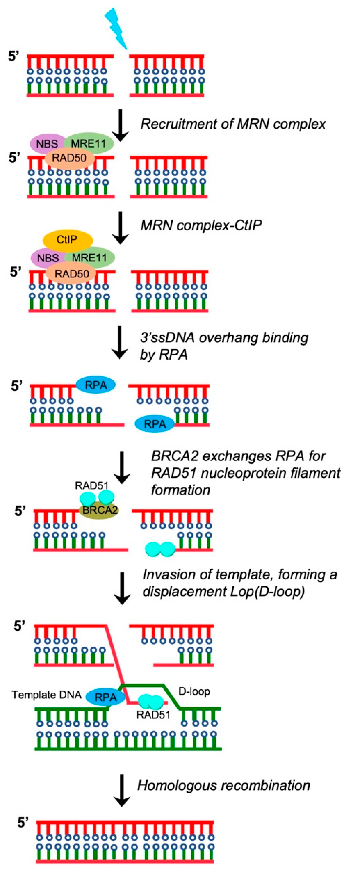
If left unrepaired or repaired incorrectly, SSBs are converted into DSBs, which are the most deleterious forms of DNA damage that lead to the loss of the majority of chromosomal regions [40,41]. Therefore, accurate reorganization and repair of DSBs are essential to maintaining genome integrity and preventing the formation of cancer. DSBs are mainly repaired using two pathways: non-homologous end joining (NHEJ) and homologous recombination (HR) [42,43,44,45]. In the HR pathway, the first step involves the initiation of DNA end resection at the break site to expose the long stretches of 3′ single-strand DNA (ssDNA) (Figure 2). Proteins that mediate DNA end resection include the MRE11-RAD50-NBS1/XRS2 (MRN/X) complex, CtIP, and BRCA1. Once the DNA ends are resected, replication protein A (RPA) efficiently binds to ssDNA to protect it from nucleases and further prevent the formation of secondary structures via the self-annealing of the ssDNA. RAD51 is then recruited to ssDNA sites to form RAD51 nucleoprotein filaments and ensure invasion of the long stretches of the 3′ ssDNA into homologous duplex DNA (Figure 2) [46,47]. Through this process, genetic information is accurately transferred to daughter cells. A study reported that germline mutations in HR-related genes, such as BRCA1, BRCA2, RAD51D, and PALB2, led to the development of EC in a subset of patients [48].

3.2. Classical Non-Homologous End Joining Repair (c-NHEJ)

In mammalian cells, DSBs that occur during the cell cycle are repaired predominantly via the classical non-homologous end joining (c-NHEJ) pathway. c-NHEJ-mediated repair involves the direct end-to-end ligation of DSB ends [49]. A biochemical analysis has demonstrated that blunt-end ligation without resection is mediated by the Ku-XRCC4-DNA ligase IV complex (Figure 3). In contrast, ligation of incompatible DNA ends is strongly stimulated by the DNA-PKcs-Artemis complex, which removes 5′ and 3′ DNA overhangs through its endonuclease activity to create DNA ends that can be ligated by the XRCC4-ligase IV complex [50,51]. Notably, XLF and PAXX were recognized as new NHEJ factors involved in ligase-complex-mediated DNA end ligation (Figure 3).

3.3. Alternative Non-Homologous End Joining (A-NHEJ)

When the c-NHEJ pathway is compromised, A-NHEJ is activated, which involves much more extensive resection of the DNA ends. A-NHEJ is also known as microhomology-mediated end joining (MMEJ) and is mostly active in the S and G2 phases of the cell cycle. A-NHEJ requires 2–20 bp microhomology [52]. The proteins involved in A-NHEJ include Pol θ, PARP1, CtIP and the MRN complex. The first step of A-NHEJ is the PARP1-mediated localization of the CtIP–MRN complex to the DNA ends [53]. Phosphorylated CtIP promotes activation of the endonuclease function of the MRN complex, which initiates resection by generating 15–100 nucleotide 3′overhangs that are recognized as microhomology sequences of the ssDNA [54]. Then PARP1, MRN complex, and Pol θ promote the alignment of ssDNA via microhomology sequences. Notably, when the annealed microhomologies are embedded within the long 3′ ssDNA, Pol θ removes non-homologous 3′ ssDNA, and the ERCC1 and XPF nucleases digest non-homologous 3′ ssDNA sequences. Finally, Pol θ mediates DNA synthesis to fill the gaps and the LIG3-XRCC1 complex is utilized to repair the remaining nicks [55,56].

4. Single-Strand Annealing (SSA)

Single-strand annealing (SSA) has more in common with A-NHEJ than c-NHEJ, because both SSA and A-NHEJ require extensive resection of DNA ends to realize microhomology [57,58,59]. Compared with A-NHEJ, SSA needs more homologous sequence exposure, and the 3′ ssDNA stretches created by the MRN complex and CtIP are further resected by nuclease EXO1, BLM, or DNA replication helicase/nuclease 2 (DNA2) to generate long stretches of 3′ ssDNA (Figure 3) [60,61,62]. The long stretches of 3′ ssDNA are stabilized via RPA binding. In contrast to HR, SSA is RAD51-independent and easily generates deletions and translocations (Figure 3). Notably, before ligation, the non-homologous 3′ ssDNA stretches must be resected and removed via nucleotide the excision repair complex XPF-XRCC1 and mismatch repair (MMR) complex MSH2-MSH3 [49,57,63].

5. Targeting DNA Damage Repair Proteins in EC

Most low-grade and early-stage endometrioid tumors have excellent prognoses and >95% five-year survival rate; however, high-grade or serous tumors have extremely poor outcomes as a result of chemoresistance. According to TCGA datasets, 25% of high grade endometrioid tumors and serous tumors exhibit widespread copy number alterations [9,17,26]. The genomic characteristics of serous tumors are common with those of high-grade serous ovarian cancers and triple-negative breast cancers. In high-grade and serous EC, mutations in DNA damage repair genes and high levels of genome instability are frequent; therefore, targeting one or more DNA damage repair proteins is a rational strategy for treating advanced EC [64,65]. The following sections summarize preclinical studies that have targeted DNA damage proteins for treating EC. Moreover, therapeutic targets are suggested to optimize the treatment efficacy of targeted therapy and immunotherapy in EC.

5.1. Targeting PARP in EC

According to one study, PARP1 protein is overexpressed in the majority of cases of EC [66]. Targeting PARP with PARP inhibitors represents a promising therapeutic strategy for treating EC. Indeed, many studies have tested the efficacy of PARP inhibitors in EC with different genetic backgrounds, including PTEN-deficient and PTEN-WT. Dedes et al. reported that the PARP inhibitor KU0058948 effectively inhibited PTEN-deficient endometrioid EC [67]. Consistently, Philip et al. reported that two PTEN-deficient EC cell lines were more sensitive to the PARP inhibitors olaparib and talazoparib than PTEN-WT cells [68]. By contrast, according to Miyasaka et al., the efficacy of the PARP inhibitor olaparib was not associated with PTEN status, because only 3/12 PTEN-deficient EC cell lines were sensitive to olaparib [23]. In line with this study, our previous study also demonstrated that PTEN-deficient endometrioid EC cells were not responsive to the PARP inhibitor olaparib in vitro and in vivo [69]. Janzen et al. demonstrated that the anti-cancer activity of olaparib in EC was negatively associated with a low estrogen microenvironment in vivo [24]. A 42-year-old woman with recurrent low-grade endometrioid EC and germline BRCA2 mutation was treated with olaparib. Because the patient responded robustly to the olaparib, PARP inhibitors may be effective in EC with deleterious BRCA1/2 mutations, especially in patients with a biallelic inactivation of BRCA1/2 [70]. Consistently, in ovarian cancer, patients with biallelic BRCA1/2 mutations showed improved outcomes than those with monoallelic BRCA1/2 mutations [71]. Two studies reported that ~20% of serous ECs harbor HR defects and may be sensitive to PARP inhibitors [25,27]. Consistently, a recent study used a patient-derived xenograft (PDX) model to examine genomic heterogeneity in EC. Given that the established PDX models include all EC molecular subtypes, the study investigated the efficacy of PARP inhibitor in these models and found that the high-copy-number (serous-like) molecular subtype was sensitive to the PARP inhibitor, which was independent of PTEN status, because patient-derived xenograft modes with PTEN mutations were unresponsive to talazoparib [22]. Thus, studies must be performed to evaluate PTEN status in determining response of PARP inhibitors in EC, and PARP inhibitors may not be sufficient as monotherapy in treating EC.
Several preclinical studies have tested PARP inhibitors in combination with other drugs to optimize treatment efficiency [72]. We previously reported that endometrioid EC models are not responsive to the use of PARP inhibitor as monotherapy, but show superior sensitivity to compound PARP-PI3K inhibition, due to the reduction in HR activity following PI3K inhibition. Our study thus provides a rational combination strategy to effectively treat endometrioid EC [69]. Consistently, Philip et al. reported that an additional PI3K inhibitor, BKM120, enhanced the efficacy of the PARP inhibitors olaparib or talazoparib in EC cells with PTEN deficiencies by inhibiting the formation of RAD51 foci [68]. A phase Ib study used AKT and PARP inhibitors to treat ovarian, endometrial, and breast cancers. The combination of AKT and PARP inhibitors had no serious adverse effects and elicited a prolonged response in ovarian, endometrial, and breast cancers, with especially superior activity in EC. This phase Ib study further recommended a phase II trial of capivasertib and olaparib in patients with advanced EC [73]. HER2 is overexpressed in ~30% of serous ECs. A recent study used uterine serous carcinoma cell lines to test the efficacy of a PARP inhibitor as monotherapy and in combination with the HER2 inhibitor neratinib. The combination of olaparib and neratinib acted synergistically against HER2-overexpressing and HR-proficient serous endometrial carcinoma [74]. Moreover, whether PTEN deficiency predicts the sensitivity of EC cells to PARP inhibitors is controversial. A study used a panel of PTEN-deficient EC cell lines to test the PARP inhibitor olaparib as monotherapy or in combination with an inhibitor of pBADS99 phosphorylation. The data in this study demonstrated that the PTEN-deficient EC cells were responsive to olaparib, and combined treatment with olaparib and pBADS99 phosphorylation inhibitor NPB resulted in synergistic antitumor effects in the PTEN-deficient ECs in vitro and in vivo. Mechanistic analysis showed that NPB treatment increased DNA damage and reduced the activity of HR, thereby improving the efficacy of the PARP inhibitor in EC with PTEN deficiency [75]. Interestingly, a recent study showed that ATAD5 deficiency can sensitize cancer cells to PARP inhibition. The study examined the cBioPortal database and found that ~10% of ECs are ATAD5-deficient; thus, ATAD5 mutation status might be a biomarker for predicting the sensitivity of PARP inhibitor in treating EC [76]. Taken together, PARP inhibitors may have an indispensable role in the management of EC and should be considered in combination with other treatments.

5.2. Targeting ATR-CHK1 Signaling in EC

EC is characterized by genome instability with replication stress (RS) due to PTEN deficiency or P53 mutation. In this genetic background, cancer cells may survive by activating the DNA damage repair and replication stress response (RSR) pathways [77]. ATR-CHK1 signaling is activated in response to RS and DSBs. In response to RS, the ATR-CHK1 axis acts as the main transducer of RSR by inhibiting late origin firing and arresting the cell cycle in the S phase. In addition, ATR-CHK1 signaling can respond to RS by (1) increasing deoxynucleotide synthesis, (2) promoting the dormant origin firing within the stalled replication fork and (3) stabilizing and resolving the stalled replication fork for a timely restart of replication [78,79]. Therefore, targeting ATR-CHK1 signaling is an attractive therapeutic strategy for the treatment of cancer with high genome instability [80]. In terms of EC, a study reported that the use of ATR or CHK1 inhibitor as monotherapy enhanced the sensitivity of EC cells to DNA-damaging agents, including cisplatin and doxorubicin [29]. Moreover, the combined use of ATR and CHK1 inhibitors synergistically induced DNA damage and inhibited cell proliferation in EC cells. Serous EC is associated with chemoresistance and poor survival. DNA sequencing data have revealed that the CCNE1 oncogene is amplified in ~50% of serous and 8% of endometrioid ECs, respectively [27,81]. A recent study by Xu et al. investigated the efficacy of an ATR inhibitor in treating EC with a CCNE1 amplification. The data indicated that the induction of CCNE1 led to activation of ATR signaling, and the use of an ATR inhibitor can reverse the inherent resistance of the WEE1 inhibitor. Moreover, the combination of a low dose of an ATR inhibitor and a WEE1 inhibitor synergistically reduced cell viability and colony formation and increased replication fork collapse in EC cells with CCNE1 amplifications. A further biomarker analysis indicated that CCNE1 copy number was a clinically tractable biomarker for predicting the sensitivity of EC to ATR-CHK1 inhibition [82].

5.3. Targeting WEE1 in EC

It is well recognized that almost all serous ECs occur with a TP53 mutation which plays an important role in G1/S and G2/M cell-cycle checkpoint regulation in response to DNA damage, thereby permitting normal and cancer cells to repair damaged-DNA before entering the S and G2 phases. P53 mutations and deficiencies in cancer cells are highly dependent on the WEE1 kinase to prevent aberrant G2/M cell-cycle entry and mitotic catastrophe [83]. WEE1 serves as a key regulator of genome integrity via inhibiting CDK1/2-mediated DNA replication during the G1-S cell-cycle transition [84]. Therefore, targeting the G2/M checkpoint via suppressing WEE1 is an attractive strategy for targeting a P53-mutant or -deficient cancer. Recent studies have shown the therapeutic efficacy of targeting WEE1 in EC. According to Meng et al., monotherapy with the WEE1 inhibitor AZD1775 was effective against P53-mutant EC via the induction of cell apoptosis. AZD1775 treatment enhanced the efficacy of a PARP inhibitor, and the combined use of AZD1775 and olaparib resulted in synergistic antitumor effects in EC cells with a P53 mutation [85]. Consistently, a recent clinical phase I trial of AZD1775 demonstrated its activity in EC, and a biomarker analysis further revealed that the expression of CCNE1 mRNA predicted the sensitivity of EC to AZD1775 [86]. The molecular characterization of serous EC revealed frequent cell-cycle dysregulation and a high level of oncogene-induced RS. Indeed, a high level of DNA replication stress is vulnerable to the inhibition of WEE1. In a phase II study, AZD1775 exhibited robust activity in recurrent serous EC, with an ORR of 29.4% and a progression-free survival (PFS) rate of 47.1% [87]. However, the evaluation could not identify a biomarker that predicted sensitivity to AZD1775. Nonetheless, co-alterations in multiple pathways and alternative measurements of RS are of interest to identify patients with serous EC who may benefit from WEE1 inhibition. Overall, this clinical study suggests that targeting WEE1 is a promising therapeutic modality for serous EC.

5.4. Ongoing Clinical Trials

Based on the efficacy of DDR inhibitors in preclinical studies, various clinical trials are evaluating the efficacy of DDR inhibitors in EC, including the inhibitors of PARP, CHK1, and WEE1 (Table 1). A PARP inhibitor is under evaluation in different stages of clinical trials (Table 1). A study of the CHK1 inhibitor BBI-355 is recruiting EC patients with EC to evaluate its efficiency in the treatment of EC (NCT05827614). Additionally, two clinical trials are recruiting patients to evaluate the efficacy of an ATR inhibitor in combination with chemotherapy or the use of a PARP inhibitor in EC (NCT04491942 and NCT03682289).

6. DDR Correlates with Cancer Cell Immunogenicity

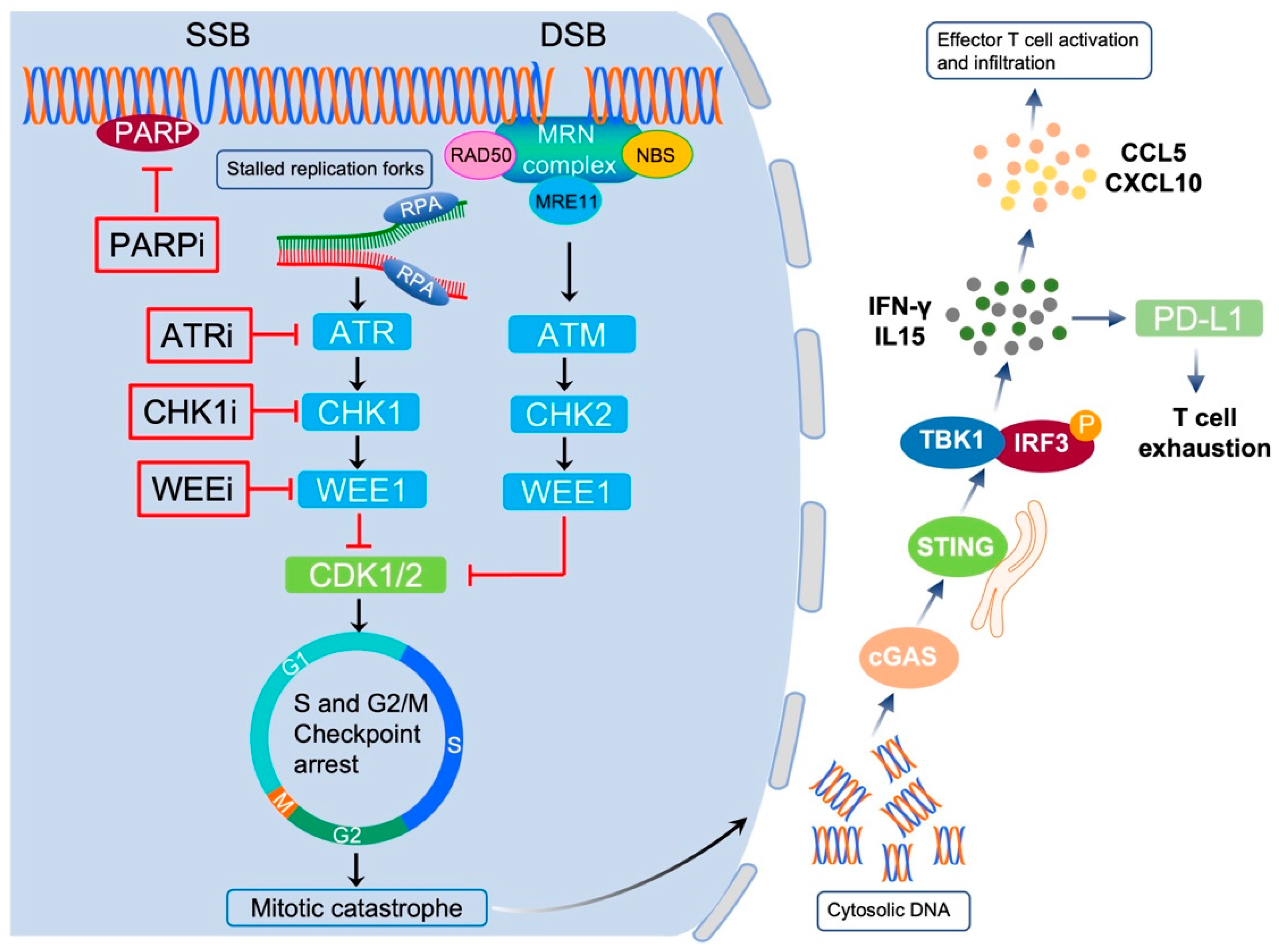
The DDR system plays key roles in maintaining the genomic integrity of cancer cells. Dysregulating DNA repair processing or uncontrolled activity of repair factors can result in DNA damage, thereby promoting genome instability in cancer cells. In recent years, targeting DDR has become an attractive therapeutic modality for cancer. However, DDR inhibitors are cytotoxic, and studies have reported induction of the innate and adaptive immune response after radiotherapy or chemotherapy. Studies in the last decades have shown that the DDR impacts several aspects of tumor cell immunogenicity, leading to the immune evasion of cancer cells [88,89]. Tumor immunogenicity is the ability of the immune system to recognize and eliminate cancer cells and mainly includes three parameters: antigenicity, adjuvanticity and reactogenicity. All these three parameters work together and define the ability of the immune system to recognize and eliminate cancer cells. DDR-mediated regulation of tumor immunogenicity is complex and involves the activation of multiple pathways, including the innate cGAS-STING immune pathway, which is activated by cytosolic DNA, generated through DNA damage induced by endogenous or exogenous factors (Figure 4) [90]. Moreover, the exogenous inhibition of DDR or inherent DDR defects have been reported to induce expression of immune checkpoint proteins in cancer cells, for example, increased PD-L1 expression in MMR-deficient (MMR-D) cells compared with MMR-proficient (MMR-P) cells (Figure 4) [88]. Under these conditions, checkpoint proteins, often activated by DDR inhibitors, are repurposed to jeopardize theantitumor immune response by binding with their ligands or receptors, and therefore, create an immune-suppressive tumor microenvironment. Thus, to circumvent these immune-suppressive effects, extensive efforts have focused on identifying potential combination therapies to overcome resistance mechanisms and reactivate the activity of T-cells toward the tumor.

7. Exploiting the Interplay between DDR and Immunity for Cancer Therapy

Recent studies suggest that pharmacological manipulation of DDR can enhance the efficacy of immunotherapy in cancer treatment. A large number of studies have shown that the implementation of DDR inhibitors elicits innate and adaptive immune response in cancer cells, leading to the immune evasion of cancer cells. Thus, the combination of DDR inhibitors with ICIs showed synergistic antitumor efficacy in cancer therapy, including ovarian cancer and breast cancers [91,92]. Immunotherapy currently represents an attractive strategy for patients with EC, mainly those with advanced or recurrent disease, with no effective treatment options available after standard chemotherapy [93]. Given the high expression of PD-1/PD-L1 in EC, it is reasonable to test inhibitors targeting the PD-1-PD-L1 axis to treat EC [94]. Indeed, many studies have tested the efficacy of PD-1 or PD-L1 antibodies in different molecular subtypes of ECs. More importantly, a combination of DDR agents with immunotherapy may expand the therapeutic antitumor effects in EC.

7.1. Pembrolizumab

The anti-PD-1 antibody has shown prolonged and beneficial responses in various human cancers, including EC. A study reported that cancers that carry mutations in POLE might serve as good candidates for ICIs. In a recent clinical trial, the efficacy of the PD-1 antibody pembrolizumab was tested in 75 patients with advanced EC. The results showed that a subset of patients was responsive to pembrolizumab. Of the three patients with complete response to pembrolizumab, one had POLE mutation, one exhibited microsatellite stability (MSS), and one had unknown microsatellite instability (MSI) status (Table 2) [95]. The findings thus pave the way to utilize the anti-PD-1 antibody in the treatment of EC. Further, in a clinical trial, the efficacy of pembrolizumab was tested in 244 patients with MSI or MMR-D, including 47 patients with EC. The results of this study showed that the majority of patients had a reduction in tumor size upon pembrolizumab treatment, especially 8 patients had complete response [96]. A longer follow-up study in patients with MMR-D or MSI-high (MSI-H) cancer demonstrated that pembrolizumab treatment had clinically meaningful effects, as well as a manageable safety profile [97]. Therefore, pembrolizumab treatment has specific effects in EC with MSI or MMR-D. Indeed, pembrolizumab was approved by the Food and Drug Administration (FDA) in 2017, for treating MSI or MMR-D solid tumors [98]. Notably, although MSI or MMR-D occur in one-third of the patients with EC, it has different molecular subgroups. Results from a clinical phase II trial revealed that patients with Lynch-like syndrome, who had high tumor mutation burdens, were highly responsive to pembrolizumab, and only 44% of the patients with sporadic MSI-H responded to pembrolizumab [99]. Consistently, a phase II trial showed that patients with EC with mutational MMR-D had higher response rates to PD-1 inhibitor pembrolizumab [100]. Together, these studies support the use of pembrolizumab in treating patients with EC, who have high tumor mutation burdens.

7.2. Nivolumab

The initial clinical trial of the human monoclonal antibody nivolumab in EC was implemented in 2016 and included two patients. WES results indicated that both patients had POLE and MSH6 mutations. According to the RECIST criteria, both patients showed significantly reduced tumor burdens, following a dose of 3 mg/kg of nivolumab every 2 weeks (Table 2) [101]. A phase II study tested the efficacy of 240 mg of nivolumab every 2 weeks in patients with solid tumors, including EC. Patients who had complete response had MSI-H, further indicating the importance of MSI status in predicting treatment response [102]. Because nivolumab showed activity in colon cancer with MMR-D, a recent clinical trial tested the efficacy of nivolumab in non-colorectal cancer with MMR-D. The results showed that nivolumab has promising activity in non-colorectal MMR-D cancers, especially in endometrioid EC [103].

7.3. Dostarlimab

A phase I nonrandomized trial has completed evaluating the efficacy of dostarlimab in recurrent EC that progressed after platinum-based chemotherapy. Patients with EC who achieved an objective response were deficient in the MMR system (Table 2) [104]. Further, objective response rate (ORR) was equal to 44.7% in the MMR-D EC compared with an ORR =3.7% in MMR-P/MSI EC. Recently, the interim results from GARNET—a phase I, single-arm study—revealed that dostarlimab exhibited robust antitumor activity in both MMR-D/MSI and MMR-P/MSS EC, as well as a manageable safety profile [105]. Based on the results of this study, the FDA approved the use of dostarlimab in advanced EC with MMR-D [106].

7.4. Durvalumab

The anti-PD-L1 efficacy of durvalumab was evaluated in the phase II PHAEDRA trial. The results revealed a PFS of 8.3 months in MMR-D versus 1.8 months in MSS, demonstrating that in patients with EC, the antitumor effects of durvalumab were dependent on MMR status [107]. The results of a recent clinical trial of durvalumab combined with olaparib in treating metastatic or recurrent EC showed that the combined use of durvalumab and olaparib was well tolerated, but did not meet 50% 6-month PFS in advanced EC [108].

7.5. Other Anti-PD-L1 Antibodies

In a phase I trial, the efficacy of an anti-PD-L1 antibody atezolizumab was evaluated in 15 patients with EC. Patients with a clinical response to atezolizumab were MMR-D (Table 2) [109]. In 2019, the efficacy of human anti-PD-L1 antibody avelumab was evaluated in patients with EC [110]. The results showed an ORR of 40% in the MMR-D group; however, these responsive patients were absent for PD-L1 expression, indicating that further studies are needed to identify biomarkers that predict sensitivity to ICIs.

7.6. Immunotherapy in Combination with Chemotherapy

Because DNA damage agents enhance the tumor mutation burden and induce neoantigen exposure, the use of chemotherapeutic agents in combination with ICIs represents a promising strategy to enhance treatment efficiency in patients with cancer. A phase II study of pembrolizumab with chemotherapy in EC demonstrated remarkable efficacy [111]. Consistently, a phase III study showed that a combination of pembrolizumab with carboplatin or paclitaxel resulted in significantly longer PFS than chemotherapy alone [112] (NCT03914612). A phase II trial has completed patient recruitment and is clinically evaluating the efficacy of pembrolizumab combined with doxorubicin in advanced, recurrent, and metastatic EC (NCT03276013). Similarly, a clinical trial is evaluating the efficacy of a combination of atezolizumab with carboplatin–paclitaxel in advanced or recurrent EC (NCT03603184). A phase III study is evaluating the combined effects of dostarlimab and carboplatin or paclitaxel in EC (NCT03981796). If positive, the findings of these studies can improve treatment options for patients with EC, regardless of their tumor stage. Consistently, a recent phase Ib/II biomarker-driven study is evaluating suitable combination agents with atezolizumab based on sequencing data from patients with persistent or recurrent EC (https://clinicaltrials.gov/ct2/show/NCT04486352 (accessed on 24 July 2020)).
Table 2. Clinical studies of the use of ICIs in monotherapy for EC.
Immune Checkpoint InhibitorPatient NumberResponse BiomarkerPhaseStatus Reference
Pembrolizumab75POLE-mutation and MSICompleted Completed[98]
Pembrolizumab47MSI or MMR-DPhase IRecruiting[99]
Pembrolizumab25MSI-highPhase IIActive, not yet recruiting[101]
pembrolizumab24MMR-DPhase IIActive, not yet recruiting[102]
Nivolumab2POLE and MSH6 mutationunknownUnknown[103]
Nivolumab2MSI-highPhase IIUnknown[104]
Nivolumab13MMR-DPhase IIActive, not yet recruiting[105]
Dostarlimab104MMR-DPhase IRecruiting[106]
Dostarlimab75MMR-DPhase IRecruiting[107]
Durvalumab71MMR-DCompleted Completed[109]
Atezolizumab15MMR-DCompleted Completed[111]
Avelumab33MMR-DPhase IIActive, not yet recruiting[112]

8. Immunotherapy in Combination with PARP Inhibitors

Collectively, the clinical results of the use of PD-1 or PD-L1 antibodies in patients with EC seem to positively correlate with the MMR-D or MSI status, and the efficacy of the PD-1 or PD-L1 antibodies in MMR-P EC remains modest. However, a clinical trial suggested that microsatellite-stable tumors with high tumor mutation burdens may benefit from immunotherapy; thus, high tumor mutation burden, but not MMR-D or MSI status, may determine the therapeutic efficacy of ICIs [113].
Combination therapy with targeted agents might increase the response rate to ICIs. Studies have shown that treatment with PARP inhibitors resulted in impaired DNA damage repair, increased tumor mutation burden, increased neoantigen exposure, and increased immune recognition of cancer cells [114,115]. In this context, a phase II study is investigating the efficacy of atezolizumab, bevacizumab, and rucaparib as monotherapy or in combination, in patients with recurrent EC (NCT03694262). Another trial is clinically investigating the efficacy of durvalumab with or without olaparib as maintenance therapy after first-line treatment in advanced or recurrent EC (NCT04269200). In addition, two combination therapies, including dostarlimab + niraparib and nivolumab + rucaparib, are now being investigated in advanced, recurrent, or metastatic EC (NCT03016338 and NCT03572478, respectively).

9. Conclusions

Although early-stage or low-grade EC have good prognoses, high-grade serous EC is highly aggressive and accounts for 40% of the deaths. Cytotoxic chemotherapy alone or in combination with radiotherapy remains the treatment of choice for high-grade serous EC. Identifying new therapeutic strategies to enhance treatment efficacy in patients with EC is an urgent clinic need. A study has demonstrated that the overexpression of DNA damage repair genes in EC was positively correlated with poor prognosis [66,116]. In terms of high-grade and serous EC, the HR repair system is deficient and tumors possess high levels of genome instability due to the amplification of oncogenes, such as CCNE1 and MYC. Therefore, modulating the repair system, such as by boosting RS or inhibiting DDR, can provide effective treatment options in serous EC. The modest efficacy of PARP inhibitors as monotherapies in EC highlights the need for combination therapy to improve the efficacy of PARP inhibitors. Targeting ATR-CHK1 signaling and WEE1 had antitumor effects in a preclinical model of EC. Strategies that improve the treatment efficacy of ATR or CHK1 inhibitors should be provided more attention to, especially in a serous histology setting. Furthermore, given the molecular heterogeneity of EC and the risk of toxicity based on DDR-targeting therapies, great attention is required to define optimal combination modality, dose concentration, and schedule of these agents. Because immune stimulation has been detected after use of DDR inhibitors in preclinical and clinical studies, understanding the immune microenvironment, in which DNA lesions can activate antitumor immunity, is important. For EC, combined use of DDR inhibitors with ICIs is now being tested in the clinic, and the results are awaited. In conclusion, understanding of the biology of EC will facilitate the development of rational combination modalities to prevent resistance to DDR-based target therapy, ultimately leading to prolonged survival in patients with EC.

10. Future Directions and Perspectives

The combination of pembrolizumab and lenvatinib has recently been approved for use in EC, regardless of different genetic background, including MSI and MSS or MMR-P. However, some patients do not respond well to this treatment due to primary or acquired resistance, as well as treatment-associated toxicity [117]. The use of DNA damage agents as monotherapy or in combination with chemotherapy has shown efficacy in small numbers of ECs, highlighting the need to identify effective treatment strategies for ECs. Targeting DNA damage repair systems, such as HR and RSR, or modulating the cell cycle is of particular interest in EC, especially with high-grade or serous EC. To our knowledge, various factors, including epigenetic regulators, such as BET family members, transcription factors, as well as some non-coding RNAs, regulate HR or RSR [118,119]. A better understanding of factors involved in DNA repair and identification of their targets is needed to develop and apply combination therapies in EC.
FDA has recently approved a combination of immunotherapy for treating EC; however, only a subset of patients that will likely benefit from it. Most patients are not responsive to immunotherapy because of the immunosuppressive tumor microenvironment. The focus is shifting toward the use of other strategies, such as DNA damage agents that expose more neoantigens to enhance the efficacy of ICI, to reduce tumor recurrence in cancer treatment. Indeed, the therapeutic efficacy of a combination of PARP inhibitors and ICI is being clinically evaluated in patients with EC, and the results are awaited [120]. Overall, preclinical and clinical studies must focus on improving treatment efficacy to benefit all molecular types of EC regardless of therapy-type-DNA damage-based targeted therapy or a combined immunotherapy.

Author Contributions

X.B. was responsible for the generation of tables and figures. X.B., C.S. and B.H. wrote this manuscript. X.B., C.S., J.C. and B.H. discussed and approved the final manuscript. All authors have read and agreed to the published version of the manuscript.

Funding

This study was supported by the National Natural Science Foundation of China (grant number 81872438 to B. Hong), and West Anhui University (grant number WGKQ2021077).

Institutional Review Board Statement

Not applicable.

Informed Consent Statement

Not applicable.

Data Availability Statement

All data generated or analyzed during this study are included in this published article.

Acknowledgments

We thank the members of the Hong laboratory for their critical reading of the manuscript and helpful discussions.

Conflicts of Interest

The authors declare no conflict of interest.

Abbreviations

EC—endometrial cancer; DDR—DNA damage response; SSBR—single-strand breaks repair; BER—base excision repair; DSBR—double-strand breaks repair; HR—homologous recombination; SSB—single-strand break; DSB—double-strand break; ICIs—immune checkpoint inhibitors; AP—apurinic/apyrimidinic site; APE1—apurinic/apyrimidinic Endonuclease 1; PNKP—polynucleotide kinase 3′-phosphatase; NHEJ—non-homologous ending joining; ssDNA—single-strand DNA; MRN/X—MRE11-RAD50-NBS1/XRS2; RPA—replication protein A; c-NHEJ—classical non-homologous end joining; Alt-EJ—alternative non-homologous end joining; MMEJ—microhomology-mediated end joining; SSA—single-strand annealing; PDX—patient-derived xenograft; RS—replication stress; RSR—replication stress response; TMB—tumor mutation burden; MSS—microsatellite stability; MSI—microsatellite instability; MMR-D—mismatch repair deficient; MMR-P—mismatch repair proficient; ORR—objective response rate. FDA—food and drug administration.

References

  1. Sung, H.; Ferlay, J.; Siegel, R.L.; Laversanne, M.; Soerjomataram, I.; Jemal, A.; Bray, F. Global Cancer Statistics 2020: GLOBOCAN Estimates of Incidence and Mortality Worldwide for 36 Cancers in 185 Countries. CA Cancer J. Clin. 2021, 71, 209–249. [Google Scholar] [CrossRef] [PubMed]
  2. Crosbie, E.J.; Kitson, S.J.; McAlpine, J.N.; Mukhopadhyay, A.; Powell, M.E.; Singh, N. Endometrial cancer. Lancet 2022, 399, 1412–1428. [Google Scholar] [CrossRef] [PubMed]
  3. Makker, V.; MacKay, H.; Ray-Coquard, I.; Levine, D.A.; Westin, S.N.; Aoki, D.; Oaknin, A. Endometrial cancer. Nat. Rev. Dis. Primers 2021, 7, 88. [Google Scholar] [CrossRef] [PubMed]
  4. Colombo, N.; Creutzberg, C.; Amant, F.; Bosse, T.; González-Martín, A.; Ledermann, J.; Marth, C.; Nout, R.; Querleu, D.; Mirza, M.R.; et al. ESMO-ESGO-ESTRO Consensus Conference on Endometrial Cancer: Diagnosis, treatment and follow-up. Ann. Oncol. 2016, 27, 16–41. [Google Scholar] [CrossRef]
  5. Calle, E.E.; Rodriguez, C.; Walker-Thurmond, K.; Thun, M.J. Overweight, obesity, and mortality from cancer in a prospectively studied cohort of U.S. adults. N. Engl. J. Med. 2003, 348, 1625–1638. [Google Scholar] [CrossRef]
  6. Bokhman, J.V. Two pathogenetic types of endometrial carcinoma. Gynecol. Oncol. 1983, 15, 10–17. [Google Scholar] [CrossRef]
  7. Matias-Guiu, X.; Prat, J. Molecular pathology of endometrial carcinoma. Histopathology 2013, 62, 111–123. [Google Scholar] [CrossRef]
  8. Secord, A.A.; Havrilesky, L.J.; Bae-Jump, V.; Chin, J.; Calingaert, B.; Bland, A.; Rutledge, T.; Berchuck, A.; Clarkepearson, D.; Gehrig, P. The role of multi-modality adjuvant chemotherapy and radiation in women with advanced stage endometrial cancer. Gynecol. Oncol. 2007, 107, 285–291. [Google Scholar]
  9. Cancer Genome Atlas Research Network; Kandoth, C.; Schultz, N.; Cherniack, A.D.; Akbani, R.; Liu, Y.; Shen, H.; Robertson, A.G.; Pashtan, I.; Shen, R.; et al. Integrated genomic characterization of endometrial carcinoma. Nature 2013, 497, 67–73. [Google Scholar]
  10. Daikoku, T.; Hirota, Y.; Tranguch, S.; Joshi, A.R.; DeMayo, F.J.; Lydon, J.P.; Ellenson, L.H.; Dey, S.K. Conditional loss of uterine Pten unfailingly and rapidly induces endometrial cancer in mice. Cancer Res. 2008, 68, 5619–5627. [Google Scholar] [CrossRef]
  11. Urick, M.E.; Rudd, M.L.; Godwin, A.K.; Sgroi, D.; Merino, M.; Bell, D.W. PIK3R1 (p85alpha) is somatically mutated at high frequency in primary endometrial cancer. Cancer Res. 2011, 71, 4061–4067. [Google Scholar] [CrossRef]
  12. Rudd, M.L.; Price, J.C.; Fogoros, S.; Godwin, A.K.; Sgroi, D.C.; Merino, M.J.; Bell, D.W. A unique spectrum of somatic PIK3CA (p110alpha) mutations within primary endometrial carcinomas. Clin. Cancer Res. 2011, 17, 1331–1340. [Google Scholar] [CrossRef]
  13. McConechy, M.K.; Ding, J.; Cheang, M.C.; Wiegand, K.C.; Senz, J.; Tone, A.A.; Yang, W.; Prentice, L.M.; Tse, K.; Zeng, T.; et al. Use of mutation profiles to refine the classification of endometrial carcinomas. J. Pathol. 2012, 228, 20–30. [Google Scholar] [CrossRef] [PubMed]
  14. Byron, S.A.; Gartside, M.; Powell, M.A.; Wellens, C.L.; Gao, F.; Mutch, D.G.; Goodfellow, P.J.; Pollock, P.M. FGFR2 point mutations in 466 endometrioid endometrial tumors: Relationship with MSI, KRAS, PIK3CA, CTNNB1 mutations and clinicopathological features. PLoS ONE 2012, 7, e30801. [Google Scholar] [CrossRef]
  15. Zighelboim, I.; Goodfellow, P.J.; Gao, F.; Gibb, R.K.; Powell, M.A.; Rader, J.S.; Mutch, D.G. Microsatellite instability and epigenetic inactivation of MLH1 and outcome of patients with endometrial carcinomas of the endometrioid type. J. Clin. Oncol. 2007, 25, 2042–2048. [Google Scholar] [CrossRef] [PubMed]
  16. Kuhn, E.; Wu, R.-C.; Guan, B.; Wu, G.; Zhang, J.; Wang, Y.; Song, L.; Yuan, X.; Wei, L.; Roden, R.B.; et al. Identification of molecular pathway aberrations in uterine serous carcinoma by genome-wide analyses. J. Natl. Cancer Inst. 2012, 104, 1503–1513. [Google Scholar] [CrossRef]
  17. Le Gallo, M.; O’Hara, A.J.; Rudd, M.L.; Urick, M.E.; Hansen, N.F.; O’Neil, N.J.; Price, J.C.; Zhang, S.; England, B.M.; Godwin, A.K.; et al. Exome sequencing of serous endometrial tumors identifies recurrent somatic mutations in chromatin-remodeling and ubiquitin ligase complex genes. Nat. Genet. 2012, 44, 1310–1315. [Google Scholar] [CrossRef]
  18. Urick, M.E.; Bell, D.W. In vitro effects of FBXW7 mutation in serous endometrial cancer: Increased levels of potentially druggable proteins and sensitivity to SI-2 and dinaciclib. Mol. Carcinog. 2018, 57, 1445–1457. [Google Scholar] [CrossRef] [PubMed]
  19. Urick, M.E.; Bell, D.W. Clinical actionability of molecular targets in endometrial cancer. Nat. Rev. Cancer 2019, 19, 510–521. [Google Scholar] [CrossRef]
  20. Hong, B.; Le Gallo, M.; Bell, D.W. The mutational landscape of endometrial cancer. Curr. Opin. Genet. Dev. 2015, 30, 25–31. [Google Scholar] [CrossRef]
  21. Groelly, F.J.; Fawkes, M.; Dagg, R.A.; Blackford, A.N.; Tarsounas, M. Targeting DNA damage response pathways in cancer. Nat. Rev. Cancer 2023, 23, 78–94. [Google Scholar] [CrossRef]
  22. Bonazzi, V.F.; Kondrashova, O.; Smith, D.; Nones, K.; Sengal, A.T.; Ju, R.; Packer, L.M.; Koufariotis, L.T.; Kazakoff, S.H.; Davidson, A.L.; et al. Patient-derived xenograft models capture genomic heterogeneity in endometrial cancer. Genome Med. 2022, 14, 3. [Google Scholar] [CrossRef]
  23. Miyasaka, A.; Oda, K.; Ikeda, Y.; Wada-Hiraike, O.; Kashiyama, T.; Enomoto, A.; Hosoya, N.; Koso, T.; Fukuda, T.; Inaba, K.; et al. Anti-tumor activity of olaparib, a poly (ADP-ribose) polymerase (PARP) inhibitor, in cultured endometrial carcinoma cells. BMC Cancer 2014, 14, 179. [Google Scholar] [CrossRef] [PubMed]
  24. Janzen, D.M.; Paik, D.Y.; Rosales, M.A.; Yep, B.; Cheng, D.; Witte, O.N.; Kayadibi, H.; Ryan, C.M.; Jung, M.E.; Faull, K.; et al. Low levels of circulating estrogen sensitize PTEN-null endometrial tumors to PARP inhibition in vivo. Mol. Cancer Ther. 2013, 12, 2917–2928. [Google Scholar] [CrossRef] [PubMed]
  25. de Jonge, M.M.; Auguste, A.; van Wijk, L.M.; Schouten, P.C.; Meijers, M.; ter Haar, N.T.; Smit, V.T.; Nout, R.A.; Glaire, M.A.; Church, D.N.; et al. Frequent Homologous Recombination Deficiency in High-grade Endometrial Carcinomas. Clin. Cancer Res. 2019, 25, 1087–1097. [Google Scholar] [CrossRef] [PubMed]
  26. Zhao, S.; Choi, M.; Overton, J.D.; Bellone, S.; Roque, D.M.; Cocco, E.; Guzzo, F.; English, D.P.; Varughese, J.; Gasparrini, S.; et al. Landscape of somatic single-nucleotide and copy-number mutations in uterine serous carcinoma. Proc. Natl. Acad. Sci. USA 2013, 110, 2916–2921. [Google Scholar] [CrossRef]
  27. Lin, D.I.; Fine, A.; Danziger, N.A.; Huang, R.S.; Mata, D.A.; Decker, B.; Killian, J.K.; Ramkissoon, S.H.; Lechpammer, M.; Janovitz, T.; et al. Molecular analysis of endometrial serous carcinoma reveals distinct clinicopathologic and genomic subgroups. Gynecol. Oncol. 2022, 164, 558–565. [Google Scholar] [CrossRef]
  28. Mukherjee, A.; Patterson, A.L.; George, J.W.; Carpenter, T.J.; Madaj, Z.B.; Hostetter, G.; Risinger, J.I.; Teixeira, J.M. Nuclear PTEN Localization Contributes to DNA Damage Response in Endometrial Adenocarcinoma and Could Have a Diagnostic Benefit for Therapeutic Management of the Disease. Mol. Cancer Ther. 2018, 17, 1995–2003. [Google Scholar] [CrossRef]
  29. Takeuchi, M.; Tanikawa, M.; Nagasaka, K.; Oda, K.; Kawata, Y.; Oki, S.; Agapiti, C.; Sone, K.; Miyagawa, Y.; Hiraike, H.; et al. Anti-Tumor Effect of Inhibition of DNA Damage Response Proteins, ATM and ATR, in Endometrial Cancer Cells. Cancers 2019, 11, 1913. [Google Scholar] [CrossRef]
  30. Lieber, M.R.; Karanjawala, Z.E. Ageing, repetitive genomes and DNA damage. Nat. Rev. Mol. Cell Biol. 2004, 5, 69–75. [Google Scholar] [CrossRef]
  31. Jackson, S.P.; Bartek, J. The DNA-damage response in human biology and disease. Nature 2009, 461, 1071–1078. [Google Scholar] [CrossRef]
  32. Caldecott, K.W. DNA single-strand break repair and human genetic disease. Trends Cell Biol 2022, 32, 733–745. [Google Scholar] [CrossRef]
  33. Lebedeva, N.A.; Rechkunova, N.I.; Endutkin, A.V.; Lavrik, O.I. Apurinic/Apyrimidinic Endonuclease 1 and Tyrosyl-DNA Phosphodiesterase 1 Prevent Suicidal Covalent DNA-Protein Crosslink at Apurinic/Apyrimidinic Site. Front. Cell Dev. Biol. 2020, 8, 617301. [Google Scholar] [CrossRef]
  34. Abbotts, R.; Wilson, D.M., 3rd. Coordination of DNA single strand break repair. Free Radic. Biol. Med. 2017, 107, 228–244. [Google Scholar] [CrossRef]
  35. Klungland, A.; Lindahl, T. Second pathway for completion of human DNA base excision-repair: Reconstitution with purified proteins and requirement for DNase IV (FEN1). EMBO J. 1997, 16, 3341–3348. [Google Scholar] [CrossRef]
  36. Frosina, G.; Fortini, P.; Rossi, O.; Carrozzino, F.; Raspaglio, G.; Cox, L.S.; Lane, D.P.; Abbondandolo, A.; Dogliotti, E. Two pathways for base excision repair in mammalian cells. J. Biol. Chem. 1996, 271, 9573–9578. [Google Scholar] [CrossRef]
  37. Palles, C.; West, H.D.; Chew, E.; Galavotti, S.; Flensburg, C.; Grolleman, J.E.; Jansen, E.A.; Curley, H.; Chegwidden, L.; Arbe-Barnes, E.H.; et al. Germline MBD4 deficiency causes a multi-tumor predisposition syndrome. Am. J. Hum. Genet. 2022, 109, 953–960. [Google Scholar] [CrossRef]
  38. Mengwasser, K.E.; Adeyemi, R.O.; Leng, Y.; Choi, M.Y.; Clairmont, C.; D’Andrea, A.D.; Elledge, S.J. Genetic Screens Reveal FEN1 and APEX2 as BRCA2 Synthetic Lethal Targets. Mol. Cell 2019, 73, 885–899.e886. [Google Scholar] [CrossRef]
  39. Long, K.; Gu, L.; Li, L.; Zhang, Z.; Li, E.; Zhang, Y.; He, L.; Pan, F.; Guo, Z.; Hu, Z. Small-molecule inhibition of APE1 induces apoptosis, pyroptosis, and necroptosis in non-small cell lung cancer. Cell Death Dis. 2021, 12, 503. [Google Scholar] [CrossRef]
  40. Shibata, A.; Jeggo, P.A. DNA double-strand break repair in a cellular context. Clin. Oncol. 2014, 26, 243–249. [Google Scholar] [CrossRef]
  41. O’Connor, M.J. Targeting the DNA Damage Response in Cancer. Mol. Cell 2015, 60, 547–560. [Google Scholar] [CrossRef] [PubMed]
  42. Hartlerode, A.J.; Scully, R. Mechanisms of double-strand break repair in somatic mammalian cells. Biochem. J. 2009, 423, 157–168. [Google Scholar] [CrossRef] [PubMed]
  43. Pardo, B.; Gomez-Gonzalez, B.; Aguilera, A. DNA repair in mammalian cells: DNA double-strand break repair: How to fix a broken relationship. Cell. Mol. Life Sci. 2009, 66, 1039–1056. [Google Scholar] [CrossRef]
  44. Moynahan, M.E.; Jasin, M. Mitotic homologous recombination maintains genomic stability and suppresses tumorigenesis. Nat. Rev. Mol. Cell Biol. 2010, 11, 196–207. [Google Scholar] [CrossRef] [PubMed]
  45. Lieber, M.R. The mechanism of double-strand DNA break repair by the nonhomologous DNA end-joining pathway. Annu. Rev. Biochem. 2010, 79, 181–211. [Google Scholar] [CrossRef]
  46. Jasin, M.; Rothstein, R. Repair of strand breaks by homologous recombination. Cold Spring Harb Perspect. Biol. 2013, 5, a012740. [Google Scholar] [CrossRef]
  47. Ranjha, L.; Howard, S.M.; Cejka, P. Main steps in DNA double-strand break repair: An introduction to homologous recombination and related processes. Chromosoma 2018, 127, 187–214. [Google Scholar] [CrossRef]
  48. Gordhandas, S.; Rios-Doria, E.; Cadoo, K.A.; Catchings, A.; Maio, A.; Kemel, Y.; Sheehan, M.; Ranganathan, M.; Green, D.; Aryamvally, A.; et al. Comprehensive analysis of germline drivers in endometrial cancer. J. Natl. Cancer Inst. 2023, 115, 560–569. [Google Scholar] [CrossRef]
  49. Chang, H.H.Y.; Pannunzio, N.R.; Adachi, N.; Lieber, M.R. Non-homologous DNA end joining and alternative pathways to double-strand break repair. Nat. Rev. Mol. Cell Biol. 2017, 18, 495–506. [Google Scholar] [CrossRef]
  50. Pannunzio, N.R.; Watanabe, G.; Lieber, M.R. Nonhomologous DNA end-joining for repair of DNA double-strand breaks. J. Biol. Chem. 2018, 293, 10512–10523. [Google Scholar] [CrossRef]
  51. Watanabe, G.; Lieber, M.R. Dynamics of the Artemis and DNA-PKcs Complex in the Repair of Double-Strand Breaks. J. Mol. Biol. 2022, 434, 167858. [Google Scholar] [CrossRef] [PubMed]
  52. Saito, S.; Kurosawa, A.; Adachi, N. Mutations in XRCC4 cause primordial dwarfism without causing immunodeficiency. J. Hum. Genet. 2016, 61, 679–685. [Google Scholar] [CrossRef] [PubMed]
  53. Xie, A.; Kwok, A.; Scully, R. Role of mammalian Mre11 in classical and alternative nonhomologous end joining. Nat. Struct. Mol. Biol. 2009, 16, 814–818. [Google Scholar] [CrossRef]
  54. Anand, R.; Ranjha, L.; Cannavo, E.; Cejka, P. Phosphorylated CtIP Functions as a Co-factor of the MRE11-RAD50-NBS1 Endonuclease in DNA End Resection. Mol. Cell 2016, 64, 940–950. [Google Scholar] [CrossRef] [PubMed]
  55. Kent, T.; Chandramouly, G.; McDevitt, S.M.; Ozdemir, A.Y.; Pomerantz, R.T. Mechanism of microhomology-mediated end-joining promoted by human DNA polymerase theta. Nat. Struct. Mol. Biol. 2015, 22, 230–237. [Google Scholar] [CrossRef]
  56. Okano, S.; Lan, L.; Tomkinson, A.E.; Yasui, A. Translocation of XRCC1 and DNA ligase IIIalpha from centrosomes to chromosomes in response to DNA damage in mitotic human cells. Nucleic Acids Res. 2005, 33, 422–429. [Google Scholar] [CrossRef]
  57. Bhargava, R.; Onyango, D.O.; Stark, J.M. Regulation of Single-Strand Annealing and its Role in Genome Maintenance. Trends Genet. 2016, 32, 566–575. [Google Scholar] [CrossRef] [PubMed]
  58. Daley, J.M.; Laan, R.L.; Suresh, A.; Wilson, T.E. DNA joint dependence of pol X family polymerase action in nonhomologous end joining. J. Biol. Chem. 2005, 280, 29030–29037. [Google Scholar] [CrossRef]
  59. Daley, J.M.; Palmbos, P.L.; Wu, D.; Wilson, T.E. Nonhomologous end joining in yeast. Annu. Rev. Genet. 2005, 39, 431–451. [Google Scholar] [CrossRef]
  60. Symington, L.S. Mechanism and regulation of DNA end resection in eukaryotes. Crit. Rev. Biochem. Mol. Biol. 2016, 51, 195–212. [Google Scholar] [CrossRef] [PubMed]
  61. Mimitou, E.P.; Symington, L.S. Sae2, Exo1 and Sgs1 collaborate in DNA double-strand break processing. Nature 2008, 455, 770–774. [Google Scholar] [CrossRef] [PubMed]
  62. Zhu, Z.; Chung, W.H.; Shim, E.Y.; Lee, S.E.; Ira, G. Sgs1 helicase and two nucleases Dna2 and Exo1 resect DNA double-strand break ends. Cell 2008, 134, 981–994. [Google Scholar] [CrossRef] [PubMed]
  63. Pannunzio, N.R.; Manthey, G.M.; Bailis, A.M. RAD59 is required for efficient repair of simultaneous double-strand breaks resulting in translocations in Saccharomyces cerevisiae. DNA Repair 2008, 7, 788–800. [Google Scholar] [CrossRef]
  64. Dasari, S.K.; Joseph, R.; Umamaheswaran, S.; Mangala, L.S.; Bayraktar, E.; Rodriguez-Aguayo, C.; Wu, Y.; Nguyen, N.; Powell, R.T.; Sobieski, M.; et al. Combination of EphA2- and Wee1-Targeted Therapies in Endometrial Cancer. Int. J. Mol. Sci. 2023, 24, 3915. [Google Scholar] [CrossRef]
  65. Teng, P.N.; Bateman, N.W.; Darcy, K.M.; Hamilton, C.A.; Maxwell, G.L.; Bakkenist, C.J.; Conrads, T.P. Pharmacologic inhibition of ATR and ATM offers clinically important distinctions to enhancing platinum or radiation response in ovarian, endometrial, and cervical cancer cells. Gynecol. Oncol. 2015, 136, 554–561. [Google Scholar] [CrossRef] [PubMed]
  66. Mhawech-Fauceglia, P.; Wang, D.; Kim, G.; Sharifian, M.; Chen, X.; Liu, Q.; Lin, Y.G.; Liu, S.; Pejovic, T. Expression of DNA repair proteins in endometrial cancer preicts disease outcome. Gynecol. Oncol. 2014, 132, 593–598. [Google Scholar] [CrossRef]
  67. Dedes, K.J.; Wetterskog, D.; Mendes-Pereira, A.M.; Natrajan, R.; Lambros, M.B.; Geyer, F.C.; Vatcheva, R.; Savage, K.; Mackay, A.; Lord, C.J.; et al. PTEN deficiency in endometrioid endometrial adenocarcinomas predicts sensitivity to PARP inhibitors. Sci. Transl. Med. 2010, 2, 53ra75. [Google Scholar] [CrossRef] [PubMed]
  68. Philip, C.A.; Laskov, I.; Beauchamp, M.C.; Marques, M.; Amin, O.; Bitharas, J.; Kessous, R.; Kogan, L.; Baloch, T.; Gotlieb, W.H.; et al. Inhibition of PI3K-AKT-mTOR pathway sensitizes endometrial cancer cell lines to PARP inhibitors. BMC Cancer 2017, 17, 638. [Google Scholar] [CrossRef]
  69. Bian, X.; Gao, J.; Luo, F.; Rui, C.; Zheng, T.; Wang, D.; Wang, Y.; Roberts, T.M.; Liu, P.; Zhao, J.J.; et al. PTEN deficiency sensitizes endometrioid endometrial cancer to compound PARP-PI3K inhibition but not PARP inhibition as monotherapy. Oncogene 2018, 37, 341–351. [Google Scholar] [CrossRef]
  70. Gockley, A.A.; Kolin, D.L.; Awtrey, C.S.; Lindeman, N.I.; Matulonis, U.A.; Konstantinopoulos, P.A. Durable response in a woman with recurrent low-grade endometrioid endometrial cancer and a germline BRCA2 mutation treated with a PARP inhibitor. Gynecol. Oncol. 2018, 150, 219–226. [Google Scholar] [CrossRef]
  71. Lheureux, S.; Bruce, J.P.; Burnier, J.V.; Karakasis, K.; Shaw, P.A.; Clarke, B.A.; Yang, S.C.; Quevedo, R.; Li, T.; Dowar, M.; et al. Somatic BRCA1/2 Recovery as a Resistance Mechanism After Exceptional Response to Poly (ADP-ribose) Polymerase Inhibition. J. Clin. Oncol. 2017, 35, 1240–1249. [Google Scholar] [CrossRef] [PubMed]
  72. Shen, K.; Yang, L.; Li, F.Y.; Zhang, F.; Ding, L.L.; Yang, J.; Lu, J.; Wang, N.N.; Wang, Y. Research Progress of PARP Inhibitor Monotherapy and Combination Therapy for Endometrial Cancer. Curr. Drug Targets 2022, 23, 145–155. [Google Scholar] [CrossRef]
  73. Westin, S.N.; Labrie, M.; Litton, J.K.; Blucher, A.; Fang, Y.; Vellano, C.P.; Marszalek, J.R.; Feng, N.; Ma, X.; Creason, A.; et al. Phase Ib Dose Expansion and Translational Analyses of Olaparib in Combination with Capivasertib in Recurrent Endometrial, Triple-Negative Breast, and Ovarian Cancer. Clin. Cancer Res. 2021, 27, 6354–6365. [Google Scholar] [CrossRef]
  74. Yadav, G.; Roque, D.M.; Bellone, S.; Manavella, D.D.; Hartwich, T.M.; Zipponi, M.; Harold, J.; Tymon-Rosario, J.; Mutlu, L.; Altwerger, G.; et al. Synergistic activity of neratinib in combination with olaparib in uterine serous carcinoma overexpressing HER2/neu. Gynecol. Oncol. 2022, 166, 351–357. [Google Scholar] [CrossRef]
  75. Zhang, X.; Huang, P.; Wang, L.; Chen, S.; Basappa, B.; Zhu, T.; Lobie, P.E.; Pandey, V. Inhibition of BAD-Ser99 phosphorylation synergizes with PARP inhibition to ablate PTEN-deficient endometrial carcinoma. Cell Death Dis. 2022, 13, 558. [Google Scholar] [CrossRef]
  76. Giovannini, S.; Weller, M.C.; Hanzlikova, H.; Shiota, T.; Takeda, S.; Jiricny, J. ATAD5 deficiency alters DNA damage metabolism and sensitizes cells to PARP inhibition. Nucleic Acids Res. 2020, 48, 4928–4939. [Google Scholar] [CrossRef] [PubMed]
  77. Berti, M.; Vindigni, A. Replication stress: Getting back on track. Nat. Struct. Mol. Biol. 2016, 23, 103–109. [Google Scholar] [CrossRef]
  78. Manic, G.; Sistigu, A.; Corradi, F.; Musella, M.; De Maria, R.; Vitale, I. Replication stress response in cancer stem cells as a target for chemotherapy. Semin. Cancer Biol. 2018, 53, 31–41. [Google Scholar] [CrossRef] [PubMed]
  79. Dobbelstein, M.; Sorensen, C.S. Exploiting replicative stress to treat cancer. Nat. Rev. Drug Discov. 2015, 14, 405–423. [Google Scholar] [CrossRef] [PubMed]
  80. da Costa, A.; Chowdhury, D.; Shapiro, G.I.; D’Andrea, A.D.; Konstantinopoulos, P.A. Targeting replication stress in cancer therapy. Nat. Rev. Drug Discov. 2023, 22, 38–58. [Google Scholar] [CrossRef]
  81. Nakayama, K.; Rahman, M.T.; Rahman, M.; Nakamura, K.; Ishikawa, M.; Katagiri, H.; Sato, E.; Ishibashi, T.; Iida, K.; Ishikawa, N.; et al. CCNE1 amplification is associated with aggressive potential in endometrioid endometrial carcinomas. Int. J. Oncol. 2016, 48, 506–516. [Google Scholar] [CrossRef] [PubMed]
  82. Xu, H.; George, E.; Kinose, Y.; Kim, H.; Shah, J.B.; Peake, J.D.; Ferman, B.; Medvedev, S.; Murtha, T.; Barger, C.J.; et al. CCNE1 copy number is a biomarker for response to combination WEE1-ATR inhibition in ovarian and endometrial cancer models. Cell Rep. Med. 2021, 2, 100394. [Google Scholar] [CrossRef]
  83. Matheson, C.J.; Backos, D.S.; Reigan, P. Targeting WEE1 Kinase in Cancer. Trends Pharmacol. Sci. 2016, 37, 872–881. [Google Scholar] [CrossRef]
  84. Lindqvist, A.; Rodriguez-Bravo, V.; Medema, R.H. The decision to enter mitosis: Feedback and redundancy in the mitotic entry network. J. Cell Biol. 2009, 185, 193–202. [Google Scholar] [CrossRef]
  85. Meng, X.; Bi, J.; Li, Y.; Yang, S.; Zhang, Y.; Li, M.; Liu, H.; Li, Y.; Mcdonald, M.E.; Thiel, K.W.; et al. AZD1775 Increases Sensitivity to Olaparib and Gemcitabine in Cancer Cells with p53 Mutations. Cancers 2018, 10, 149. [Google Scholar] [CrossRef]
  86. Takebe, N.; Naqash, A.R.; O’Sullivan Coyne, G.; Kummar, S.; Do, K.; Bruns, A.; Juwara, L.; Zlott, J.; Rubinstein, L.; Piekarz, R.; et al. Safety, Antitumor Activity, and Biomarker Analysis in a Phase I Trial of the Once-daily Wee1 Inhibitor Adavosertib (AZD1775) in Patients with Advanced Solid Tumors. Clin. Cancer Res. 2021, 27, 3834–3844. [Google Scholar] [CrossRef] [PubMed]
  87. Liu, J.F.; Xiong, N.; Campos, S.M.; Wright, A.A.; Krasner, C.; Schumer, S.; Horowitz, N.; Veneris, J.; Tayob, N.; Morrissey, S.; et al. Phase II Study of the WEE1 Inhibitor Adavosertib in Recurrent Uterine Serous Carcinoma. J. Clin. Oncol. 2021, 39, 1531–1539. [Google Scholar] [CrossRef]
  88. Chabanon, R.M.; Rouanne, M.; Lord, C.J.; Soria, J.C.; Pasero, P.; Postel-Vinay, S. Targeting the DNA damage response in immuno-oncology: Developments and opportunities. Nat. Rev. Cancer 2021, 21, 701–717. [Google Scholar] [CrossRef] [PubMed]
  89. Pilger, D.; Seymour, L.W.; Jackson, S.P. Interfaces between cellular responses to DNA damage and cancer immunotherapy. Genes Dev. 2021, 35, 602–618. [Google Scholar] [CrossRef]
  90. Reislander, T.; Groelly, F.J.; Tarsounas, M. DNA Damage and Cancer Immunotherapy: A STING in the Tale. Mol. Cell 2020, 80, 21–28. [Google Scholar] [CrossRef] [PubMed]
  91. Ding, L.; Kim, H.-J.; Wang, Q.; Kearns, M.; Jiang, T.; Ohlson, C.E.; Li, B.B.; Xie, S.; Liu, J.F.; Stover, E.H.; et al. PARP Inhibition Elicits STING-Dependent Antitumor Immunity in Brca1-Deficient Ovarian Cancer. Cell Rep. 2018, 25, 2972–2980.e5. [Google Scholar] [CrossRef]
  92. Pantelidou, C.; Sonzogni, O.; De Oliveria Taveira, M.; Mehta, A.K.; Kothari, A.; Wang, D.; Visal, T.; Li, M.K.; Pinto, J.; Castrillon, J.A.; et al. PARP Inhibitor Efficacy Depends on CD8(+) T-cell Recruitment via Intratumoral STING Pathway Activation in BRCA-Deficient Models of Triple-Negative Breast Cancer. Cancer Discov. 2019, 9, 722–737. [Google Scholar] [CrossRef] [PubMed]
  93. Marin-Jimenez, J.A.; Garcia-Mulero, S.; Matias-Guiu, X.; Piulats, J.M. Facts and Hopes in Immunotherapy of Endometrial Cancer. Clin. Cancer Res. 2022, 28, 4849–4860. [Google Scholar] [CrossRef]
  94. Zhang, T.; Liu, Q.; Zhu, Y.; Zhang, S.; Peng, Q.; Strickland, A.L.; Zheng, W.; Zhou, F. PD-L1 Expression in Endometrial Serous Carcinoma and Its Prognostic Significance. Cancer Manag. Res. 2021, 13, 9157–9165. [Google Scholar] [CrossRef]
  95. Ott, P.A.; Bang, Y.-J.; Berton-Rigaud, D.; Elez, E.; Pishvaian, M.J.; Rugo, H.S.; Puzanov, I.; Mehnert, J.M.; Aung, K.L.; Lopez, J.; et al. Safety and Antitumor Activity of Pembrolizumab in Advanced Programmed Death Ligand 1-Positive Endometrial Cancer: Results From the KEYNOTE-028 Study. J. Clin. Oncol. 2017, 35, 2535–2541. [Google Scholar] [CrossRef]
  96. Marabelle, A.; Le, D.T.; Ascierto, P.A.; Di Giacomo, A.M.; De Jesus-Acosta, A.; Delord, J.-P.; Geva, R.; Gottfried, M.; Penel, N.; Hansen, A.R.; et al. Efficacy of Pembrolizumab in Patients With Noncolorectal High Microsatellite Instability/Mismatch Repair-Deficient Cancer: Results From the Phase II KEYNOTE-158 Study. J. Clin. Oncol. 2020, 38, 1–10. [Google Scholar] [CrossRef]
  97. Maio, M.; Ascierto, P.; Manzyuk, L.; Motola-Kuba, D.; Penel, N.; Cassier, P.; Bariani, G.; Acosta, A.D.J.; Doi, T.; Longo, F.; et al. Pembrolizumab in microsatellite instability high or mismatch repair deficient cancers: Updated analysis from the phase II KEYNOTE-158 study. Ann. Oncol. 2022, 33, 929–938. [Google Scholar] [CrossRef] [PubMed]
  98. Casak, S.J.; Marcus, L.; Fashoyin-Aje, L.; Mushti, S.L.; Cheng, J.; Shen, Y.L.; Pierce, W.F.; Her, L.; Goldberg, K.B.; Theoret, M.R.; et al. FDA Approval Summary: Pembrolizumab for the First-line Treatment of Patients with MSI-H/dMMR Advanced Unresectable or Metastatic Colorectal Carcinoma. Clin. Cancer Res. 2021, 27, 4680–4684. [Google Scholar] [CrossRef]
  99. Bellone, S.; Roque, D.M.; Siegel, E.R.; Buza, N.; Hui, P.; Bonazzoli, E.; Guglielmi, A.; Zammataro, L.; Nagarkatti, N.; Zaidi, S.; et al. A phase 2 evaluation of pembrolizumab for recurrent Lynch-like versus sporadic endometrial cancers with microsatellite instability. Cancer 2022, 128, 1206–1218. [Google Scholar] [CrossRef] [PubMed]
  100. Chow, R.D.; Michaels, T.; Bellone, S.; Hartwich, T.M.; Bonazzoli, E.; Iwasaki, A.; Song, E.; Santin, A.D. Distinct Mechanisms of Mismatch-Repair Deficiency Delineate Two Modes of Response to Anti-PD-1 Immunotherapy in Endometrial Carcinoma. Cancer Discov. 2023, 13, 312–331. [Google Scholar] [CrossRef] [PubMed]
  101. Santin, A.D.; Bellone, S.; Buza, N.; Choi, J.; Schwartz, P.E.; Schlessinger, J.; Lifton, R.P. Regression of Chemotherapy-Resistant Polymerase epsilon (POLE) Ultra-Mutated and MSH6 Hyper-Mutated Endometrial Tumors with Nivolumab. Clin. Cancer Res. 2016, 22, 5682–5687. [Google Scholar] [CrossRef]
  102. Tamura, K.; Hasegawa, K.; Katsumata, N.; Matsumoto, K.; Mukai, H.; Takahashi, S.; Nomura, H.; Minami, H. Efficacy and safety of nivolumab in Japanese patients with uterine cervical cancer, uterine corpus cancer, or soft tissue sarcoma: Multicenter, open-label phase 2 trial. Cancer Sci. 2019, 110, 2894–2904. [Google Scholar] [CrossRef]
  103. Azad, N.S.; Gray, R.J.; Overman, M.J.; Schoenfeld, J.D.; Mitchell, E.P.; Zwiebel, J.A.; Sharon, E.; Streicher, H.; Li, S.; McShane, L.M.; et al. Nivolumab Is Effective in Mismatch Repair-Deficient Noncolorectal Cancers: Results From Arm Z1D-A Subprotocol of the NCI-MATCH (EAY131) Study. J. Clin. Oncol. 2020, 38, 214–222. [Google Scholar] [CrossRef] [PubMed]
  104. Oaknin, A.; Tinker, A.V.; Gilbert, L.; Samouëlian, V.; Mathews, C.; Brown, J.; Barretina-Ginesta, M.P.; Moreno, V.; Gravina, A.; Abdeddaim, C.; et al. Clinical Activity and Safety of the Anti-Programmed Death 1 Monoclonal Antibody Dostarlimab for Patients with Recurrent or Advanced Mismatch Repair-Deficient Endometrial Cancer: A Nonrandomized Phase 1 Clinical Trial. JAMA Oncol. 2020, 6, 1766–1772. [Google Scholar] [CrossRef]
  105. Oaknin, A.; Gilbert, L.; Tinker, A.V.; Brown, J.; Mathews, C.; Press, J.; Sabatier, R.; O’Malley, D.M.; Samouelian, V.; Boni, V.; et al. Safety and antitumor activity of dostarlimab in patients with advanced or recurrent DNA mismatch repair deficient/microsatellite instability-high (dMMR/MSI-H) or proficient/stable (MMRp/MSS) endometrial cancer: Interim results from GARNET-a phase I, single-arm study. J. Immunother. Cancer 2022, 10, e003777. [Google Scholar] [PubMed]
  106. Markham, A. Dostarlimab: First Approval. Drugs 2021, 81, 1213–1219. [Google Scholar] [CrossRef] [PubMed]
  107. Antill, Y.; Kok, P.-S.; Robledo, K.; Yip, S.; Cummins, M.; Smith, D.; Spurdle, A.; Barnes, E.; Lee, Y.C.; Friedlander, M.; et al. Clinical activity of durvalumab for patients with advanced mismatch repair-deficient and repair-proficient endometrial cancer. A nonrandomized phase 2 clinical trial. J. Immunother. Cancer 2021, 9, e002255. [Google Scholar] [CrossRef]
  108. Post, C.; Westermann, A.; Boere, I.; Witteveen, P.; Ottevanger, P.; Sonke, G.; Lalisang, R.; Putter, H.; Kranenbarg, E.M.-K.; Braak, J.; et al. Efficacy and safety of durvalumab with olaparib in metastatic or recurrent endometrial cancer (phase II DOMEC trial). Gynecol. Oncol. 2022, 165, 223–229. [Google Scholar] [CrossRef] [PubMed]
  109. Liu, J.F.; Gordon, M.; Veneris, J.; Braiteh, F.; Balmanoukian, A.; Eder, J.P.; Oaknin, A.; Hamilton, E.; Wang, Y.; Sarkar, I.; et al. Safety, clinical activity and biomarker assessments of atezolizumab from a Phase I study in advanced/recurrent ovarian and uterine cancers. Gynecol. Oncol. 2019, 154, 314–322. [Google Scholar] [CrossRef]
  110. Konstantinopoulos, P.A.; Luo, W.; Liu, J.F.; Gulhan, D.C.; Krasner, C.; Ishizuka, J.J.; Gockley, A.A.; Buss, M.; Growdon, W.B.; Crowe, H.; et al. Phase II Study of Avelumab in Patients with Mismatch Repair Deficient and Mismatch Repair Proficient Recurrent/Persistent Endometrial Cancer. J. Clin. Oncol. 2019, 37, 2786–2794. [Google Scholar] [CrossRef] [PubMed]
  111. Barber, E.L.; Chen, S.; Pineda, M.J.; Robertson, S.E.; Hill, E.K.; Teoh, D.; Schilder, J.; O’Shea, K.L.; Kocherginsky, M.; Zhang, B.; et al. Clinical and Biological Activity of Chemoimmunotherapy in Advanced Endometrial Adenocarcinoma: A Phase II Trial of the Big Ten Cancer Research Consortium. Cancer Res. Commun. 2022, 2, 1293–1303. [Google Scholar] [CrossRef]
  112. Eskander, R.N.; Sill, M.W.; Beffa, L.; Moore, R.G.; Hope, J.M.; Musa, F.B.; Mannel, R.; Shahin, M.S.; Cantuaria, G.H.; Girda, E.; et al. Pembrolizumab plus Chemotherapy in Advanced Endometrial Cancer. N. Engl. J. Med. 2023, 388, 2159–2170. [Google Scholar] [CrossRef]
  113. Goodman, A.M.; Sokol, E.S.; Frampton, G.M.; Lippman, S.M.; Kurzrock, R. Microsatellite-Stable Tumors with High Mutational Burden Benefit from Immunotherapy. Cancer Immunol. Res. 2019, 7, 1570–1573. [Google Scholar] [CrossRef] [PubMed]
  114. Jiao, S.; Xia, W.; Yamaguchi, H.; Wei, Y.; Chen, M.K.; Hsu, J.M.; Hsu, J.L.; Yu, W.H.; Du, Y.; Lee, H.H.; et al. PARP Inhibitor Upregulates PD-L1 Expression and Enhances Cancer-Associated Immunosuppression. Clin. Cancer Res. 2017, 23, 3711–3720. [Google Scholar] [CrossRef]
  115. Mouw, K.W.; Goldberg, M.S.; Konstantinopoulos, P.A.; D’Andrea, A.D. DNA Damage and Repair Biomarkers of Immunotherapy Response. Cancer Discov. 2017, 7, 675–693. [Google Scholar] [CrossRef] [PubMed]
  116. Zighelboim, I.; Schmidt, A.P.; Gao, F.; Thaker, P.H.; Powell, M.A.; Rader, J.S.; Gibb, R.K.; Mutch, D.G.; Goodfellow, P.J. ATR mutation in endometrioid endometrial cancer is associated with poor clinical outcomes. J. Clin. Oncol. 2009, 27, 3091–3096. [Google Scholar] [CrossRef] [PubMed]
  117. Arora, S.; Balasubramaniam, S.; Zhang, W.; Zhang, L.; Sridhara, R.; Spillman, D.; Mathai, J.P.; Scott, B.; Golding, S.J.; Coory, M.; et al. FDA Approval Summary: Pembrolizumab plus Lenvatinib for Endometrial Carcinoma, a Collaborative International Review under Project Orbis. Clin. Cancer Res. 2020, 26, 5062–5067. [Google Scholar] [CrossRef] [PubMed]
  118. Yang, L.; Zhang, Y.; Shan, W.; Hu, Z.; Yuan, J.; Pi, J.; Wang, Y.; Fan, L.; Tang, Z.; Li, C.; et al. Repression of BET activity sensitizes homologous recombination-proficient cancers to PARP inhibition. Sci. Transl. Med. 2017, 9, eaal1645. [Google Scholar] [CrossRef]
  119. Liu, L.; Chen, Y.; Huang, Y.; Cao, K.; Liu, T.; Shen, H.; Cui, J.; Li, B.; Cai, J.; Gao, F.; et al. Long non-coding RNA ANRIL promotes homologous recombination-mediated DNA repair by maintaining ATR protein stability to enhance cancer resistance. Mol. Cancer 2021, 20, 94. [Google Scholar] [CrossRef] [PubMed]
  120. Konstantinopoulos, P.A.; Gockley, A.A.; Xiong, N.; Krasner, C.; Horowitz, N.; Campos, S.; Wright, A.A.; Liu, J.F.; Shea, M.; Yeku, O.; et al. Evaluation of Treatment with Talazoparib and Avelumab in Patients with Recurrent Mismatch Repair Proficient Endometrial Cancer. JAMA Oncol. 2022, 8, 1317–1322. [Google Scholar] [CrossRef]
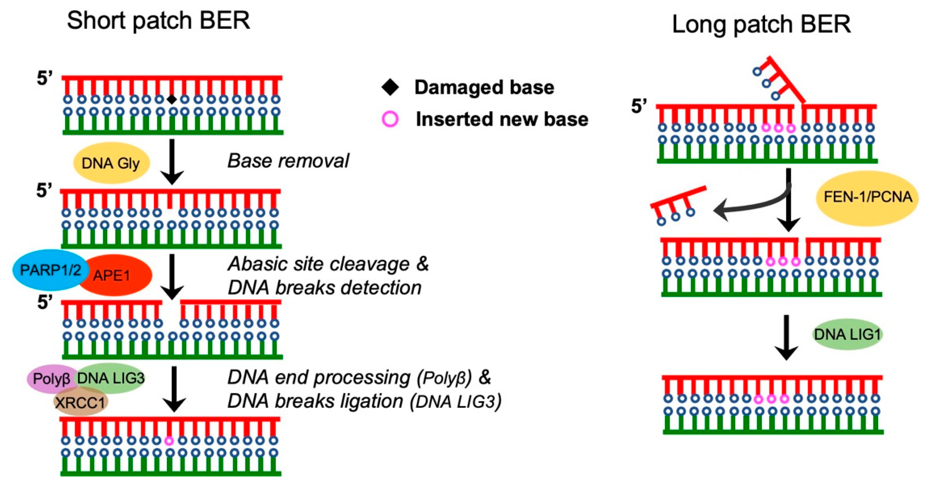
Figure 1. The repair of single-strand DNA breaks via BER. (Left: short-patch BER) The damaged bases are removed by DNA glycosylase, then the remaining abasic site (Ab) is cleaved by an apurinic/apyrimidinic endonuclease, leaving a 1-bp DNA gap. The 5′ abasic sugar is removed by DNA polymerase β (POLβ), which inserts a new nucleotide into the DNA gap. Finally, the nick is ligated by DNA ligase 3 (LIG3). (Right: long-patch BER) In long-patch repair, about 2–30 nucleotides are replaced. Polyβ induces the formation of an extended gap, then the gap displaces the 5′-terminus to create a flap that is excised by FEN1. Finally, the DNA ligase 1 ligates 5′ and 3′ nicks.
Pharmaceutics 15 02241 g001
Figure 2. DNA double-strand breaks by homologous recombination. The first step for homologous recombination includes the initiation of end resection, which is mediated by the CtIP—MRN complex (short-patch) or the EXO1/DNA2 (long-patch) nuclease. DNA end resection leads to the generation of 3′ssDNA overhangs, which are bound by replication protein A. Then, BRCA2 exchanges replication protein A on the DNA ends to promote the formation of RAD51 nucleoprotein filaments. The RAD51–ssDNA nucleoprotein filaments mediates homology search by invasion of template dsDNA. Furthermore, the RAD51–ssDNA nucleoprotein filaments form a synaptic complex that contains a three-stranded DNA helix intermediate. In this process, DNA polymerase δ (Pol δ) plays an important role in the synthesis of nascent strands.
Pharmaceutics 15 02241 g002
Figure 3. Non-homologous end joining repair mediates the repair of DNA double-strand breaks. DNA double-strand breaks can be repaired via c-NHEJ (left), A-NHEJ (middle), and single-strand annealing (SSA) (right). 53BP1 is a positive regulator of c-NHEJ, and the process usually requires 4-bp microhomology. A-NHEJ, also called microhomology-mediated end joining, requires 2–20 bp microhomology for break repair. PARP1 and Pol θ are important for A-NHEJ. Higher levels of resection can further promote SSA repair pathway, which requires >25 bp microhomology. BLM and EXO1 account for additional resections in SSA. Replication protein A binds and stabilizes ssDNA and promotes SSA. RAD52-mediated annealing of a homologous sequence is important for SSA. ERCC1/XPF cuts the remaining 3′ nonhomologous ssDNA prior to ligation by DNA LIG1.
Pharmaceutics 15 02241 g003
Figure 4. DNA damage repair blockade stimulates innate antitumor immunity via the cGAS-STING pathway. Upon DNA damage, DNA repair proteins are recruited to DNA damage sites for DNA repair. Inhibition of components of the repair pathway leads to cell-cycle checkpoint abrogation and inappropriate mitotic entry, and ultimately, induces mitotic catastrophe. In addition to being cytotoxic, DDR inhibitors exhibit antitumor immunity. PARPi, ATRi, or CHK1i-induced DSBs generate cytosolic dsDNA fragments, which activate the cGAS-STING innate immune pathway to initiate the IFN-γ response. This innate immune response upregulates chemokines, such like CCL5 or CXCL10, to enhance T cell recruitment. Moreover, PD-L1 expression is upregulated by IFN-γ that may lead to T cell exhaustion, an effect that can be abrogated using PD-1 or PD-L1 antibodies.
Pharmaceutics 15 02241 g004
Table 1. Ongoing clinical trials of the use of DDR inhibitors in EC.
Inhibitor Combination withPhaseStatusNCT Number
OlaparibMonotherapyPhase INot yet recruitingNCT05320757
OlaparibCYH33Phase IRecruitingNCT04586335
NiraparibMonotherapyPhase IIRecruitingNCT04716686
RucaparibNivolumabPhase I and IITerminatedNCT03572478
NiraparibTSR-042Phase IIActiveNCT03016338
OlaparibCarboplatinPhase ICompletedNCT01237067
OlaparibSelumetinibPhase IIRecruitingNCT05554328
AZD5305Paclitaxel or
Carboplatin or
T-Dxd or
Dato-Dxd or
Camizestrant
Phase I and IIRecruitingNCT04644068
OlaparibAZD2014 or AZD5363Phase I and IIActiveNCT02208375
RucaparibBevacizumabPhase IIActiveNCT03476798
NiraparibCopanlisibPhase IActiveNCT03586661
NiraparibDostarlimabPhase IINot yet recruitingNCT05870761
Olaparib or AZD6738AZD6738 or DurvalumabPhase IIRecruitingNCT03682289
OlaparibDS-8201aPhase IRecruitingNCT04585958
BBI-355MonotherapyPhase IRecruitingNCT05827614
BAY1895344ChemotherapyPhase IRecruitingNCT04491942
ART0380MonotherapyPhase IINot yet recruitingNCT05798611
AZD1775Radiotherapy and chemotherapyPhase IActiveNCT03345784
Disclaimer/Publisher’s Note: The statements, opinions and data contained in all publications are solely those of the individual author(s) and contributor(s) and not of MDPI and/or the editor(s). MDPI and/or the editor(s) disclaim responsibility for any injury to people or property resulting from any ideas, methods, instructions or products referred to in the content.