1. Introduction
The imidazole group’s wide-spectrum antifungal drug MCZ is frequently used to treat oral candidiasis, an opportunistic fungal infection of the oral cavity brought on by an overabundance of Candida species [
1]. Of them, the most common species found in oral cavity isolates from both healthy and sick people is
Candida albicans. MCZ is typically taken as an oral gel to treat oral candidiasis. Unfortunately, the limited aqueous solubility of this antifungal drug may limit its efficacy when preparing solid formulations for oral administration [
2]. The creation of MCZ-loaded nano carrier systems, such nanoparticles, is one possible way to get over this hurdle [
3]. The encapsulation of drugs into nanoparticles has been demonstrated to enhance the antifungal efficacy of these systems, making them ideal carriers for the administration of weakly water soluble antifungals such as clotrimazole, MCZ nitrate, and amphotericin B.
One kind of extracellular polysaccharide that is secreted by the bacteria
Xanthomonas campestris is called XGM. A terminal D-mannose unit is connected to a pyruvate group, D-glucose, D-mannose, and D-glucoronic acid in a ratio of 2:2:1. The structure is made up of a linear D-glucose backbone connected to a trisaccharide side chain containing D-mannose with an acetyl group, D-glucuronic acid. XGM is resistant to enzymatic degradation and is very stable over a broad pH and temperature range [
4]. It is widely utilized in the food and pharmaceutical industries because of its non-toxic and biocompatible qualities. Although the mucoadhesion duration is quite short, XGM is widely employed in mucoadhesive drug delivery systems [
5]. Due to less mucoadhesion time, the availability of the drug for absorption is low which ultimately lowers the drug’s bioavailability. In order to improve the mucoadhesion qualities, mercaptobutyric acid was used to thiolate the XGM in this work.
Many studies have been conducted recently on the development of bioadhesive/mucoadhesive controlled release systems [
6]. It is commonly acknowledged that raising a drug’s viscosity to impede clearance and extending the duration of contact through mucoadhesive interactions may increase the drug’s, or any therapeutic’s, bioavailability [
7]. For these kinds of applications, polymeric enhancers or mucoadhesive polymers might be a good option. They have use in the oral distribution of medications because they can improve permeability [
8]. One useful technique for drug immobilizationor localization is mucoadhesion, which is the binding of a synthetic or natural polymer to a biological surface. This is an essential component in controlled drug delivery. Although mucoadhesion is not a novel topic, a spike in curiosity has occurred in the use of mucoadhesive polymers for medication administration in recent years [
8,
9]. Recently there has been a lot of work on focusing a medication or formulation in one area of the body over extended periods of time [
10]. Medications that are absorbed via the mucosal lining of tissues can enter the blood stream directly and not be rendered inactive by enzymatic degradation in the gastrointestinal tract, which is why this is necessary not only for targeting medications but also for the improved regulation of systemic drug distribution [
8]. These polymers’ mucoadhesive qualities or the mucoadhesion mechanism can be described by the ionic interactions and production of non-covalent bonds, such as hydrogen bonds, between the mucoadhesive polymer and the mucus layer. Previous research on polymers has involved thiolation with mercaptopropionic and thioglycolic acid, which, when compared to the original polysaccharide, demonstrated good mucoadhesion capabilities [
11,
12].
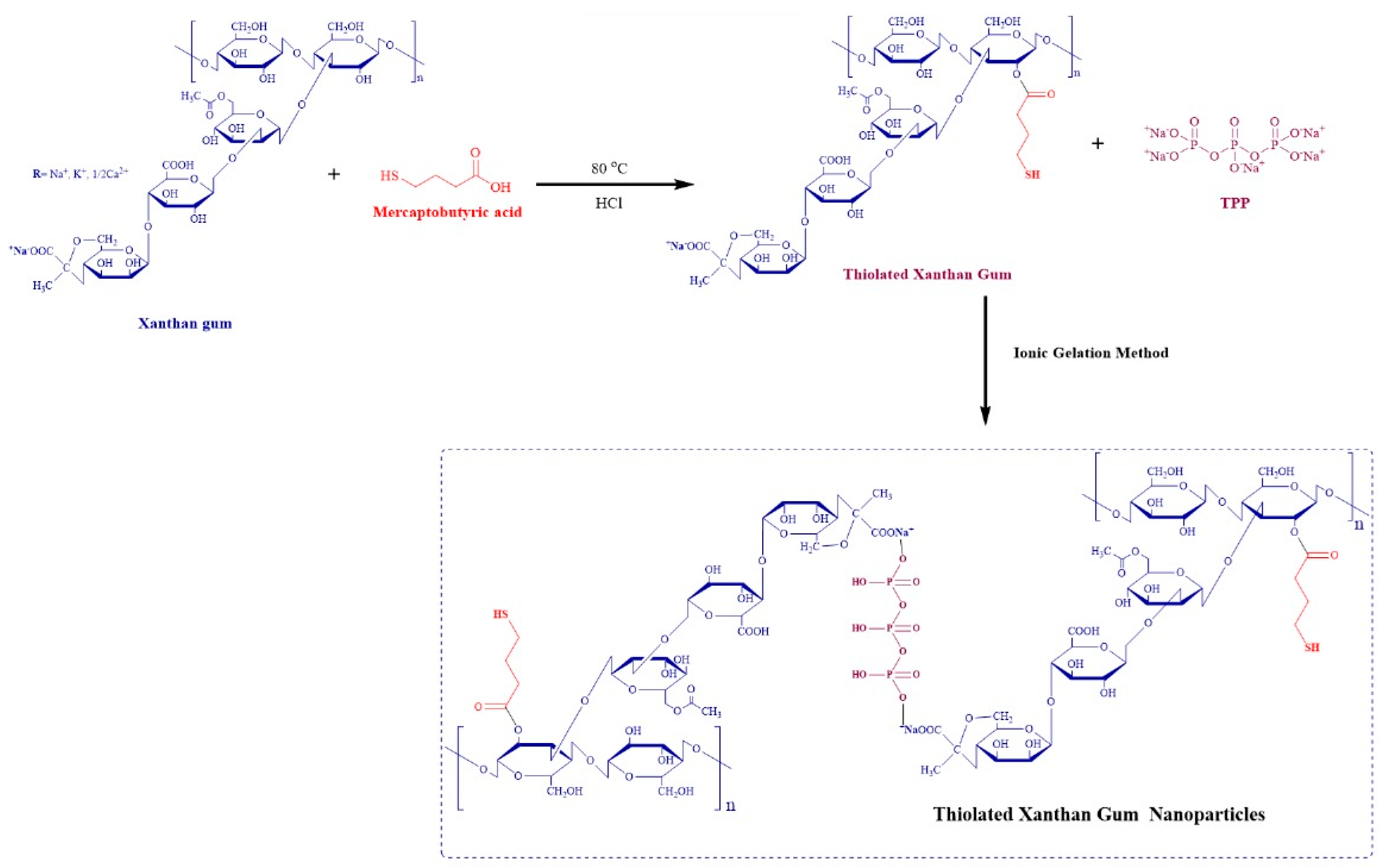
In this research work, XGM was thiolated with mercaptobutyric acid for the enhancement of mucoadhesion. Mercaptobutyric acid has excellent optical properties and is safe for humans due to its use in foods and beverages. Mercaptobutyric acid has an appropriate chain length [
13] and improves the mucoadhesive properties of XGM by incorporating the thiol group. Due to the presence of a sulfhydryl group in mucin, TXGM forms a disulfide bond which enhances mucoadhesion strength. According to our best knowledge from the literature search, mercaptobutyric acid is being used for the first time for the thiolation of xanthan gum to enhance the mucoadhesion time. The developed nanoparticles were characterized by Fourier transforms infrared spectroscopy (FTIR), differential scanning calorimetry (DSC), thermogravimetric analysis (TGA), X-ray diffraction (XRD) and scanning electron microscopy (SEM). The nanoparticles were also evaluated for drug release, stability, antifungal activity and pharmacokinetics of MCZ using albino rats after oral delivery.
2. Materials and Methods
2.1. Materials
MCZ was gifted by Saffron Pharmaceuticals, Pvt. Ltd., Faisalabad, Pakistan. Xanthan gum (XGM), mercaptobutyric acid, methanol and HCl were purchased from Sigma Aldrich Gmbh, Darmstadt, Germany. Trypsin and potassium dihydrogen phosphate were purchased from Merck, Darmstadt, Germany.
2.2. Thiolation of XGM
Using mercaptobutyric acid (MBA) and a catalytic quantity of hydrochloric acid, the esterification reaction was utilized to thiol-functionalize XGM. Two moles of MBA were added to each hydroxyl group in one mole of XGM to carry out the reaction. A catalytic amount of HCl (7 N) and MBA (4.2 mL) were added to 250 mL of hot water to dissolve 8 g of XGM. For 150 min, the reaction mixture was refluxed at 80 °C. Methanol was added to the reaction mixture above, allowing it to cool and precipitate. The resulting precipitates were then dried in an oven set to 50 °C after being rinsed with methanol twice [
12].
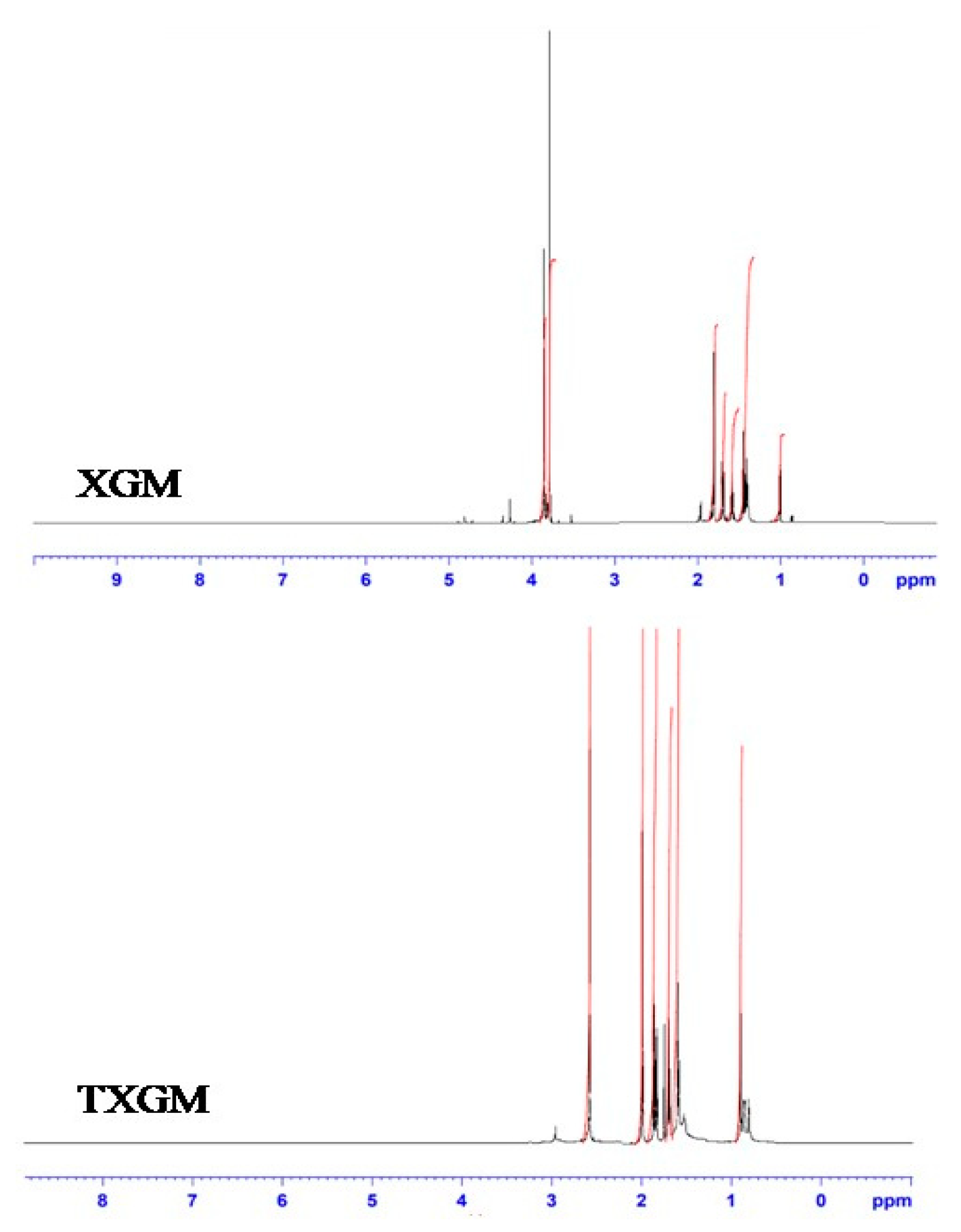
2.3. 1H NMR Analysis
1H NMR spectra of XGM and TXGM were obtained by dissolving them in deuterated NMR solvents at 400-MHz spectrometer using a 1H NMR spectrometer (Bruker Avance III) Harwell, UK.
2.4. Determination of Thiol Contents
Ellman’s reagent [DTNB] was used in a colorimetric process to quantify the contents of thiol of TXGM [
14]. The free thiol groups of TXGM react with DTNB in this reaction to generate a colored conjugate, whose absorbance may be measured at 412 nm using a UV/vis spectrometer (UNICAM 8700 series). To put it briefly, 500 µL of 0.5 M phosphate buffer pH 8.0 was used to hydrate 0.5 mg of each conjugate. The samples absorbance was evaluated at 412 nm following a 2-h incubation period at 25 °C with 500 µL of Ellman’s reagent (0.03% (
w/
v) DTNB in 0.5 M phosphate buffer pH 8.0). With the help of linearity curve resulting from the sulfhydryl group measurement of solutions containing different amounts of cysteine (0.003–0.05 mg/mL), the contents of thiol groups in TXGM were determined. With the help of linearity curve resulting from the sulfhydryl group measurement of solutions containing different amounts of cysteine holding a constant concentration of unmodified XGM (0.5 mg/mL), the quantity of thiol groups of TXGM was determined.
2.5. Preparation of Nanoparticles
Prepare 1% solution of previously dried xanthan gum powder that makes it 0.5 g of previously dried and processed thiolated xanthan gum in 50 mL of deionized water and stir for 30 min. Then prepare tri-sodium polyphosphate (STPP) solution with the concentration of 0.7 mg/mL and with the final volume of 50 mL. After 30 min add 50 mL of STPP solution in the xanthan gum solution drop wise and stir for about an hour. After 1 h, centrifuge the mixture for about 60 min at 6000 rpm. Separate the supernatant, dry the precipitates, and again grind it into a fine powder. Five formulations were prepared by changing the concentration of STPP shown in
Table 1. The particle size was reduced by using a high pressure homogenizer (Camsdorfer Ufer 12 07749, Jena, Germany) at 23,000 rpm for 18 min with 3 intervals of 6 min and pausing for 1 min after every interval. Then centrifuge (De Novo Tech, 27AFUPN0722K1Z2, Pune, India) the mixture at 12,000 rpm for 15 min. After 15 min, separate the supernatant from the dried particles and grind it to fine powder [
15].
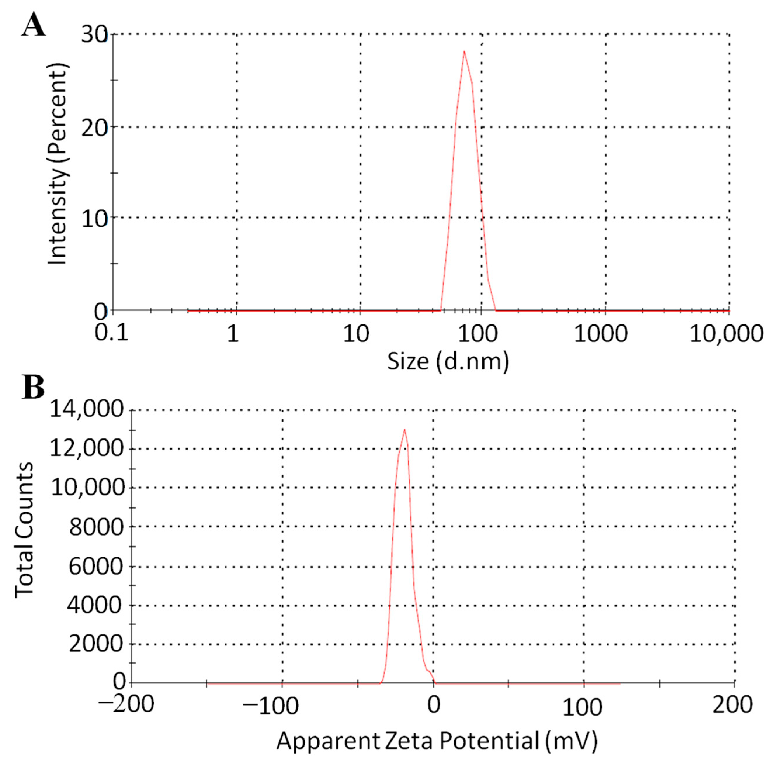
2.6. Size and Surface Charge Analysis
Particle size and polydispersity index (PDI) were measured by diluting specimens in a polystyrene cuvette using double-distilled water [
16]. Dynamic light scattering (DLS) was used to measure the samples at a temperature of 25 °C and with a scattering angle of 90° using a Beckman Coulter N5 Particle Size Analyzer (Beckman, Brea, CA, USA). In order to describe the size distribution, PDI was also measured. The particles’ zeta potential was measured at 25 °C using a Zetasizer (Malvern Instrument Ltd., Malvern, UK, ZS 90) after an appropriate dispersion was made using double-distilled, de-ionized water. The average particle size, PDI and zeta potential were obtained by repeating each sample three times (
n = 3).
2.7. Loading and Entrapment Efficiency of MCZ
A precisely measured quantity of nanoparticles was mixed with 1 mL of phosphate buffer saline (pH 7.4). The addition of trypsin (0.1 mg/mg of NPs) degraded the particles in the solution [
17]. After centrifuging the mixture and stirring it for the entire night, the drug concentration of the supernatant was examined (Equation (1)). All the measurements of % MCZ loading were triplicated (
n = 3).
A 1 mL sample was spun at 40,000 rpm for 20 min using a Beckman-Coulter Optima MAX-XP ultra-centrifuge (Beckman, Brea, CA, USA) in order to evaluate the MCZ entrapment in nanoparticles. The MCZ content in the supernatant was examined, and the entrapment efficiency (
n = 3) was computed using the equation below:
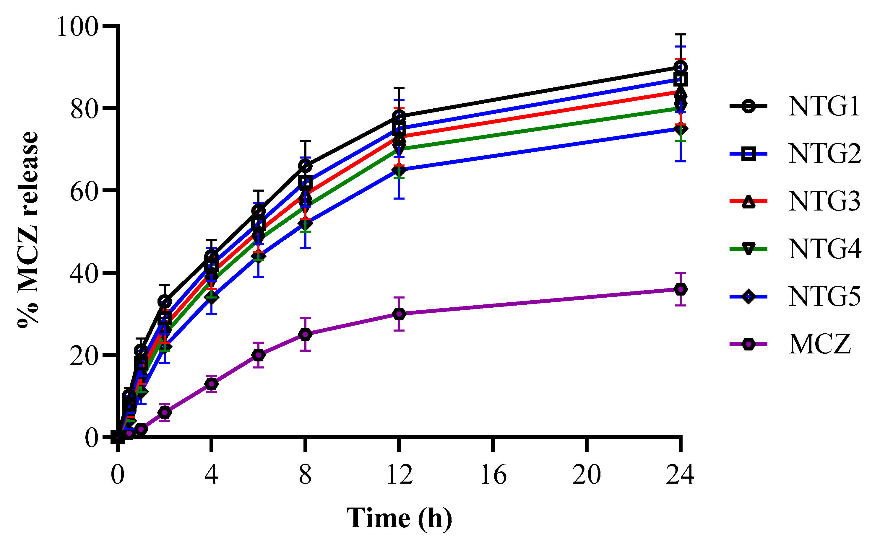
2.8. Release of MCZ from Nanoparticles
MCZ-loaded nanoparticles containing 10 mg of the MCZ were placed inside the dialysis bags (dialyzing membrane-150, molecular weight cutoff 12,000–14,000 Dalton) in order to evaluate the MCZ release from the nanoparticles. Before use, the membrane was soaked in water for 30 min. The dialysis bag was incubated for 24 h at 37 ± 1 °C with constant stirring at 100 rpm in 900 mL of pH 6.8 phosphate buffer. To keep the sink condition, 2 mL of sample volume were removed and replaced with fresh buffer at predetermined intervals. Using HPLC, the amount of MCZ released in the dissolution media was measured [
18]. The release of MCZ from all prepared nanoparticulate formulations was repeated six times (
n = 6).
2.9. Characterization of Nanoparticles
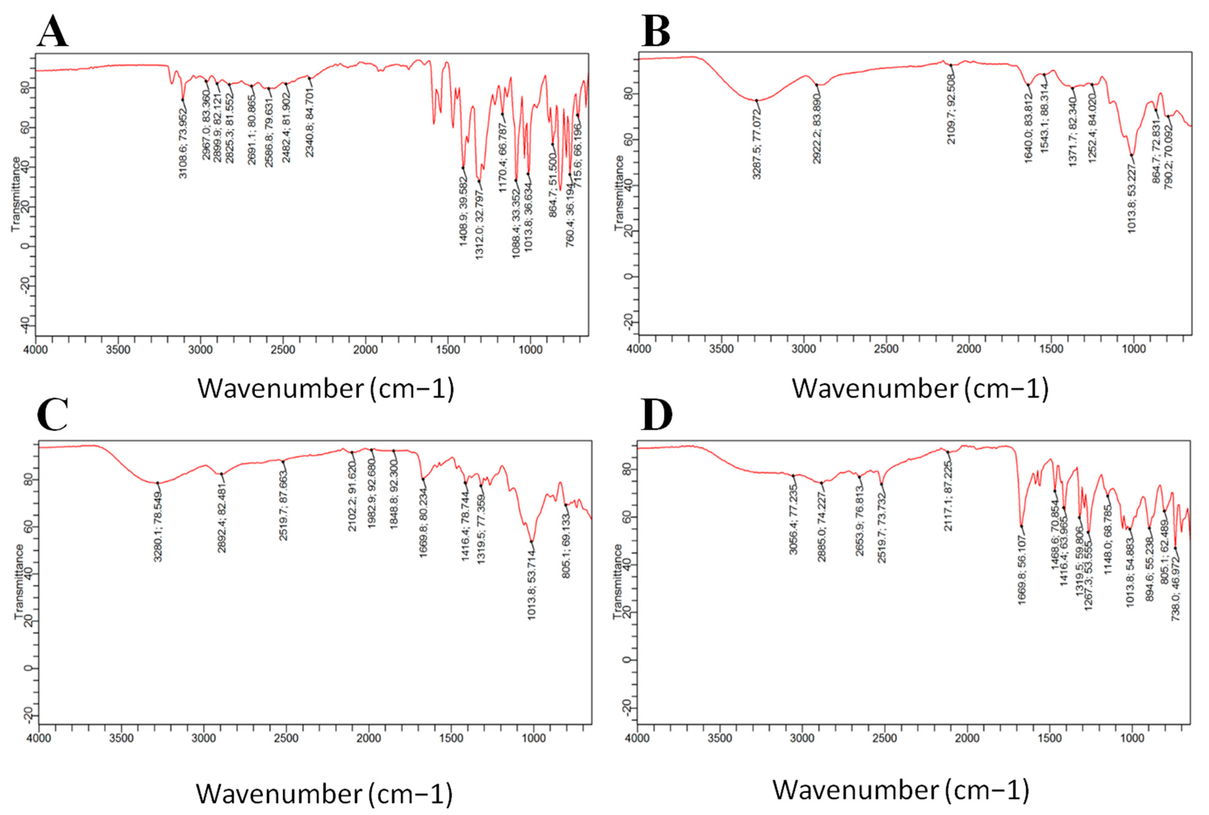
2.9.1. Fourier Transforms Infrared Spectroscopy (FTIR)
MCZ, XGM, TXGM, blank and MCZ-loaded nanoparticles werechecked for determination of thiol and other functional groups using ATR-FTIR Bruker, Alpha, Germany at 400–4000 cm−1 wavelength with a resolution of 1 cm−1.
2.9.2. Differential Scanning Calorimetry (DSC)
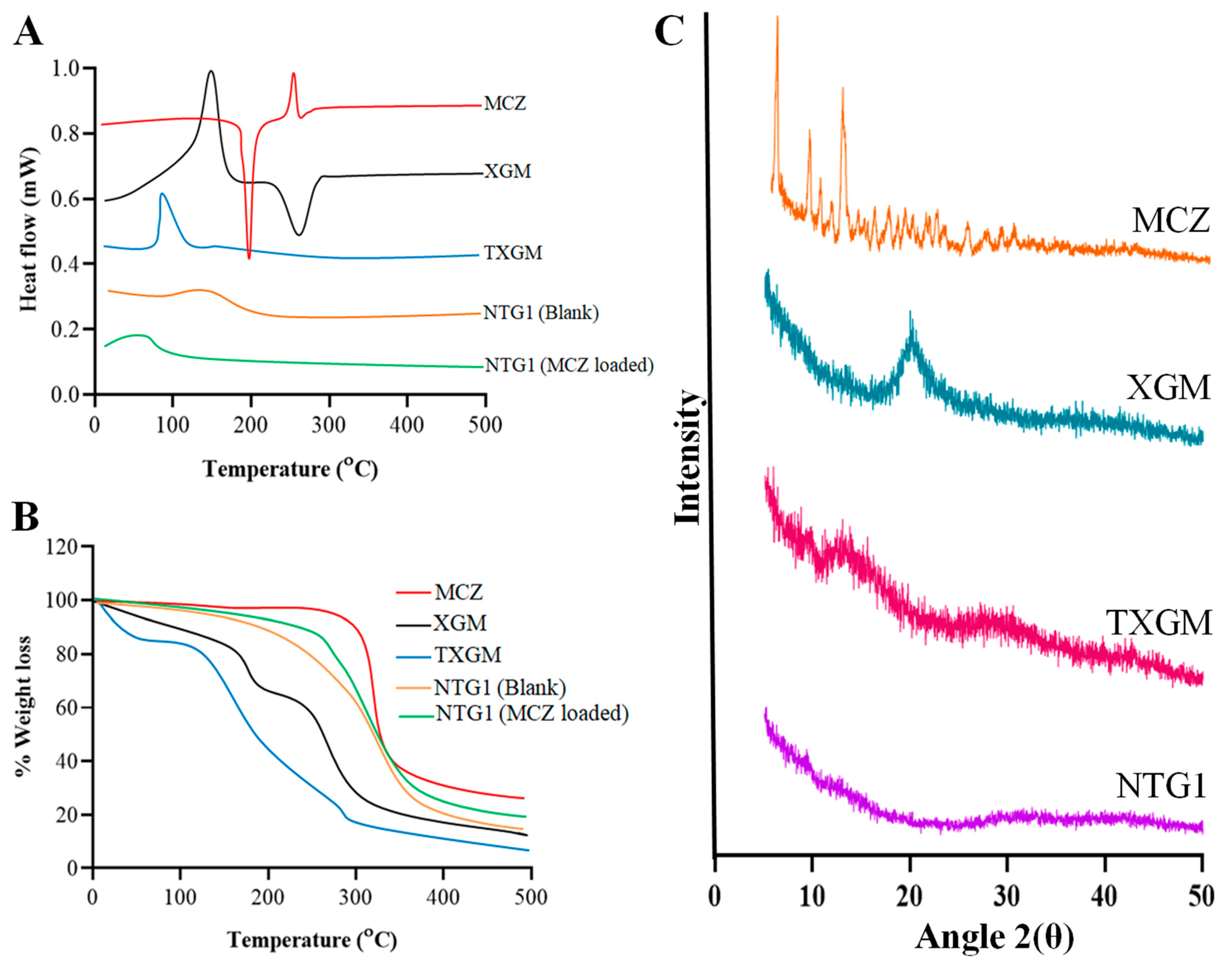
A Mettler DSC 823 (Mettler-Toledo, Greifensee, Switzerland) was used to execute a DSC analysis of MCZ, XGM, TXGM, and lyophilized nanoparticles of NTG1. Weighed and tightly sealed, the milled samples were placed within an aluminum pan. Both the reference cell and the sample were evacuated with the flow of nitrogen during the measurement. The samples were put inside the cell of the sample holder and heated to 500 °C, with a 10 °C/min increase in temperature while thermograms were taken [
19].
2.9.3. Thermogravimetric Analysis (TGA)
TG curves were obtained for MCZ, XGM, TXGM, blank, and MCZ-loaded nanoparticles using an alumina crucible and a Shimadzu DTG60 thermobalance set to heat at a rate of 10 °C min−1 from 30 to 500 °C, with a dynamic atmosphere of nitrogen at 50 mL min−1. The mass of the sample was precisely weighted, weighing approximately 2.5 mg.
2.9.4. X-ray Diffraction Analysis (XRD)
The X-ray diffractometer (PANalytical B.V., Almelo, The Netherlands) was used to measure X-ray diffraction of MCZ, XGM, TXGM, blank and MCZ-loaded nanoparticles. The setup for the generator was 40 kv and 40 Ma. The sample’s reflected radiation was measured at 25 °C and within the 2θ angle ranges of 2° to 80°, using a test speed of 1°/min.
2.9.5. Scanning Electron Microscopy (SEM)
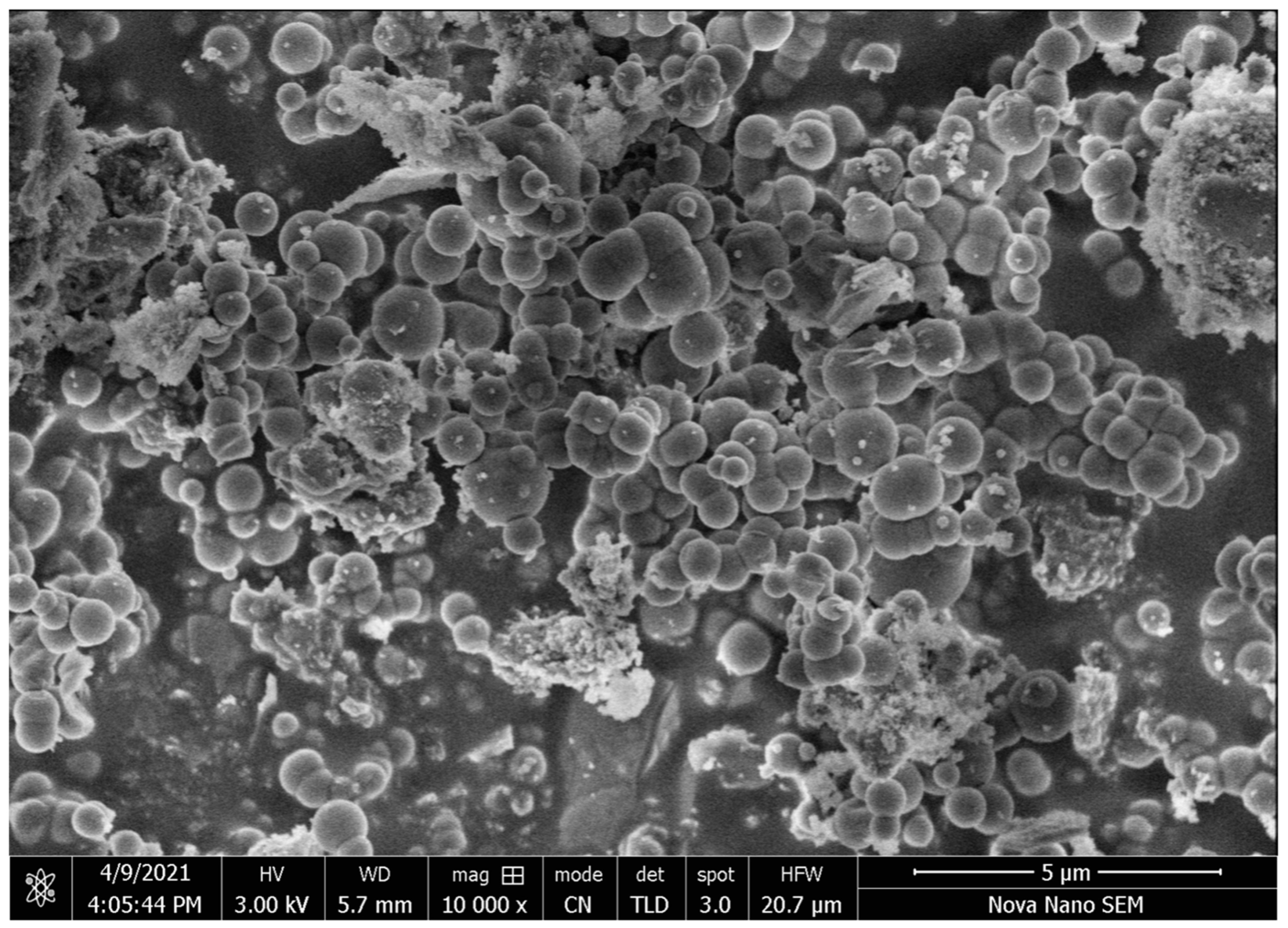
Using SEM, the surface morphology of the particles was investigated. A small conductive layer of gold was applied to the samples for this purpose, and their morphology was then examined under a scanning electron microscope (SEM-JEOL, JSM-7600F, JEOL Inc., Tokyo, Japan) operating at an accelerating voltage of 10–20 kW.
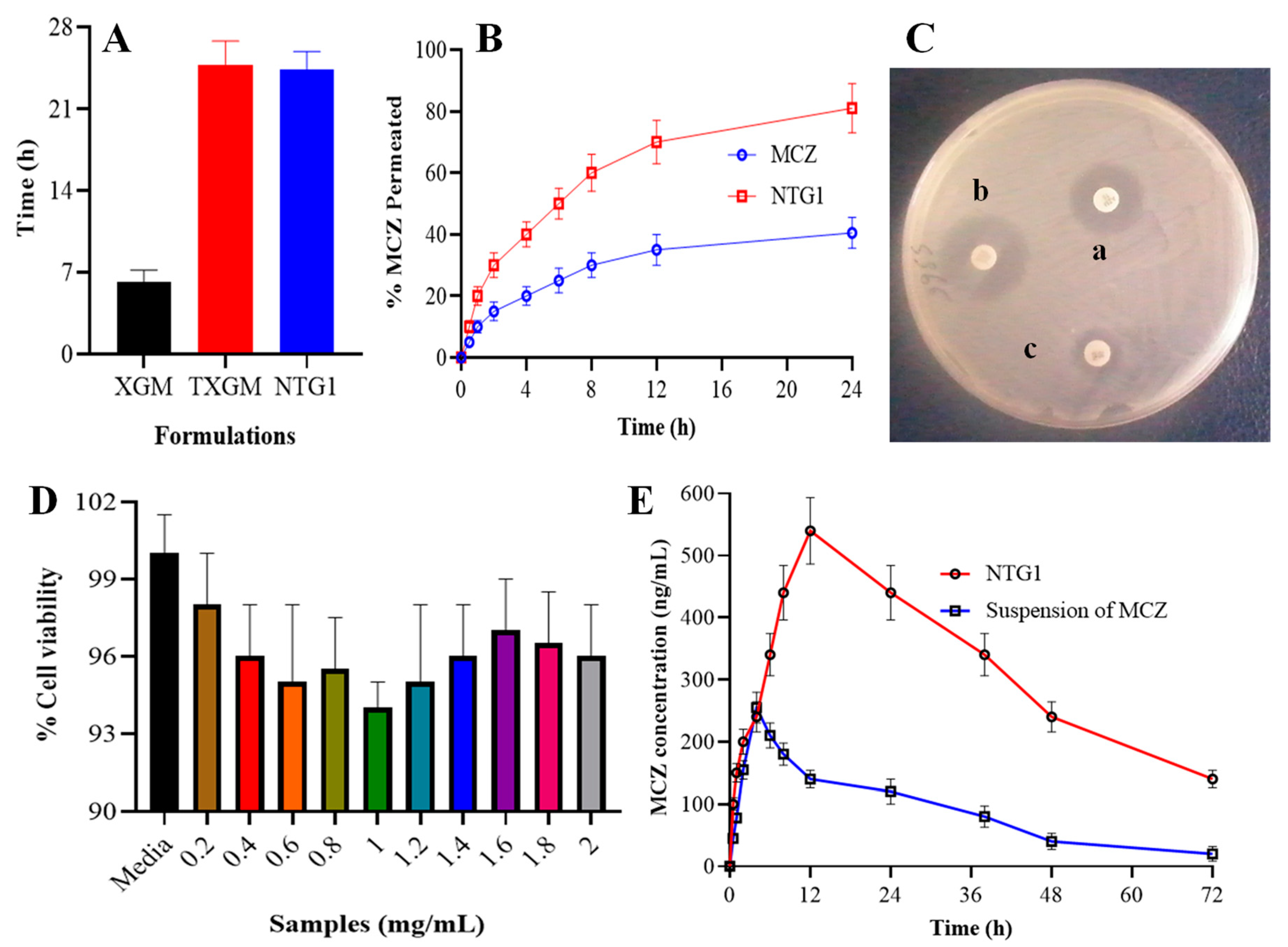
2.10. Mucoadhesion Study
The ex vivo adhesion time of TXGM and developed nanoparticles was measured using the intestinal mucosa of sheep as the membrane of biological origin in order to evaluate their mucoadhesive capabilities [
20]. Within 30 min of the slaughter, the intestinal mucosa of the sheep was taken and washed with distilled water, followed by a 37 °C simulation of intestinal fluid (pH 6.8). Using cyanoacrylate glue, XGM, TXGM, and developed nanoparticles moistened with one drop of artificial intestinal fluid (pH 6.8), the mucosa was attached on the inside of a beaker. The mucosa was then pasted on using a light fingertip force for a duration of 20 s. Dissolution apparatus type II was used to keep the beaker, which held 900 mL of the simulated intestinal fluid, at 37 °C. Nanoparticle attachment was observed when the liquid was agitated at 100 rpm using a paddle to mimic the intestinal environment. The adhesion time is the time needed for the XGM, TXGM, and developed nanoparticles to separate from the intestinal mucosa. The average mucoadhesion time was obtained by repeating each sample three times (
n = 3)
2.11. Permeation Study
The Franz diffusion cell, which has an interior diameter of 2.5 cm, was used for permeability investigations. As the model membrane, albino rat intestinal mucosa was employed [
21]. Dissection was used to get the intestine of the recently dead albino rats. After being carefully removed and clipped from the sides, the intestinal mucosa was cleaned in an isotonic phosphate buffer with a pH of 7.4. In the space between the donor and receptor compartments, the mucosa was mounted. A magnetic bead revolving at 100 rpm was used to stir 30 mL of isotonic phosphate buffer with a pH of 7.4, which was kept at 37 ± 0.5 °C, into the receptor compartment. Following their prior moistening with 2 mL of buffer, the mucosal surface of the membrane came into close contact with the nanoparticles and suspension in water containing 4 mg of MCZ. Samples were taken out at appropriate intervals for up to 24 h, and the HPLC method was used to analyze them after replacing the same amount with fresh media. The measurements of permeability studies from each formulation repeated three times (
n = 3).
Coefficients of apparent-permeability (
Papp, cm/h) and steady state diffusion (
D, cm
2/h) were considered using Equations (3) and (4);
The calculation of Jss involved graphing the total amount of MCZ permeability per unit area versus time (hours), with the slope of the linear section of the curve representing steady state flux. The initial total donor chamber concentration of MCZ is denoted by Cd. L is the diffusion path length, and K is the MCZ partition coefficient (logP).
2.12. Antifungal Activity of Nanoparticles Containing MCZ
Candida albicans (ATCC No. 10231, Sigma-Aldrich Production GmbH, Buchs, Switzerland) was used in the agar-well diffusion method to measure the antifungal activity [
22]. For the preparation of inoculum, 24-h-cultures of Candida albicans were used. The concentration of yeast was adjusted to 0.5 McFarland by turbidimetry (Densimat, bioMérieux, Marcy-l’Étoile, France) on saline [
23]. This procedure will yield a yeast stock suspension of 1 × 10
6 to 5 × 10
6 cells per mL. To prepare 150 mg/mL concentrations of MCZ, NTG1 nanoparticles and MCZ gel are suspended in DMSO along with MCZ, the positive control. In the tests, the DMSO concentration did not go above 0.4%. A typical inoculum of fungal culture was inoculated onto a plate holding Muller-Hinton agar. A, B and C, three 6-mm-diameter wells, were punched into the agar. Wells A and B were filled with the MCZ gel and the prepared MCZ-containing nanoparticles, respectively, and C indicated the control group. The plates were incubated at 37 °C for 48 h. To assess the antifungal activity, the zone of inhibition’s diameter was assessed. Minimum inhibitory concentration (MIC) values of MCZ gel and NTG1 dispersion were measured by making dispersion in DMSO. The lowest concentration that completely stopped yeast growth was identified as the MIC value. Three (
n = 3) MIC studies were conducted.
2.13. In Vitro Cytocompatibility Studies
The MTT assay was used to determine the in vitro cytocompatibility of the synthesized NTG1 nanoparticles in the Caco-2 cell line (ATCC 2112 M St, Washington, DC, USA). MTT assay: a colorimetric examination that relies on live cells’ specific capability to convert MTT into formazan crystals with a purple color [
24]. The absorbance maxima of these formazan crystals are located at 570 nm. After dissolving the formed formazan crystals in a solubilization buffer, the absorbance at 570 nm was measured. The quantity of live cells is exactly proportional to the absorbance that was obtained. The percentage of cell viability versus the amount of NTG1 nanoparticles in mg/mL was then shown on a graph. Using particle-free Caco-2 cell culture wells as a negative control, the MTT test was performed at doses varying from 0.2 to 2 mg/mL. The cells and particles were then cultured for 48 h. Next, 10% MTT solution was added, and the mixture was incubated for 4 h. After that, solubilization buffer was added, and the mixture was incubated for an additional hour. With the help of a Beckmann-Coulter Elisa plate reader (Bio-Tek Power Wave XS, Hampton, NH, USA), the absorbance was measured at 570 nm after the MTT had completely dissolved. For every experiment, three duplicate samples were examined.
2.14. Stability Study of Nanoparticles
Size, PDI, and surface charge are used to characterize the stability of nanoparticles. Because surface energy and the surface-to-volume ratio both rise at the nanoscale, the stability of nanoparticles is a crucial parameter [
25]. Important factors for physical targeting or drug delivery to particular regions are size, PDI, surface charge and %EE. The stability tests were conducted in closed glass vials for 6 months at 4 °C and 25 °C. The size, PDI, surface charge and %EE of NTG1 to NTG5 formulations were repeated three times (
n = 3).
2.15. HPLC Method for MCZ Estimation
The HPLC method was developed for the quantification of MCZ using mobile phase consists of methanol and water in a ratio of 75:25 v/v. The Hitachi L-2000 series HPLC system, made in Japan, was furnished with a UV-Vis detector L2420 (Leuven, Belgium), L injection loop, and a Model L-2130 pump. A C18 column was used for separation with a detection wavelength of 313 nm with 1 mL/min flow rate of used mobile phase. The method was validated via calculating the percentage recoveries, precision, system suitability parameters, limit of detection and quantification.
2.16. Pharmacokinetic Analysis
The pharmacokinetics of MCZ was assessed in albino rats weighing between 250–450 g. The twelve albino rats were taken and divided into two groups (n = 6). Group 1 (the control group) received a suspension of MCZ in distilled water, and nanoparticles of thiolated XGM containing MCZ were administered to group2 (the test group) orally. After administration, blood samples from the rat tail vein were withdrawn at regular time intervals (0, 0.5, 1, 2, 4, 8, 12, 24, 36, 48 and 72 h) and placed in heparinized tubes. The blood samples were centrifuged at 4500 rpm for 20 min, the plasma was separated, deproteinizer was added and centrifuged again at 4500 rpm for 20 min. The clear supernatant was separated and injected into the HPLC system for analysis. The study determined several pharmacokinetic parameters, including maximum concentration of MCZ in plasma (Cmax), the moment at which plasma concentration peaks appear (tmax), and area under the plasma concentration–time curve from 0 h to 24 h (AUC0–t) and 0 to infinity (AUC0–∞), and mean residence time (MRT).
2.17. Statistical Analysis
The release pattern of MCZ from nanoparticles was statistically analyzed by one way ANOVA. The student t test was also applied to the pharmacokinetics parameters.
4. Conclusions
TXGM, a mucoadhesive polymer, was synthesized and examined. Using Ellman’s procedure, the degree of thiol substitution was determined to be 61%. FTIR, DSC, TGA, XRD, SEM, and DLS analyses were used to characterize the synthesized TXGM nanoparticles. The produced nanoparticles have a spherical shape and range in size from 87 to 156 nm. Zeta potential measurements were used to study the system’s stability. It became apparent that the nanoparticles had a negative surface charge, and that the system was remarkably stable. Using the MTT assay, the produced nanoparticles’ in vitro cytocompatibility with cancer cell lines was investigated. According to these findings, the produced nanoparticles are less harmful (almost 93% of the cells remain viable in the TXGM nanoparticle samples at all dosages). Therefore, the produced TXGM nanoparticles can be applied as a useful biomaterial in biomedical applications including gene and mucoadhesive medication delivery.


 ,
, 


