Next Issue
Volume 12, March
Previous Issue
Volume 11, September


Cardiogenetics, Volume 11, Issue 4 (December 2021) – 8 articles

Cover Story (view full-size image): 
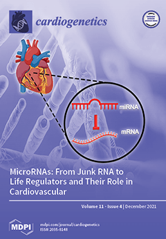
MicroRNAs (miRNAs) are single-stranded small non-coding RNA that have been considered junk RNA. Thanks to advances in molecular biology, miRNAs have become increasingly important as regulators of life. Post-transcriptional binding of miRNAs determines the inhibition of specific target genes. In the heart, miRNAs are responsible for regulating cardiomyocyte contractility, maintenance of cardiac rhythm, angiogenesis, and other functions. In pathological conditions, such as cardiomyopathy, these non-coding RNAs have been found to be differentially expressed in respect to physiological conditions causing cardiomyocyte damage, cardiac hypertrophy, cardiac fibrosis. Therefore, the study of circulating and tissue-specific miRNAs could lead to the identification of new therapeutic strategies and early diagnosis not only of cardiomyopathy but also of numerous other diseases. View this paper
  • Issues are regarded as officially published after their release is announced to the table of contents alert mailing list.
  • You may sign up for e-mail alerts to receive table of contents of newly released issues.
  • PDF is the official format for papers published in both, html and pdf forms. To view the papers in pdf format, click on the "PDF Full-text" link, and use the free Adobe Reader to open them.
Order results
Result details
Section
Select all
Export citation of selected articles as:
27 pages, 3895 KiB  
Review
Pathogenesis, Diagnosis and Risk Stratification in Arrhythmogenic Cardiomyopathy
by Maria Teresa Florio, Filomena Boccia, Erica Vetrano, Marco Borrelli, Thomas Gossios and Giuseppe Palmiero
Cardiogenetics 2021, 11(4), 263-289; https://doi.org/10.3390/cardiogenetics11040025 - 8 Dec 2021
Viewed by 3602
Abstract
Arrhythmogenic cardiomyopathy (ACM) is a genetically determined myocardial disease associated with sudden cardiac death (SCD). It is most frequently caused by mutations in genes encoding desmosomal proteins. However, there is growing evidence that ACM is not exclusively a desmosome disease but rather appears [...] Read more.
Arrhythmogenic cardiomyopathy (ACM) is a genetically determined myocardial disease associated with sudden cardiac death (SCD). It is most frequently caused by mutations in genes encoding desmosomal proteins. However, there is growing evidence that ACM is not exclusively a desmosome disease but rather appears to be a disease of the connexoma. Fibroadipose replacement of the right ventricle (RV) had long been the hallmark of ACM, although biventricular involvement or predominant involvement of the left ventricle (LD-ACM) is increasingly found, raising the challenge of differential diagnosis with arrhythmogenic dilated cardiomyopathy (a-DCM). A-DCM, ACM, and LD-ACM are increasingly acknowledged as a single nosological entity, the hallmark of which is electrical instability. Our aim was to analyze the complex molecular mechanisms underlying arrhythmogenic cardiomyopathies, outlining the role of inflammation and autoimmunity in disease pathophysiology. Secondly, we present the clinical tools used in the clinical diagnosis of ACM. Focusing on the challenge of defining the risk of sudden death in this clinical setting, we present available risk stratification strategies. Lastly, we summarize the role of genetics and imaging in risk stratification, guiding through the appropriate patient selection for ICD implantation. Full article
(This article belongs to the Special Issue Cardiogenetics: Feature Papers 2021)
Show Figures

Figure 1

8 pages, 1001 KiB  
Article
Understanding the Function of a Locus Using the Knowledge Available at Single-Nucleotide Polymorphisms
by Majid Nikpay, Sepehr Ravati, Robert Dent and Ruth McPherson
Cardiogenetics 2021, 11(4), 255-262; https://doi.org/10.3390/cardiogenetics11040024 - 7 Dec 2021
Cited by 2 | Viewed by 2447
Abstract
Understanding the function of a locus is an issue in molecular biology. Although numerous molecular data have been generated in the last decades, it remains difficult to grasp how these data are related at a locus. In this study, we describe an analytical [...] Read more.
Understanding the function of a locus is an issue in molecular biology. Although numerous molecular data have been generated in the last decades, it remains difficult to grasp how these data are related at a locus. In this study, we describe an analytical workflow that can solve this problem using the knowledge available at the single-nucleotide polymorphism (SNP) level. The underlying algorithm uses SNPs as connectors to link biological entities and identify correlations between them through a joint bioinformatics/statistics approach. We demonstrate its application in finding the mechanism whereby a mutation causes a phenotype and in revealing the path whereby a gene is regulated and impacts a phenotype. We translate our workflow into publicly available shell scripts. Our approach provides a basic framework to solve the information overload problem in biology surrounding the annotation of a locus and is a step toward repurposing GWAS data for new applications. Full article
(This article belongs to the Special Issue Cardiogenetics: Feature Papers 2021)
Show Figures

Figure 1

25 pages, 860 KiB  
Review
MicroRNAs: From Junk RNA to Life Regulators and Their Role in Cardiovascular Disease
by Federica Amodio, Martina Caiazza, Fabio Fimiani, Paolo Calabrò and Giuseppe Limongelli
Cardiogenetics 2021, 11(4), 230-254; https://doi.org/10.3390/cardiogenetics11040023 - 29 Nov 2021
Cited by 1 | Viewed by 3282
Abstract
MicroRNAs (miRNAs) are single-stranded small non-coding RNA (18–25 nucleotides) that until a few years ago were considered junk RNA. In the last twenty years, they have acquired more importance thanks to the understanding of their influence on gene expression and their role as [...] Read more.
MicroRNAs (miRNAs) are single-stranded small non-coding RNA (18–25 nucleotides) that until a few years ago were considered junk RNA. In the last twenty years, they have acquired more importance thanks to the understanding of their influence on gene expression and their role as negative regulators at post-transcriptional level, influencing the stability of messenger RNA (mRNA). Approximately 5% of the genome encodes miRNAs which are responsible for regulating numerous signaling pathways, cellular processes and cell-to-cell communication. In the cardiovascular system, miRNAs control the functions of various cells, such as cardiomyocytes, endothelial cells, smooth muscle cells and fibroblasts, playing a role in physiological and pathological processes and seeming also related to variations in contractility and hereditary cardiomyopathies. They provide a new perspective on the pathophysiology of disorders such as hypertrophy, fibrosis, arrhythmia, inflammation and atherosclerosis. MiRNAs are differentially expressed in diseased tissue and can be released into the circulation and then detected. MiRNAs have become interesting for the development of new diagnostic and therapeutic tools for various diseases, including heart disease. In this review, the concept of miRNAs and their role in cardiomyopathies will be introduced, focusing on their potential as therapeutic and diagnostic targets (as biomarkers). Full article
(This article belongs to the Special Issue Cardiogenetics: Feature Papers 2021)
Show Figures

Figure 1

11 pages, 12430 KiB  
Case Report
Arrhythmogenic Cardiomyopathy—Further Insight into the Clinical Spectrum of Desmoplakin Disease
by Joanne Simpson, Joan Anusas, Denise Oxnard, Sylvia Wright, Ruth McGowan and Caroline Coats
Cardiogenetics 2021, 11(4), 219-229; https://doi.org/10.3390/cardiogenetics11040022 - 10 Nov 2021
Viewed by 2712
Abstract
Arrhythmogenic cardiomyopathy is a familial heart muscle disease characterized by structural, electrical, and pathological abnormalities. Recognition of left ventricular (LV) involvement in arrhythmogenic right ventricular cardiomyopathy (ARVC) has led to the newer term of arrhythmogenic cardiomyopathy (ACM). We report on a family with [...] Read more.
Arrhythmogenic cardiomyopathy is a familial heart muscle disease characterized by structural, electrical, and pathological abnormalities. Recognition of left ventricular (LV) involvement in arrhythmogenic right ventricular cardiomyopathy (ARVC) has led to the newer term of arrhythmogenic cardiomyopathy (ACM). We report on a family with autosomal dominant desmoplakin (DSP) related ACM to illustrate the broad clinical spectrum of disease. The importance of evaluation of relatives with cardiac magnetic resonance imaging and consideration of genetic testing in the absence of Task Force diagnostic criteria is discussed. The practical and ethical issues of access to the Guthrie collection for deoxyribonucleic acid (DNA) testing are considered. Full article
(This article belongs to the Special Issue Genetic Diagnostics in Inherited Cardiomyopathies)
Show Figures

Figure 1

15 pages, 4042 KiB  
Article
Analysis of ABC Transporter Gene Expression in Atherosclerosis
by Stanislav Kotlyarov and Anna Kotlyarova
Cardiogenetics 2021, 11(4), 204-218; https://doi.org/10.3390/cardiogenetics11040021 - 4 Nov 2021
Cited by 7 | Viewed by 3288
Abstract
ABC transporters are a large family of membrane proteins that transport chemically diverse substrates across the cell membrane. Disruption of transport mechanisms mediated by ABC transporters causes the development of various diseases, including atherosclerosis. Methods: A bioinformatic analysis of a dataset from Gene [...] Read more.
ABC transporters are a large family of membrane proteins that transport chemically diverse substrates across the cell membrane. Disruption of transport mechanisms mediated by ABC transporters causes the development of various diseases, including atherosclerosis. Methods: A bioinformatic analysis of a dataset from Gene Expression Omnibus (GEO) was performed. A GEO dataset containing data on gene expression levels in samples of atherosclerotic lesions and control arteries without atherosclerotic lesions from carotid, femoral, and infrapopliteal arteries was used for analysis. To evaluate differentially expressed genes, a bioinformatic analysis was performed in comparison groups using the limma package in R (v. 4.0.2) and the GEO2R and Phantasus tools (v. 1.11.0). Results: The obtained data indicate the differential expression of many ABC transporters belonging to different subfamilies. The differential expressions of ABC transporter genes involved in lipid transport, mechanisms of multidrug resistance, and mechanisms of ion exchange are shown. Differences in the expression of transporters in tissue samples from different arteries are established. Conclusions: The expression of ABC transporter genes demonstrates differences in atherosclerotic samples and normal arteries, which may indicate the involvement of transporters in the pathogenesis of atherosclerosis. Full article
Show Figures

Figure 1

13 pages, 2122 KiB  
Article
Spectrum of Clinical Features and Genetic Profile of Left Ventricular Noncompaction Cardiomyopathy in Children
by Agata Paszkowska, Alicja Mirecka-Rola, Dorota Piekutowska-Abramczuk, Elżbieta Ciara, Łukasz Mazurkiewicz, Katarzyna Bieganowska and Lidia Ziółkowska
Cardiogenetics 2021, 11(4), 191-203; https://doi.org/10.3390/cardiogenetics11040020 - 22 Oct 2021
Cited by 3 | Viewed by 2860
Abstract
Background: Left ventricular noncompaction (LVNC) is a genetically determined cardiomyopathy that occurs following a disruption of endomyocardial morphogenesis. The purpose of this study was to identify the clinical characteristics and genetic profile of children with LVNC. Methods: From February 2008 to July 2020, [...] Read more.
Background: Left ventricular noncompaction (LVNC) is a genetically determined cardiomyopathy that occurs following a disruption of endomyocardial morphogenesis. The purpose of this study was to identify the clinical characteristics and genetic profile of children with LVNC. Methods: From February 2008 to July 2020, a total of 32 children (median 11.5 years) with LVNC were prospectively enrolled and followed up for a median of 4.02 years. Diagnosis was made based on characteristic features of LVNC in echocardiography and cardiovascular magnetic resonance (CMR). Patients’ clinical symptoms, family history, ECG, Holter ECG, and genetic tests were also evaluated. Results: The most common presenting symptom was heart failure (31% of children). ECG abnormalities were noted in 56% of patients. The most prominent features were ventricular arrhythmias, sinus bradycardia, and paroxysmal third-degree atrioventricular block. Most of the patients (94%) met the criteria for LVNC and CMR confirmed this diagnosis in 82% of cases. The molecular etiology was found in 53% of children. Conclusion: Although heart failure and arrhythmias were very frequent in our study group, thromboembolic events and genetic syndromes were rare. For the accurate and reliable assessment of children with LVNC, it is necessary to get to know their family history and detailed clinical profile. Full article
Show Figures

Figure 1

6 pages, 2095 KiB  
Case Report
Azygos Vein ICD Lead Implantation Lowers Defibrillation Threshold in a Patient with Hypertrophic Cardiomyopathy
by Giovanni Quarta, Paola Ferrari, Andrea Giammarresi, Giovanni Malanchini, Cristina Leidi, Michele Senni and Paolo De Filippo
Cardiogenetics 2021, 11(4), 185-190; https://doi.org/10.3390/cardiogenetics11040019 - 7 Oct 2021
Viewed by 3377
Abstract
A 14-year-old boy with hypertrophic cardiomyopathy (HCM) diagnosed at the age of 1 year and with massive left ventricular hypertrophy suffered an episode of ventricular fibrillation during mild effort. He underwent a dual-chamber implantable cardioverter defibrillator (ICD) implantation. The defibrillation threshold testing (DFT) [...] Read more.
A 14-year-old boy with hypertrophic cardiomyopathy (HCM) diagnosed at the age of 1 year and with massive left ventricular hypertrophy suffered an episode of ventricular fibrillation during mild effort. He underwent a dual-chamber implantable cardioverter defibrillator (ICD) implantation. The defibrillation threshold testing (DFT) was ineffective. Subcutaneous multi-coli arrays tunneled into the left postero-lateral position and connected to the superior vena cava (SVC) port of the dual-chamber ICD were added to increase the myocardial mass involved in the defibrillation shock pathway. A new DFT was unsuccessful. The patient was transferred to our hospital for myectomy. An epicardial defibrillation patch was placed on the left ventricular lateral wall, but again, DFT testing was ineffective using the right ventricular (RV) coil to lateral patch as shock pathway. Another epicardial defibrillation patch was then placed on the inferior wall. In this case, DFT testing was effective with a defibrillation pathway between the two patches and the can. In November 2015, a high shock impedance alarm was recorded through remote monitoring, thus compromising the safety of the ICD shock pathway. The patient underwent the implant of a new trans-venous defibrillation coil lead in the azygos vein. After few months, the patient developed symptomatic severe aortic regurgitation and underwent an aortic valve replacement. During the operation, DFT testing was performed and was successful. Our case illustrates that azygous vein ICD lead implantation is efficacious in HCM with massive hypertrophy and high DFT, and prompts further studies to systematically investigate its efficacy in this particular subgroup of the HCM population. Full article
(This article belongs to the Special Issue Cardiogenetics: Feature Papers 2021)
Show Figures

Figure 1

21 pages, 6391 KiB  
Article
Specific Deletion of the FHA Domain Containing SLMAP3 Isoform in Postnatal Myocardium Has No Impact on Structure or Function
by Taha Rehmani, Jana Mlynarova, Joseph Byers, Maysoon Salih and Balwant S. Tuana
Cardiogenetics 2021, 11(4), 164-184; https://doi.org/10.3390/cardiogenetics11040018 - 4 Oct 2021
Cited by 2 | Viewed by 3047
Abstract
Sarcolemmal membrane-associated proteins (SLMAPs) belong to the superfamily of tail-anchored membrane proteins known to regulate diverse biological processes, including protein trafficking and signal transduction. Mutations in SLMAP have been linked to Brugada and defective sodium channel Nav1.5 shuttling. The SLMAP gene is alternatively [...] Read more.
Sarcolemmal membrane-associated proteins (SLMAPs) belong to the superfamily of tail-anchored membrane proteins known to regulate diverse biological processes, including protein trafficking and signal transduction. Mutations in SLMAP have been linked to Brugada and defective sodium channel Nav1.5 shuttling. The SLMAP gene is alternatively spliced to generate numerous isoforms, broadly defined as SLMAP1 (~35 kDa), SLMAP2 (~45 kDa) and SLMAP3 (~80–95 kDa), which are highly expressed in the myocardium. The SLMAP3 isoform exhibits ubiquitous expression carrying an FHA domain and is believed to negatively regulate Hippo signaling to dictate cell growth/death and differentiation. Using the αMHC-MerCreMer-flox system to target the SLMAP gene, we specifically deleted the SLMAP3 isoform in postnatal mouse hearts without any changes in the expression of SLMAP1/SLMAP2 isoforms. The in vivo analysis of mice with SLMAP3 cardiac deficiency revealed no significant changes to heart structure or function in young or aged mice without or with isoproterenol-induced stress. SLMAP3-deficient hearts revealed no obvious differences in cardiac size, function or hypertrophic response. Further, the molecular analysis indicated that SLMAP3 loss had a minor impact on sodium channel (Nav1.5) expression without affecting cardiac electrophysiology in postnatal myocardium. Surprisingly, the loss of SLMAP3 did not impact Hippo signaling in postnatal myocardium. We conclude that the FHA domain-containing SLMAP3 isoform has no impact on Hippo signaling or sodium channels in postnatal myocardium, which is able to function and respond normally to stress in its absence. Whether SLMAP1/SMAP2 isoforms can compensate for the loss of SLMAP3 in the affairs of the postnatal heart remains to be determined. Full article
(This article belongs to the Section Molecular Genetics)
Show Figures

Figure 1

Previous Issue
Next Issue
Back to TopTop