Article

Evaluation of Outer Surface Protein Vaccine Candidates of Borrelia burgdorferi for Lyme Disease

Latvian Biomedical Research and Study Centre, Rātsupītes 1, k-1, LV1067 Riga, Latvia
*
Author to whom correspondence should be addressed.
Microbiol. Res. 2023, 14(4), 2022-2033; https://doi.org/10.3390/microbiolres14040136
Submission received: 18 October 2023 / Revised: 21 November 2023 / Accepted: 27 November 2023 / Published: 28 November 2023

Abstract

:
Lyme disease affects several hundred thousand people worldwide annually, yet there is no registered vaccine for the disease available for human use. The disease is caused by Borrelia burgdorferi sensu lato complex bacteria, which harbor numerous outer surface proteins, and many of which have been targeted for vaccine development. However, to effectively combat various Borrelia species, the target protein should ideally be conserved and located in the chromosome. In this study, we evaluated the potential of seven conservative, chromosome-encoded outer surface proteins as vaccine candidates. Unfortunately, four of the initial candidates could not be produced in E. coli. The remaining BB0028, BB0158, and BB0689 proteins were administered to mice in both the free form and as conjugates with virus-like particles (VLPs). In most cases, high antibody titers were obtained, confirming the good immunogenicity of the selected proteins. However, for BB0158 and BB0689 proteins, adverse effects were observed following the injection of free proteins, which were not observed when they were coupled to VLPs. Bactericidity tests of the obtained antibodies suggested that none of the vaccine candidates could induce the production of bactericidal antibodies.

1. Introduction

Lyme disease is caused by several genospecies present in the Borrelia burgdorferi sensu lato complex [1]. In North America, the disease is primarily caused by Borrelia burgdorferi sensu stricto, while in Europe, it is predominantly caused by Borrelia garinii and Borrelia afzelii [2]. Lyme disease is the most common tick-borne infection in the Northern Hemisphere. According to a recent meta-analysis, the estimated seroprevalence of Borrelia burgdorferi sensu lato in the global population, or the number of people who have been exposed to Lyme disease during their lifetime, accounts for approximately 14.5% of the world’s population [3]. It is also essential to note that in the last few decades, the number of cases of the disease has significantly increased compared to the period from 2001 to 2010 [4]. According to estimates, Lyme disease occurs in more than 200,000 people every year in Western Europe [2], while in the USA, this figure is approximately 476,000 [5].
Antibacterial therapy can be used to treat Lyme disease, and Borrelia burgdorferi is susceptible to three classes of antibiotics: beta-lactam antibiotics, tetracyclines, and, to a lesser extent, macrolides [6]. Although the disease can be cured with antibiotics, symptoms may persist for years even after antibiotic treatment, and hence, the obvious need for prophylactic vaccination.
The surface of Borrelia bacteria is adorned with numerous surface proteins that can serve as targets for vaccine development. In the 1990s, two Lyme disease vaccines were developed that based on the Borrelia outer surface protein OspA: “LYMErix™” (SmithKline Beecham, London, UK) and “ImuLyme™” (Pasteur Mérieux Connaught, Toronto, ON, Canada). Phase III trials of both vaccines showed that they are safe and effective. However, “ImuLyme™” was not licensed, while “LYMErix™,” after being introduced to the market in 1998, was withdrawn in 2002 due to insufficient consumer demand and reports of side effects such as arthritis [7]. It should be noted that reported side effects have no real scientific bases as recently reviewed by [8]. However, OspA is expressed in bacteria only when residing in ticks, not in host; therefore, high antibody titers should be maintained. Also, since OspA does not reside in host, no amnesic response is activated. Many other outer surface proteins have also been targeted, including OspC [9], BB0405 [10], BBA64 [11], BBI16 [12], BBA52 [13], BBK32 [14], BbHtrA [15], RevA [16,17], and CspZ [18,19]. New vaccine prototypes are also being developed using the OspA protein. The vaccine VLA15, which utilizes the C-terminal segments of six OspA serotypes, has shown effectiveness in mouse models against four clinically significant Borrelia species: B. burgdorferi, B. afzelii, B. garinii, and B. bavariensis [20]. This vaccine is currently in phase III clinical trials (Pfizer Inc. and Valneva SE, 2022). A somewhat similar approach, using protein subunits as vaccine targets, has also been used for other tick-borne pathogens, such as rBdP0 [21] and rBdEBP [22] from Babesia protozoa.
The aim of our study was to create a VLP vaccine candidate, based on conserved, chromosome-encoded B. burgdorferi surface proteins. We explored the immunogenic properties of several uncharacterized (BB0158, BB0171, BB0213, BB0352, BB0689, and BB0823) or partially characterized (BB0028) B. burgdorferi surface proteins for their potential as new anti-Lyme disease vaccine candidates. One of the approaches in developing new vaccines involves using virus-like particles (VLPs). VLPs are self-assembling structures of viral capsid proteins that mimic the viral structure, enhancing the immune recognition of exposed antigens [23]. VLPs from ssRNA phages like MS2, AP205, and Qβ are particularly popular in vaccine development due to their robustness and their ease of production in bacteria [24]. VLPs have been used in Lyme disease vaccine development, e.g., coupling OspA and OspC to hepatitis B core antigen [25] and CspZ to ssRNA bacteriophage Qβ VLPs [26]. We have generated several VLP-based vaccine candidates—AP205d-BB0689, QβVLP-BB0028, QβVLP-BB0158, and QβVLP-BB0689. To this end, mice were immunized with vaccine candidates and uncoupled proteins. Subsequently, immunogenicity and in vitro antibody bactericidal assays were determined using established methods. No bactericidal effect was observed in any of the cases, neither for VLP conjugates nor uncoupled proteins.

2. Materials and Methods

2.1. Production and Purification of Recombinant Proteins

Production and purification of B. burgdorferi proteins were carried out as previously described [27]. Protein sequences from B. burgdorferi strain B31 were supplemented with a cleavable 6xHis tag at the N-terminal for purification. The corresponding codon-optimized genes were ordered and cloned into the pETduet-1 vector by the company Biocat.

2.2. Generation of Vaccine Candidates and Coupling to Bacteriophage Qβ VLPs

The purification of Qβ VLPs and the chemical coupling of B. burgdorferi proteins to the purified VLPs followed a basic protocol described previously [28], with slight modifications. Briefly, Qβ VLPs were labeled with tetrazine-NHS by adding it to the protein solution in PBS from a stock solution in DMSO at a 0.9:1 molar ratio with respect to Qβ VLPs. The mixture was incubated at room temperature for 2 h. The unreacted tetrazine-NHS label was removed, and the buffer was changed to PBS (pH 7.2) using a desalting column. B. burgdorferi proteins were labeled with BCN-NHS ester by adding them to the protein solution to achieve a final concentration of 2 mM from a 50× stock solution in DMSO. Subsequently, the unreacted BCN-NHS ester was removed, and the buffer was changed to PBS using a desalting column. Labelled Qβ VLPs-tetrazine-NHS and B. burgdorferi proteins-BCN-NHS were mixed at mass ratios of 1:1, incubated for 16 h at +4 °C, and the excess free B. burgdorferi proteins were separated from the Qβ-protein conjugates using dialysis through a 1000 kDa cutoff membrane against PBS. Protein concentrations were determined using a NanoDrop spectrophotometer (ThermoFisher Scientific, Waltham, MA, USA). The protein purity and efficiency of chemical coupling were verified using SDS-PAGE followed by Coomassie staining and native agarose gel electrophoresis.

2.3. Animal Studies

The experimental procedures in animals were approved by the National Animal Welfare and Ethics Committee (permit no. 102/2019, for mice infection study; and permit no. 136/2022, for antibody response evaluation studies) and were conducted in compliance with the Directive 2010/63/EU as adopted by the national legislation.
For infection studies with B. burgdorferi, we used five-week-old female C3H/HeNHsd mice (n = 8), and for immunization studies, we employed seven-week-old female BALB/cOlaHsd mice (Envigo, Melderslo, The Netherlands) (n = 8). Female mice have been used in most studies of Lyme disease vaccine candidates [19,29,30,31,32]; thus, we also chose the same sex. For infection, mice were intradermally infected with 105 B. burgdorferi (same dose as reported previously [29,31,33]) suspended in 100 µL of BSK-H incomplete medium (BioConcept, Allschwil, Switzerland). Negative control mice were injected with BSK-H incomplete medium. The joint diameters of both tibiotarsus joints were measured with a digital caliper on days 0, 7, 14, 21, and 28 after infection, in a similar manner as reported before [29,33]. For immunization, the vaccines with squalene-based adjuvant AddaVax (Invivogen, vac-adx-10, San Diego, CA, USA) were mixed in a 1:1 (v/v) ratio immediately before injection. Each animal received three doses of vaccine or control, 14 days apart, with a dose of 25 µg administered subcutaneously per injection per animal in a total volume of 100 µL, which is the vaccine dose that we usually use in our vaccine studies [19,26,28,34]. Animals were weighed every three days and monitored for possible vaccination-induced side effects. At the end of the experiment, animals were humanely euthanized under deep surgical isoflurane anesthesia (5%), and cardiac puncture was performed to collect final blood samples.

2.4. Evaluation of Vaccine-Induced Antibody Responses

For the evaluation of the total IgG response against B. burgdorferi proteins, ELISA plates (Greiner, Meiningen, Germany) were coated with 1 µg per well of recombinant B. burgdorferi proteins overnight at 4 °C. Subsequently, wells were blocked with 1% BSA in PBST and then incubated with two-fold serially diluted serum samples in 1% BSA in PBST for 1 h at 37 °C. The wells were washed three times with PBST and dH2O between the steps. Plates were then incubated with a rabbit anti-mouse IgG HRP conjugate (1:5000, Sigma-Aldrich A9044, Burlington, MA, USA) for 1 h at 37 °C, and then were washed, and the serum antibody binding was visualized using colorimetric O-phenylenediamine dihydrochloride detection (Sigma, P6912-100TAB, Burlington, MA, USA). Optical density was measured spectrophotometrically at 492 nm using an ELISA plate reader (BDSL Immunoskan MS, Finland). For the assessment of the vaccine-induced IgG1, IgG2a, and IgG2b antibody responses, goat anti-mouse IgG subclass-specific secondary antibody HRP conjugates were used (Abcam ab98693, ab986998, and ab98703). The vaccine-induced IgG antibody responses against the Qβ VLP carrier were assessed using the same protocol with the following modifications: the ELISA plate was coated with recombinantly expressed Qβ coat protein, and the serum samples were three-fold serially diluted. A positive signal was defined relative to the negative control, i.e., the mean OD429 values from the wells probed with serum samples from animals injected with PBS.

2.5. B. burgdorferi Strain and Bactericidal Activity of Serum from Immunized Mice

The B. burgdorferi strain B31-A3 used in this study is a clonal isolate of B31 [26] grown at 33 °C in BSK II complete medium (Sigma-Aldrich, Burlington, MA, USA). Mouse sera from immunized mice and infected mice were used to determine bactericidal activity against B. burgdorferi with serum bactericidal assay [26]. Prior to determining the bactericidal activity, these mouse sera were heat-treated at 56 °C for 30 min to inactivate the complement system. Then, 50 µL of diluted mouse serum (1:20, 1:40, 1:80, 1:160, 1:320, 1:640, 1:1280, and 1:2560) was mixed with 10 µL of complement-preserved guinea pig serum (Sigma-Aldrich, Burlington, MA, USA) and B. burgdorferi (105 cells/mL) in 40 µL of BSK II complete medium and then incubated at 33 °C for 24 h. Surviving spirochetes were quantified by directly counting only motile spirochetes using phase-contrast microscopy (ZEISS Axio Lab.A1, Jena, Germany). The survival percentage was calculated as the proportion of serum-treated-to-untreated B. burgdorferi.

3. Results

3.1. Choice of Vaccine Candidate Proteins

As vaccine candidates, we selected several surface proteins that had to meet specific criteria. Firstly, the corresponding gene had to reside in the bacterial chromosome rather than in plasmids, as some plasmids are known to be lost in specific isolates. Secondly, the protein had to be reasonably conserved, preferably with more than an 80% identity across different Borrelia burgdorferi species. Lastly, the candidate had to be confirmed as a surface protein in previous experimental studies. To fulfil this last criterion, we referred to a paper by Dowdell et al. [35].
Overall, based on the above criteria, we selected seven proteins for further studies: BB0028 (UniProt Proteome ID UP000001807), BB0158 (UP000001807), BB0171 (UP000001807), BB0213 (UP000001807), BB0352 (UP000001807), BB0689 (UP000001807), and BB0823 (UP000001807).
BB0028 has been previously shown to be a part of the beta-barrel assembly complex proteins [36] and resides in the periplasm. Nevertheless, we included it in our study due to the convincing surface exposure data provided by Dowdell et al. BBH0689 is a chromosome-encoded borrelial outer surface protein of unknown function, but it has been structurally characterized by our group [27]. Some proteins with similar folds are involved in binding lipophilic substances such as cholesterol and fatty acids. BBH0689 shares approximately 87% identity with various Borrelia species. Presumably, BBH0689 plays an active role during the infection process since its production is upregulated after the transfer of bacteria from the tick vector to the mammalian host. BB0158, BB0171, BB0213, BB0352, and BB0823 are uncharacterized proteins with unknown functions.
To enhance the immunogenicity of the proteins, we chose to utilize the virus-like particle (VLP) platform. Several approaches can be employed to display proteins or peptides on the VLP surface, e.g., genetic fusion, chemical conjugation, and bio-glue. Primarily, we attempted the genetic fusion method using a single-chain dimer of the AP205 phage coat protein, enabling a single foreign protein insertion per dimer. As a secondary approach, we utilized chemical conjugation to Qβ virus-like particles. Based on our previous experience, Qβ VLPs proved to be more efficient for chemical coupling compared to other VLPs available in our lab.

3.2. Production of Proteins for Vaccination

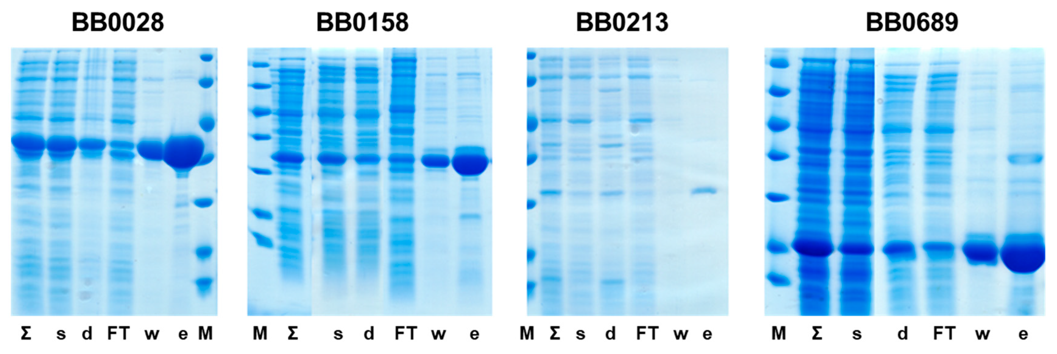
Nearly all chimeric proteins produced through the genetic fusion method were either insoluble or inadequately produced in E. coli cells. The exception was AP205d-BB0689, which was both soluble and capable of forming VLPs. The production and purification of AP205d-BB0689 are depicted in Figure 1.
Although it was possible to produce proteins BB0213 and BB0352 in their soluble form, their production level was extremely low. Consequently, both proteins were deemed unsuitable for further development. Additionally, no detectable production was observed for proteins BB0171 and BB0823. Both BB0028 and BB0158 could be produced and purified in a soluble form. Therefore, based on the production results, we proceeded with three proteins: BB0028, BB0158, and BB0689.
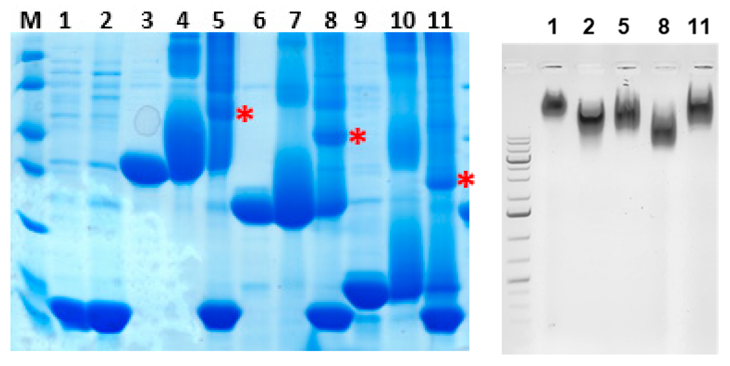
We successfully coupled BB0028, BB0158, and BB0689 proteins to Qβ VLPs using the tetrazine/BCN-8PEG linker. The efficiency of in vitro chemical conjugation was determined using SDS-PAGE and native agarose gel electrophoresis. The appearance of new bands in the higher molecular weight range on SDS-PAGE, and a shift in the position of VLP bands on native agarose gel, indicated the successful conjugation of the antigens to the VLPs. The production and purification of stand-alone proteins are presented in Figure 2, while their coupling to phage Qβ VLPs is illustrated in Figure 3.

3.3. Presence of Antibodies against Produced Antigens in Infected Mice

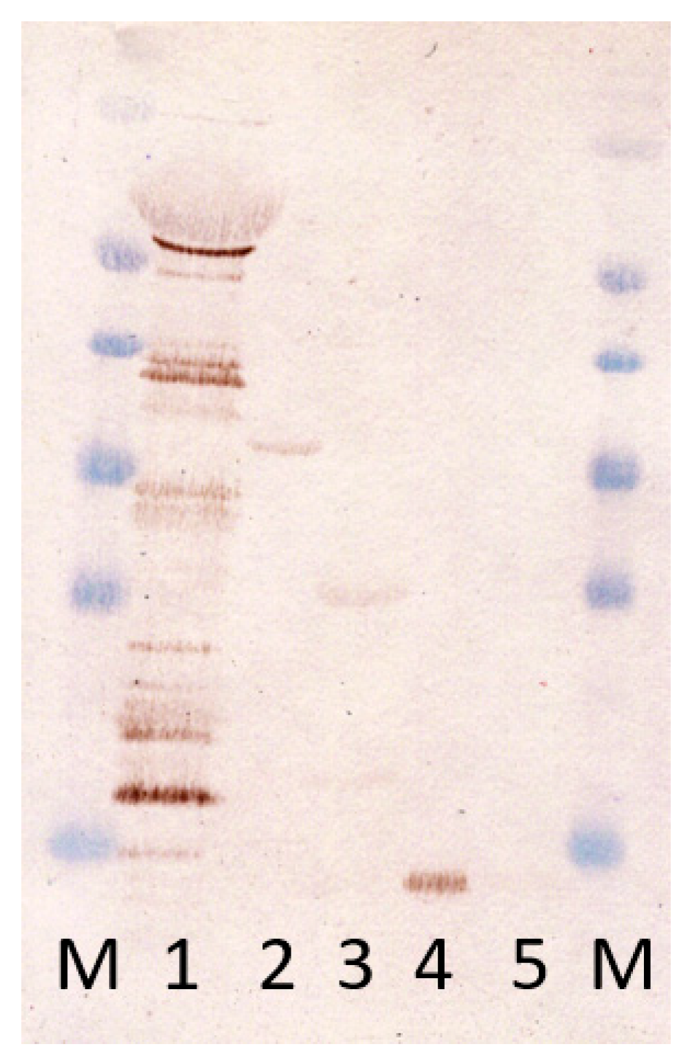
After the successful production of antigens, we wanted to check whether antibodies against them are produced during the infection of animals with B. burgdorferi. To test this, we performed a Western Blot, where sera from infected animals indeed displayed some reactivity with all three chosen proteins—BB0689, BB0158, and BB0028 (Figure 4). The strongest reactivity was observed with BB0689, while reactivity with BB0158 and BB0028 was somewhat weaker.

3.4. Immunogenicity Studies in Mice

The initial immunization experiment, followed by Borrelia burgdorferi infection, was conducted in C3H mice. These mice developed arthritis during the infection [37]. Therefore, this murine model is commonly employed in anti-Lyme disease vaccine studies because it closely mimics the chronic presentation of Lyme borreliosis in humans [19,26,38,39,40].
At the outset of the study, only the BB0689 protein and its genetically coupled version with the AP205 single-chain dimer were available. Consequently, we established five groups of mice, each consisting of eight animals: PBS, BB0689, AP205d-BB0689, AP205d VLP, and the infection group.
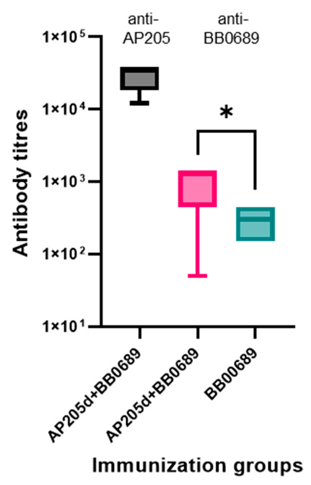
Following the third immunization, all the mice in the BB0689 group experienced an anaphylactic shock, resulting in the loss of half of the animals. Two weeks after immunization, we obtained blood serum samples and determined antibody titers against BB0689 and AP205d VLP proteins. The sera from the AP205d-BB0689 and AP205d VLP groups exhibited significantly high anti-AP205d VLP titers, approximately 3 × 10⁴, while the sera from the AP205d-BB0689 and BB0689 groups displayed anti-BB0689 titers below 1 × 10³. Notably, anti-BB0689 antibody titers were statistically significantly higher, as illustrated in Figure 5.
Due to low antibody titers in C3H mice, we conducted further immunogenicity tests on the stand-alone proteins BB0028, BB0158, and BB0689, as well as their chemical conjugates with bacteriophage Qβ VLPs in BALB/c mice. While the initial immunization was well tolerated by all animals, we observed that the second and third immunizations with stand-alone BB0689 and BB0158 proteins caused apparent illness in mice. Within 30 min, half of the mice in groups immunized with BB0158 and BB0689 became less mobile, alert, and social, failed the grip test, and did not avoid being handled by the caretakers. Additionally, we observed a decrease in their body temperature by palpation. These effects lasted for about one to two hours. In contrast, the BB0028 stand-alone vaccine and all Qβ VLP conjugate vaccines were well tolerated throughout the experiment. Notably, the resulting antibody titers were high in all cases; however, exposure to antigens present on Qβ VLPs did not lead to an increase in antibody levels (Figure 6). Similar observations were made in our previous studies of the CspZ vaccine, where antigen coupling to Qβ VLPs did not result in an overall increase in antibody titers. Nevertheless, the Qβ VLP conjugates exhibited enhanced bactericidal activity and protective efficacy [26].

3.5. Bactericidal Efficacy Testing of Obtained Sera

To evaluate the bactericidal effect of antibodies from immunized mice, we conducted a bactericidal test. As controls, we utilized B. burgdorferi cultures without sera or complement, sera from mice immunized with Qβ VLPs as a negative control, and serum from non-immunized C3H mice infected with B. burgdorferi as a positive control. Qβ VLPs are non-infectious nanoparticles that elicit a robust immune response but do not cross-react with B. burgdorferi antigens. The results revealed no bactericidal effect on spirochetes in any of the groups, even at minimal serum dilution. The number and morphology of surviving bacteria exhibited no differences between immunization groups and were consistent with the monitoring and negative control samples. The sera from the positive control group exhibited a bactericidal effect. These results indicate that the serum antibodies from immunized mice do not possess bactericidal activity against B. burgdorferi.

3.6. Confirmation of the Presence of Selected Antigens in B. burgdorferi Bacteria

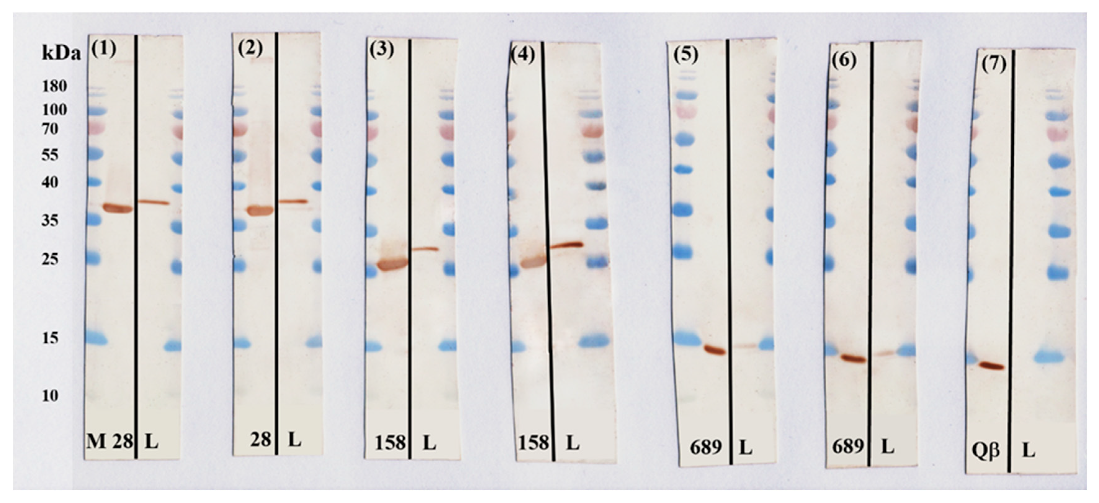
To rule out the possibility that our antigens were not being produced by the bacteria during cultivation, we investigated the synthesis of proteins BB0028, BB0158, and BB0689 in B. burgdorferi grown at +33 °C. Western Blot results demonstrated that antibodies from both the control and vaccine group mouse sera were bound to proteins corresponding to the detectable bands of the correct molecular weight (Figure 7). Conversely, antibodies from the Qβ VLP group did not bind to proteins in the B. burgdorferi lysate. By evaluating the bands visible in the Western Blot images, it can be seen that the synthesis of the BB0689 protein in B. burgdorferi is detectable, but somewhat lower than that of the BB0028 and BB0158 proteins.

4. Discussion

The B. burgdorferi proteins selected as potential anti-Lyme vaccine candidates had to meet specific criteria: they had to be exposed on the bacterial cell surface, be conserved across different Borrelia genospecies, be produced during bacterial transmission from ticks to mammals or during mammalian infection [41], and be chromosomally encoded. Unfortunately, out of the seven proteins selected in the study, only three had sufficient production in E. coli cells: BB0028, BB0158, and BB0689. Furthermore, the genetic fusion of the selected antigens with the phage AP205 coat protein dimer was successful only in the case of BB0689, which was soluble and formed VLPs. For vaccine construction, we opted for the chemical conjugation of Borrelia surface proteins to Qβ VLPs, a well-established technology with proven efficacy [42,43,44]. We successfully chemically attached these proteins to Qβ VLPs.
An initial study of the vaccine candidate AP205d-BB0689’s immunogenicity was performed in the C3H mouse line. Following immunization, Borrelia burgdorferi infection was induced with the intention of studying the vaccine’s protective effects. Notably, animals in the control group, which received only the BB0689 protein as an antigen in the second dose, experienced anaphylactic shock, leading to the death of half of the animals. Interestingly, both the vaccine and control groups exhibited very low anti-BB0689 antibody titers in their sera. In contrast, the AP205d-BB0689 vaccine did not induce any side effects in vaccinated mice.
We conducted a bactericidal assay by incubating a pure culture of B. burgdorferi with sera from immunized mice in the presence of complement. During the testing of the bactericidal effect in all immunization groups, both vaccine and control antibodies, it was discovered that the antibodies in the serum did not possess bactericidal properties. Changes in the morphology and the number of B. burgdorferi were examined through phase-contrast microscopy, involving the manual counting of motile bacteria. These findings, indicating the absence of a bactericidal effect for the BB0689 protein, contradicted studies by other authors, which claimed that antibodies targeting BB0689 effectively inhibited bacterial growth and division and killed the bacteria [45].
In general, several factors could contribute to the lack of bactericidal activity in surface protein-targeted antibodies. Firstly, the targeted protein must genuinely be a surface protein. While the surface exposure of BB0689, BB0158, and BB0028 was evident in the study by Dowdell et al., it has also been conclusively demonstrated that BB028 is one of the beta-barrel assembly complex proteins and should predominantly reside in the periplasm. In such cases, antibodies against it are unlikely to harm the bacteria. Another reason for antibody inefficiency could be their potential interaction with host proteins, which may mask some epitopes essential for the production of protective antibodies. This was observed in our earlier studies with the CspZ protein, which binds complement factor H (FH) and factor H-like protein 1 (FHL-1) in the mammalian organism. A CspZ mutant, incapable of binding factor H, proved much more effective in inducing bactericidal and protective antibodies [19,26]. However, it should be noted that despite considerable efforts, we failed to detect any binding partners for BB0689 or BB0158 proteins in mammalian organisms. Additionally, when attached to Borrelia’s surface, not all parts of surface proteins are accessible to antibodies. For instance, prior research on OspA demonstrated that a certain minimum level of antibodies recognizing parts of the C-terminal domain, rather than the total OspA antibody titer, is required for protection [46]. Whether the masking of epitopes is indeed the reason for the lack of bactericidal activity in antibodies produced during our studies could be investigated in the future. Nevertheless, it appears that BB0689, BB0158, and BB0028 are unlikely candidates for further vaccine development.

Author Contributions

K.T. and I.L. designed the study and wrote the manuscript, I.L. performed all animal experiments, A.K. performed bactericidity experiments, M.Š. and K.B. produced the proteins, and I.A. performed the microbiological work. All authors have read and agreed to the published version of the manuscript.

Funding

This study was supported by European Research and Development Foundation (grant number: 1.1.1.1/20/A/048).

Institutional Review Board Statement

The experimental procedures in animals were approved by the National Animal Welfare and Ethics Committee (permit no. 102/2019, for mice infection study; and permit no. 136/2022, for antibody response evaluation studies) and were conducted in compliance with the Directive 2010/63/EU as adopted by the national legislation.

Informed Consent Statement

Not applicable.

Data Availability Statement

Data are contained within the article.

Conflicts of Interest

The authors declare no conflict of interest.

References

  1. Burgdorfer, W. Discovery of the Lyme disease spirochete and its relation to tick vectors. Yale J. Biol. Med. 1984, 57, 515–520. [Google Scholar]
  2. Marques, A.R.; Strle, F.; Wormser, G.P. Comparison of Lyme Disease in the United States and Europe. Emerg. Infect. Dis. 2021, 27, 2017–2024. [Google Scholar] [CrossRef] [PubMed]
  3. Bobe, J.R.; Jutras, B.L.; Horn, E.J.; Embers, M.E.; Bailey, A.; Moritz, R.L.; Zhang, Y.; Soloski, M.J.; Ostfeld, R.S.; Marconi, R.T.; et al. Recent Progress in Lyme Disease and Remaining Challenges. Front. Med. 2021, 8, 666554. [Google Scholar] [CrossRef] [PubMed]
  4. Dong, Y.; Zhou, G.; Cao, W.; Xu, X.; Zhang, Y.; Ji, Z.; Yang, J.; Chen, J.; Liu, M.; Fan, Y.; et al. Global seroprevalence and sociodemographic characteristics of Borrelia burgdorferi sensu lato in human populations: A systematic review and meta-analysis. BMJ Glob. Health 2022, 7, e007744. [Google Scholar] [CrossRef] [PubMed]
  5. Kugeler, K.J.; Schwartz, A.M.; Delorey, M.J.; Mead, P.S.; Hinckley, A.F. Estimating the Frequency of Lyme Disease Diagnoses, United States, 2010–2018. Emerg. Infect. Dis. 2021, 27, 616–619. [Google Scholar]
  6. Wright, W.F.; Riedel, D.J.; Talwani, R.; Gilliam, B.L. Diagnosis and management of Lyme disease. Am. Fam. Physician 2012, 85, 1086–1093. [Google Scholar]
  7. Aronowitz, R.A. The rise and fall of the lyme disease vaccines: A cautionary tale for risk interventions in American medicine and public health. Milbank Q. 2012, 90, 250–277. [Google Scholar] [CrossRef]
  8. Dattwyler, R.J.; Gomes-Solecki, M. The year that shaped the outcome of the OspA vaccine for human Lyme disease. Npj Vaccines 2022, 7, 10. [Google Scholar] [CrossRef]
  9. Pfeifle, A.; Raman, S.N.T.; Lansdell, C.; Zhang, W.; Tamming, L.; Cecillon, J.; Laryea, E.; Patel, D.; Wu, J.; Gravel, C.; et al. DNA lipid nanoparticle vaccine targeting outer surface protein C affords protection against homologous Borrelia burgdorferi needle challenge in mice. Front. Immunol. 2023, 14, 1020134. [Google Scholar] [CrossRef]
  10. Kung, F.; Kaur, S.; Smith, A.A.; Yang, X.; Wilder, C.N.; Sharma, K.; Buyuktanir, O.; Pal, U. A Borrelia burgdorferi Surface-Exposed Transmembrane Protein Lacking Detectable Immune Responses Supports Pathogen Persistence and Constitutes a Vaccine Target. J. Infect. Dis. 2016, 213, 1786–1795. [Google Scholar] [CrossRef]
  11. Brandt, K.S.; Patton, T.G.; Allard, A.S.; Caimano, M.J.; Radolf, J.D.; Gilmore, R.D. Evaluation of the Borrelia burgdorferi BBA64 protein as a protective immunogen in mice. Clin. Vaccine Immunol. 2014, 21, 526–533. [Google Scholar] [CrossRef] [PubMed]
  12. Labandeira-Rey, M.; Baker, E.A.; Skare, J.T. VraA (BBI16) protein of Borrelia burgdorferi is a surface-exposed antigen with a repetitive motif that confers partial protection against experimental Lyme borreliosis. Infect. Immun. 2001, 69, 1409–1419. [Google Scholar] [CrossRef] [PubMed]
  13. Kumar, M.; Kaur, S.; Kariu, T.; Yang, X.; Bossis, I.; Anderson, J.F.; Pal, U. Borrelia burgdorferi BBA52 is a potential target for transmission blocking Lyme disease vaccine. Vaccine 2011, 29, 9012–9019. [Google Scholar] [CrossRef] [PubMed]
  14. Fikrig, E.; Feng, W.; Barthold, S.W.; Telford, S.R.; Flavell, R.A. Arthropod- and host-specific Borrelia burgdorferi bbk32 expression and the inhibition of spirochete transmission. J. Immunol. 2000, 164, 5344–5351. [Google Scholar] [CrossRef] [PubMed]
  15. Ullmann, A.J.; Russell, T.M.; Dolan, M.C.; Williams, M.; Hojgaard, A.; Weiner, Z.P.; Johnson, B.J.B. Evaluation of Borrelia burgdorferi BbHtrA Protease as a Vaccine Candidate for Lyme Borreliosis in Mice. PLoS ONE 2015, 10, e0128868. [Google Scholar] [CrossRef] [PubMed]
  16. Brissette, C.A.; Bykowski, T.; Cooley, A.E.; Bowman, A.; Stevenson, B. Borrelia burgdorferi RevA antigen binds host fibronectin. Infect. Immun. 2009, 77, 2802–2812. [Google Scholar] [CrossRef]
  17. Floden, A.M.; Gonzalez, T.; Gaultney, R.A.; Brissette, C.A. Evaluation of RevA, a fibronectin-binding protein of Borrelia burgdorferi, as a potential vaccine candidate for lyme disease. Clin. Vaccine Immunol. 2013, 20, 892–899. [Google Scholar] [CrossRef]
  18. Rogers, E.A.; Abdunnur, S.V.; McDowell, J.V.; Marconi, R.T. Comparative analysis of the properties and ligand binding characteristics of CspZ, a factor H binding protein, derived from Borrelia burgdorferi isolates of human origin. Infect. Immun. 2009, 77, 4396–4405. [Google Scholar] [CrossRef]
  19. Marcinkiewicz, A.L.; Lieknina, I.; Yang, X.; Lederman, P.L.; Hart, T.M.; Yates, J.; Chen, W.-H.; Bottazzi, M.E.; Mantis, N.J.; Kraiczy, P.; et al. The Factor H-Binding Site of CspZ as a Protective Target against Multistrain, Tick-Transmitted Lyme Disease. Infect. Immun. 2020, 88, e00956-19. [Google Scholar] [CrossRef]
  20. Comstedt, P.; Schüler, W.; Meinke, A.; Lundberg, U. The novel Lyme borreliosis vaccine VLA15 shows broad protection against Borrelia species expressing six different OspA serotypes. PLoS ONE 2017, 12, e0184357. [Google Scholar] [CrossRef]
  21. El-Sayed, S.A.; Rizk, M.A.; Eldoumani, H.; Sorour, S.S.; Terkawi, M.A.; AbouLaila, M.; Igarashi, I.; Sayed-Ahmed, M.Z. Identification and Characterization of P0 Protein as a Vaccine Candidate Against, Blood Parasite of Veterinary and Zoonotic Importance. Front. Vet. Sci. 2022, 8, 795906. [Google Scholar] [CrossRef] [PubMed]
  22. El-Sayed, S.A.E.-S.; Rizk, M.A.; Terkawi, M.A.; Yokoyama, N.; Igarashi, I. Molecular identification and antigenic characterization of Babesia divergens Erythrocyte Binding Protein (BdEBP) as a potential vaccine candidate. Parasitol. Int. 2017, 66, 721–726. [Google Scholar] [CrossRef] [PubMed]
  23. Grgacic, E.V.; Anderson, D.A. Virus-like particles: Passport to immune recognition. Methods 2006, 40, 60–65. [Google Scholar] [CrossRef] [PubMed]
  24. Pumpens, P.; Renhofa, R.; Dishlers, A.; Kozlovska, T.; Ose, V.; Pushko, P.; Tars, K.; Grens, E.; Bachmann, M.F. The True Story and Advantages of RNA Phage Capsids as Nanotools. Intervirology 2016, 59, 74–110. [Google Scholar] [CrossRef]
  25. Nassal, M.; Skamel, C.; Vogel, M.; Kratz, P.A.; Stehle, T.; Wallich, R.; Simon, M.M. Development of hepatitis B virus capsids into a whole-chain protein antigen display platform: New particulate Lyme disease vaccines. Int. J. Med. Microbiol. 2008, 298, 135–142. [Google Scholar] [CrossRef]
  26. Marcinkiewicz, A.L.; Lieknina, I.; Kotelovica, S.; Yang, X.; Kraiczy, P.; Pal, U.; Lin, Y.-P.; Tars, K. Eliminating Factor H-Binding Activity of Borrelia burgdorferi CspZ Combined with Virus-Like Particle Conjugation Enhances Its Efficacy as a Lyme Disease Vaccine. Front. Immunol. 2018, 9, 181. [Google Scholar] [CrossRef]
  27. Brangulis, K.; Jaudzems, K.; Petrovskis, I.; Akopjana, I.; Kazaks, A.; Tars, K. Structural and functional analysis of BB0689 from Borrelia burgdorferi, a member of the bacterial CAP superfamily. J. Struct. Biol. 2015, 192, 320–330. [Google Scholar] [CrossRef]
  28. Brune, K.D.; Liekniņa, I.; Sutov, G.; Morris, A.R.; Jovicevic, D.; Kalniņš, G.; Kazāks, A.; Kluga, R.; Kastaljana, S.; Zajakina, A.; et al. N-Terminal Modification of Gly-His-Tagged Proteins with Azidogluconolactone. Chembiochem 2021, 22, 3199–3207. [Google Scholar] [CrossRef]
  29. Pine, M.; Arora, G.; Hart, T.M.; Bettini, E.; Gaudette, B.T.; Muramatsu, H.; Tombácz, I.; Kambayashi, T.; Tam, Y.K.; Brisson, T.; et al. Development of an mRNA-lipid nanoparticle vaccine against Lyme disease. Mol. Ther. 2023, 31, 2702–2714. [Google Scholar] [CrossRef]
  30. Chen, Y.L.; Marcinkiewicz, A.L.; Nowak, T.A.; Kundu, R.T.; Liu, Z.; Strych, U.; Bottazzi, M.E.; Chen, W.-H.; Lin, Y.-P. CspZ FH-Binding Sites as Epitopes Promote Antibody-Mediated Lyme Borreliae Clearance. Infect. Immun. 2022, 90, e0006222. [Google Scholar] [CrossRef]
  31. Klouwens, M.J.; Salverda, M.; Trentelman, J.; Ersoz, J.; Wagemakers, A.; Gerritzen, M.; van der Ley, P.; Hovius, J. Vaccination with meningococcal outer membrane vesicles carrying OspA protects against experimental Lyme borreliosis. Vaccine 2021, 39, 2561–2567. [Google Scholar] [CrossRef] [PubMed]
  32. Klouwens, M.J.; Trentelman, J.J.; Ersoz, J.I.; Nieves Marques Porto, F.; Sima, R.; Hajdusek, O.; Thakur, M.; Pal, U.; Hovius, J.W. Investigating BB0405 as a novel vaccination candidate in Lyme borreliosis. Sci. Rep. 2021, 11, 4775. [Google Scholar] [CrossRef] [PubMed]
  33. Guibinga, G.H.; Sahay, B.; Brown, H.; Cooch, N.; Chen, J.; Yan, J.; Reed, C.; Mishra, M.; Yung, B.; Pugh, H.; et al. Protection against infection mediated by a synthetically engineered DNA vaccine. Hum. Vaccines Immunother. 2020, 16, 2114–2122. [Google Scholar] [CrossRef] [PubMed]
  34. Dishlers, A.; Petrovskis, I.; Skrastina, D.; Zarina, I.; Lieknina, I.; Jansons, J.; Akopjana, I.; Zakova, J.; Ose, V.; Sominskaya, I. PreS1 Containing HBc VLPs for the Development of a Combined Therapeutic/Prophylactic Hepatitis B Vaccine. Microorganisms 2023, 11, 972. [Google Scholar] [CrossRef] [PubMed]
  35. Dowdell, A.S.; Murphy, M.D.; Azodi, C.; Swanson, S.K.; Florens, L.; Chen, S.; Zückert, W.R. Comprehensive Spatial Analysis of the Borrelia burgdorferi Lipoproteome Reveals a Compartmentalization Bias toward the Bacterial Surface. J. Bacteriol. 2017, 199, 10–1128. [Google Scholar] [CrossRef] [PubMed]
  36. Lenhart, T.R.; Kenedy, M.R.; Yang, X.; Pal, U.; Akins, D.R. BB0324 and BB0028 are constituents of the Borrelia burgdorferi beta-barrel assembly machine (BAM) complex. BMC Microbiol. 2012, 12, 60. [Google Scholar] [CrossRef] [PubMed]
  37. Ma, Y.; Seiler, K.P.; Eichwald, E.J.; Weis, J.H.; Teuscher, C.; Weis, J.J. Distinct characteristics of resistance to Borrelia burgdorferi-induced arthritis in C57BL/6N mice. Infect. Immun. 1998, 66, 161–168. [Google Scholar] [CrossRef]
  38. Klouwens, M.J.; Trentelman, J.J.; Wagemakers, A.; Ersoz, J.I.; Bins, A.D.; Hovius, J.W. Tick-Tattoo: DNA Vaccination Against or Tick Proteins. Front. Immunol. 2021, 12, 615011. [Google Scholar] [CrossRef]
  39. Gaber, A.M.; Mandric, I.; Nitirahardjo, C.; Piontkivska, H.; Hillhouse, A.E.; Threadgill, D.W.; Zelikovsky, A.; Rogovskyy, A.S. Comparative transcriptome analysis of and C3H mice infected with the Lyme disease pathogen. Front. Cell. Infect. Microbiol. 2023, 13, 33. [Google Scholar] [CrossRef]
  40. Kamp, H.D.; Swanson, K.A.; Wei, R.R.; Dhal, P.K.; Dharanipragada, R.; Kern, A.; Sharma, B.; Sima, R.; Hajdusek, O.; Hu, L.T.; et al. Design of a broadly reactive Lyme disease vaccine. Npj Vaccines 2020, 5, 1–19. [Google Scholar] [CrossRef]
  41. Kenedy, M.R.; Lenhart, T.R.; Akins, D.R. The role of Borrelia burgdorferi outer surface proteins. FEMS Immunol. Med. Microbiol. 2012, 66, 1–19. [Google Scholar] [CrossRef] [PubMed]
  42. Chang, X.; Krenger, P.; Krueger, C.C.; Zha, L.; Han, J.; Yermanos, A.; Roongta, S.; Mohsen, M.O.; Oxenius, A.; Vogel, M.; et al. TLR7 Signaling Shapes and Maintains Antibody Diversity Upon Virus-Like Particle Immunization. Front. Immunol. 2021, 12, 827256. [Google Scholar] [CrossRef] [PubMed]
  43. Doucet, M.; El-Turabi, A.; Zabel, F.; Hunn, B.H.; Bengoa-Vergniory, N.; Cioroch, M.; Ramm, M.; Smith, A.M.; Gomes, A.C.; de Miranda, G.C.; et al. Preclinical development of a vaccine against oligomeric alpha-synuclein based on virus-like particles. PLoS ONE 2017, 12, e0181844. [Google Scholar] [CrossRef] [PubMed]
  44. Schmitz, N.; Dietmeier, K.; Bauer, M.; Maudrich, M.; Utzinger, S.; Muntwiler, S.; Saudan, P.; Bachmann, M.F. Displaying Fel d1 on virus-like particles prevents reactogenicity despite greatly enhanced immunogenicity: A novel therapy for cat allergy. J. Exp. Med. 2009, 206, 1941–1955. [Google Scholar] [CrossRef]
  45. Brooks, C.S.; Vuppala, S.R.; Jett, A.M.; Akins, D.R. Identification of Borrelia burgdorferi outer surface proteins. Infect. Immun. 2006, 74, 296–304. [Google Scholar] [CrossRef]
  46. Golde, W.T.; Piesman, J.; Dolan, M.C.; Kramer, M.; Hauser, P.; Lobet, Y.; Capiau, C.; Desmons, P.; Voet, P.; Dearwester, D.; et al. Reactivity with a specific epitope of outer surface protein A predicts protection from infection with the Lyme disease spirochete, Borrelia burgdorferi. Infect. Immun. 1997, 65, 882–889. [Google Scholar] [CrossRef]
Figure 1. Production, purification, and characterization of AP205d-BB0689 VLPs as visualized on SDS-PAGE gel (on the left) and in electron micrography (on the right). M—marker; 1—total cell lysate; 2—soluble fraction; 3—insoluble fraction; and 4–11—gel filtration fractions of lysate. The position of AP205d-BB0689 protein is indicated with an arrow.
Microbiolres 14 00136 g001
Figure 2. Production and purification of BB0028, BB0158, BB0213, and BB0689 proteins monitored using SDS-PAGE. All proteins were purified with his-tag affinity chromatography. Σ—total lysate; s—soluble part of lysate; d—insoluble part of lysate; FT—flow-through fraction during His-tag purification; w—wash fraction; e—eluate; and M—marker. As it can be seen, it was possible to purify high amounts of BB0028, and BB0158 and originally planned BB0689, but not BB0213 (other unsuccessful proteins are not shown).
Microbiolres 14 00136 g002
Figure 3. Coupling of proteins to bacteriophage Qβ VLPs visualized on SDS-PAGE (on the right) and native agarose gel (in the left). M—marker; 1—Qβ VLP; 2—Qβ VLP/tetrazine; 3—BB0028; 4—BB0028/BCN; 5—Qβ VLP+BB0028; 6—BB0158; 7—BB0158/BCN; 8—Qβ VLP+BB0158; 9—BB0689; 10—BB0689/BCN; and 11—Qβ VLP+BB0689. Chemical conjugate bands on SDS-PAGE are shown with red asterisks.
Microbiolres 14 00136 g003
Figure 4. Determination of antigenicity of proteins using Western Blot. 1—B. burgdorferi lysate; 2—BB0028; 3—BB0158; 4—BB0689; and 5—Qβ; M—PageRuler™ Prestained Protein Ladder, 10 to 180 kDa, ThermoFisher (Waltham, MA, USA).
Microbiolres 14 00136 g004
Figure 5. Anti-AP205 VLP carrier and anti-BB0689 antibody titers of vaccinated C3H mice. *—p < 0.05 in Mann–Whitney two-tailed test.
Microbiolres 14 00136 g005
Figure 6. Anti-antigen and anti-Qβ VLP carrier antibody titers of vaccinated BALB/c animals: (A)—compared anti-stand-alone antibody titers in QβVLP conjugate and control groups; (B)—anti-Qβ antibody titers in all immunization groups, and the Qβ VLP conjugate groups are statistically compared with the control Qβ VLP group; and (C)—evaluation of different IgG subtypes in Qβ VLP conjugate and control groups. For statistical analysis, Mann–Whitney two-tailed test was used; ns—the difference is not significant; *—p < 0.05; **—p < 0.01; and ***—p < 0.001.
Microbiolres 14 00136 g006
Figure 7. Western Blot to determine the binding of antibodies from immunized mice sera to the B. burgdorferi BB0028, BB0158, and BB0689 proteins, Qβ phage VLP, and proteins in the lysate of the B. burgdorferi strain B31-A3. The ladder (PageRuler™ Prestained Protein Ladder, 10 to 180 kDa) was used as a reference. L denotes B. burgdorferi lysate, while 28, 158, and 689 denote BB0028, BB0158, and BB0689 transferred to the membrane, respectively. Qβ denotes Qβ VLP transferred to the membrane. The membrane was incubated with antibodies from the following immunization groups: (1) BB0028; (2) QβVLP-BB0028; (3) BB0158; (4) QβVLP-BB0158; (5) BB0689; (6) QβVLP-BB0689; and (7) Qβ VLP.
Microbiolres 14 00136 g007
Disclaimer/Publisher’s Note: The statements, opinions and data contained in all publications are solely those of the individual author(s) and contributor(s) and not of MDPI and/or the editor(s). MDPI and/or the editor(s) disclaim responsibility for any injury to people or property resulting from any ideas, methods, instructions or products referred to in the content.