Brief Report

The Detection of Circulating Cell-Free DNA for the Diagnosis of Schistosoma in Immigrants from African Countries in Italy

by
Valentina Dimartino
1,2,
Fernanda Scopelliti
1,*,
Caterina Cattani
1,
Gianluca Nicolella
1 and
Andrea Cavani
1
1
National Institute for Health, Migration and Poverty INMP/NIHMP, via di S.Gallicano 25/a, 00153 Rome, Italy
2
National Institute for Infectious Diseases Lazzaro Spallanzani IRCCS, via Portuense 292, 00149 Rome, Italy
*
Author to whom correspondence should be addressed.
Microbiol. Res. 2023, 14(4), 2034-2040; https://doi.org/10.3390/microbiolres14040137
Submission received: 3 November 2023 / Revised: 23 November 2023 / Accepted: 29 November 2023 / Published: 1 December 2023

Abstract

:
The rising migration and travel from and towards endemic areas has brought renewed concerns about many parasitic infections, including neglected tropical diseases, such as schistosomiasis. Although serology is the most widely used method for the screening of schistosomiasis in non-endemic countries, this technique lacks sensitivity, especially to distinguish between past and ongoing infections. More recently, a molecular test based on the detection of Schistosoma cell-free DNA in the serum has been proposed as a diagnostic procedure for parasitosis. To test the performance of a blood PCR assay, this work investigated 102 serum samples collected from migrants coming from endemic areas by using primers specific to genomic regions of S. mansoni and S. haematobium patients. The results were then compared with the detection of specific IgG Abs with serological tests. Molecular analysis detected Schistosoma DNA in 32 patients. Among them, we characterized nine S. haematobium, 20 S. mansoni, and three coinfections. Compared with molecular assay, serological analysis detected specific antibodies against Schistosoma antigens in 52 out of 102 patients. Concordance between the two tests was found in 76 out of 102 patients (74.51%): in particular, both diagnostic tests were positive in 29 patients (28.43%) and negative in 47 (46.08%). The specificity of the molecular test was 94%. Overall, our data suggest that serological diagnosis could be combined with the molecular approach, providing the clinician with the serotyping of the parasite and useful information about the infection as well as the required further diagnostic procedures.

1. Introduction

Schistosomiasis is a neglected tropical disease (NTD) that occurs due to a parasitic worm of the genus Schistosoma, affecting over 250 million people worldwide. Humans can be infected by at least six different species of Schistosoma. S. mansoni and S. haematobium are responsible for most of the cases in the sub-Saharan African population [1]. Infection occurs through contact with cercariae-infested water when cercariae penetrate the skin, undergo maturation, and migrate in the mesenteric veins in the case of S. mansoni or in vesical plexus in the case of S. haematobium. Worms may persist for decades within the human host, producing eggs that exit the body with stool and urine or could be trapped in the liver or the urogenital district, leading to the formation of granuloma, fibrosis, and functional dysfunction. Infection with S. haematobium can cause bladder cancer at the site of egg implantation. As a result of migration flows to Europe and international travel, NTDs are increasingly common in Italy, often representing a diagnostic challenge both because the disease in its chronic phase may be asymptomatic or pauci-symptomatic and because a gold standard for the diagnosis of chronic schistosomiasis is missing [2]. The Kato–Katz method, based on the direct detection of the parasite eggs by microscopy in stool and urine, to determine the presence of S. mansoni and S. haematobium, respectively, remains the reference diagnostic procedure in endemic areas [3]. In non-endemic areas and in patients with chronic disease, the intermittent or limited release of eggs by the host reduces the sensitivity of the tests [3]. For this reason, in non-endemic areas, where patients have low egg release, single or combined serological tests are often used to detect IgG against egg antigens or to detect schistosome antigens for the immunological diagnosis. Accordingly, the Italian guidelines recommend serological testing for schistosomiasis in migrants who have lived in or traveled through endemic areas [4]. The finding of positive serology for Schistosoma spp. should be considered as an ongoing infection and, as such, deserving of treatment [5]. Unfortunately, serological tests have several limitations because they are unable to discriminate between active and past or cured parasitosis, and importantly, antibody detection may be affected by immunosuppression conditions. Among immunological tests, the detection of the Schistosoma-specific cathodic antigen in urine, stool, or serum has also been documented, with contradictory results depending on the species of Schistosoma involved [6,7]. More recently, molecular diagnostic methods have been developed with promising results in terms of sensitivity and specificity.
These approaches are based on the detection of specific genes amplified after DNA extraction from eggs released with the stool, urine, or biopsies or the extraction of circulating cell-free DNA (cf-DNA) of the parasite from serum or plasma [8,9,10,11,12,13,14,15].
The detection of circulating cell-free DNA in human plasma and in the serum of patients has long been used for the non-invasive diagnosis of a variety of pathologies. However, the presence of cell-free DNA in the blood may be a consequence of apoptosis as a result of the physiological and pathological turnover of tissue. Particularly in schistosomias, a high turnover of parasites depends on their ability to quickly replicate in the host. Several groups have reasoned about the possibility of finding cell-free parasite DNA (cf-DNA) circulating in plasma and serum [16,17] and that this could be used to diagnose schistosomiasis. As an extension of this rationale, different groups have developed strategies to identify cf-DNA of Schistosoma in serum samples. Several studies on animal models using host serum to detect Schistosoma cf-DNA demonstrated that a positive diagnosis can also be made in the early stages of infection. Subsequently, different retrospective studies evaluated the accuracy of a specific Schistosoma PCR assay for the diagnosis of schistosomiasis [8,9,10,11,12,13,14,15].
Compared to DNA extraction from stool or urine, real-time PCR for parasite circulating cf-DNA detection benefits both DNA equally distributed in the plasma and the test, which is not limited by day-by-day differences in egg depositions [18]. Additionally, an important advantage of the molecular approach using cf-DNA is the possibility of identifying the Schistosoma species in a single diagnostic test [19,20]. The aim of this work was to set up a protocol for the speciation of S. mansoni and S. haematobium based on the detection of circulating cf-DNA in African migrants admitted to the National Institute for Health Migration and Poverty (INMP) outpatient clinic in Rome and previously screened for the presence of Schistosoma. The present study was designed to investigate whether the detection of circulating parasitic DNA by real-time PCR performed on a limited amount of serum (1 mL) could be a feasible diagnostic procedure for the identification of Schistosoma species based on specific sequences of S. mansoni and S. haematobium in patients chronically affected from schistosomiasis and residing in a low-endemic area. Secondly, we were interested in determining the specificity of our molecular test as compared to the results obtained with the detection of Schistosoma.

2. Materials and Methods

Between October 2020 and December 2021, a 1-year cross-sectional study was carried out in our institute to assess the diagnostic performance of a real-time PCR used to identify different species of Schistosoma in patients with or without clinical symptoms. A written informed consent form was signed by all subjects included in the study prior to participation and for the donation of biological samples.
A total of 102 patients were randomly selected to have their serum samples examined. Serum was obtained from blood sample of each patient and was split in two parts in the lab: the first part was sent to the Department of Infectious Disease and Microbiology of the IRCCS Sacro Cuore-Don Calabria di Negrar (Verona), which provided Schistosoma diagnosis, while 1 mL serum was used to extract free-circulating DNA using QIAmp Circulating Nucleic Acid kit (Qiagen, Hilden, Germany). Schistosomiasis was diagnosed using serology Western blot (WB IgG) containing a pool of antigen of worms of both species (SCHISTO II WB IgG, LDBIO, Lyon, France), and a rapid diagnostic test (RDT): Schistosoma ICT IgG-IgM (SCHISTOSOMA ICT IgG-IgM, LDBIO, France). Because of the lack of a gold standard, the guidelines indicate that a patient can be considered infected only if positive for at least two serologic techniques; so, in our study, a subject was classified as infected if both serological tests were positive. Primers and probes used for genotyping were designed using best practices [21]. Specific genomic regions were chosen as targets for the analysis, one for S. mansoni (GenBank: M61098.1) and one for S. haematobium (GenBank: DQ157698.1). In addition, analysis was performed on BLAST® blastn suite to verify whether the genomic regions of Schistosoma chosen for genotyping were not evolutionarily shared with other organisms. Specific primers were used for S. mansoni (fw CCGACCAACCGTTCTATGA, rv CACGCTCTCGCAAATAATCTAAA [22]), for S. haematobium (fw GATCTCACCTATCAGACGAAACAA rv TTTCACAACGATACGACCAACCA), as well as human hemoglobin B gene (NCBI Reference Sequence: NC_000011.10), and the primers HBB FW AGTGAGCTGCACTGTGACAA were used. HBB RV CATCAAGCGTCCCATAGACTCA was used to evaluate the quality of the extracted cf-DNA and as internal control (IC). For this purpose, primers were processed using the primer blast program Primer-BLAST (NCBI). Purified plasmids (GenScript®_DNA construct in pUC57) were used to obtain synthetic genes corresponding to the genomic regions of interest, set the standard curve, and define the limit of detection (LoD) of our method. The analysis of samples for each PCR reaction required 100 ng of total DNA, 400 nM of each specific primer, 10 μL of SYBR GREEN Master Mix (SMOBIO® 2X Fast Q-PCR), and H2O DEPC, which were used to make a final volume of 20 μL. The PCR conditions on Roche LightCycler® were as follows:
95   C   two   minutes Cycling 95   C   15   s 60   C   60   s } × 40
Concordance rate is the number of subjects concordant across the overall number of subjects assessed; specificity of the test is the proportion of negative results detected by the molecular analysis over the negative patients. Data were analyzed using GraphPad Prism software version 9.

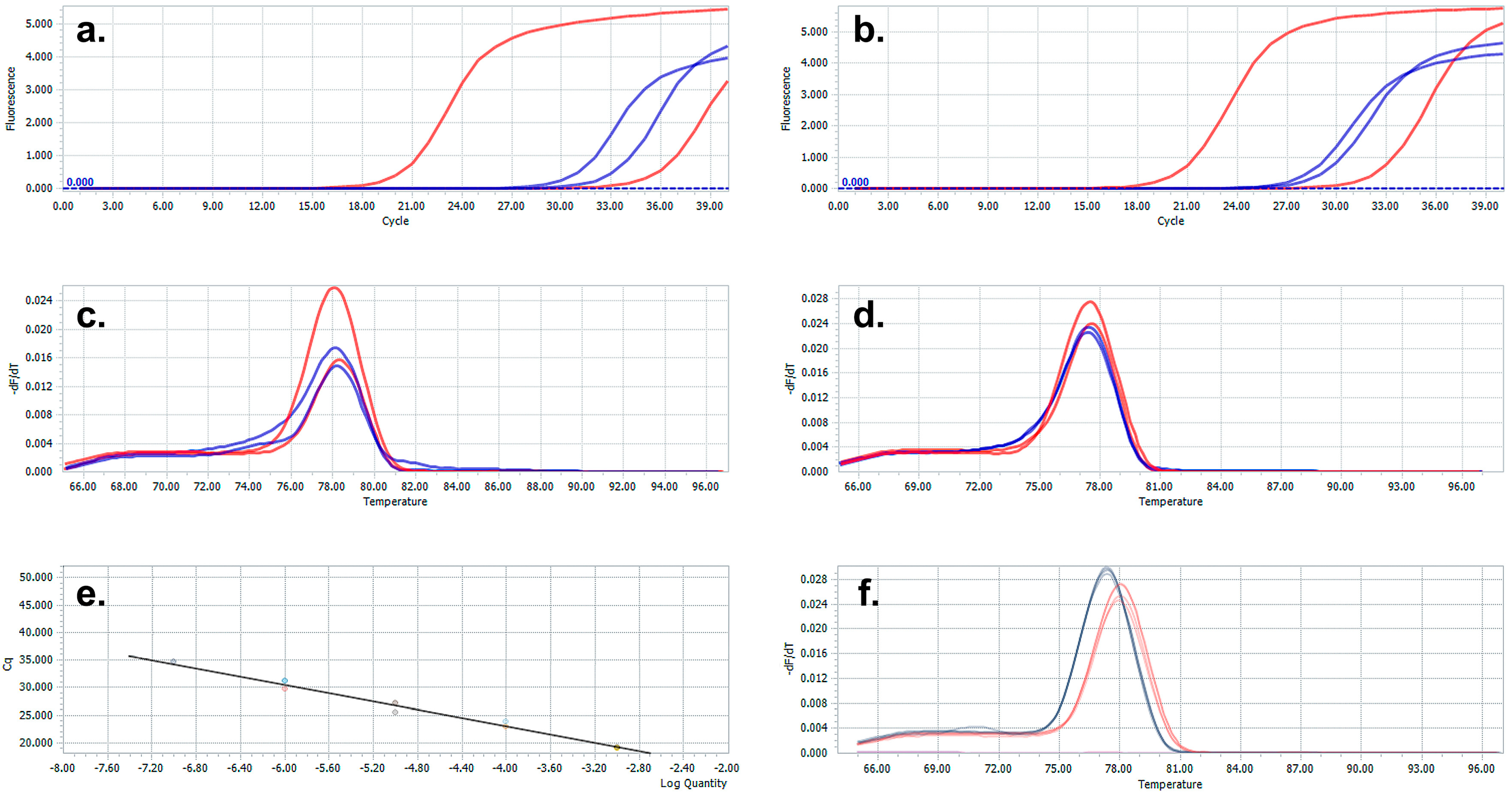
3. Results and Discussion

African patients of all ages, refugees, and asylum seekers coming from sub-Saharan areas and whose stay in Italy ranges from a few months to a few years were included in this study. Countries of origin with the highest frequency were Mali, Senegal, Nigeria, the Ivory Coast, Gambia, Guinea, and Ghana. The cell-free DNA of the parasite was obtained from 1 mL of serum obtained from a patient’s venous blood sample. The method involves the extraction of all circulating free DNA in the serum, both the host DNA and the pathogen DNA. The extraction was performed with the ‘QIAamp Circulating Nucleic Acid’ kit (Qiagen). A characteristic of this kit is the use of a vacuum pump connected to an extraction tube holder (QIAvac 24 Plus, Qiagen). In our protocol, the elution volume was set at 30 μL. Following the extraction, the minimum value measured with Thermo Scientific™ NanoDrop™ One was 15 ng/µL, and the maximum obtained was 69 ng/µL, with an average yield of 30 ± 11 ng/µL; as a result, an average of 900 ng cf-DNA could be extracted from 1 mL of serum. The starting concentration of synthetic genes was 40 ng/µL; tenfold serial dilutions were made to generate the standard curve and the yield of the primers on the sequences. Through the standard curve, we have established the useful range for assessing the positivity of our samples and the lowest point of DNA concentration that can be amplified with our method. In parallel with the PCR of the samples, a standard curve was made for each, with tenfold dilutions of the synthetic genes S. mansoni and S. haematobium. In addition to the amplification curves, the melting curves of the samples were compared to the melting of the synthetic genes, confirming that the specificity of the PCR product represents the target genomic portion of our interest (Figure 1). Molecular analysis was performed on 102 sera from African patients previously evaluated for the presence of antibodies against Schistosoma antigens. The Schistosoma ICT IgG-IgM POC test and WB blot test simultaneously detected IgG and IgM antibodies against S. mansoni and S. haematobium antigen based on lateral flow methodology.
Molecular analysis detected Schistosoma DNA in 32 patients. Among them, we characterized nine S. haematobium, 20 S. mansoni, and three coinfections. On the other hand, serological analysis revealed specific antibodies against Schistosoma antigens in 52 out of 102 patients. Concordance of data in 76 out of 102 patients (74.51%) was measured by comparing the results of the serological test with those of real-time PCR; in particular, both diagnostic tests were positive in 29 patients (28.43%) and negative in 47 (46.89%). The specificity of the molecular test was 94%. Discordance was observed in 26 patients (25.49%); in particular, in 3 patients with a negative serology, the molecular analysis detected cell-free DNA of Schistosoma, two S. haematobium, and one S. mansoni, while 23 patients had a positive serology versus a negative real-time PCR (Table 1).
Amplification of the human HBB gene was also analyzed for all samples as a quality control measure for cf-DNA extraction. Results showed the detection of the human HBB gene in all samples.
We provided evidence that real-time PCR on cf-DNA obtained from serum could be a fast, money-saving diagnostic method for the detection and species determination of Schistosoma parasitosis in chronic patients. Here, our primary objective was to determine whether it was possible to obtain enough schistosome DNA from 1 mL of serum. Indeed, 1 mL of serum provided an average of 900 ng of cf-DNA, sufficient to perform the molecular analysis. This minimized amount of sample is of the most importance as blood donation still represents a barrier for sub-Saharan Africans. The second objective was to determine the species of the parasite with the same analysis. The primer pairs were selected to distinguish between the genomes of the two strains of the parasite, being highly specific for tandem regions of approximately 121 bp, which have different nucleotide sequences in the two genomes. Synthetic genes obtained from plasmids containing the above-mentioned characteristic regions for the two Schistosoma genomes were used to have positive control of the PCR amplifications and determine the LoD of our method.
To determine the specificity of the molecular test, results obtained with the serological test were compared with those of real-time PCR; the two diagnostic tests showed correspondence in 76 out of 102 patients, while 23 patients had a positive serology versus a negative real-time PCR. Comparison of two different approaches, involving the detection of the parasite DNA versus the detection of the IgG antibody response, makes the sensitivity of the molecular test hardly measurable. Indeed, discordance may be a consequence of either a limited sensitivity of the molecular analysis due to the scarce amount of DNA released in the peripheral blood or the consequence of immunological memory in patients previously exposed to the parasite but without active infection. IgG remains positive for years, whereas serum DNA declines more rapidly over time [21]. Finally, three patients showed a negative serology and a positive molecular analysis, which detected cell-free DNA of Schistosoma with two S. haematobium and one S. mansoni. False negative immunologic test or immunodepression of the patient, or, alternatively, a false positive detection of parasite DNA, should be considered in those patients.
Serum Schistosoma PCRs were previously shown to be mainly contributive to the diagnosis of early infections in different studies [8,9,10,11,12,13,14,15]. Wichmann and colleagues conducted a prospective European-wide multicenter study on 38 patients with acute schistosomiasis, finding that S. mansoni-specific real-time PCR in serum was more sensitive (92%) than serology (70%) [11]. Moreover, Guegain et al. evaluated the accuracy of a specific Schistosoma PCR assay for the diagnosis of schistosomiasis by retrospectively studying a large cohort of migrants and travelers returning from endemic areas, which confirmed the relevance of serum Schistosoma PCR [23]. While reinforcing these data, in addition, our study is able to implement the technique and identify the type of Schistosoma: this study reasoned that it might also be possible to confirm the elimination of Schistosoma after treatment with Praziquantel.
Currently, several challenges still face the diagnosis of schistosomiasis; one of these is the difficulty for clinicians to detect parasitosis in affected persons because it is often paucisymptomatic or asymptomatic. Especially in Europe, where it is not very prevalent, or at least underestimated, the clinical aspect is not easily associated with Schistosoma infection. Molecular analysis, if more easily managed by both the laboratory and the patient, is more likely to speed up the clinical diagnostic pathway for positive persons. The sensitivity of the molecular test remains to be established. Therefore, at the moment, combining the molecular approach aimed at detecting the species of the parasite and the serological screening (currently recommended by the Italian guidelines for health checks upon arrival for refugees who have lived or traveled in endemic areas) could provide the clinician with more accurate knowledge about the infection and integrate it into a broader diagnostic algorithm.

Author Contributions

V.D. Conceptualization, data curation, writing. F.S. analysis, review and editing. C.C. data curation. N.G. analysis. A.C. supervision. All authors have read and agreed to the published version of the manuscript.

Funding

Part of a project financed by the Ministry of Health (cup: J86C18001110005).

Institutional Review Board Statement

The study was approved by the ethical committee of the Istituto Superiore di Sanità (AOO-ISS 02/08/2019 0024068).

Informed Consent Statement

Informed consent was obtained from all subjects involved in the study.

Data Availability Statement

Data available on request due to restrictions, e.g., privacy or ethical.

Acknowledgments

We thank Eleonora Rizzi, Stefano Tais, and Monica Degani for Western blot diagnosis. We thank Angela Forese for the critical revision of the English language.

Conflicts of Interest

The authors declare no conflict of interest.

References

  1. Rey, O.; Webster, B.L.; Huyse, T.; Rollinson, D.; Van den Broeck, F.; Kincaid-Smith, J.; Onyekwere, A.; Boissier, J. Population genetics of African Schistosoma species. Infect. Genet. Evol. 2021, 89, 104727. [Google Scholar] [CrossRef]
  2. Colley, D.G.; Bustinduy, A.L.; Secor, W.E.; King, C.H. Human schistosomiasis. Lancet 2014, 383, 2253–2264. [Google Scholar] [CrossRef]
  3. Engels, D.; Sinzinkayo, E.; Gryseels, B. Day-to-day egg count fluctuation in Schistosoma mansoni infection and its operational implications. Am. J. Trop. Med. Hyg. 1996, 54, 319–324. [Google Scholar] [CrossRef]
  4. Wickmann, D.; Panning, M.; Quack, T.; Kramme, S.; Burchard, G.; Grevelding, C.; Drosten, C. Diagnosing schistosomiasis by detection of cell-free parasite DNA in human plasma. PLoS Negl. Trop. Dis. 2009, 3, e422. [Google Scholar] [CrossRef] [PubMed]
  5. INMP; ISS; SIMM. I Controlli Alla Frontiera. La Frontiera dei Controlli. In Border Checks Kept in Check. Health Checks and Protection Pathways for Migrants upon Arrival and While Hosted in Reception Centers; Ministry of Health: Rome, Italy, 2017. Available online: http://www.salute.gov.it/imgs/C_17_pubblicazioni_2624_allegato.pdf (accessed on 1 December 2022).
  6. Hinz, R.; Norbert, G.; Schwarz, C.; Hahn, A.; Frickmann, H. Serological approaches for the diagnosis of schistosomiasis—A review. Mol. Cell. Probes 2017, 31, 2–21. [Google Scholar] [CrossRef]
  7. Fuss, A.; Mazigo, H.D.; Tappe, D.; Kasang, C.; Mueller, A. Comparison of sensitivity and specificity of three diagnostic test to detect Schistosoma mansoni infections in school children in Mwanza region, Tanzania. PLoS ONE 2018, 13, e020249. [Google Scholar] [CrossRef]
  8. Utzinger, J.; Becker, S.; Van Lieshout, L.; Van Dam, J.; Knopp, S. New diagnostic tools in schistosomiasis. Clin. Microbiol. Infect. 2015, 21, 529–542. [Google Scholar]
  9. Aryeetey, Y.A.; Essien-Baidoo, S.; Larbi, I.A.; Ahmed, K.; Amoah, A.S.; Obeng, B.B.; Van Lieshout, L.; Yazdanbakhsh, M.; Boakye, D.A.; Verweij, J.J. Molecular diagnosis of Schistosoma infections in urine samples of school children in Ghana. Am. J. Trop. Med. Hyg. 2013, 88, 1028–1031. [Google Scholar] [CrossRef]
  10. Pontes, L.A.; Dias-Neto, E.; Rabello, A. Detection by polymerase chain reaction of Schistosoma mansoni DNA in human serum and feces. Am. J. Trop. Med. Hyg. 2002, 66, 157–162. [Google Scholar] [CrossRef]
  11. Wichmann, D.; Poppert, S.; Von Thien, H.; Clerinx, J.; Dieckmann, S.; Mogens, J.; Parola, P.; Richter, J.; Shunk, M.; Zanger, A.; et al. Prospective European-wide multicentre study on a blood based real-time PCR for the diagnosis of acute schistosomiasis. BMC Infect. Dis. 2013, 13, 55. [Google Scholar] [CrossRef]
  12. Berriman, M.; Haas, B.J.; LoVerde, P.T.; Wilson, R.A.; Dillon, G.P.; Cerqueira, G.C.; Mashiyama, S.T.; Al-Lazikani, B.; Andrade, L.F.; Ashton, P.D.; et al. The genome of the blood fluke Schistosoma mansoni. Nature 2009, 460, 352–358. [Google Scholar] [CrossRef]
  13. Weerakoon, K.G.; McManus, D.P. Cf-DNA as a Diagnostic Tool for Human Parasitic Infections. Trends Parasitol. 2016, 32, 378–391. [Google Scholar] [CrossRef]
  14. Weerakoon, K.G.; Gordon, A.C.; Williams, G.M.; Cai, P.; Gobert, G.N.; Olveda, R.M.; Ross, A.G.; Olveda, D.U.; McManus, D.P. Droplet digital PCR diagnosis of human schistosomiasis: Parasite cf-DNA detection in diverse clinical samples. J. Infect. Dis. 2015, 216, 1611–1622. [Google Scholar] [CrossRef]
  15. Weerakoon, K.G.; Gordon, A.C.; Cai, P.; Gobert, G.N.; Duke, M.; Williams, G.M.; McManus, D.P. A novel duplex ddPCR assay for the diagnosis of schistosomiasis japonica: Proof of concept in an experimental mouse model. Parasitology 2017, 144, 1005–1015. [Google Scholar] [CrossRef] [PubMed]
  16. Lo, Y.M.; Chiu, R.W. Prenatal diagnosis: Progress through plasma nucleic acids. Nat. Rev. Genet. 2016, 8, 71–77. [Google Scholar]
  17. Goessl, C. Diagnostic potential of circulating nucleic acids for oncology. Expert Rev. Mol. Diagn. 2003, 3, 431–442. [Google Scholar] [CrossRef] [PubMed]
  18. Kato-Hayashi, N.; Yasuda, M.; Yuasa, J.; Isaka, S.; Haruki, K.; Ohmae, H.; Osada, Y.; Kanazawa, Y. Use of cell-free circulating schistosome DNA in serum, urine, semen, and saliva to monitor a case of refractory imported schistosomiasis hematobia. J. Clin. Microbiol. 2013, 51, 3435–3438. [Google Scholar] [CrossRef]
  19. Ullah, H.; Qadeer, A.; Giri, B.R. Detection of circulating cf-DNA to diagnose Schistosoma japonicum infection. Acta Trop. 2020, 211, 105604. [Google Scholar] [CrossRef] [PubMed]
  20. Ullah, H.; Arbab, S.; Inayat Ullah Khan, M.; Li, K.; Muhammad, N.; Qadeer, A.; Dan, W.; Otake Sato, M. Circulating cell-free mitochondrial DNA fragment: A possible marker for early detection of Schistosoma japonicum. Infect. Genet. Evol. 2021, 88, 104683. [Google Scholar] [CrossRef] [PubMed]
  21. Real-Time PCR Guide: Design, Validation, Analysis, and Troubleshooting. Integrated DNA Technologies. September 2020. Available online: https://go.idtdna.com/202009qPCRCampaignConsolidatedGuideParts1-qpcrguide-part-3.htm (accessed on 1 December 2022).
  22. Hamburger, J.; Xu, X.; Xin, Y.; Ramzy, R.; Jourdane, A.; Ruppel, A. A polymerase chain reaction assay for detecting snails infected with bilharzia parasites (Schistosoma mansoni) from very early prepatency. Am. J. Trop. Med. Hyg. 1998, 59, 872–876. [Google Scholar] [CrossRef]
  23. Guegan, H.; Fillaux, J.; Charpentier, E.; Robert-Gangneux, F.; Chauvin, P.; Guemas, E. Real-time PCR for diagnosis of imported schistosomiasis. PLoS Negl. Trop. Dis. 2019, 13, e0007711. [Google Scholar] [CrossRef] [PubMed]
Figure 1. Representative cases positive for S. haematobium (a) and S. mansoni (b); the positivity is provided by the amplification with a CT lower than the amplification curve of the minimum concentration of the control gene for S. haematobium and S. mansoni (the sample in blue; the sample in red is the synthetic gene). The melting curve of S. haematobium (c) and S. mansoni (d); red pick corresponding to the amplification of the synthetic gene and blue pick to the amplification of the samples. (e) Representative standard curve; (f) melting curve of the synthetic genes.
Microbiolres 14 00137 g001
Table 1. Comparison of molecular analysis on cf-DNA and serology shows 74.5% of concordance.
Serology +Serology −Total Number
Cell-fee DNA PCR +29332
Cell-free DNA PCR −234770
Total5250102
Specificity %94 (47/50)
Concordance rate %74.51 ((29 + 47)/102)
Disclaimer/Publisher’s Note: The statements, opinions and data contained in all publications are solely those of the individual author(s) and contributor(s) and not of MDPI and/or the editor(s). MDPI and/or the editor(s) disclaim responsibility for any injury to people or property resulting from any ideas, methods, instructions or products referred to in the content.

