Review

A Review of Bacterial Biofilm Components and Formation, Detection Methods, and Their Prevention and Control on Food Contact Surfaces

1
Department of Clinical Nutrition and Dietetics, Faculty of Applied Medical Sciences, The Hashemite University, Zarqa 13133, Jordan
2
Department of Nutrition and Integrative Health, Faculty of Allied Medical Sciences, Middle East University, Amman 11831, Jordan
3
Department of Nutrition and Food Technology, Faculty of Agriculture, Jordan University of Science and Technology, Irbid 22110, Jordan
4
Department of Clinical Nutrition and Dietetics, College of Health Sciences, University of Sharjah, Sharjah 2727, United Arab Emirates
5
Science of Nutrition and Dietetics Program, College of Pharmacy, Al Ain University, Abu Dhabi P.O. Box 64141, United Arab Emirates
6
Department of Food, Nutrition and Health, College of Food and Agriculture, United Arab Emirates University, Al Ain P.O. Box 15551, United Arab Emirates
7
Department of Food and Human Nutritional Sciences, University of Manitoba, Winnipeg, MB R3T 2N2, Canada
*
Author to whom correspondence should be addressed.
Microbiol. Res. 2024, 15(4), 1973-1992; https://doi.org/10.3390/microbiolres15040132
Submission received: 8 August 2024 / Revised: 17 September 2024 / Accepted: 26 September 2024 / Published: 28 September 2024

Abstract

:
The microbial biofilms are a community of microorganisms that adhere to each other and to surfaces, typically in a mucilaginous or gel-like matrix composed of extracellular polymeric substances, including polysaccharides, proteins, lipids, and DNA. In the food industry, the bacterial biofilms may be formed on different surfaces and cause post-processing contamination or cross-contamination from the food contact surfaces to food products. Conventional cleaning and sanitizing methods are often ineffective at removing bacterial biofilms. Among more recent alternative methods proposed to address this problem are the use of hydrolytic enzymes, essential oils, and bacteriocins. These methods show promise since their antibacterial and antibiofilm actions involve degradation of the extracellular polymeric matrix of the biofilm and lead to inhibition of the foodborne pathogens present. Understanding the limitations and mechanisms of action of enzymes, bacteriocins, and essential oils in controlling bacterial biofilms on foods and food contact surfaces is essential for developing solutions to prevent and control biofilm formation. This review critically summarizes the current knowledge of bacterial biofilm components, their formation, detection methods, prevention, and removal from food contact surfaces.

1. Introduction

Foodborne diseases are a major problem that create public health and economic consequences. Most foodborne illness outbreaks that result in a significant rate of morbidity and mortality worldwide are caused by bacteria, and many of these outbreaks have been linked to biofilm formation [1,2]. A biofilm is a community of microorganisms (MOs) that adhere to each other and to surfaces, typically in a sticky, gel-like matrix composed of extracellular polymeric substances (EPS). These substances are secreted by the MOs within the biofilm and form a protective environment that helps the biofilm adhere to most surfaces [3]. In nature, microbes more frequently cohabit in multi-species communities and form mixed biofilms on various food contact surfaces or food products [4]. Mixed biofilms of spoilage and pathogenic Gram-negative and Gram-positive bacteria were detected on different food products, including leafy green vegetables and meat products [4,5,6,7,8]. For example, Aeromonas hydrophila, Listeria monocytogenes, Salmonella enterica, or Vibrio spp. form biofilms in the fishing industry and fresh fish products [8]. Furthermore, Escherichia coli, Burkholderia caryophylli, and Ralstonia insidiosa are synergistically interacted to form mixed biofilms in a fresh-cut produce-processing plant [9]. In the last few years, the biofilm-forming ability of foodborne pathogens has been well studied in the clinical environment as well as the food industry [1]. Biofilm formation on food plant equipment surfaces that contact products has become a major source of food cross-contamination [10].
Many pathogenic MOs can form biofilms, including Salmonella spp., E. coli, L. monocytogenes, and Cronobacter sakazakii [11]. Formation of biofilms can compromise the safety of products in the food industry and cause serious problems. A bacterial cell inside a biofilm has a significant advantage over an isolated planktonic cell because the biofilm provides the cells present within it with aqueous environments that concentrate nutrients close to a solid surface, providing better access to nutrients and facilitating resource capture [12]. It also increases the overall fitness and genetic diversity of bacterial cells within the biofilm, by virtue of the high level of gene exchange cells go through within a biofilm [13]. Although C. sakazakii can be found in a variety of food products, illness outbreaks are usually associated with powdered infant milk formula (PIMF). This is because it can survive and form biofilms on the production line equipment and even in baby feeding bottles [14]. In contrast, many serotypes of Salmonella enterica have the ability to form biofilms in a variety of environments, and they are most often associated with meats and meat products, mainly poultry products but also dairy products and even fresh produce [15]. Furthermore, the most important aspect of biofilm formation is antimicrobial resistance. The EPS matrix is the first barrier to encounter an antibiotic or other antimicrobial agent and, subsequently, the various biopolymers present bind to the antimicrobial, or they may render the agent partially or completely inactive, thus altering its effectiveness [16]. Other barriers to antimicrobial action in biofilms are the protective compounds that are secreted by the cells and diffuse through the EPS, such as catalases and antioxidants, which may act together, often synergistically, to protect the bacterial cells [17]. For example, Stewart and Owkes [18] studied the catalase-dependent tolerance of biofilm-forming MOs to H2O2 and found that catalase was able to deactivate and neutralize H2O2 in water. The study used a lethal dose of H2O2 for planktonic cells, but the treated biofilm became thicker. Studies on Salmonella biofilms showed that chlorine concentrations must be at least 10 times higher to have the same inhibitory effect as when free bacterial cells are treated [19].
Unfortunately, traditional sanitizing materials have often proven to be ineffective against biofilm-forming bacteria on food contact surfaces. This illustrates the need for alternative agents with better antibiofilm activity [20]. Consequently, many strategies for their control have been proposed, including the use of biodegradable, eco-compatible enzymes that can degrade crucial components of the biofilm matrix, resulting in biofilm disruption, followed by cell lysis [1,21]. Work has shown that enzymatic treatment does not consistently eliminate biofilms and pathogenic cells, and many studies suggested the need for the use of an additional approach along with enzymatic treatments [21]. One method has been the inclusion of essential oils (EOs), which have value as antimicrobials due to their content of flavonoids and phenolic compounds, which are bactericidal against a number of pathogenic bacteria [22]. This review aims to critically summarize the current knowledge of bacterial biofilm components and formation and discuss the proposed methods for prevention and control of biofilms on food contact surfaces.

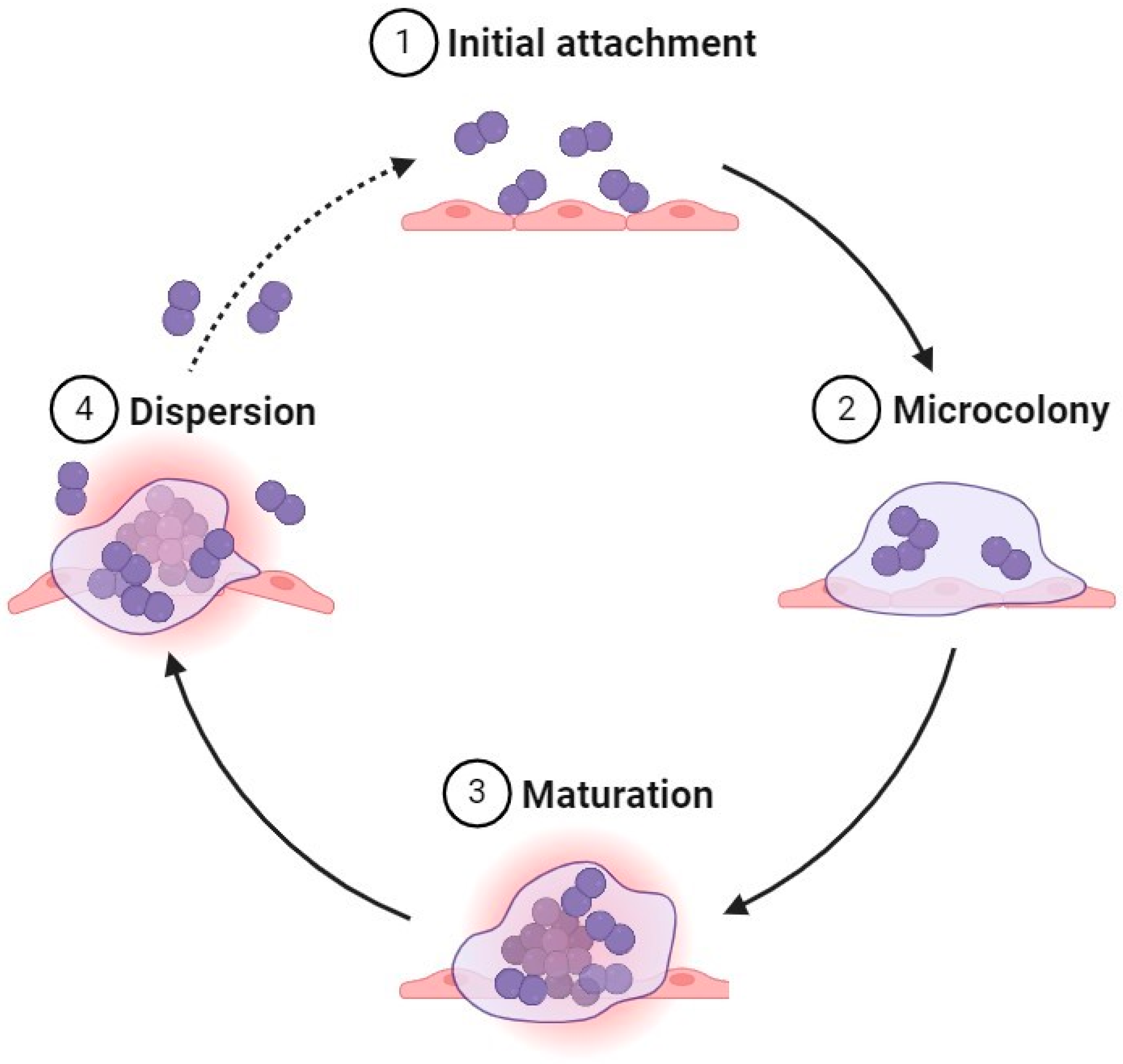
2. Biofilm Formation

Biofilms are the result of a natural survival strategy and consist of bacterial cells embedded within an EPS that offers stability and enhances stress resistance [23]. In nature, microbial cells tend to form these collections of organisms by attaching to a surface, forming a micro- then macro-colony, enlarging further, cooperating as a functional community, and finally sloughing off planktonic cells that move to new sites and repeat the cycle [24,25].
The EPS consists of polysaccharides, such as cellulose, and other components, including nucleic acids, proteins, and lipids. It also contains bacterial structures, such as pili, flagella, and fimbriae. The major roles of EPS include providing a stable anchor for bacterial attachment to materials, serving to sequester substrates for bacterial growth, as well as serving as a barrier against antimicrobial penetration and enhancing antimicrobial resistance by enabling the production of protective compounds, such as catalases [26,27]. Moreover, the regulation of EPS production affords the bacterial community many advantages, including adaptation to a wide range of antimicrobial agents, environmental stresses, and enabling attachment to different types of surfaces, such as steel, plastic, concrete, or even food [14,28].
It has been reported that most MOs can form biofilms and live within them. When bacterial cells form biofilms, major changes in their lifestyles will occur, including changes in protein production, gene expression, and phenotype. During biofilm establishment, bacterial cells transition from motile planktonic organisms into sessile cell communities that exhibit multicellular behavior [29,30]. Furthermore, a variety of factors are responsible for the formation of biofilms, including benzo(a)pyrene, which is a polycyclic aromatic hydrocarbon that has been reported to stimulate biofilm formation by acting on the MO as a stress factor. It also enables increased adaptation and tolerance because it can serve as a substrate. Other factors include the presence of environmental DNA, including the gene curli, cellulose, and flagella. Biofilms can be formed in a variety of unfavorable environments, and if unnoticed on a food contact surface, they may become a repeated source of apparently unrelated foodborne illness outbreaks not linked to a common food product [24,25].
Food-processing environments are considered as a favorable environment for biofilm formation, mainly on food products and food contact surfaces, due to several factors, including long periods of production, large probable areas for biofilm growth, complicated processing steps, and mass product generation [9,31]. Several microorganisms have been reported to form biofilms in food-processing areas, including pathogenic bacteria, such as L. monocytogenes, Yersinia enterocolitica, Campylobacter jejuni, Salmonella spp., Staphylococcus spp., Bacillus cereus, and E. coli O157:H7, and spoilage bacteria, such as Geobacillus stearothermophilus, Pseudomonas spp., Anoxybacillus flavithermus, and Pectinatus spp. [9].

2.1. Formation Steps

In general, the biofilm lifecycle contains four main stages (Figure 1), starting from the initial attachment of a bacterial cell to a biotic or abiotic surface, which yields a microcolony containing only non-motile cells. After microcolony formation, the cycle continues until a mature biofilm forms, limited in size by the diffusion of nutrients into and metabolic waste out of the biofilm matrix. Upon nutrient depletion or other unfavorable environmental conditions, motile planktonic bacterial cells will be formed, detach, and then colonize a surface to form another biofilm [24,25].

2.1.1. Initial Attachment

This step includes two stages: the first is reversible, followed by irreversible attachment to the surface. During reversible attachment, the bacterial cell uses its extracellular organelles, such as pili and flagella, in combination with electrostatic and hydrophobic interactions, as well as van der Waals forces, to cling to surfaces. At this stage, the bacterial cell may be easily detached by shear forces and start to secrete EPS, which initiates a strong interaction with the surface. The biofilm then enters the second stage within a few minutes [32,33,34]. Irreversible attachment occurs after a monolayer of EPS is formed, and cells aggregate to form a microcolony. In contrast, the cells at this stage are firmly adhered to the surface and require a strong chemical intervention or shear force to be removed [34,35].

2.1.2. Microcolony

After attachment, the bacterial cells continue reproduction and secrete more extracellular polymeric material to become a microcolony under appropriate growth conditions. The biofilm continues its expansion, and within it the cells grow, divide, and develop complex structures by using cell-to-cell signaling or quorum sensing [33,34]. Quorum sensing (QS) is an intercellular communication system used when the bacterial population density reaches a certain threshold, which, in this case, leads to the secretion of molecules that regulate the expression of the gene(s) responsible for EPS production [34,35,36].

2.1.3. Maturation

After the first biofilm layer is established to create a microcolony, the bacterial cell continues the secretion of EPS components, and with the development of pores and channels within the community, the biofilm transforms into a three-dimensional (3D), mushroom-like structure [33,34]. The role of pores and channels is to transport water, nutrients, and oxygen, as well as act as conduits for waste removal [13,37]. It has become apparent that the mature community contains three layers: the first is represented by a network structure on the surface, the middle layer exists as a basement membrane for non-motile, compact bacterial cells, and the upper one contains planktonic cells. Overall, the biofilm is composed of a stable community characterized by high antimicrobial resistance and resists removal [34].

2.1.4. Dispersion

Eventually, some planktonic cells in the outer layers acquire motility, move away from the parent colony through channels in the biofilm, and become attached to another surface to begin the process again. The detachment process is affected by several factors, including nutrient limitation, catabolite repression, and secretory proteins [34].

3. The Biofilm Matrix Components

The biofilm matrix plays a critical role in enhancing the virulence and resilience of bacterial biofilms, particularly those foodborne pathogens renowned for their biofilm-forming abilities within food-processing and distribution settings [24,38]. This matrix is a complex mixture of EPS, encompassing polysaccharides, proteins, lipids, and extracellular DNA (eDNA), collectively serving as a protective barrier for encased bacterial cells. With Salmonella, the biofilm matrix not only provides mechanical stability but also acts as a formidable barrier against environmental stressors and antimicrobial agents, substantially elevating the challenge of eradicating the pathogen from industrial food contact surfaces [39,40]. Similarly, C. sakazakii, acknowledged for its tenacity to persist in environments where powdered infant formula is manufactured, exploits the matrix to ensure its survival during the manufacture and storage of these products, underlining the critical role played by the matrix in its survival [38]. A comprehensive understanding of the composition and functions of the biofilm matrix produced by these pathogens is vital for the development of targeted strategies for disrupting biofilm formation and enhancing the safety of food [38,40].
The EPS produced by the bacterial cell is considered the most critical characteristic of the biofilm matrix. This is “the house” that covers and surrounds the bacterial cells and accounts for ≥90% of the whole biofilm dry mass. In contrast, the bacterial cells only account for about 10% of the mass in most cases [24,26].

3.1. Polysaccharides

Biofilm polysaccharides, also known as extracellular polysaccharides (PSs), represent a significant component within bacterial biofilms, exerting a multifaceted influence on the formation, integrity, and resilience of these structured microbial communities [26]. PSs consist of long chains of sugar molecules secreted by bacterial cells, forming an intricate matrix that encapsulates and binds the constituent cells of the biofilm [41].
The significance of biofilm PSs in bacterial biofilms can be demonstrated through their functions in providing structural support and stability, where polysaccharides serve as the architectural scaffolding of the biofilm, creating a complex network that imparts structural integrity to the biofilm [26,42]. This matrix provides the biofilm with its characteristic 3D structure, enhancing its resistance to mechanical disruption and shear forces [41]. In addition, the adhesion and attachment of the PSs promote the initial attachment of bacterial cells to surfaces and host cells during biofilm formation. These polysaccharides can also interact with surfaces, facilitating the initial reversible attachment of bacterial cells through mechanisms such as van der Waals forces and electrostatic interactions [26]. A third aspect of PSs’ function is their pivotal role in shielding bacterial cells from various environmental stressors. PSs act as a physical barrier, preventing desiccation as well as the penetration of chemical disinfectants and antimicrobial agents into the biofilm [41].
The PSs in the EPS enhance water retention and nutrient trapping via their high water-holding capacity, thus maintaining a hydrated microenvironment within the biofilm [41]. This is particularly advantageous for bacterial cells in environments with limited nutrient availability and can contribute to their long-term survival because they are protected within the biofilm [26]. Finally, they can play a role in biofilm detachment when required. Some biofilm-forming bacteria produce enzymes that target and degrade the polysaccharide matrix, facilitating the release of cells and enabling biofilm dispersal and colonization of new environments [43].

3.2. Proteins

Proteins in biofilms comprise of adhesion and enzymatic proteins. Adhesion proteins enhance the microbial attachment to food products or food contact surfaces and their attachment to each other via fimbria, curli, and pili [24], while enzymatic proteins, such as proteases, play a critical role in nutrient acquisition and substances’ degradation within the biofilm matrix. Proteins in biofilm may also participate in signaling pathways (e.g., quorum sensing) that regulate biofilm development and maintenance [44].

3.3. Lipids and Biosurfactants

Lipids and biosurfactants in biofilms, including phospholipids, lipid A surfactin, emulsan, viscosin, and rhamnolipids, contribute to the structure and function of the biofilm matrix in adhering to and colonizing some hydrophobic surfaces, such as Teflon and waxy surfaces. In addition, they play a role in binding the heavy metals, producing virulence factors, and contributing to the resistance of biofilms to solvents, sanitizers, and detergents [44,45].

3.4. Extracellular DNA

Usually, eDNA is released through bacterial secretion systems due to the cell lysis that may occur because of autolysis of bacterial cells, presence of bacteriophages, or its release into outer-membrane vesicles. It has been reported that eDNA plays a critical role in the initiation of biofilm formation, mainly via motility and facilitating the cells’ interaction in biofilms via binding with the positive charges on proteins. In addition, it has a role in biofilm stability, antibiotic resistance, and horizontal gene transfer [44,46,47].

4. Biofilm Issues in Food Processing

The formation of biofilms both in natural and artificial environments is a constant challenge to humans in many ways. Pathogenic MOs may enter municipal water supply systems and form biofilms, sometimes causing illness, especially in developing countries, which frequently have unreliable water supplies [26,48]. In the pharmaceutical industry, biofilms may be formed on machine and equipment surfaces and have been known to cause chronic diseases, such as pneumonia and cystic fibrosis, in patients taking contaminated medications. Biofilms can also be formed on medical devices in hospitals and when functional in patients [42,49]. In the food industry, the situation is quite similar to other environments, but the type of MO is highly dependent on the type of food manufactured as well as the nature of extrinsic environmental factors, such as the type of surfaces present [50,51].
One of the most common surfaces used in the food industry is stainless steel, but other surfaces made of rubber, glass, Teflon, and plastic are also used. Stepanovic et al. [52] studied the ability of 122 Salmonella spp. and 48 L. monocytogenes strains to produce biofilms on plastic surfaces using different growth media, including meat broth, brain heart infusion broth (BHIB), tryptic soy broth (TSB), and 1/20 diluted TSB. They found that all bacterial strains tested formed biofilms, but the quantities of Salmonella spp. biofilms were higher than those of L. monocytogenes, and the diluted TSB was the most productive medium for Salmonella spp. biofilms, presumably due to its limited content of available nutrients. This pathogen has been identified as the second most frequent cause of food- and water-borne illness outbreaks in the European Union [15,50].
It has become apparent that C. sakazakii has the ability to form biofilms on plastic surfaces. Oh et al. [53] investigated the ability of 72 C. sakazakii strains to generate biofilms on plastic surfaces designed to mimic the infant feeding bottle. Test bottles contained several types of nutritive media, including different concentrations of infant milk formula (IMF), nutrient broth (NB), brain heart infusion (BHIB), and tryptone soya (TSB). They found that the normal concentration of IMF was able to allow 56 of the C. sakazakii strains to form biofilms on the plastic surfaces.
During processing, foods go through a variety of changes that affect the microbial load, and some of these processing steps may cause stress to the microbial population present and can lead to biofilm formation, which in turn may serve as a source for cross-contamination of processed products [54,55]. Further, the variety of surfaces used in the food industry provides additional opportunities for bacterial cells to colonize. Stainless steel, rubber, and plastic are often used in the food environment, while other plant parts, such as concrete walls, glass windows, and metal pipes, may be difficult to clean because of cracks and crevices normally present to varying degrees [56]. In general, hydrophobic, energetic surfaces with a rough texture enhance the attachment of bacterial cells, thus increasing the likelihood of biofilm development. For example, Salmonella was found to attach better to plastic surfaces than rubber and stainless steel [23]. Consequently, stainless steel is the surface most often used in the food industry, with grades 304 and 316 being the most frequent types due to their stability during thermal processing. They are also considered the easiest to clean and are resistant to corrosion [57].
It was understood as early as 1996 that Salmonella can form biofilms on a variety of food contact surfaces, and studies also indicate that their formation can easily occur on steel [58]. Salmonella also has the ability to survive in dry food and form biofilms, thus causing cross-contamination of food products [15,58]. For example, in 2007 in the USA, a multistate illness outbreak occurred, with over 150 confirmed cases of S. Typhimurium infection. The outbreak was linked to different dairy products, including milk, yogurt, and cheese, produced at a dairy-processing plant in the Midwest, USA. The investigation revealed that Salmonella had formed robust biofilms on various processing equipment surfaces, and the biofilms acted as a persistent source of microorganisms, leading to contamination of the dairy products during processing [59]. Nonetheless, poultry production is the main source of Salmonella outbreaks, and it has been shown that about 50% of Salmonella strains isolated from poultry farms can produce biofilms [15]. As noted earlier, C. sakazakii is also a biofilm-forming pathogen. Besides its ability to survive in low-water-activity food as well as at refrigerator and ambient temperatures, it was able to produce biofilms on different abiotic surfaces, such as those of silicone and stainless steel [50]. Studies by Hurrell et al. [60] and Kim et al. [61] demonstrated that C. sakazakii may colonize and form biofilms in the infant feeding tube.

5. Detection Methods of Biofilm Formation

The importance of biofilm formation in food and other industries as one of the leading causes of infections, along with their societal and economic benefits from another side, raises the importance of describing several quantitative and qualitative measurement methods [62]. With no golden standard method for biofilm quantification, many methods have been proposed [63,64]. Furthermore, the complexity of this community and the several factors that affect and promote its formation, such as food and surfaces, among others, make it difficult to determine a standard way to evaluate and control biofilm formation [62].
Certainly, the quantitative method is one of the most common methods used to determine the number of cells either for planktonic or inside biofilm MOs. Moreover, the quantitative methods may be either direct or indirect [62]. Plate counts, Coulter cell counting, flow cytometry, microscopic cell counts, and fluorescence microscopy are among the direct methods [65,66]. These methods allow to count the cells that can be cultured [62]. On the other hand, indirect methods, such as determination of dry mass, microtiter plate assays, total protein, and total organic carbon, were also used [66,67].
The viable cell enumeration or aerobic plate count by plate count is a standard basic method used to determine the viable cell number [68,69]. In this method, there is no need to dye or stain the cell, the cell should only be plated on agar under optimal conditions (time, temperatures, and agar type), thus allowing the live cell to grow [68]. The main advantage of this method is its simplicity, as there is no need to advance equipment to perform it. On the other hand, this method is considered labor-intensive and time-consuming [70]. Another direct method used to evaluate biofilm formation is the use of light and fluorescence microscopy, as this method may yield a 3D shape for the formed biofilm [71]. It enables detailed visualization of biofilm structures and bacterial cells, with fluorescence microscopy allowing for enhanced contrast and specific staining to distinguish between live and dead cells [72]. However, the main disadvantages of this method are its cost and complexity [62,73].
Flow cytometry is a sophisticated analytical technique used for the quantitative measurement and detailed characterization of cell populations as they flow in a single file through a laser beam [62]. This method offers several advantages, including high-resolution analysis of cell subpopulations, allowing researchers to simultaneously assess various parameters, such as cell viability, size, and surface properties [63,74]. The speed and efficiency of flow cytometry enable rapid processing of large numbers of cells, which significantly reduces the time required compared to manual counting methods. Additionally, this technique provides accurate quantification of cell populations and can differentiate between live and dead cells using specific fluorescent dyes, offering a precise evaluation of biofilm composition [63,74]. However, flow cytometry also has notable limitations, as the initial cost of flow cytometers is quite high [62]. Sample preparation is another challenge, as cells must be suspended and homogenized before analysis. This process can be problematic for biofilms or cells with strong adherence, potentially altering their original structure [74].
On the other hand, the growth of biofilms may be determined indirectly by using a specific marker, thus inferring the biofilm quantity. Dry mass or biofilm density is a type of indirect method that is expressed as mass per unit [75]. This method uses a marker that quantifies the biofilm formation quickly by using heat, usually between 60 and 105 °C [62,75]. However, this method is unable to differentiate between the cell mass from other film components [75].
Crystal violet (CV) staining is another indirect method that is widely used for evaluating biofilm formation, involving the staining of biofilms with a CV solution, followed by destaining with ethanol and measuring the optical absorbance of the reclaimed dye [64]. This approach provides an indirect measure of biofilm biomass by quantifying the amount of CV retained, which binds to both bacterial cells and the extracellular matrix [64,76]. However, CV staining does not differentiate between living and dead cells, and thus it does not provide information on biofilm viability [62,76]. Moreover, CV staining is relatively simple, reproducible, and cost-effective, though it is limited by its inability to assess cell viability and its susceptibility to variability from factors such as incubation times and temperatures [62].

6. Biofilm Prevention and Treatment in the Food Industry

Foodborne pathogens are responsible for a wide range of global public health concerns. More than 600 million people are affected by foodborne illnesses annually. In the USA, more than 50% of persistent infections are caused by biofilm-forming bacteria [2]. Because the bacterial biofilm is a complex community, it is considered very difficult to eliminate due to its physical and chemical resistance. Biofilms affect food quality and safety largely by causing post-processing contamination, resulting in a decreased food product shelf life and spread of foodborne pathogens [11,50].
It is understood that cells inside biofilms are more resistant to antimicrobials and sanitizers than planktonic bacteria of the same species because they are protected by the EPS matrix surrounding them. This barrier interferes with sanitizer penetration and antibiotic diffusion to reduce their effectiveness [2]. Additionally, cells within biofilms exhibit changes in the physiological state and largely reduced metabolic activity, which can make them more resistant to antibiotics [77]. On the other hand, sanitizing materials can easily penetrate planktonic cells and cause disruption, which leads to effective control of the MO [78]. It is also true that planktonic bacteria are more sensitive to antibiotics, which can directly target and kill them.

6.1. Conventional Sanitizing Materials

In general, traditional disinfection methods used in the food industry include the application of heat treatment (frequently steam or hot water) or the use of liquid chemicals, such as quaternary ammonium compounds (QACs), chlorine, other halogens, acids, amphoteric products, biguanides, iodophores, and peroxygens [2,11]. Chlorine in the form of sodium hypochlorite (NaClO) is a common disinfectant in food plants due to its high oxidizing power, and it is generally inexpensive compared to other chemicals [2,11]. While these chemical sanitizers are effective against planktonic bacterial cells, they cannot proficiently disinfect surfaces contaminated with bacteria inside biofilms. It was found that a 10-fold greater concentration of chlorine was needed to achieve the same effect on Salmonella inside biofilms compared to planktonic cells [11,19]. Moreover, pathogens, such as L. monocytogenes and E. coli O26, showed greater resistance to certain sanitizers, even with extended exposure times. Additionally, some cells may possess natural resistance to the sanitizing agent, while others can acquire resistance through genetic exchanges or mutations [79]. A study by Aryal and Muriana [79] examined the efficacy of five different commercial sanitizers against seven-day-old robust biofilms of L. monocytogenes, S. Montevideo FSIS 051, and E. coli O26 using a microplate protocol. They reported that both chlorine-based sanitizers (NaClO and quaternary ammonium chloride (QAC)) at 200 ppm for 2 h achieved only a 1–2 log CFU/mL reduction. Moreover, a commercial product, namely Decon7 solution, containing a combination of three different sanitizers, including QAC (5.5–6.5%), diacetin (30–60%), and H2O2 (<8%), at a concentration of 10% was needed to obtain a 7 log CFU/mL reduction of all tested pathogens.
In another study, Meesilp and Mesil [80] evaluated the efficiency of chlorine and oxisan against the biofilm formers Staphylococcus aureus and Pseudomonas aeruginosa, isolated from ultra-high-temperature milk. They reported that 4% oxisan for 1 h was needed to reduce 68% of S. aureus numbers and 47% of P. aeruginosa counts in 48-hour-old biofilms on a stainless-steel surface. However, when using lower concentrations (0.3%), only 53% and 48% reductions were achieved in numbers of S. aureus and P. aeruginosa, respectively, under the same conditions. On the other hand, chlorine at 4% and 0.3% for 1 h reduced numbers of S. aureus in 48-hour-old biofilm on stainless steel by 66% and 44%, respectively. When used against 48-hour-old P. aeruginosa biofilms, the same treatments reduced the microbial load by 55% and 45%, respectively. However, the concentrations of sanitizers in this study were higher than the accepted standard level of 200 ppm chlorine permitted for the disinfection of food contact surfaces [81].
In addition to their generally low activity as antibiofilm agents, commercial chemical sanitizers may possess adverse effects when used. For example, chlorine sanitizers are known for their cost-effectiveness and ease of use. However, the indiscriminate use of chlorine in food sanitation can lead to the formation of halogenated by-products that are carcinogenic, including haloacetic acids and trihalomethanes. Exposure to these substances has been linked to cancer and adverse reproductive outcomes in humans [82]. Additionally, the adverse effects may involve toxic by-products, development of microbial resistance, corrosiveness, residue formation, reactivity with biofilm EPS, discoloration, and the potential release of explosive gas. On the other hand, H2O2 is generally recognized as a safe and common sanitizer used in milk, instant tea, and wine. However, H2O2 may cause metal corrosion in production line equipment and utensils, and high concentrations are required to be effective against foodborne pathogens [82,83]. Another alternative, QACs, also have many adverse effects both on humans and the environment [58]. In humans, QACs may lead to disruption of metabolic function, developmental and reproductive toxicity, and impairment of mitochondrial function, and may also impart dermal and respiratory effects. QACs are also linked to the development of antimicrobial resistance and are considered ineffective against Gram-negative bacteria [83,84]. Because each of the commonly used disinfectants has substantial limitations when it comes to biofilms, it would be prudent to explore the ability of alternative methods for controlling biofilm formation in the food environment. Many methods have been proposed to address this issue, including the use of enzymes and essential oils (EOs), bacteriophages, biosurfactants, electrolyzed oxidizing water, ultrasound, and ozone. In the past few decades, the use of less environmentally challenging biocides to overcome the biofilm problem in the food industry has received significant interest [2,11].

6.2. Enzymes

Enzymes are selectively active proteins that act as biological catalysts via interactions with cofactors (non-protein materials) involved in biofilm synthesis, and they can inhibit biofilm formation. Enzymes accelerate the rate of reaction without altering the chemical equilibrium [1]. They can be organized into six main groups based on their functionality: oxidoreductases, transferases, hydrolases, lyases, isomerases, and ligases. Oxidoreductases are a group of enzymes that catalyze the electron transfer reaction. In other words, they transfer hydrogen or oxygen atoms through the redox reaction, which directly or indirectly targets bacterial growth. Transferases transmit single atoms or a group of atoms between two compounds, with the target here being the EPS matrix [1,2,21]. Hydrolases are enzymes that target different sites in the biofilm community, such as the EPS matrix, bacterial growth, and QS molecules, by catalyzing the breakdown of different bonds between molecules. Lyases target the EPS and can eliminate atoms [1,85]. Isomerases can catalyze the isomerization process, and ligases join two different molecules together via a covalent bond [1]. However, both hydrolases and oxidoreductases were found to be enzymes with the most promising antibiofilm activity [85].
As stated previously, the EPS is a critical component of biofilms, providing both protection and structural support for bacterial cells. Hydrolases can degrade this matrix, making the cells inside the biofilm community more susceptible to other antimicrobial agents [26]. Included among these enzymes are proteases that hydrolyze specific peptide bonds between amino acids, leading to the breakdown of proteins present in the EPS matrix [1,85]. These types of enzymes have previously been used in the food production environment to clean (mainly for protein removal) pipelines and ultrafiltration units, among other utensils, as well as equipment in food plants. Different types of proteases have been used alone or in combination with other enzymes as antibiofilm agents. For example, a study on the activity of 1% serine protease against the biofilms of Pseudomonas fluorescens, Bacillus mycoides, and B. cereus on stainless-steel surfaces indicated that the treatment was able to achieve 3.9, 2.6, and 2.3 log CFU/cm2 reductions, respectively, at 45 °C after 30 min [10]. In addition, the use of polysaccharide-degrading enzymes, such as amylase and lyases, seems promising. These compounds may be used in combination with other enzymes to increase their efficacy against bacterial biofilms [1]. α-amylase is a common antibiofilm enzyme that hydrolyzes the α-1,4 glycosidic linkage in carbohydrates. The enzyme is available commercially from different sources, including bacteria, mainly Bacillus subtilis. In a study by Kalpana et al. [86] regarding the efficiency of B. subtilis α-amylase against S. aureus, Vibrio cholerae, and P. aeruginosa, it was found that the enzyme was able to restrict biofilm formation and degrade polysaccharides by 51.8% to 73.1% at 30 °C for 2 h; however, the authors recommended the use of this enzyme in combination with other enzymes to adequately enhance its antibiofilm activity.
There has also been some interest in the use of lipolytic enzymes for biofilm control. This group of enzymes that degrade lipids works against biofilms by targeting the fat present in the biofilm EPS [87]. Lipases from different species of a marine bacterium, Oceanobacillus, have been tested for their action against biofilms of several MOs, including P. fluorescens, E. coli, Listeria spp., B. cereus, and Vibrio parahemolyticus. The results showed that 150 µL/mL was able to achieve 90–95% disruption of biofilms on a glass surface for all tested bacteria after 1 h of treatment [1,85].
It has been found in many studies that because of biofilm matrix heterogeneity, a single enzyme is usually not sufficient to completely eradicate biofilms and kill the pathogenic cells contained inside. Effective enzymatic treatment often requires a combination of different enzymes that target different components. The main advantages of using enzyme combinations over a single enzyme are: (1) it ensures extensive disruption of the matrix, (2) it provides an opportunity for additive or synergistic inhibitory enzyme interactions to occur, (3) it reduces the possibility that adaptation to a single enzyme may occur, and (4) it enhances the penetration of the biofilm matrix [16,88,89]. In addition, the biofilm-forming microorganisms are unable to develop resistance to these enzymes, which have a high specificity, biocompatibility, and selectivity [90]. However, some disadvantages of using enzyme combinations have been reported, including the higher cost and enhanced complexity associated with setting up optimal conditions (e.g., temperature and pH) for the simultaneous activity of a multi-enzyme system [16].
For example, the use of a lipase, amylase, cellulase, and protease combination in an ultrafiltration membrane was found to be effective against Klebsiella oxytoca, where a 6.2 log CFU/cm2 reduction was achieved [91]. In another study by Ripolles-Avila et al. [20], the antibiofilm activity of two different combinations of hydrolytic enzymes was investigated. The first was preventive and involved the use of 5% protease, 0.5% lipase, and 2.5% amylase, tested at 45 and 50 °C at pH 7.0. The second was aggressive and used 10% protease, 1% lipase, and 5% amylase, tested at 50 °C and a pH of 7.0. Both were used against S. enterica serovar Typhimurium CCUG 29478 and C. sakazakii ATCC BAA-894 on stainless-steel coupons and polystyrene surfaces during a 30 min treatment. It was found that the preventive treatment caused 31.0–46.7% biofilm detachment compared to the initial microbial load, with the maximum biofilm detachment of 46.7% of S. Typhimurium from polystyrene surfaces. However, the reduction of S. Typhimurium from stainless steel was lower, at 37.5%. In contrast, C. sakazakii numbers were reduced by 34.5% and 31.0% on stainless-steel and polystyrene surfaces, respectively. The aggressive treatment was more effective against S. Typhimurium on the two surfaces, with a 61.7% and 54.2% reduction on stainless-steel and polystyrene surfaces, respectively. However, against C. sakazakii, on stainless-steel and polystyrene surfaces, the aggressive treatment reductions were 27.6% and 50%, respectively. In another study, Olaimat et al. [71] investigated the antibiofilm activity of two mixtures of different enzymes (5% protease, 2.5% α-amylase, and 0.5% lipase, or 10% protease, 5% α-amylase, and 1% lipase) against a S. enterica cocktail on stainless-steel and plastic surfaces at 50 °C for 30 min. The antibiofilm activity increased as the concentration of enzymes increased, where the enzyme mixture of 10% protease, 5% α-amylase, and 1% lipase reduced the numbers of S. enterica in the biofilm by 2.1–2.2 log CFU/coupon on both surfaces, while the 5% protease, 2.5% α-amylase, and 0.5% lipase mixture reduced S. enterica numbers in the biofilm by 1.2–1.3 log CFU/coupon on both surfaces, compared to the samples treated with sterilized distilled water. No significant differences were observed between the enzyme activity on stainless-steel and plastic surfaces. It is worth mentioning that limited information about the safety of hydrolytic enzymes, mainly on food products, are available. Selected studies about the antibiofilm activity of different hydrolytic enzymes are summarized in Table 1.
It has been recommended that physical or chemical methods be used in conjunction with enzymes, particularly in therapeutic applications, where ultrasound, buffers, chelating agents, and surfactants have proven valuable. This type of treatment enhances the effectiveness of the enzymes by improving their penetration, facilitating multisite targeting, and contributing the additive/synergistic interaction between the two treatments [92,93]. For example, when pronase and DNase I were combined, the effectiveness of benzalkonium chloride was enhanced against L. monocytogenes and E. coli 48 h biofilms [94]. Wang et al. [95] investigated the use of surfactants in combination with enzymes against biofilms formed by seven Salmonella enterica serotypes (S. Typhimurium, S. Enteritidis, S. Infantis, S. Stanley, S. Agona, S. Derby, and S. Indiana) on stainless-steel surfaces. Four different types of surfactants were used (cetyltrimethylammonium bromide (CTAB), sodium dodecyl sulfate (SDS), rhamnolipid, and tween-80) combined with three proteases (proteinase K, dispase, and subtilisin) and two glycosidases (cellulase (R-10) and Yakult, glucoside amylase). For the enzymatic treatment only, the highest biofilm reduction was 80%, and it was observed with 20 mg/mL of cellulase at 2 h and 40 °C. When the biofilm was immersed in a mixed solution of cellulase and CTAB for 2 h at 40 °C, it achieved 100% biofilm removal. Other results where only enzymes were used in groups were not as dramatic. Proteinase K, glucoside amylase, subtilisin, dispase II, and cellulase reached a maximum reduction of 85% with a 2 h treatment at 40 °C. While for a single surfactant treatment, both tween-80 and rhamnolipid removed <60% of biofilm within 1 h at 20 °C at a concentration of 100 mg/mL. Recently, Olaimat et al. [71] reported that the two-step sequential cleaning treatments based on enzyme mixtures and Aleppo pine essential oil (APEO) were effective in controlling the biofilms of S. enterica on stainless-steel and plastic surfaces. The enzyme mixture of 10% protease, 5% α-amylase, and 1% lipase at 50 °C for 30 min, followed by 4× MIC (2000 µg/mL) of APEO at 25 °C for 30 min, reduced the numbers of S. enterica in biofilms on stainless-steel and plastic surfaces by 4.3–4.6 log CFU/coupon, compared to the control samples treated with sterilized distilled water.

6.3. Essential Oils

EOs are natural plant extracts of leaves, seeds, roots, stems, and petals. EOs have been used in medicine and for therapeutic purposes since ancient times. They are also used as well-known antioxidant, anti-fungal, and antibacterial agents in the food industry due to their content of terpenes, aldehydes, and phenolic compounds. Terpenes are hydrocarbons, such as pinene, terpinene, and limonene, which are known for their antimicrobial activity, as they can disrupt cell membranes and inhibit bacterial growth. In addition, those that contain aldehydes inhibit bacterial growth by interfering with cell functions. EOs that contain phenolic compounds, which are the strongest antibacterial agents, are able to kill bacterial cells by damaging cell membranes and proteins [96].
The use of EOs as potential environmentally friendly antibiofilm agents has gained attention in the past years due to their ability to disturb the bacterial biofilm by inhibiting the initial attachment, as well as the destruction of the EPS matrix [97]. They also may act as anti-quorum-sensing agents by interfering with bacterial cell-to-cell communication [98]. Several studies have shown the efficiency of using EOs alone as antibiofilm agents or in combination with other treatments (Table 2). For example, Čabarkapa et al. [99] studied the antibiofilm activities of carvacrol and thymol EOs against two strains of S. Enteritidis in 96-well polystyrene microtiter plates, and they found that these EOs were effective against biofilm formation at a sub-minimum inhibitory concentration (MIC) of the tested EOs after 48 h at room temperature. The MIC for both carvacrol and thymol EOs against S. Enteritidis was 0.156 µL/mL, and 2 MIC of carvacrol was able to reduce the biofilm formation by 90% compared to the control. Additional work showed that 0.25 MIC carvacrol and thymol was able to reduce biofilm formation by 50%.
In other work, Valeriano et al. [74,100] investigated the antibiofilm activity of peppermint and lemongrass EOs against biofilm-forming S. Enteritidis on 304 stainless-steel surfaces. The stainless-steel surfaces with 240-hour-old biofilm were treated with 7.8 µL/mL (1 MIC) peppermint or lemongrass EOs for 10, 20, and 40 min. They found that the EOs of peppermint and lemongrass were able to reduce the bacterial count by 4.0 and 4.2 log CFU/cm2, respectively, after 10 min, and after 20 and 40 min, adhered cells were not found in samples treated with either EO. Olaimat et al. [65] investigated the antibiofilm activity of APEO at 1× MIC (500 µg/mL), 2× MIC (1000 µg/mL), or 4× MIC (2000 µg/mL) with exposure times of 10, 20, or 30 min against S. enterica biofilm on stainless-steel and plastic surfaces. The antibiofilm activity of APEO increased as the concentrations and exposure time increased, with the highest reductions of 1.6 and 1.8 log CFU/coupon obtained at 4× MIC APEO for 30 min on plastic and stainless steel, respectively.
Somrani et al. [101] investigated the antibiofilm and antibacterial activity of clove EO against S. Enteritidis and L. monocytogenes. The results showed that the clove EO MIC was higher for S. Enteritidis (0.1 mg/mL) than for L. monocytogenes (0.05 mg/mL). The initial biofilm attachment in microtiter plates of each pathogen was inhibited by 61.8% and 49.8% for L. monocytogenes and S. Enteritidis, respectively, when the wells of the microtiter plate were treated with 1 MIC of clove EO. They also tested the effect of clove EO on the eradication of the biofilm after its formation to develop clove EO-based sanitizers, and they found that at 1 MIC, clove EO for 1 h reduced the biofilm formation of S. Enteritidis and L. monocytogenes by 20.3% and 30.2%, respectively. Results indicated that the EO was better able to prevent biofilm formation than remove already formed biofilms.
The EO antibiofilm activity against C. sakazakii has also been studied. Wang et al. [95] examined the antibiofilm activity of Litsea cubeba (mountain pepper) EO at concentrations of 0, 2.5, 5, 10, and 20 MIC for 0–4 h against the 48-hour-old biofilms of 8 strains of C. sakazakii on stainless-steel surfaces at 25 °C. The results showed that the MIC of LC-EO for all strains ranged from 1.5 to 4 µL/mL, and 10 MIC for 4 h or 20 MIC for 3 h were able to reduce the population to below the detection level (1 CFU/cm). However, both 2.5 and 5 MIC for 4 h reduced the numbers by 1.9 and 3.0 log CFU/cm2, respectively.
Since EOs have been shown to prevent as well as remove biofilms to varying extents, it is likely that at least some EOs have anti-adhesion and perhaps anti-quorum-sensing activities that influence biofilm establishment and persistence. Shi et al. [102] studied the effectiveness of thymoquinone, the active material in Nigella sativa seeds (black seed) oil, against 9 strains of biofilm-forming C. sakazakii, and they found that thymoquinone had an MIC of 1800 to 3600 µmol/L against all 9 strains of the organism. Their work also showed that 600 µmol/L was able to reduce the extent of biofilm formation by 68.3%, 44.4%, and 45.6% after 72, 48, and 24 h at 25 °C, respectively, while a reduction of 81.4%, 73.5%, and 31.5% was obtained at 12 °C after 72, 48, and 24 h, respectively.
Although several studies reported the efficiency of EOs in eradicating bacterial biofilms, some drawbacks are reported, including the variations in the EOs’ chemical composition, which depends on the degree of plant ripeness, season of harvesting, and geographical origin. Further, EOs are less stable when they are exposed to some environmental factors, such as high temperatures or UV light. In addition, some EOs may cause human toxicity, hypersensitivity reactions, headache, nausea, or dizziness [103,104].

6.4. Bacteriocins

Bacteriocins are considered as ribosomal-synthesized antimicrobial peptides that are produced by various bacterial groups to inhibit similar or non-related strains [105]. Bacteriocins’ interest has expanded to include their potential use as a natural food preservative in the food industry and their anti-biofilm activities in controlling biofilm-related infections [106,107]. In general, bacteriocins may be classified according to different factors, such as enzymatic sensitivity, chemical structure of the producer microorganism, heat stability, amino acid sequence homologies, presence of modified amino acids, and mechanisms of action [105,107]. Bacteriocins are classified into Class I (small post-translationally modified peptides), Class II (unmodified bacteriocins), and Class III (larger peptides) [107,108]. Another classification was proposed to divide the bacteriocins from Gram-negative microorganisms into Class I (low-molecular-mass peptides; <5 kDa) and Class II (larger peptides; 5–10 kDa) [105].
In general, bacteriocins have a high antimicrobial activity due to their extremely cationic amphipathic nature. The bactericidal action of bacteriocins against pathogens is due to their activity on the cell envelope, interfering with cytoplasmic membrane permeability by inducing pore formation and inhibiting the biosynthesis of the bacterial cell wall, in addition to affecting protein synthesis and gene expression inside the cell [107,109]. Unlike antibiotics, bacteriocins are very specific, as they may target only a specific MO, while antibiotics may cause harm to the intestinal microbiota [110]. On the other hand, bacteriocins may also be used as a growth and health promoter for intestinal microbiota [107,110].
Bacteriocins exert anti-biofilm activity through various mechanisms, targeting the biofilm formation process and established biofilms [111]. These antimicrobial peptides disrupt biofilm formation primarily by increasing membrane permeability, which impairs bacterial cell viability and alters the biofilm structure [105,111]. Also, they induce pores or channels in bacterial membranes, leading to ion leakage, ATP efflux, and subsequent cell death [106,111]. Bacteriocins can act against a broad range of bacteria, including Gram-positive and Gram-negative strains, and their effectiveness is often enhanced when used in combination with other antimicrobials [110,111]. This multifaceted approach allows bacteriocins to reduce biofilm formation, disrupt established biofilms, and ultimately inhibit bacterial growth and persistence [111,112]. Table 3 shows the antimicrobial and antibiofilm activity of bacteriocins.

7. Conclusions

In conclusion, biofilm formation is a natural survival method that hugely influences the economy and public health. The greater resistance of MOs inside the biofilm to traditional chemicals reveals the need for alternative methods to overcome this problem. The use of hydrolytic enzymes, including lipase, protease, and amylase, has the potential of reducing the bacterial load and destroying biofilms of several Gram-positive and Gram-negative bacteria, and partially reducing the pathogens on food contact surfaces. It is of interest that the use of EOs and bacteriocins has been proposed as a preventative or treatment strategy for already-existing bacterial biofilms. Furthermore, studies examining the stability and suitability of these treatments are needed to setup a starting point for developing less environmentally challenging, E-number-free sanitizing agents.

Author Contributions

A.N.O., conceptualization, supervision, writing—original draft, writing—review and editing; A.M.A., writing—original draft; M.A.-H., writing—review and editing; A.A.-N., writing—review and editing; T.O., writing—review and editing; M.A. (Mahmoud Abughoush), writing—review and editing; M.A. (Mutamed Ayyash), writing—review and editing; R.A.H., writing—review and editing. All authors have read and agreed to the published version of the manuscript.

Funding

This research received no external funding.

Institutional Review Board Statement

Not applicable.

Informed Consent Statement

Not applicable.

Data Availability Statement

No new data were created.

Conflicts of Interest

The authors declare no conflicts of interest.

References

  1. Borges, A.; Meireles, A.; Mergulhão, F.; Melo, L.; Simões, M. Biofilm control with enzymes. In Recent Trends in Biofilm Science and Technology; Elsevier: Amsterdam, The Netherlands, 2020; pp. 249–271. [Google Scholar] [CrossRef]
  2. Zhu, T.; Yang, C.; Bao, X.; Chen, F.; Guo, X. Strategies for controlling biofilm formation in food industry. Grain Oil Sci. Technol. 2022, 5, 179–186. [Google Scholar] [CrossRef]
  3. Bamford, N.C.; MacPhee, C.E.; Stanley-Wall, N.R. Microbial primer: An introduction to biofilms—What they are, why they form and their impact on built and natural environments. Microbiology 2023, 169, 8. [Google Scholar] [CrossRef] [PubMed]
  4. Ma, Z.; Tang, X.; Stanford, K.; Chen, X.; McAllister, T.A.; Niu, Y.D. Single- and dual-species biofilm formation by Shiga toxin-producing Escherichia coli and Salmonella, and their susceptibility to an engineered peptide WK2. Microorganisms 2021, 9, 2510. [Google Scholar] [CrossRef] [PubMed]
  5. Nan, Y.; Rodas-Gonzalez, A.; Stanford, K.; Nadon, C.; Yang, X.; McAllister, T.; Narváez-Bravo, C. Formation and transfer of multi-species biofilms containing E. coli O103:H2 on food contact surfaces to beef. Front. Microbiol. 2022, 13, 863778. [Google Scholar] [CrossRef]
  6. Wagner, E.M.; Pracser, N.; Thalguter, S.; Fischel, K.; Rammer, N.; Pospíšilová, L.; Alispahic, M.; Wagner, M.; Rychli, K. Identification of biofilm hotspots in a meat processing environment: Detection of spoilage bacteria in multi-species biofilms. Int. J. Food Microbiol. 2020, 328, 108668. [Google Scholar] [CrossRef]
  7. Jahid, I.K.; Ha, S.D. A review of microbial biofilms of produce: Future challenge to food safety. Food Sci. Biotechnol. 2012, 21, 299–316. [Google Scholar] [CrossRef]
  8. Mizan, M.F.; Jahid, I.K.; Ha, S.D. Microbial biofilms in seafood: A food-hygiene challenge. Food Microbiol. 2015, 49, 41–55. [Google Scholar] [CrossRef]
  9. Carrascosa, C.; Raheem, D.; Ramos, F.; Saraiva, A.; Raposo, A. Microbial biofilms in the food industry—A comprehensive review. Int. J. Environ. Res. Public Health 2021, 18, 2014. [Google Scholar] [CrossRef]
  10. Lequette, Y.; Boels, G.; Clarisse, M.; Faille, C. Using enzymes to remove biofilms of bacterial isolates sampled in the food industry. Biofouling 2010, 26, 421–431. [Google Scholar] [CrossRef]
  11. Giaouris, E.E.; Simões, M.V. Pathogenic biofilm formation in the food industry and alternative control strategies. In Foodborne Diseases; Academic Press: New York, NY, USA, 2018; pp. 309–377. [Google Scholar] [CrossRef]
  12. Penesyan, A.; Paulsen, I.T.; Kjelleberg, S.; Gillings, M.R. Three faces of biofilms: A microbial lifestyle, a nascent multicellular organism, and an incubator for diversity. npj Biofilms Microbiomes 2021, 7, 80. [Google Scholar] [CrossRef]
  13. Klausen, M.; Aaes-Jørgensen, A.; Molin, S.; Tolker-Nielsen, T. Involvement of bacterial migration in the development of complex multicellular structures in Pseudomonas aeruginosa biofilms. Mol. Microbiol. 2003, 50, 61–68. [Google Scholar] [CrossRef] [PubMed]
  14. Beuchat, L.R.; Kim, H.; Gurtler, J.B.; Lin, L.; Ryu, J.H.; Richards, G.M. Cronobacter sakazakii in foods and factors affecting its survival, growth, and inactivation. Int. J. Food Microbiol. 2009, 136, 204–213. [Google Scholar] [CrossRef] [PubMed]
  15. Merino, L.E.; Procura, F.; Trejo, F.M.; Bueno, D.J.; Golowczyc, M.A. Biofilm formation by Salmonella sp. in the poultry industry: Detection, control and eradication strategies. Food Res. Int. 2019, 119, 530–540. [Google Scholar] [CrossRef] [PubMed]
  16. Flemming, H.; Wingender, J.; Szewzyk, U.; Steinberg, P.D.; Rice, S.A.; Kjelleberg, S. Biofilms: An emergent form of bacterial life. Nat. Rev. Microbiol. 2016, 14, 563–575. [Google Scholar] [CrossRef]
  17. Stewart, P.S.; Roe, F.; Rayner, J.; Elkins, J.G.; Lewandowski, Z.; Ochsner, U.A.; Hassett, D.J. Effect of catalase on hydrogen peroxide penetration into Pseudomonas aeruginosa biofilms. Appl. Environ. Microbiol. 2000, 66, 836–838. [Google Scholar] [CrossRef]
  18. Stewart, P.S.; Owkes, M. Simulation of catalase-dependent tolerance of microbial biofilm to hydrogen peroxide with a biofilm computer model. npj Biofilms Microbiomes 2023, 9, 60. [Google Scholar] [CrossRef]
  19. Trmcic, A.; Chen, H.; Trzaskowska, M.; Tamber, S.; Wang, S. Biofilm-forming capacity of five Salmonella strains and their fate on postharvest mini cucumbers. J. Food Prot. 2018, 81, 1871–1879. [Google Scholar] [CrossRef] [PubMed]
  20. Ripolles-Avila, C.; Ríos-Castillo, A.G.; Fontecha-Umaña, F.; Rodríguez-Jerez, J.J. Removal of Salmonella enterica serovar Typhimurium and Cronobacter sakazakii biofilms from food contact surfaces through enzymatic catalysis. J. Food Saf. 2020, 40, 2. [Google Scholar] [CrossRef]
  21. Meireles, A.; Borges, A.; Giaouris, E.; Simões, M. The current knowledge on the application of anti-biofilm enzymes in the food industry. Food Res. Int. 2016, 86, 140–146. [Google Scholar] [CrossRef]
  22. Gómez-Sequeda, N.; Cáceres, M.; Stashenko, E.; Hidalgo, W.; Ortíz, C. Antimicrobial and antibiofilm activities of essential oils against Escherichia coli O157:H7 and methicillin-resistant Staphylococcus aureus (MRSA). Antibiotics 2020, 9, 730. [Google Scholar] [CrossRef]
  23. Donlan, R.M. Biofilms: Microbial life on surfaces. Emerg. Infect. Dis. 2002, 8, 881–890. [Google Scholar] [CrossRef] [PubMed]
  24. Bai, X.; Nakatsu, C.H.; Bhunia, A.K. Bacterial biofilms and their implications in pathogenesis and food safety. Foods 2021, 10, 2117. [Google Scholar] [CrossRef] [PubMed]
  25. Tasneem, U.; Yasin, N.; Nisa, I.; Shah, F.; Rasheed, U.; Momin, F.; Zaman, S.; Qasim, M. Biofilm producing bacteria: A serious threat to public health in developing countries. J. Food Sci. Nutr. 2018, 1, 25–31. [Google Scholar] [CrossRef]
  26. Flemming, H.; Wingender, J. The biofilm matrix. Nat. Rev. Microbiol. 2010, 8, 623–633. [Google Scholar] [CrossRef]
  27. Fux, C.A.; Costerton, J.W.; Stewart, P.S.; Stoodley, P. Survival strategies of infectious biofilms. Trends Microbiol. 2005, 13, 34–40. [Google Scholar] [CrossRef] [PubMed]
  28. Steenackers, H.; Hermans, K.; Vanderleyden, J.; De Keersmaecker, S.C.J. Salmonella biofilms: An overview on occurrence, structure, regulation and eradication. Food Res. Int. 2012, 45, 502–531. [Google Scholar] [CrossRef]
  29. Costerton, J.W.; Lewandowski, Z.; Caldwell, D.E.; Korber, D.R.; Lappin-Scott, H.M. Microbial biofilms. Annu. Rev. Microbiol. 1995, 49, 711–745. [Google Scholar] [CrossRef]
  30. Watnick, P.; Kolter, R. Biofilm, city of microbes. J. Bacteriol. 2000, 182, 2675–2679. [Google Scholar] [CrossRef]
  31. Lindsay, D.; von Holy, A. Bacterial biofilms within the clinical setting: What healthcare professionals should know. J. Hosp. Infect. 2006, 64, 313–325. [Google Scholar] [CrossRef]
  32. Garrett, T.R.; Bhakoo, M.; Zhang, Z. Bacterial adhesion and biofilms on surfaces. Prog. Nat. Sci. Mater. Int. 2008, 18, 1049–1056. [Google Scholar] [CrossRef]
  33. Rabin, N.; Zheng, Y.; Opoku-Temeng, C.; Du, Y.; Bonsu, E.; Sintim, H.O. Biofilm formation mechanisms and targets for developing antibiofilm agents. Future Med. Chem. 2015, 7, 493–512. [Google Scholar] [CrossRef] [PubMed]
  34. Zhao, X.; Zhao, F.; Wang, J.; Zhong, N. Biofilm formation and control strategies of foodborne pathogens: Food safety perspectives. RSC Adv. 2017, 7, 36670–36683. [Google Scholar] [CrossRef]
  35. O’Toole, G.A.; Kaplan, H.B.; Kolter, R. Biofilm formation as microbial development. Annu. Rev. Microbiol. 2000, 54, 49–79. [Google Scholar] [CrossRef]
  36. Waters, C.M.; Lu, W.; Rabinowitz, J.D.; Bassler, B.L. Quorum sensing controls biofilm formation in Vibrio cholerae through modulation of cyclic di-GMP levels and repression of vpsT. J. Bacteriol. 2008, 190, 2527–2536. [Google Scholar] [CrossRef]
  37. Tolker-Nielsen, T.; Molin, S. Spatial organization of microbial biofilm communities. Microb. Ecol. 2000, 40, 75–84. [Google Scholar] [CrossRef]
  38. Lepuschitz, S.; Ruppitsch, W.; Pekard-Amenitsch, S.; Forsythe, S.J.; Cormican, M.; Mach, R.L.; Piérard, D.; Allerberger, F. Multicenter study of Cronobacter sakazakii infections in humans, Europe, 2017. Emerg. Infect. Dis. 2019, 25, 515–522. [Google Scholar] [CrossRef] [PubMed]
  39. Flemming, H.; Van Hullebusch, E.D.; Neu, T.R.; Nielsen, P.H.; Seviour, T.; Stoodley, P.; Wingender, J.; Wuertz, S. The biofilm matrix: Multitasking in a shared space. Nat. Rev. Microbiol. 2022, 21, 70–86. [Google Scholar] [CrossRef]
  40. Solomon, E.B.; Niemira, B.A.; Sapers, G.M. Annihilation of Escherichia coli O157:H7 on lettuce by electrolyzed oxidizing water. J. Food Prot. 2005, 68, 173–178. [Google Scholar] [CrossRef]
  41. Limoli, D.H.; Jones, C.J.; Wozniak, D.J. Bacterial extracellular polysaccharides in biofilm formation and function. Microbiol. Spectr. 2015, 3, 223–247. [Google Scholar] [CrossRef]
  42. Singh, S.; Datta, S.; Narayanan, K.B.; Rajnish, K.N. Bacterial exo-polysaccharides in biofilms: Role in antimicrobial resistance and treatments. J. Genet. Eng. Biotechnol. 2021, 19, 140. [Google Scholar] [CrossRef]
  43. Lear, G.; Lewis, G.D.; Cenci, G. Rheological characterization of the extracellular polymeric substances produced by five species of environmental aerobic biofilm-forming bacteria. Water Res. 2010, 44, 3862–3870. [Google Scholar] [CrossRef]
  44. Rather, M.A.; Gupta, K.; Mandal, M. Microbial biofilm: Formation, architecture, antibiotic resistance, and control strategies. Braz. J. Microbiol. 2021, 52, 1701–1718. [Google Scholar] [CrossRef] [PubMed]
  45. Adetunji, A.I.; Olaniran, A.O. Production and potential biotechnological applications of microbial surfactants: An overview. Saudi J. Biol. Sci. 2021, 28, 669–679. [Google Scholar] [CrossRef]
  46. Ibáñez de Aldecoa, A.L.; Zafra, O.; González-Pastor, J.E. Mechanisms and regulation of extracellular DNA release and its biological roles in microbial communities. Front. Microbiol. 2017, 8, 1390. [Google Scholar] [CrossRef] [PubMed]
  47. Uruén, C.; Chopo-Escuin, G.; Tommassen, J.; Mainar-Jaime, R.C.; Arenas, J. Biofilms as promoters of bacterial antibiotic resistance and tolerance. Antibiotics 2020, 10, 3. [Google Scholar] [CrossRef]
  48. Kumpel, E.; Nelson, K.L. Comparing microbial water quality in an intermittent and continuous piped water supply. Water Res. 2013, 47, 5176–5188. [Google Scholar] [CrossRef]
  49. Francolini, I.; Donelli, G. Prevention and control of biofilm-based medical-device-related infections. FEMS Immunol. Med. Microbiol. 2010, 59, 227–238. [Google Scholar] [CrossRef]
  50. Galié, S.; Garcia-Gutierrez, C.; Miguélez, E.M.; Villar, C.J.; Lombó, F. Biofilms in the food industry: Health aspects and control methods. Front. Microbiol. 2018, 9, 89. [Google Scholar] [CrossRef]
  51. Srey, S.; Jahid, I.K.; Ha, S. Biofilm formation in food industries: A food safety concern. Food Control 2013, 31, 572–585. [Google Scholar] [CrossRef]
  52. Stepanović, S.; Ćirković, I.; Ranin, L.; Švabić-Vlahović, M. Biofilm formation by Salmonella spp. and Listeria monocytogenes on plastic surface. Lett. Appl. Microbiol. 2004, 38, 428–432. [Google Scholar] [CrossRef]
  53. Oh, S.; Chen, P.; Kang, D. Biofilm formation by Enterobacter sakazakii grown in artificial broth and infant milk formula on plastic surface. J. Rapid Methods Autom. Microbiol. 2007, 15, 311–319. [Google Scholar] [CrossRef]
  54. Brooks, J.D.; Flint, S. Biofilms in the food industry: Problems and potential solutions. Int. J. Food Sci. Technol. 2008, 43, 2163–2176. [Google Scholar] [CrossRef]
  55. Reij, M.; Aantrekker, E.D. Recontamination as a source of pathogens in processed foods. Int. J. Food Microbiol. 2004, 91, 1–11. [Google Scholar] [CrossRef] [PubMed]
  56. Corcoran, M.; Morris, D.; De Lappe, N.; O’Connor, J.; Lalor, P.; Dockery, P.; Cormican, M. Commonly used disinfectants fail to eradicate Salmonella enterica biofilm from food contact surface materials. Appl. Environ. Microbiol. 2013, 80, 1507–1514. [Google Scholar] [CrossRef]
  57. Van Houdt, R.; Michiels, C.W. Biofilm formation and the food industry: A focus on the bacterial outer surface. J. Appl. Microbiol. 2010, 109, 1117–1131. [Google Scholar] [CrossRef] [PubMed]
  58. Shi, X.; Zhu, X. Biofilm formation and food safety in food industries. Trends Food Sci. Technol. 2009, 20, 407–413. [Google Scholar] [CrossRef]
  59. Smith, A.B.; Johnson, C.D. Investigation of a Salmonella Typhimurium outbreak associated with biofilm formation at a dairy processing plant. J. Food Saf. 2009, 29, 189–202. [Google Scholar]
  60. Hurrell, E.; Kucerova, E.; Loughlin, M.; Caubilla-Barron, J.; Forsythe, S. Biofilm formation on enteral feeding tubes by Cronobacter sakazakii, Salmonella serovars and other Enterobacteriaceae. Int. J. Food Microbiol. 2009, 136, 227–231. [Google Scholar] [CrossRef]
  61. Kim, H.; Ryu, J.-H.; Beuchat, L.R. Attachment of and biofilm formation by Enterobacter sakazakii on stainless steel and enteral feeding tubes. Appl. Environ. Microbiol. 2006, 72, 5846–5856. [Google Scholar] [CrossRef]
  62. Wilson, C.; Lukowicz, R.; Merchant, S.; Valquier-Flynn, H.; Caballero, J.; Sandoval, J.; Okuom, M.; Huber, C.; Brooks, T.D.; Wilson, E.; et al. Quantitative and qualitative assessment methods for biofilm growth: A mini-review. Res. Rev. J. Eng. Technol. 2017, 6, 1–9. [Google Scholar]
  63. Haney, E.; Trimble, M.; Cheng, J.; Vallé, Q.; Hancock, R. Critical assessment of methods to quantify biofilm growth and evaluate antibiofilm activity of host defence peptides. Biomolecules 2018, 8, 29. [Google Scholar] [CrossRef] [PubMed]
  64. Mandakhalikar, K.D.; Rahmat, J.N.; Chiong, E.; Neoh, K.G.; Shen, L.; Tambyah, P.A. Extraction and quantification of biofilm bacteria: Method optimized for urinary catheters. Sci. Rep. 2018, 8, 8069. [Google Scholar] [CrossRef] [PubMed]
  65. Koo, H. Inhibition of Streptococcus mutans biofilm accumulation and polysaccharide production by apigenin and tt-farnesol. J. Antimicrob. Chemother. 2003, 52, 782–789. [Google Scholar] [CrossRef] [PubMed]
  66. Ambriz-Aviña, V.; Contreras-Garduño, J.A.; Pedraza-Reyes, M. Applications of flow cytometry to characterize bacterial physiological responses. Biomed. Res. Int. 2014, 2014, 461941. [Google Scholar] [CrossRef]
  67. Schwartz, K.; Stephenson, R.; Hernandez, M.; Jambang, N.; Boles, B. The use of drip flow and rotating disk reactors for Staphylococcus aureus biofilm analysis. J. Vis. Exp. 2010, 46, 2470. [Google Scholar] [CrossRef]
  68. Adetunji, V.O.; Odetokun, I.A. Assessment of biofilm in E. coli O157:H7 and Salmonella strains: Influence of cultural conditions. Am. J. Food Technol. 2012, 7, 582–595. [Google Scholar] [CrossRef]
  69. Cloete, T.E.; Brözel, V.S.; Von Holy, A. Practical aspects of biofouling control in industrial water systems. Int. Biodeterior. Biodegrad. 1992, 29, 299–341. [Google Scholar] [CrossRef]
  70. Buchholz, F.; Harms, H.; Maskow, T. Biofilm research using calorimetry—A marriage made in heaven? Biotechnol. J. 2010, 5, 1339–1350. [Google Scholar] [CrossRef]
  71. Olaimat, A.N.; Ababneh, A.M.; Al-Holy, M.; Al-Nabulsi, A.; Qatatsheh, A.A.; Jaradat, Z.W.; Albiss, B.A.; Osaili, T.; Holley, R.A. Antimicrobial and antibiofilm activities of Aleppo pine essential oil and enzymes against Salmonella enterica biofilms formed on stainless steel and plastic surfaces. Food Control 2024, 164, 110587. [Google Scholar] [CrossRef]
  72. Relucenti, M.; Familiari, G.; Donfrancesco, O.; Taurino, M.; Li, X.; Chen, R.; Artini, M.; Papa, R.; Selan, L. Microscopy methods for biofilm imaging: Focus on SEM and VP-SEM pros and cons. Biology 2021, 10, 51. [Google Scholar] [CrossRef]
  73. Chen, Y.; Chauhan, S.; Gong, C.; Dayton, H.; Xu, C.; De, D.; Tsai, Y.-Y.W.; Datta, M.S.; Rosoklija, G.B.; Dwork, A.J.; et al. Low-cost and scalable projected light-sheet microscopy for the high-resolution imaging of cleared tissue and living samples. Nat. Biomed. Eng. 2024, 8, 1109–1123. [Google Scholar] [CrossRef] [PubMed]
  74. Bogachev, M.I.; Volkov, V.Y.; Markelov, O.A.; Trizna, E.Y.; Baydamshina, D.R.; Melnikov, V.; Murtazina, R.R.; Zelenikhin, P.V.; Sharafutdinov, I.S.; Kayumov, A.R. Fast and simple tool for the quantification of biofilm-embedded cells sub-populations from fluorescent microscopic images. PLoS ONE 2018, 13, e0193267. [Google Scholar] [CrossRef] [PubMed]
  75. Yokoi, N.; Yuasa, T.; Niskanen, I.; Hibino, K.; Funamizu, H.; Aizu, Y. Monitoring of the formation of biofilm inside a glass tube using light scattering patterns. Opt. Rev. 2024, 31, 225–235. [Google Scholar] [CrossRef]
  76. Welch, K.; Cai, Y.; Strømme, M. A method for quantitative determination of biofilm viability. J. Funct. Biomater. 2012, 3, 418–431. [Google Scholar] [CrossRef]
  77. Mah, T.F.C.; O’Toole, G.A. Mechanisms of biofilm resistance to antimicrobial agents. Trends Microbiol. 2001, 9, 34–39. [Google Scholar] [CrossRef]
  78. Gilbert, P.; Maira-Litrán, T.; McBain, A.J.; Rickard, A.H.; Whyte, F.W. The physiology and collective recalcitrance of microbial biofilm communities. In Advances in Microbial Physiology; Elsevier: Amsterdam, The Netherlands, 2002; pp. 203–256. [Google Scholar] [CrossRef]
  79. Aryal, M.; Muriana, P.M. Efficacy of commercial sanitizers used in food processing facilities for inactivation of Listeria monocytogenes, E. Coli O157:H7, and Salmonella biofilms. Foods 2019, 8, 639. [Google Scholar] [CrossRef]
  80. Meesilp, N.; Mesil, N. Effect of microbial sanitizers for reducing biofilm formation of Staphylococcus aureus and Pseudomonas aeruginosa on stainless steel by cultivation with UHT milk. Food Sci. Biotechnol. 2018, 28, 289–296. [Google Scholar] [CrossRef]
  81. Marriott, N.G.; Schilling, M.W.; Gravani, R.B. Principles of Food Sanitation. In Food Science Text Series; Springer International Publishing: Cham, Switzerland, 2018. [Google Scholar] [CrossRef]
  82. Yoon, J.; Lee, S. Review: Comparison of the effectiveness of decontaminating strategies for fresh fruits and vegetables and related limitations. Crit. Rev. Food Sci. Nutr. 2017, 58, 3189–3208. [Google Scholar] [CrossRef]
  83. Wirtanen, G.; Salo, S. Disinfection in food processing—Efficacy testing of disinfectants. Rev. Environ. Sci. Bio/Technol. 2003, 2, 293–306. [Google Scholar] [CrossRef]
  84. Arnold, W.A.; Blum, A.; Branyan, J.; Bruton, T.A.; Carignan, C.C.; Cortopassi, G.A.; Datta, S.; DeWitt, J.C.; Doherty, A.; Halden, R.U.; et al. Quaternary ammonium compounds: A chemical class of emerging concern. Environ. Sci. Technol. 2023, 57, 7645–7665. [Google Scholar] [CrossRef]
  85. Nahar, S.; Mizan, M.F.R.; Ha, A.J.W.; Ha, S. Advances and future prospects of enzyme-based biofilm prevention approaches in the food industry. Compr. Rev. Food Sci. Food Saf. 2018, 17, 1484–1502. [Google Scholar] [CrossRef]
  86. Kalpana, B.J.; Aarthy, S.; Pandian, S.K. Antibiofilm activity of α-amylase from Bacillus subtilis S8-18 against biofilm forming human bacterial pathogens. Appl. Biochem. Biotechnol. 2012, 167, 1778–1794. [Google Scholar] [CrossRef] [PubMed]
  87. Lentner, M.; Otto, H. Lipolytic enzymes as antibiofilm agents. In Enzymes in Action; Springer: Berlin/Heidelberg, Germany, 2013; pp. 65–87. [Google Scholar]
  88. Karygianni, L.; Ren, Z.; Koo, H.; Thurnheer, T. Biofilm matrixome: Extracellular components in structured microbial communities. Trends Microbiol. 2020, 28, 668–681. [Google Scholar] [CrossRef] [PubMed]
  89. Koo, H.; Allan, R.N.; Howlin, R.P.; Stoodley, P.; Hall-Stoodley, L. Targeting microbial biofilms: Current and prospective therapeutic strategies. Nat. Rev. Microbiol. 2017, 15, 740–755. [Google Scholar] [CrossRef] [PubMed]
  90. Ramakrishnan, R.; Singh, A.K.; Singh, S.; Chakravortty, D.; Das, D. Enzymatic dispersion of biofilms: An emerging biocatalytic avenue to combat biofilm-mediated microbial infections. J. Biol. Chem. 2022, 298, 102352. [Google Scholar] [CrossRef]
  91. Tang, X.; Flint, S.H.; Bennett, R.J.; Brooks, J.D.; Morton, R.H. Biofilm growth of individual and dual strains of Klebsiella oxytoca from the dairy industry on ultrafiltration membranes. J. Ind. Microbiol. Biotechnol. 2009, 36, 1491–1497. [Google Scholar] [CrossRef]
  92. Miao, L.; Wang, P.; Hou, J.; Yao, Y.; Liu, Z.; Liu, S.; Li, T. Distinct community structure and microbial functions of biofilms colonizing microplastics. Sci. Total Environ. 2019, 650, 2395–2402. [Google Scholar] [CrossRef]
  93. Percival, S.L.; Hill, K.E.; Malic, S.; Thomas, D.W.; Williams, D.W. Antimicrobial tolerance and the significance of persister cells in recalcitrant chronic wound biofilms. Wound Repair Regener. 2011, 19, 1–9. [Google Scholar] [CrossRef]
  94. Rodríguez-López, P.; Puga, C.H.; Orgaz, B.; Cabo, M.L. Quantifying the combined effects of pronase and benzalkonium chloride in removing late-stage Listeria monocytogenes–Escherichia coli dual-species biofilms. Biofouling 2017, 33, 690–702. [Google Scholar] [CrossRef]
  95. Wang, H.; Xing, T.; Wu, N.; Xu, X. Removal of Salmonella biofilm formed under meat processing environment by surfactant in combination with bio-enzyme. LWT 2016, 66, 298–304. [Google Scholar] [CrossRef]
  96. Tajkarimi, M.M.; Ibrahima, S.A.; Cliver, D.O. Antimicrobial herb and spice compounds in food. Food Control 2010, 21, 1199–1218. [Google Scholar] [CrossRef]
  97. Nazzaro, F.; Fratianni, F.; De Martino, L.; Coppola, R.; De Feo, V. Effect of essential oils on pathogenic bacteria. Pharmaceuticals 2013, 6, 1451–1474. [Google Scholar] [CrossRef] [PubMed]
  98. Rasamiravaka, T.; Labtani, Q.; Duez, P.; Jaziri, M.E. The formation of biofilms by Pseudomonas aeruginosa: A review of the natural and synthetic compounds interfering with control mechanisms. BioMed Res. Int. 2015, 2015, 759348. [Google Scholar] [CrossRef]
  99. Čabarkapa, I.; Čolović, R.; Ðuragić, O.; Popović, S.; Kokić, B.; Dubravka, M.; Pezo, L. Anti-biofilm activities of essential oils rich in carvacrol and thymol against Salmonella Enteritidis. Biofouling 2019, 35, 361–375. [Google Scholar] [CrossRef]
  100. Valeriano, C.; De Oliveira, T.L.C.; Carvalho, S.M.; Cardoso, M.D.G.; Alves, E.; Piccoli, R.H. The sanitizing action of essential oil-based solutions against Salmonella enterica serotype Enteritidis S64 biofilm formation on AISI 304 stainless steel. Food Control 2012, 25, 673–677. [Google Scholar] [CrossRef]
  101. Somrani, M.; Debbabi, H.; Palop, A. Antibacterial and antibiofilm activity of essential oil of clove against Listeria monocytogenes and Salmonella Enteritidis. Food Sci. Technol. Int. 2021, 28, 331–339. [Google Scholar] [CrossRef]
  102. Shi, C.; Yan, C.; Sui, Y.; Sun, Y.; Guo, D.; Chen, Y.; Jin, T.; Peng, X.; Ma, L.; Xia, X. Thymoquinone inhibits virulence related traits of Cronobacter sakazakii ATCC 29544 and has anti-biofilm formation potential. Front. Microbiol. 2017, 8, 2220. [Google Scholar] [CrossRef] [PubMed]
  103. Nuță, D.C.; Limban, C.; Chiriță, C.; Chifiriuc, M.C.; Costea, T.; Ioniță, P.; Nicolau, I.; Zarafu, I. Contribution of essential oils to the fight against microbial biofilms—A review. Processes 2021, 9, 537. [Google Scholar] [CrossRef]
  104. Glinel, K.; Thebault, P.; Humblot, V.; Pradier, C.M.; Jouenne, T. Antibacterial surfaces developed from bio-inspired approaches. Acta Biomater. 2012, 8, 1670–1684. [Google Scholar] [CrossRef]
  105. Santos, V.; Drummond, R.N.; Dias-Souza, M. Bacteriocins as antimicrobial and antibiofilm agents. In Elsevier eBooks; Elsevier: Amsterdam, The Netherlands, 2017; pp. 403–436. [Google Scholar] [CrossRef]
  106. Chopra, L.; Singh, G.; Jena, K.K.; Sahoo, D.K. Sonorensin: A new bacteriocin with potential of an anti-biofilm agent and a food biopreservative. Sci. Rep. 2015, 5, 13412. [Google Scholar] [CrossRef]
  107. Kirtonia, K.; Salauddin, M.; Bharadwaj, K.K.; Pati, S.; Dey, A.; Shariati, M.A.; Tilak, V.K.; Kuznetsova, E.; Sarkar, T. Bacteriocin: A new strategic antibiofilm agent in food industries. Biocatal. Agric. Biotechnol. 2021, 36, 102141. [Google Scholar] [CrossRef]
  108. Duraisamy, S.; Balakrishnan, S.; Ranjith, S.; Husain, F.; Sathyan, A.; Peter, A.S.; Prahalathan, C.; Kumarasamy, A. Bacteriocin—A potential antimicrobial peptide towards disrupting and preventing biofilm formation in the clinical and environmental locales. Environ. Sci. Pollut. Res. 2020, 27, 44922–44936. [Google Scholar] [CrossRef]
  109. Darbandi, A.; Asadi, A.; Mahdizade Ari, M.; Ohadi, E.; Talebi, M.; Halaj Zadeh, M.; Darb Emamie, A.; Ghanavati, R.; Kakanj, M. Bacteriocins: Properties and potential use as antimicrobials. J. Clin. Lab. Anal. 2022, 36, e24093. [Google Scholar] [CrossRef] [PubMed]
  110. Lamas, A.; Regal, P.; Sanjulián, L.; López-Santamarina, A.; Franco, C.M.; Cepeda, A. An Overview of Salmonella Biofilms and the Use of Bacteriocins and Bacteriophages as New Control Alternatives; IntechOpen eBooks; InTech Open: Rijeka, Croatia, 2021. [Google Scholar] [CrossRef]
  111. Mathur, H.; Field, D.; Rea, M.C.; Cotter, P.D.; Hill, C.; Ross, R.P. Fighting biofilms with lantibiotics and other groups of bacteriocins. npj Biofilms Microbiomes 2018, 4, 9. [Google Scholar] [CrossRef]
  112. Xiang, Y.-Z.; Zhang, Y.-M.; Liu, Y.-Y.; Zhang, M.; Lin, L.-B.; Zhang, Q.-L. Purification, characterization, and antibacterial and antibiofilm activity of a novel bacteriocin against Salmonella Enteritidis. Food Control 2021, 127, 108110. [Google Scholar] [CrossRef]
  113. Luo, L.; Yi, L.; Chen, J.; Liu, B.; Lü, X. Antibacterial mechanisms of bacteriocin BM1157 against Escherichia coli and Cronobacter sakazakii. Food Control 2021, 123, 107730. [Google Scholar] [CrossRef]
  114. 114. Burgos, M.J.G.; López, R.L.; del Carmen López Aguayo, M.; Pulido, R.P.; Gálvez, A. Inhibition of planktonic and sessile Salmonella enterica cells by combinations of enterocin AS-48, polymyxin B and biocides. Food Control 2013, 30, 214–221. [Google Scholar] [CrossRef]
  115. Kim, N.; Kim, W.J.; Kang, S. Anti-biofilm effect of crude bacteriocin derived from Lactobacillus brevis DF01 on Escherichia coli and Salmonella Typhimurium. Food Control 2019, 98, 274–280. [Google Scholar] [CrossRef]
  116. Pérez-Ibarreche, M.; Castellano, P.; Leclercq, A.; Vignolo, G. Control of Listeria monocytogenes biofilms on industrial surfaces by the bacteriocin-producing Lactobacillus sakei CRL1862. FEMS Microbiol. Lett. 2016, 363, 12. [Google Scholar] [CrossRef]
Figure 1. The biofilm formation stages: attachment, microcolony, maturation, and detachment (dispersion). Created at BioRender.com (accessed on 4 September 2024).
Microbiolres 15 00132 g001
Table 1. Selected studies about the antibiofilm activity of different hydrolytic enzymes.
EnzymeConcentrationMicrobial BiofilmContact SurfaceResultsReferences
Serine protease from B. subtilis1%P. fluorescens, B. mycoides, and B. cereusstainless steel2.3–3.9 log CFU/cm2 reductions at 45 °C after 30 min[10]
α-Amylase from B. subtilis50–250 μL/mLS. aureus, V. cholerae, and P. aeruginosapolystyrene51.8% to 73.1% reductions at 30 °C after 2 h[86]
Lipases from marine bacterium Oceanobacillus150 µL/mLP. fluorescens, E. coli, Listeria spp., B. cereus, and V. parahemolyticusGlass90–95% disruption of biofilms after 1 h[85]
Protease, lipase, and amylase combination5–10% protease, 0.5–1.0% lipase, and 2.5–5% amylaseS. Typhimurium and C. sakazakiistainless steel and polystyrene27.6–61.7% biofilm detachment at 50 °C after 30 min[20]
Protease, lipase, and amylase combination5–10% protease, 0.5–1.0% lipase, and 2.5–5% amylaseS. enterica and C. sakazakiistainless steel and plastic1.2–2.2 log CFU/coupon reduction at 50 °C after 30 min[71]
Table 2. Selected studies about the antibiofilm activity of different essential oils.
Essential OilConcentrationMicrobial BiofilmContact SurfaceResultsReferences
Carvacrol and thymol0.156 µL/mLS. Enteritidispolystyrenereduce the biofilm formation by 90%[73]
Peppermint and lemongrass7.8 µL/mLS. Enteritidisstainless steel4.0–4.2 log CFU/cm2 reductions after 10 min[74]
Aleppo pine2000 µg/mLS. entericaplastic and stainless steel1.6 and 1.8 log CFU/coupon reductions after 30 min[65]
Clove0.05–0.1 mg/mLS. Enteritidis and L. monocytogenespolystyrenereduce the biofilm formation by 49.8–61.8%[75]
Mountain pepper15–40 µL/mLC. sakazakiistainless steelreduce the cell to below the detection level (1 CFU/cm).[69]
Table 3. Selected studies about the antimicrobial and antibiofilm activity of different bacteriocins.
BacteriocinsMOMedia/SurfaceTreatment ConditionsResultsReference
Bacteriocin BM1157E. coli ATCC25922LB broth/96-well plate72 h/37 °C83% reduction in comparison with the control[113]
Bacteriocin BM1157C. sakazakii ATCC29544LB broth/96-well plate72 h/37 °C80% reduction in comparison with the control[113]
Enterocin AS-48 at 25 mg/L from Enterococcus faecalis4 strains S. Enteritidis UJ3449TSB broth/96-well plate1 h/30 °CReduce the number from 1 to 2 log[114]
Enterocin AS-48 at 50 g/L from Enterococcus faecalisS. EnteritidisTSB broth/96-well plate1 h/30 °CReduce the number from 1.5 to 3.5 log[114]
DF01 bacteriocin from Lactobacillus brevis DF01E. coli KCTC 103996-well microtiter plates24 h/37 °CInhibit the biofilm formation by 60% in comparison with control[115]
DF01 bacteriocin from Lactobacillus brevis DF01S. Typhimurium KCTC 192596-well microtiter plates24 h/37 °CInhibit the biofilm formation by 50% in comparison with control[115]
Bacteriocin AMYX6 at 36 μg/mL from B. amyloliquefaciens JDF-17S. Enteritidis 35LB medium/24-well plates24 h/37 °CReduce the biofilm formation by 52% in comparison with the control[112]
Bacteriocin AMYX6 at 72 μg/mL from B. amyloliquefaciens JDF-17S. Enteritidis 35LB medium/24-well plates24 h/37 °CReduce the biofilm formation by 79% in comparison with the control[112]
Bacteriocin from Lactobacillus sakei CRL1862 (800 μL)L. monocytogenes FBUNT and Scott AStainless steel (SS)96 h/10 °C on 6-day-old biofilm3.1 log reduction in comparison with the control[116]
Bacteriocin from Lactobacillus sakei CRL1862 (800 μL)L. monocytogenes FBUNT and Scott APolytetrafluoroethylene (PTFE)96 h/10 °C on 6-day-old biofilm3.6 log reduction in comparison with the control[116]
Disclaimer/Publisher’s Note: The statements, opinions and data contained in all publications are solely those of the individual author(s) and contributor(s) and not of MDPI and/or the editor(s). MDPI and/or the editor(s) disclaim responsibility for any injury to people or property resulting from any ideas, methods, instructions or products referred to in the content.

