Article

A Metabarcoding Approach for Investigating the Stomach Microbiota of the Corallivorous Snail Coralliophila meyendorffii (Muricidae, Coralliophilinae) and Its Venomous Host, the Sea-Anemone Parazoanthus axinellae (Zoanthidea, Parazoanthidae)

1
Department of Biology and Biotechnologies “Charles Darwin”, Sapienza University of Rome, 00185 Rome, Italy
2
Istituto Zooprofilattico Sperimentale del Lazio e della Toscana “M. Aleandri”, 00178 Rome, Italy
3
Department of Biology and Evolution of Marine Organisms, Stazione Zoologica Anton Dohrn, 00198 Naples, Italy
4
Department of Technological Innovations and Safety of Plants, Products and Anthropic Settlements (DIT), INAIL, Research Area, Via R. Ferruzzi 38/40, 00143 Rome, Italy
*
Author to whom correspondence should be addressed.
Microbiol. Res. 2024, 15(4), 2341-2357; https://doi.org/10.3390/microbiolres15040157
Submission received: 10 October 2024 / Revised: 6 November 2024 / Accepted: 13 November 2024 / Published: 21 November 2024

Abstract

:
The corallivorous snails Coralliophila meyendorffii and its coral host Parazoanthus axinellae are appealing candidates for studying symbiotic interactions at the microbiome level. In this study, we investigated for the first time the microbial community in the stomach of C. meyendorffii and in the polyps of its coral host P. axinellae using as markers multiple regions of the 16S rRNA gene. The bacterial community in the stomach of another corallivorous snail, Babelomurex cariniferus, that feeds on Cladocora hexacorals, was also investigated for comparison. The obtained results indicated the phylum Proteobacteria as the most abundant among the analysed samples, with Alphaproteobacteria and Gammaproteobacteria as the main classes. Among the investigated communities, some bacterial taxa were recognised in line with previous findings in the microbiota of marine invertebrates. As both organisms are exposed to the same bacteria in their habitats, this might suggest shared environmental influences for their microbiota composition. Most of the detected taxa found exclusively or predominantly in P. axinellae samples suggest the presence of holobiont components within the microbial community of this coral, mirroring those identified in other corals, while the stomach microbiome of C. meyendorffii did not indicate a primary role in parasitism. Finally, we provide evidence that many of these bacterial taxa are horizontally transferred between Parazohantus and Corallliophila.

1. Introduction

Symbiotic interactions play crucial roles in ecosystems and are fundamental in shaping community dynamics [1,2]. This is particularly evident in coral reefs, an exceptionally diverse ecosystems, sustaining multiple symbiotic interactions, serving as habitat for approximately one-third of all marine species including fish, invertebrates, algae, and microbes [3]. Among coral reefs Cnidaria, the class Anthozoa (including sea anemones, coral, corallimorphs, sea pens and tube anemones), comprises organisms that have evolved specialized mechanisms to produce and deliver venom, that includes a wide array of toxins used for predation, defence, and intraspecific competition [4]. One of the most common Anthozoa of the Mediterranean Sea and the northeastern Atlantic is the yellow cluster sea-anemone Parazoanthus axinellae (Schmidt, 1862), belonging to the family Parazoanthidae of the order Zoantharia. It is a colonial hexacoral lacking a calcareous skeleton, with an encrusting colony of soft polyps.
Despite their inherent toxicity, cnidarian serve as a food source for various organisms, including different lineages of marine gastropods [5]. Several of these well-known parasites are marine snails of the neogastropod family Muricidae Rafinesque, 1815, and particularly the entire subfamily Coralliophilinae Chenu, 1859 [6], which are able to inactivate coral venom components [5,7]). An intriguing model for studying the association between Anthozoa and their parasitic snails is Coralliophila meyendorffii Calcara, 1845, a coralliophiline species commonly found in shallow waters of the northeastern Atlantic, including the Mediterranean Sea [8,9]. Unlike the typical specialised trophic ecology of most coralliophiline species, C. meyendorffii exhibits a broad diet, feeding on various cnidarian parasites from five different hexacorals families: Parazoanthidae, Cladocoridae, Sagartiidae, Dendrophylliidae and Hormathiidae [10]. The feeding behaviour of this snail involves the insertion of the proboscis either into a hole drilled on the stalk of large polyps to suck the internal tissues/fluids, or into the digestive cavity (throughout the stomodeum) to feed on the pre-digested content [9], a process that may be prolonged for days [11].
The distinctive feeding behaviour of corallivorous species such as C. meyendorffii raise the question of whether their gut hosts a specialised symbiotic microbiome that has evolved to support this diet, even if the diversity and potential adaptive role of microbiota in the digestive system of corallivorous gastropods have been largely unexplored. Similarly, the microbiota of Parazoanthus remains poorly known [12], and overall, there is limited information available on the microbiome of Anthozoa.
In this study, we investigated for the first time the microbial community in the stomach of the corallivorous snail C. meyendorffii and in soft polyps of its coral host P. axinellae (Figure 1) using a bacterial metabarcoding approach.
We aimed to answer the following questions: (i) Is the coralliophiline microbiome involved in facilitating adaptation to feeding on venomous corals like P. axinellae?; (ii) Is the bacterial community hosted in the gut of C. meyendorffii similar to that characterising the polyps of P. axinellae?
Additionally, the bacterial community in the stomach of another coralliophiline species, Babelomurex cariniferus (G. B. Sowerby II, 1834), which feeds on two distinct colonial scleractinian families of Anthozoa (Cladocoriidae and Dendrophylliidae [13]), was also investigated for comparison.
By combining the amplification of several different hypervariable regions of the barcoding 16S rRNA gene and the resolution power of next-generation sequencing (NGS) with different approaches, we explored, for the first time, the bacterial communities at different taxonomic levels in the stomach of the corallivorous snail C. meyendorffii and in the soft polyps of its host P. axinellae.
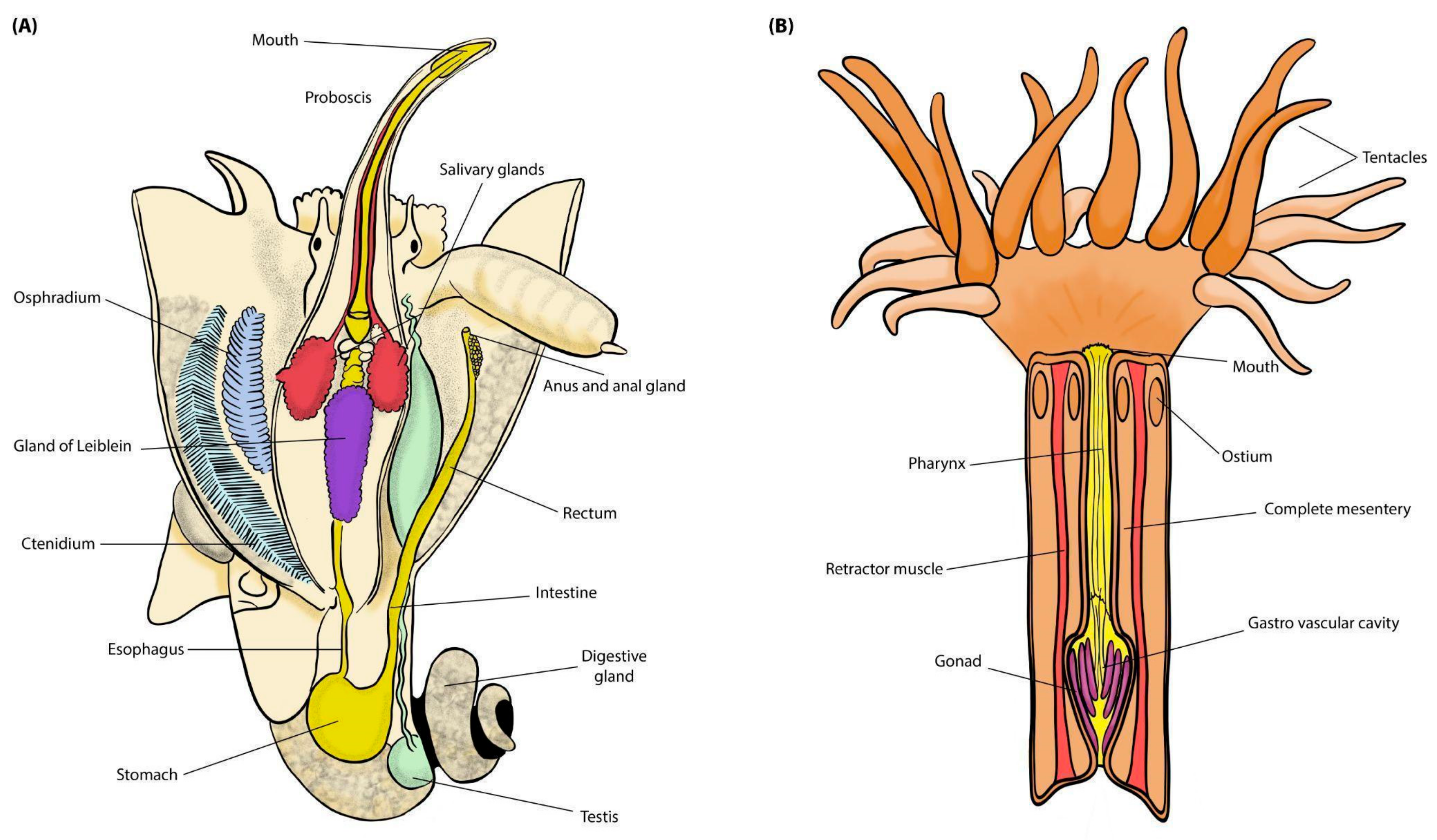
As in many Muricidae, the digestive system in C. meyendorffii comprises a pleuroembolic proboscis which, together with the oesophagus (that includes an anterior, middle, and posterior region) represents the foregut; a stomach (midgut); and a tubular intestine ending in rectum (hindgut) which opens on the right side of the pallial cavity [14] (Figure 2A). As for other Neogastropoda, these snails increase their parasitic efficiency through complex chemical secretions produced in a pair of acinous salivary glands. Two thin salivary ducts run along the oesophagus, opening into the roof of the buccal cavity. The secretory epithelium of the glands comprises two mixed cell types: superficial ciliated cells secreting mucus, and basal cells with apocrine secretion [7]. Another relevant foregut structure is the gland of Leiblein, a conspicuous lobular gland directly connected to the mid-oesophagus through a slit [15]. The function of the gland of Leiblein in secreting digestive enzymes was demonstrated by Mansour-Bek (1934) [16] by identifying four proteolytic enzymes. Lau and Leung (2004) [17] hypothesised that it was also engaged in absorption and intracellular digestion in the foregut. However, the biochemical composition of the secretions of salivary glands and gland of Leiblein in C. meyendorffii remains to be investigated [11].
The anatomy of Zoantharia (Figure 2B) typically features a cylindrical body, with a buccal aperture surrounded by a ring of tentacles at one end. This body is excavated by a mesentery, which connects with the mouth through an ectodermal oesophageal region comprising a short and wide tube. Between the oesophageal tube and the walls of the body are the septa, which terminate freely by the lower part of their inner margin in the mesenteric cavity [18]. The colonies of P. axinellae are yellow or orange in colour and are connected by a continuous layer of tissue called coenenchyme. It is known that several pigments, including carotenoids [19], biosynthesized by symbiotic marine microalgae, can be responsible for the colour of coral, with putative functional roles [20].
Figure 2. Generalised anatomical schemes of the studied invertebrates. (A) Coralliophila meyendorffii (male). Modified after (2010) [21]. (B) Polyp of Parazoanthus axinellae.
Microbiolres 15 00157 g002

2. Material and Methods

2.1. Samples and Collecting Data

Samples were collected at two localities of the Mediterranean Sea (Figure 3, created using QGIS, version 3.28.11): Ponza Island for C. meyendorffii and its host P. axinellae (40°56′48″ N–13°01′28″ E), and Giannutri Island for the single Babelomurex cariniferus sample (42°15′47″ N–11°06′33″ E). Coralliophila meyendorffii and P. axinellae were manually collected on vertical walls under small overhangs at 3–9 m depth; Babelomurex cariniferus was collected free ranging in the proximity of colonies of Cladocora coespitosa, at 5 m depth. Samples were preserved in molecular grade ethanol upon collection. Vouchers are stored in the collection of the Department of Biology and Biotechnologies ‘Charles Darwin’ of Sapienza University of Rome (BAU acronym).
Morphological identification of our samples involved observing shell shape, as well as the arrangement and distribution of polyps, under a Wild-M6 microscope. The stomach of C. meyendorffii (samples BAU-3165.1S, BAU-3165.2S, BAU-3165.5S, BAU-3165.7S, BAU-3165.8S, BAU-3165.10S) was excised with sterile forceps and then stored separately from the whole bodies in EtOH 100%. Samples BAU-3166.1, BAU-3166.2 and BAU-3166.3 were three polyps of the same colony.

2.2. DNA Extraction and Molecular Analyses

All samples were processed at the Molecular Systematic Laboratory, Department of Biology and Biotechnologies ‘Charles Darwin’, Sapienza University of Rome, Italy. Total genomic DNA was extracted from the stomach of each snail and from the whole body of each polyp and the mucus with a proteinase K/phenol–chloroform extraction protocol [22].
Considering the reported issues of phenotypic plasticity in Coralliophilinae (Nocella et al. 2024), we tested that all the C. meyendorffii specimens belonged to the same species, employing an integrative taxonomy approach. The 658 bp barcode region of the mitochondrial cytochrome oxidase subunit 1, cox1, was amplified following the protocol outlined by Nocella et al. (2024) [10]. Polymerase chain reactions were conducted following [23]. The resulting PCR products were purified using ExoSAP-IT, and both strands were sequenced at Macrogen Europe, Inc. The cox1 sequences of two samples of C. meyendorffii (BAU-3165.1S and BAU-3165.2S) and of Babelomurex cariniferus (BAU-3180.S) were published in Nocella et al. [10] (2024; accession numbers: PP093888, PP093889 and PP093890). All sequences were compared with those available in GenBank using the NCBI BLAST web interface https://blast.ncbi.nlm.nih.gov/Blast.cgi, accessed on 20 June 2024). Taxonomic identifications were checked for consistency with morphology.
Cnidarians samples were observed under a Wild-M6 microscope for the examination on the arrangement and distribution of polyps. Taxa were identified following [24].

2.3. Metabarcoding Analysis

For microbiota analysis, the Ion 16S™ Metagenomics Kit, which employed two primer pools (primer set V2–4–8; primer set V3–6, 7–9, ThermoFisher; see [25] was used to PCR amplify several hypervariable regions (V2, V3, V4, V6–V7, V8, and V9) of the bacterial 16S rRNA gene of four coralliophiline samples (BAU-3165.1S, BAU-3165.5S, BAU-3165.8S, BAU-3180.S). Following library construction and template preparation, libraries generated with the Ion 16S Metagenomics Kit were then sequenced using the Ion S5 XL System at the Eurofins Genoma Group (Rome, Italy). Data analysis, annotation, and taxonomic assignment were performed using Ion Reporter™ Software version 5.6, (ThermoFisher, Waltham, MA, USA), which provided a workflow specifically developed for analysing bacterial 16S rRNA gene. Raw reads were trimmed from the primer sequence and filtered to discard reads shorter than 150 bp. The remaining reads were clustered in amplicon sequence variant (ASVs) for the identification assignment. A consensus approach based on combining information from different hypervariable regions of the 16S rRNA gene and developed by Life Technologies (AA.VV. 2014) was employed following Russini et al. (2021) [6].
Based on the results of the sequencing findings indicating that the V3–V4 region yielded the most successfully amplified sequences (see below), universal primers Pro341F and Pro805R [26] were specifically employed for the remaining samples (BAU-3165.2S, BAU-3165.5S, BAU-3165.10S, BAU-3166.2, BAU-3166.3, and BAU-3166.4) to target only the V3-V4 region of 16S rDNA. The sequencing of 16S V3–V4 fragments was performed on an Illumina MiSeq platform (Illumina, San Diego, CA, USA) with pair-end 2 × 300 paired ends run with double indexing of the library. DNA library was prepared using Nextera XT kit (Illumina, San Diego, CA, USA) and validated with Bioanalyzer 2100 (Agilent Technologies, Palo Alto, CA, USA) and were then sequenced using the MiSeq at the Bio-Fab research srl (Rome, Italy). QIIME 2 (v2019.10) pipeline (https://qiime2.org/; Bolyen et al. 2019) was used for the V3–V4 region of 16S rDNA sequence analysis.
The sequencing reads were assigned to taxonomic identities using 16S reference library v2013 by Thermofisher and Greengenes v.13.5 (BAU-3165.1S, .5S, .8S and 3180.S) or Silva database (release 138) for the remaining samples. For the DNA sequence-based identification of bacteria, the following criteria were used following [25]: minimum 90% coverage to assign a read to a database sequence; minimum 99% percentage of identity to assign a read to a genus and 97% to assign a read to a family.
To ensure an unbiased statistical approach, all the analyses have been conducted exclusively using the sequences obtained from the V3–V4 region, for all samples (Supplementary Materials, Tables S1 and S2).
The taxonomic bar plots were generated after removing all ASVswith a relative count below 1% in each sample. However, all other statistical and bioinformatic analyses were conducted on the complete dataset obtained from the V3–V4 amplified bacterial sequences. To comparatively assess diversity among sampled individuals, diversity was evaluated using α- and β-metrics. The diversity analyses were performed among sample groups, in particular between P. axinellae, C. meyendorffii and B. cariniferus. Alpha diversity estimates were evaluated using the Kruskal–Wallis test [27]. Alpha rarefaction curves (created using the plugin diversity alpha-rarefaction in QIIME2) were utilised to assess bacterial communities’ diversity across different sequencing depths. These curves are generated by randomly subsampling sequencing reads at various depths, altering the number of reads or analysed samples, and computing diversity metrics at each level. This method enables the estimation of how bacterial community diversity changes according to sequencing efforts. PERMANOVA (Permutational multivariate analysis of variance [28]) and ANOSIM (Analysis of similarities) were employed for assessing the statistical significance of the observed differences. Given that simultaneous testing of multiple hypotheses increases the likelihood of observing false positives, the q-value correction was employed in this context to provide a measure of the false positives proportion among all significant results, granting more accurate results. The q-value, akin to the p-value but adjusted to accommodate multiple comparisons, plays a crucial role in addressing the false discovery rate (FDR) in multiple hypothesis testing scenarios.
The R software (version 4.2.1, package readxl) was utilised to identify the bacterial genera shared between Coralliophila and Babelomurex, those shared between Coralliophila and Parazoanthus, as well as those shared among at least four out of six Coralliophila samples. This allowed for generating of a heatmap depicting common bacterial genera abundance in QIIME2 [29] and a Venn diagram illustrating the number of shared bacterial genera using the R package ggVennDiagram. The heatmap provided a graphical representation of data in a matrix format, where rows represent the analysed samples and columns indicate the relative frequency of shared bacterial variants identified as genera.

3. Results

3.1. Overview of Sequencing Information

All the four cox1 sequences correctly blasted with C. meyendorffii sequences available in the Genbank nucleotide database with an e-value = 0.0 and a percentage of identity ≥ 98.04%.
For the microbial analyses, 960,092 reads were correctly merged, and 659,424 (~70%) were retained for subsequent analyses, with an average of 65,942.4 reads per sample; the remaining sequences were discarded. More specifically, low-quality bases were trimmed from both ends of the obtained raw sequencing reads, and sequences that fell below a minimum quality threshold were excluded from the data set, ensuring higher accuracy in upcoming analyses. Chimeric reads were also removed from the total reads, to avoid including in the definitive ASV table any artefact formed by a wrong joining of distinct biological sequences during PCR amplification.
The abundance of sequences by primer barplots from Ion System output revealed that the V3–V4 region yielded the most successfully amplified sequences (Supplementary Figure S1).
The microbial DNA sequencing data obtained in the present study were submitted to the Sequence Read Archive (SRA) at GenBank database (National Center for Biotechnology Information, NCBI, https://www.ncbi.nlm.nih.gov/, accessed on 20 June 2024): bioproject PRJNA1101417.
The new cox1 sequences of C. meyendorffii are available on the NCBI nucleotide database (accession numbers PP625322-PP625323-PP625324-PP625325).

3.2. Bacterial Communities

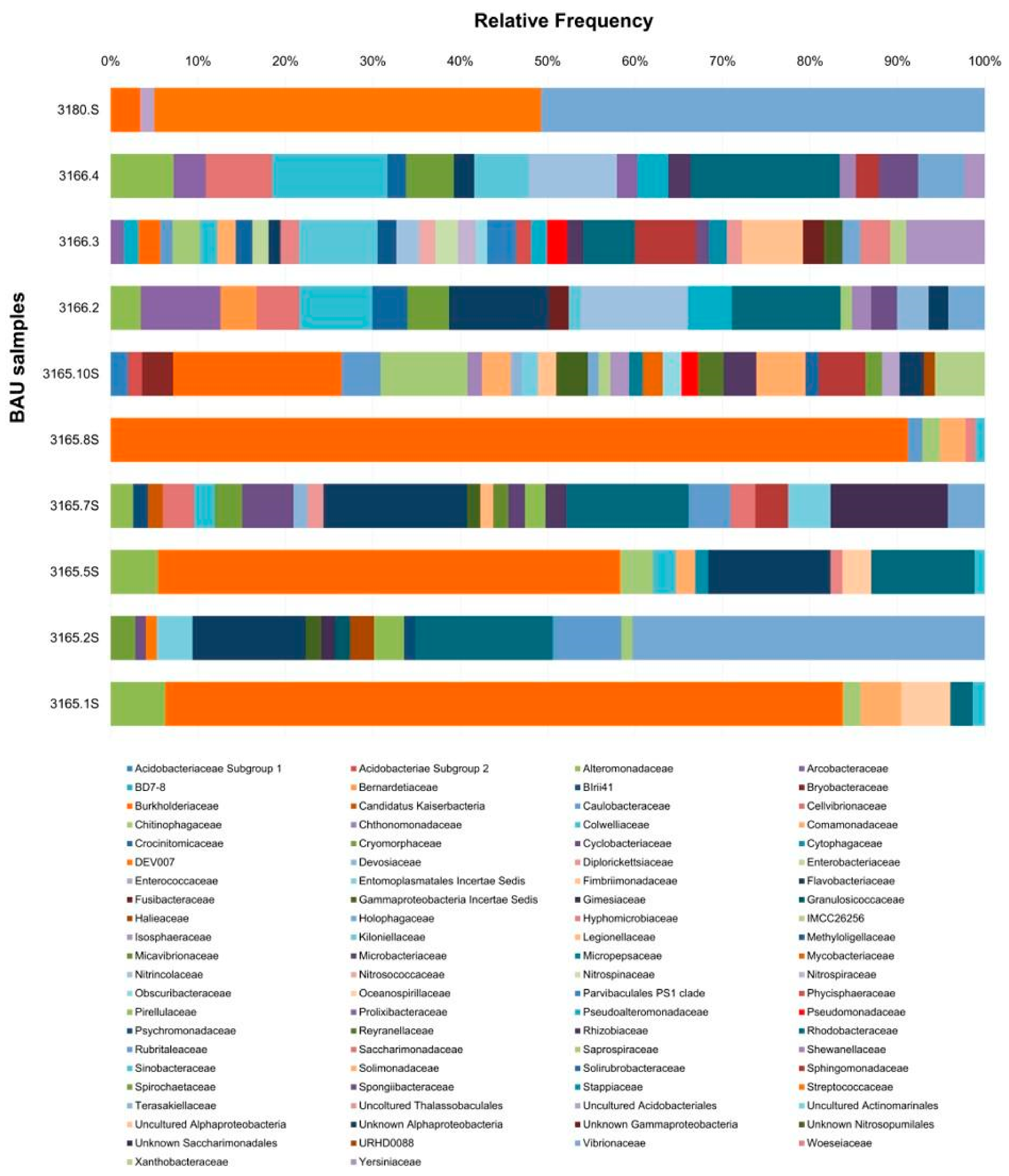
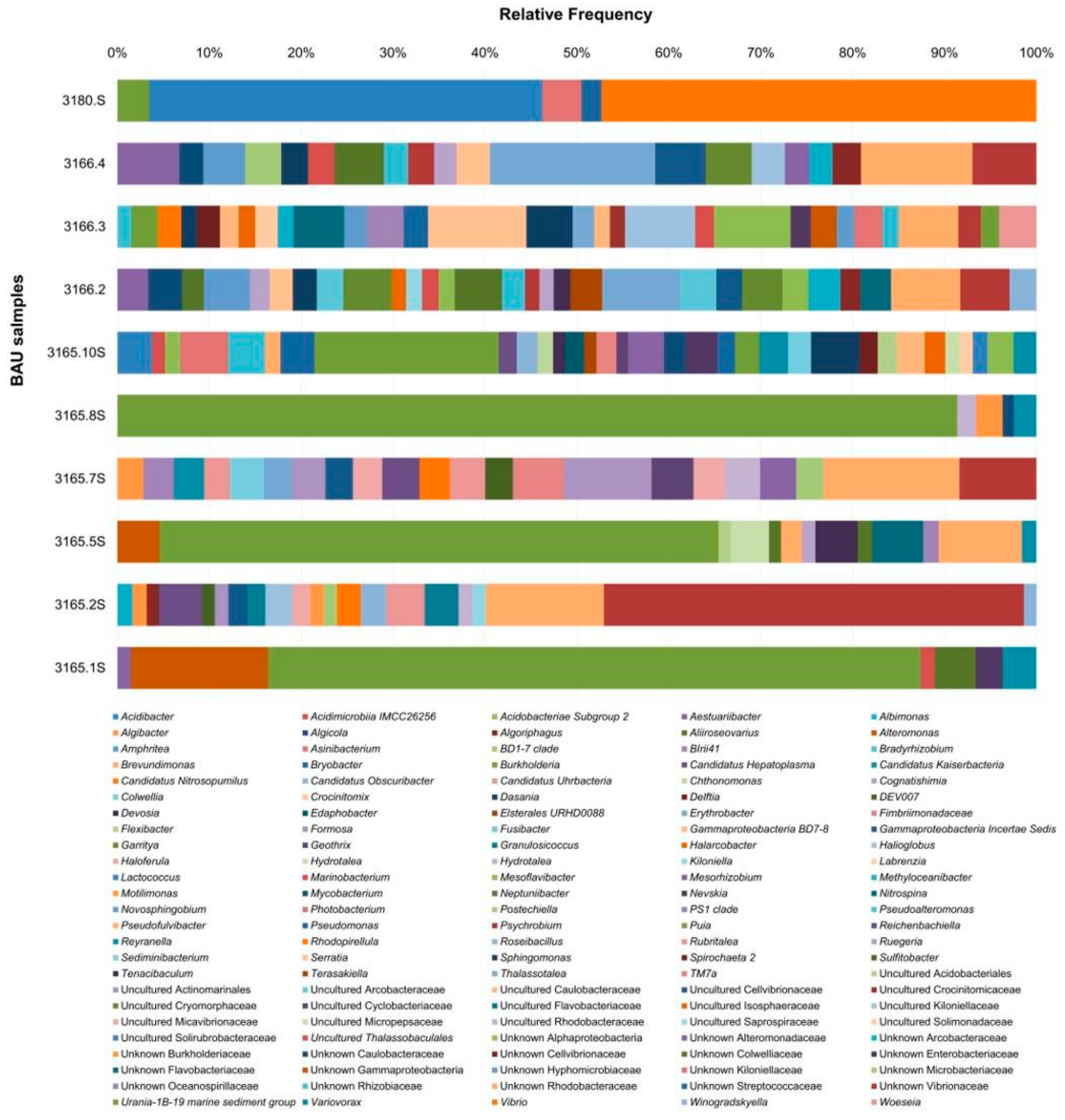
We compared microbial communities detected by means of NGS methods based on 16S rRNA gene sequences from the polyp of P. axinellae and the stomach of C. meyendorffii. A total diversity of 97 ASVsat the family level (Figure 4) and 176 ASVsat the genus level (Figure 5) were observed with a relative abundance ≥0.1% in each sample. Proteobacteria families were overwhelmingly predominant with Burkholderiaceae (genus Burkholderia), Vibrionaceae (genus Vibrio), and Rhodobacteraceae (unknown genus) being the most abundant, followed by the Firmicutes family Streptococcaceae (genus Lactococcus). Barplots representing ASVs identified with amplification of regions V2, V3, V4, V6–V7, V8, and V9 are available in Supplementary Materials, Tables S3 and S4.

3.2.1. Family Level

Several microbial families analysed in this study were found exclusively in the mucus/tissue of P. axinellae polyps (samples BAU-3166.2, .3, .4), in particular Bernardetiaceae (Bacteroidota, Cytophagales), Prolixibacteraceae, and Marinifilaceae (Bacteroidota; Marinilabiliales), Kiloniellaceae and Terasakiellaceae (Alphaproteobacteria; Rhodospirillales), and Kordiimonadaceae (Alphaproteobacteria; Kordiimonadales).
Among the most abundant ASVss, some bacterial families mainly detected in P. axinellae were also found in the stomach of C. meyendorffii, the majority of which belonged to the Pseudomonadota. Examples are Nitrincolaceae, Spongiibacteraceae and Pseudoalteromonadaceae (Gammaproteobacteria), and Rhodobacteraceae (Alphaproteobacteria). Moreover, ASVs affiliated with the family Arcobacteraceae (Campylobacterota; Epsilonproteobacteria) and the family Crocinitomicaceae (Bacteroidota, Flavobacteriales) were detected, but more abundant in P. axinellae than in C. meyendorffii.
The bacterial 16S rRNA gene sequences that were detected exclusively in the stomach of samples BAU-3165.2S and BAU-3165.7S of C. meyendorffii belonged to an unidentified family of the order Verrucomicrobiales.

3.2.2. Genus Level

Considering the results of this study at the genus level, a particularly noteworthy finding is that the sequences of some genera were detected exclusively in the three samples of P. axinellae (BAU-3166.2-3-4) (Figure 5). An example at the genus level of Gammaproteobacteria (Pseudomonadota) was Algicola (Alteromonadales; Pseudoalteromonadaceae). Some microbial genera were detected only in two samples of P. axinellae. Examples of Gammaproteobacteria were Psychrobium (Alteromonadales; Shewanellaceae), Pseudoteredinibacter (Cellvibrionales; Cellvibrionaceae), and Marinomonas (Oceanospirillales; Oceanospirillaceae). Examples of Alphaproteobacteria were the genera Cognatishimia (Rhodobacterales; Paracoccaceae), Terasakiella (Rhodospirillales; Terasakiellaceae), and Filomicrobium (Hyphomicrobiales; Hyphomicrobiaceae). Also present in the polyps of P. axinellae was Halarcobacter of Arcobacteraceae, a bacterial family within the Epsilonproteobacteria (Campylobacterota), and Draconibacterium (Bacteroidota; Marinilabiliales; Prolixibacteraceae).
The bacterial 16S rRNA gene sequences that were detected only in the stomach of samples BAU-3165.2S and BAU-3165.7S of C. meyendorffii, belong to the Verrucomicrobiota genus Haloferula (family Verrucomicrobiaceae of the order Verrucomicrobiales), and to the Planctomycetota genus Rhodopirellula (family Pirellulaceae). Examples of the genera found in BAU-3165.5S and BAU-3165.7S were Formosa and Algibacter. Unknown bacterial genera of the family Flavobacteriaceae in the Flavobacteriia (Bacteroidota) were detected in some stomach samples of C. meyendorffii.
Identified genera shared by at least four coralliophiline samples, between gastropods and corals, and between the two coralliophiline species, are reported in the paragraph below.

3.3. Microbiome Similarity of Snails and Corals

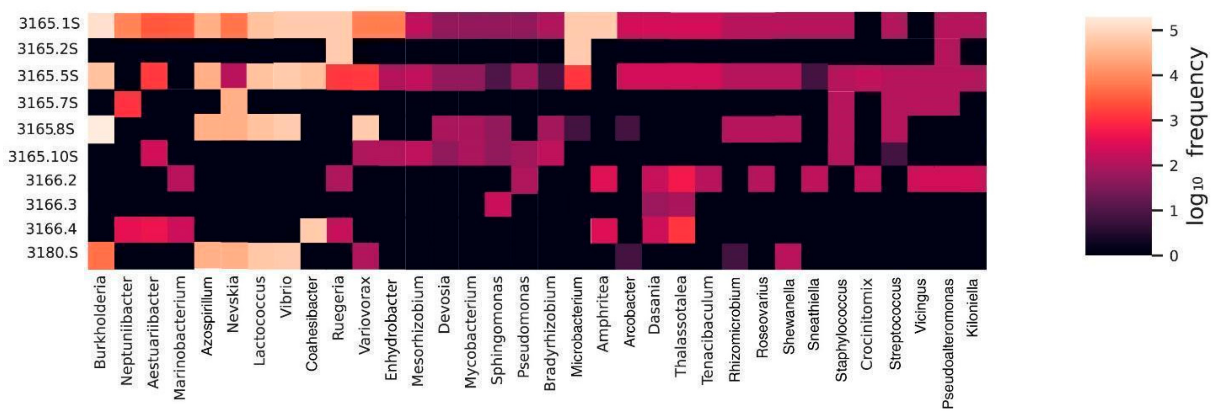
All the 34 genera shared between Coralliophila and Babelomurex, Coralliophila and Parazoanthus, or among at least four Coralliophila samples, along with their normalised count frequency, are reported in the heatmap (Figure 6).
Looking at the genus level, the heatmap showed that some microbial genera detected in the polyps of P. axinellae were also found in stomachs of C. meyendorffii. Among them, we found three Flavobacteriia genera: Tenacibaculum (Flavobacteriaceae), Vicingus (Vicingaceae) and Crocinitomix (Crocinitomicaceae). Other gastropod/coral common genera are the Gammaproteobacteria Dasania (Spongiibacteraceae), Neptuniibacter, Amphritea, and Marinobacterium (Oceanospirillaceae), Thalassotalea (Colwelliaceae), and Aestuariibacter (Alteromonadaceae), as well as the Alphaproteobacteria genera Roseovarius (Roseobacteraceae) and Ruegeria (Roseobacteraceae).
The heatmap analysis also showed that some microbial genera were found in at least four out of six stomach samples of C. meyendorffii. Among the genera of Hyphomicrobiales (Alphaproteobacteria), we found Enhydrobacter, Mesorhizobium (Phyllobacteriaceae), Devosia (Devosiaceae), and Bradyrhizobium (Nitrobacteraceae). Also, the Microbacteriaceae genera Mycobacterium and Microbacterium were present.
The bacterial 16S rRNA gene sequences that were detected in both neogastropods, C. meyendorffii and B. cariniferus, belonged to Pseudomonadota genus. Specifically, for the Gammaproteobacteria, the genera Shewanella (Shewanellaceae) and Nevskia (Xanthomonadaceae), and for the Alphaproteobacteria, the genera Azospirillum (Azospirillaceae) and Rhizomicrobium (Micropepsaceae). Also, the Campylobacterota genus Arcobacter (Arcobacteraceae) within the Epsilonproteobacteria was detected. Among the order Vibrionales (Gammaproteobacteria), the genus Photobacterium (Vibrionaceae) was found in B. cariniferus and in few samples of C. meyendorffii.

3.4. Alpha and Beta Diversity Analysis

The permutation-based statistical test PERMANOVA was used to detect significant differences in microbial structure among sample groups. This method calculates a statistical test based on the partitioning of total variation in the data into between-group and within-group; in this case the permutation approach estimates the likelihood that any observed difference between the groups has occurred by chance or if it has statistical significance. It relied on a previously created Bray-Curtis dissimilarity matrix of beta diversity distance values between groups: Parazoanthus, Coralliophila, and Babelomurex. The value of the Pseudo-F statistic in the PERMANOVA analysis indicates the strength of the differences between the groups. PERMANOVA revealed significant differences in bacterial ASV structure among the three selected sample groups (Table 1) at both taxonomic levels assessed: families and genera. In both cases, the p-values associated to the respective statistics (family: pseudo-F statistic 1.527733, p-value of 0.038; genera: pseudo-F statistic is 1.520639, p-value of 0.039), were smaller than 0.05, suggesting differences in variance between groups, considering both the location (centroid) and dispersion (scatter) of the data points. Overall, the obtained results are coherent with the PCoA plot, which shows a lack of distinct clustering among most of the samples, emphasising the heterogeneity of the microbial communities in the dataset (Figure 7). As evidenced by the non-significant ANOSIM test results (R = 0.014403, p-value = 0.416 at family level; R = 0.024691, p-value = 0.403 at genus level), the dissimilarities between groups were not statistically significant when considering ranked distances alone. However, as previously mentioned, PERMANOVA detected significant differences in multivariate variance (p-values < 0.05). This discrepancy suggests that the observed differences between groups are more pronounced when considering multivariate dispersion, as measured by PERMANOVA, compared to the ranked dissimilarities assessed by ANOSIM.
The alpha diversity results indicate that both the overall Kruskal-Wallis tests and pairwise comparisons for the families and genera levels did not indicate a statistically significant difference between groups: H statistic = 3.036, q-value = 0.219 for families; H statistic of 3.964, q-value = 0.138 for genera. The sampling effort was sufficient to capture the diversity of the community, as evidenced through alpha diversity rarefaction curves (Supplementary Figure S2), which ensured that the sequencing was adequate for covering the majority of the bacterial diversity in the sample.

4. Discussion

4.1. Structure of Bacterial Communities

The results from sequencing across different hypervariable regions of the 16S rRNA gene conducted on the first four samples analysed (three Coralliophila meyendorffii and the Babelomurex cariniferus stomachs) showed that the V3–V4 regions of the 16S rRNA gene represented the greatest variability in microbial community composition at different taxonomic levels, as already reported in previous studies (e.g., [30,31]). These two regions were then selected to conduct all the 16S metabarcoding analyses to assess the microbial communities of C. meyendorffii and P. axinellae. However, we are aware that all the conclusions drawn here are based on the absence of an environmental sample, which was not collected due to logistical sampling constraints. Therefore, our findings should be interpreted with caution, as the absence of environmental data means that certain bacteria found exclusively in one species may, in fact, originate from the surrounding environment.
Proteobacteria were the dominant phylum (mainly Alphaproteobacteria and Gammaproteobacteria), present both in the two coralliophiline species and in Parazoanthus axinellae (Figure 4).
Looking at the most abundant ASVs at the family level, some families were detected exclusively, or mostly in the polyp of P. axinellae (samples BAU-3166.2-4; Figure 4). This suggests that distinct components of the microbial community might be uniquely adapted within the coral, with bacterial features probably well-suited for the mucus or tissue of P. axinellae.
Some families detected in the polyps of P. axinellae were also found in the stomachs of C. meyendorffii (Figure S3). Most of these belonged to the phylum Pseudomonadota. Among them, some families have been previously detected as members of the microbiome of marine invertebrates. In particular, previous studies have shown that the Spongiibacteraceae mainly dominate the core microbiomes of corals [32], even though they have never been found in gastropods before. Additionally, members of the lineage Rhodobacteraceae were detected in coral-bacteria symbiosis [33] and in the gut of the freshwater gastropod Bellamya aeruginosa [34]. Moreover, members of Nitrincolaceae have been previously found in the coenenchyme of Corallium rubrum [35] and in the pedal mucus of the marine gastropod species Echinolittorina peruviana [36]). Additionally, shared bacterial communities include the family Crocinitomicaceae, which resulted abundant in the sea anemone Exaiptasia diaphana [37] and in the pedal mucus of the grazer molluscs Fissurella crassa and Chiton granosus [36] In these same study [36] other two bacterial families (Cyclobacteriaceae and Rubritaleaceae) resulted in association with Fissurella crassa and Chiton granosus, which in our study were found only with the stomachs of C. meyendorffii. Similarly, the family Pirellulaceae, previously found in the faecal microbiome of the freshwater snail Biomphalaria glabrata [38], was also identified.
With regards to the ASVs identified at the genus level, according to our analyses certain bacterial taxa were found exclusively, or mostly, in the polyp of P. axinellae (Figure 5). Among them, the genera Algicola (Pseudoalteromonadaceae) and Draconibacterium (Prolixibacteraceae) have been found in different species of stony corals [39,40]. Additionally, the genus Crocinitomix (Crocinitomicaceae) has previously been found in the bacterial community inhabiting the sea anemone Exaiptasia diaphana [37]. All the aforementioned genera are known to typically live in symbiotic relationships with their coral hosts [41]. It is reasonable to suggest that some forms of symbiotic association exist between the coral P. axinellae and the coral-resident bacterial genera observed in this study. These associations may enable the coral to utilise microbial biosynthetic pathways, such as those providing nutritional resources, as occurred in other reported coral-bacteria associations. However, experimental validation would be required to confirm this hypothesis.
A few bacterial genera were detected only in the C. meyendorffii stomachs, such as the genera Devosia and Algibacter (Flavobacteriaceae), that had previously been isolated from freshwater gastropods of the family Lymnaeidae and from intertidal gastropods of the family Onchidiidae, respectively [42,43]. In particular, genomic investigation of aquatic Devosia strains revealed genes functional in the metabolism of aromatic compounds, alkylphosphonate and organic nitrogen, potentially beneficial for nutrient acquisition, while the natural role of Algibacter is centred around the decomposition of algal biomass, as it was shown to hydrolyse galantine, alginate and starch, indicating its ecological significance in breaking down algal biopolymers [44].
Several bacterial genera identified in the stomach of Coralliophila meyendorffii were also present in the stomach of Babelomurex cariniferus (Figure 6), some of which are known to associate specifically with marine invertebrates, including Mollusca. For example, Shewanella (Shewanellaceae) and Arcobacter (Campylobacteraceae) were isolated from the gut of the abalone Haliotis diversicolor [45] and Haliotis gigantea, respectively [46,47] while Vibrio (Vibrionaceae) was found in high percentages in the muricid Rapana venosa [48] While Shewanella is associated with oxidase activity [45], Arcobacter and Vibrio are known to be potentially pathogenic [46,48].
These findings suggest that the bacterial genera present in both Coralliophilinae species, yet absent in Parazoanthus, likely function as symbionts within gastropods. Furthermore, the presence of these taxa in other marine molluscs indicates that their association is more likely driven by molluscan physiology rather than by specific host species. This could explain their absence in Parazoanthus, which may engage in different ecological interactions.
Among bacteria found only in Babelomurex cariniferus, the Pseudomonadota genus Photobacterium (Vibrionaceae) is distributed in marine habitats worldwide and is known to include pathogenic species for aquatic organisms, including fish, molluscs and crustaceans, and even for humans [49]. Its species are unique even for their ability to produce luminescence in fish and squids [50], although this ability has never been observed in Coralliophilinae. It is likely that the bacteria might have been acquired from the surrounding environment and integrated into the microbiota of B. cariniferus.
The molecular evidence emerging from our study suggests that certain typically environmental bacteria are shared between the polyps of P. axinellae and the stomach of C. meyendorffii (Figure 7). Most of them are known to be part of the marine environment, such as Aestuariibacter (Alteromonadaceae) [51], Cohaesibacter (Cohaesibacteraceae) [52], Dasania (Spongiibacteraceae) [53], Thalassotalea (Colwelliaceae) [54], Sneathiella (Sneathiellaceae) (Lee 2019), Vicingus (Cryomorphaceae) [55] and Pseudoalteromonas [56]. Pseudomonas (Pseudoalteromonadaceae) was also found, even if it is primarily associated with terrestrial habitats [57].
Additionally, we identified in both Coralliophila and Parazohantus some bacteria typically associated with marine organisms, such as Tenacibaculum (Flavobacteriaceae), known to cause diseases in fish [58]; Neptunibacter (Oceanospirillaceae), found in bivalves [59] and sea cucumber larvae [60] but never in corals; Crocinitomix (Crocinitomicaceae), which has been isolated from algae [61]; Amphritea (Oceanospirillaceae), found in sea cucumbers and sponges [62,63]); and Kiloniella (Kiloniellaceae), whose members are often associated with living organisms such as sponges, corals, squid eggs, lobsters, fish intestines, and macroalgae [64].
Of particular interest among the bacteria shared between Coralliophila and Parazohantus, are the genera typically identified in previous studies as corals holobionts. Marinobacterium (Alteromonadaceae), for instance, was found to produce compounds displaying UV spectra in scleractinians [65]; Ruegeria (Rhodobacteraceae), found in colonial Scleractinia [66] are considered to contribute to carbon and sulphur cycling [67]; Roseovarius (Roseobacteraceae), a component of the scleractinian holobiont, exhibits the potential to produce two antioxidants [68] Sphingomonas (Sphingomonadaceae) is often described as a coral pathogen, causing tissue damage to at least 16 varieties of corals and hydrocorals [69].
Regarding the first two groups of shared bacteria, those typically found in the environment and those found in other marine organisms, we suggest that their presence in C. meyendorffii and P. axinellae is due to environmental origin: these bacteria may have been acquired by the two organisms through horizontal transmission from the surrounding environment and have become part of the genetic pool of the two invertebrates. What we cannot ascertain is whether they were transmitted independently to Parazoanthus and Coralliophila or if the latter acquired them through feeding.
In contrast, the presence of genera typically described in literature as coral holobionts suggests the possibility of horizontal bacterial transfer. It is likely that some specific bacterial genera are primarily hosted by the coral and subsequently acquired by Coralliophilinae during its feeding activities on coral tissues and mucus. However, these hypotheses need to be confirmed by including water samples as controls in future analyses.

4.2. Microbial Diversity

The permutational based PERMANOVA test, applied to estimate beta diversity values, showed differences in microbial structure among coral and gastropod samples at both family and genus levels. In the context of beta group significance, the divergence between ANOSIM and PERMANOVA outputs might depend on the different focuses of those two methods, since ANOSIM could overlook the minor differences in multivariate dispersion. The q-value correction method was used for addressing multiple hypothesis testing, as to better control false positives in beta diversity values when comparing different sample groups (Parazoanthus, Coralliophila, and Babelomurex) and taxonomic levels (genus and family). The presence of specific bacterial taxa within the microbiota of each sample likely reflects adaptation to the unique biological and physiological needs of the hosts. This resulting heterogeneity in bacterial communities highlights the complex interactions occurring within marine ecosystems, where microbial groups can be influenced by environmental disturbances. Factors such as host-microbiota interactions and habitat characteristics may contribute to shaping the microbiota composition in Parazoanthus, Coralliophila, and Babelomurex samples. The absence of variation in alpha diversity values could be due to host-specific factors, including immune responses, metabolic pathways, or chemical secretions. Future research could explore whether microbial community structures shift across different habitats and examine the stability of these communities over time.

5. Conclusions

This study represents the first investigation into the microbial communities inhabiting the stomach of Coralliophila meyendorffii and its coral host Parazoanthus axinellae using 16S rDNA metabarcoding (V3–V4 regions). Our analysis has led to the following key findings:
-
The bacterial taxa found exclusively or predominantly in the P. axinellae samples suggest the presence of holobiont components within the microbial community of this coral, as has been observed in other coral species.
-
Our findings indicate that the stomach microbiome of C. meyendorffii shows similarities to that of its coral host, P. axinellae, suggesting a possible influence of the coral’s microbiome on the gastropod’s gut. This is further supported by the observation that Babelomurex cariniferus and Parazoanthus axinellae, despite living in similar environments, exhibit distinct microbiome compositions. These differences highlight those specific ecological interactions, including potential horizontal transfer of bacterial taxa between organisms, significantly shape the microbiomes of these coral-associated species.
-
The microbial community identified in the stomachs of C. meyendorffii does not provide evidence for a primary role in parasitism, as we did not detect any bacteria known to be directly associated with corallivory, particularly those involved in neutralising or digesting the venomous substances of P. axinellae. This observation suggests that the microbiota responsible for toxin degradation may not be present in the digestive system but rather within the salivary glands that the Coralliophilinae have evolved over time. Future research should focus on investigating these glands to elucidate their potential role in toxin processing.

Supplementary Materials

The following supporting information can be downloaded at: https://www.mdpi.com/article/10.3390/microbiolres15040157/s1, Figure S1: The abundance of sequences by primer barplots; Figure S2: Alpha rarefaction curves for ASVs at the family level; Figure S3: Venn diagram depicting all the 34 shared genera. Table S1: The total number of ASVs identified at the family level. Table S2: The total number of ASVs identified at the genus level. Table S3: The total number of ASVs identified at the family level with all the primers. Table S4: The total number of ASVs identified at the genus level with all the primers. Table S5: The total number of ASVs identified at the family level ≥1%. Table S6: The total number of ASVs identified at the genus level ≥1%.

Author Contributions

Conceptualization, D.D.; methodology, D.D. and G.F.; software, V.R.; validation, V.R., D.D. and C.B.; formal analysis, E.N. and C.B.; investigation, E.N.; resources, M.O. and M.V.M.; data curation, E.N. and C.B.; writing—original draft preparation, E.N.; writing—review and editing, C.B., G.F., V.R., D.D. and M.O.; visualization, E.N.; supervision, D.D.; project administration, D.D.; funding acquisition, D.D. All authors have read and agreed to the published version of the manuscript.

Funding

The research was funded by the Sapienza 2015 “Medi Progetti Universitari” grant.

Data Availability Statement

The datasets presented in this study can be found in online repositories. The names of the repository/repositories and accession number(s) can be found in the article, in the Section 3.

Acknowledgments

We are grateful to Paolo Mariottini (University of Roma Tre) for providing precious photographic material. We are thankful to Giulia Maiello (University of Rome Tor Vergata) and Brian Cunningham (University of Malaga) for their help in processing specimens, and to Andrea Lepri (Sapienza University of Rome) for his help with the bioinformatic analysis.

Conflicts of Interest

The authors declare no conflict of interest.

References

  1. Stella, J.; Pratchett, M.; Hutchings, P.; Jones, G. Coral-associated invertebrates: Diversity, ecological importance and vulnerability to disturbance. Oceanogr. Mar. Biol. Annu. Rev. 2011, 49, 43–104. [Google Scholar] [CrossRef]
  2. Pita, L.; Rix, L.; Slaby, B.M.; Franke, A.; Hentschel, U. The sponge holobiont in a changing ocean: From microbes to ecosystems. Microbiome 2018, 6, 46. [Google Scholar] [CrossRef] [PubMed]
  3. Plaisance, L.; Matterson, K.; Fabricius, K.; Drovetski, S.; Meyer, C.; Knowlton, N. Effects of low pH on the coral reef cryptic invertebrate communities near CO2 vents in Papua New Guinea. PLoS ONE 2021, 16, e0258725. [Google Scholar] [CrossRef] [PubMed]
  4. Ashwood, L.M.; Norton, R.S.; Undheim, E.A.B.; Hurwood, D.A.; Prentis, P.J. Characterising functional venom profiles of anthozoans and medusozoans within their ecological context. Mar. Drugs 2020, 18, 202. [Google Scholar] [CrossRef]
  5. Robertson, R. Review of the predators and parasites of stony corals, with special reference to symbiotic prosobranch gastropods. Pac. Sci. 1970, 24, 43–54. [Google Scholar]
  6. Russini, V.; Fassio, G.; Nocella, E.; Houart, R.; Barco, A.; Puillandre, N.; Lozouet, P.; Modica, M.V.; Oliverio, M. Whelks, rock-snails, and allied: A new phylogenetic framework for the family Muricidae (Mollusca: Gastropoda). Eur. Zool. J. 2023, 90, 856–868. [Google Scholar] [CrossRef]
  7. Ponte, G.; Modica, M.V. Salivary glands in predatory mollusks: Evolutionary considerations. Front. Physiol. 2017, 8, 266744. [Google Scholar] [CrossRef]
  8. Oliverio, M.; Mariottini, P. A molecular framework for the phylogeny of Coralliophila and related muricoids. J. Molluscan Stud. 2001, 67, 215–224. [Google Scholar] [CrossRef]
  9. Oliverio, M.; Mariottini, P. Contrasting morphological and molecular variation in Coralliophila meyendorffii (Muricidae, Coralliophilinae). J. Molluscan Stud. 2001, 67, 243–246. [Google Scholar] [CrossRef]
  10. Nocella, E.; Fassio, G.; Zuccon, D.; Puillandre, N.; Modica, M.V.; Oliverio, M. From coral reefs into the abyss: The evolution of corallivory in the Coralliophilinae (Neogastropoda, Muricidae). Coral Reefs 2024, 43, 1285–1302. [Google Scholar] [CrossRef]
  11. Ritcher, A.; Amor, M.J.; Dufort, M. The anatomy and ultrastructure of the gland of Leiblein of Bolinus brandaris and Coral-liophila meyendorffii, two neogastropod species with different ecology and feeding strategies. In Proceedings of the SEB Annual Main Meeting, Prague, Czech Republic, 30 June–3 July 2010. [Google Scholar]
  12. Guillen, P.O.; Jaramillo, K.B.; Genta-Jouve, G.; Thomas, O.P. Marine natural products from zoantharians: Bioactivity, biosynthesis, systematics, and ecological roles. Nat. Prod. Rep. 2020, 37, 515–540. [Google Scholar] [CrossRef] [PubMed]
  13. Nocella, E.; Zvonareva, S.S.; Fassio, G.; Pica, D.; Buge, B.; Villa, R.; Puillandre, N.; Modica, M.V.; Oliverio, M. Spicy food for the egg-cowries: The evolution of corallivory in the Ovulidae (Gastropoda: Cypraeoidea). Front. Mar. Sci. 2024, 10, 1323156. [Google Scholar] [CrossRef]
  14. Simone, L.R.L. A new species of Thaisella (Neogastropoda: Muricidae) from Caribbean Guatemala, with accounts on the anatomy and taxonomy of the genus in the western Atlantic. Arch. Molluskenkd. 2017, 146, 111–120. [Google Scholar] [CrossRef]
  15. Andrews, E.B.; Thorogood, K.E. An Ultrastructural Study of the gland of Leiblein of muricid and nassariid neogastropods in relation to function, with a discussion on its homologies in other caenogastropods. J. Molluscan Stud. 2005, 71, 269–300. [Google Scholar] [CrossRef]
  16. Mansour-Bek, J.J. Über die proteolytischen Enzyme von Murex anguliferus Lamk. Z. Vgl. Physiol. 1934, 20, 343–369. [Google Scholar] [CrossRef]
  17. Lau, D.C.; Leung, K.M. Feeding physiology of the carnivorous gastropod Thais clavigera (Kuster): Do they eat “soup”? J. Exp. Mar. Biol. Ecol. 2004, 312, 43–66. [Google Scholar] [CrossRef]
  18. Jourdan, M. On the Zoantharia Malacodermata of the shores of Marseilles. Ann. Mag. Nat. Hist. 1879, 4, 325–326. [Google Scholar] [CrossRef]
  19. Galasso, C.; Corinaldesi, C.; Sansone, C. Carotenoids from marine organisms: Biological functions and industrial applications. Antioxidants 2017, 6, 96. [Google Scholar] [CrossRef]
  20. Glynn, P.W. Coral reef bleaching: Ecological perspectives. Coral Reefs 1993, 12, 1–17. [Google Scholar] [CrossRef]
  21. Modica, M.V.; Holford, M. The Neogastropoda: Evolutionary Innovations of Predatory Marine Snails with Remarkable Pharmacological Potential. In Evolutionary Biology—Concepts, Molecular and Morphological Evolution; Pontarotti, P., Ed.; Springer: Berlin/Heidelberg, Germany, 2010. [Google Scholar] [CrossRef]
  22. Gorson, J.; Fassio, G.; Lau, E.S.; Holford, M. Diet diversity in carnivorous terebrid snails is tied to the presence and absence of a venom gland. Toxins 2021, 13, 108. [Google Scholar] [CrossRef]
  23. Fassio, G.; Russo, P.; Bonomolo, G.; Fedosov, A.E.; Modica, M.V.; Nocella, E.; Oliverio, M. A molecular framework for the systematics of the Mediterranean spindle-shells (Gastropoda, Neogastropoda, Fasciolariidae, Fusininae). Mediterr. Mar. Sci. 2022, 23, 623–636. [Google Scholar] [CrossRef]
  24. Veron, J.E.N. Corals of the World; AIMS: Townsville, Australia, 2000; Volumes 1–3, p. 1410. [Google Scholar]
  25. Russini, V.; Fassio, G.; Chimenti, C.; Davolos, D. Discovering symbiosis in the supralittoral: Bacterial metabarcoding analysis from the hepatopancreas of Orchestia and Tylos (Crustacea). Symbiosis 2021, 83, 225–236. [Google Scholar] [CrossRef]
  26. Takahashi, S.; Tomita, J.; Nishioka, K.; Hisada, T.; Nishijima, M. Development of a Prokaryotic Universal Primer for Simultaneous Analysis of Bacteria and Archaea Using Next-Generation Sequencing. PLoS ONE 2014, 9, e105592. [Google Scholar] [CrossRef] [PubMed]
  27. Kruskal, W.H.; Wallis, W.A. Use of Ranks in One-Criterion Variance Analysis. J. Am. Stat. Assoc. 1952, 47, 583–621. [Google Scholar] [CrossRef]
  28. Anderson, M.J. Permutational Multivariate Analysis of Variance (PERMANOVA). In Wiley StatsRef: Statistics Reference Online; Wiley: Hoboken, NJ, USA, 2017; pp. 1–15. [Google Scholar]
  29. Bolyen, E.; Rideout, J.R.; Dillon, M.R.; Bokulich, N.A.; Abnet, C.C.; Al-Ghalith, G.A.; Alexander, H.; Alm, E.J.; Arumugam, M.; Asnicar, F.; et al. Reproducible, interactive, scalable and extensible microbiome data science using QIIME 2. Nat. Biotechnol. 2019, 37, 852–857. [Google Scholar] [CrossRef] [PubMed]
  30. Yang, F.; Xiao, Z.; Wei, Z.; Long, L. Bacterial Communities Associated with Healthy and Bleached Crustose Coralline Alga Porolithon onkodes. Front. Microbiol. 2021, 12, 646143. [Google Scholar] [CrossRef]
  31. Zhou, Z.; Wu, H.; Li, D.; Zeng, W.; Huang, J.; Wu, Z. Comparison of gut microbiome in the Chinese mud snail (Cipangopaludina chinensis) and the invasive golden apple snail (Pomacea canaliculata). PeerJ 2022, 10, e13245. [Google Scholar] [CrossRef]
  32. Quintanilla, E.; Rodrigues, C.F.; Henriques, I.; Hilário, A. Microbial associations of abyssal gorgonians and anemones (>4,000 m depth) at the Clarion-Clipperton Fracture Zone. Front. Microbiol. 2022, 13, 828469. [Google Scholar] [CrossRef]
  33. Bernasconi, R.; Stat, M.; Koenders, A.; Paparini, A.; Bunce, M.; Huggett, M.J. Establishment of coral-bacteria symbioses reveal changes in the core bacterial community with host ontogeny. Front. Microbiol. 2019, 10, 1529. [Google Scholar] [CrossRef]
  34. Lyu, T.; Zhu, J.; Yang, X.; Yang, W.; Zheng, Z. Responses of Gut Microbial Community Composition and Function of the Freshwater Gastropod Bellamya aeruginosa to Cyanobacterial Bloom. Front. Microbiol. 2022, 13, 906278. [Google Scholar] [CrossRef]
  35. van de Water, J.A.J.M.; Allemand, D.; Ferrier-Pagès, C. Bacterial symbionts of the precious coral Corallium rubrum are differentially distributed across colony-specific compartments and differ among colormorphs. Environ. Microbiol. Rep. 2024, 16, e13236. [Google Scholar] [CrossRef] [PubMed]
  36. Arboleda-Baena, C.; Pareja, C.B.; Pla, I.; Logares, R.; De la Iglesia, R.; Navarrete, S.A. Hidden interactions in the intertidal rocky shore: Variation in pedal mucus microbiota among marine grazers that feed on epilithic biofilm communities. PeerJ 2022, 10, e13642. [Google Scholar] [CrossRef] [PubMed]
  37. Wuerz, M.; Lawson, C.A.; Oakley, C.A.; Possell, M.; Wilkinson, S.P.; Grossman, A.R.; Weis, V.M.; Suggett, D.J.; Davy, S.K. Symbiont identity impacts the microbiome and volatilome of a model cnidarian-dinoflagellate symbiosis. Biology 2023, 12, 1014. [Google Scholar] [CrossRef] [PubMed]
  38. Osório, J.B.; Pereira, L.d.M.; Giongo, A.; Marconatto, L.; Potriquet, J.; Candido, R.R.F.; Mulvenna, J.; Jones, M.; Graeff-Teixeira, C.; Morassutti, A.L. Mollusk microbiota shift during Angiostrongylus cantonensis infection in the freshwater snail Biomphalaria glabrata and the terrestrial slug Phillocaulis soleiformis. Parasitol. Res. 2020, 119, 2495–2503. [Google Scholar] [CrossRef] [PubMed]
  39. Becker, C.C.; Brandt, M.; Miller, C.A.; Apprill, A. Microbial bioindicators of stony coral tissue loss disease identified in corals and overlying waters using a rapid field-based sequencing approach. Environ. Microbiol. 2022, 24, 1166–1182. [Google Scholar] [CrossRef]
  40. Messer, L.F.; Bourne, D.G.; Robbins, S.J.; Clay, M.; Bell, S.C.; McIlroy, S.J.; Tyson, G.W. A genome-centric view of the role of the Acropora kenti microbiome in coral health and resilience. Nat. Commun. 2024, 15, 2902. [Google Scholar] [CrossRef]
  41. Pollock, F.J.; McMinds, R.; Smith, S.; Bourne, D.G.; Willis, B.L.; Medina, M.; Thurber, R.V.; Zaneveld, J.R. Coral-associated bacteria demonstrate phylosymbiosis and cophylogeny. Nat. Commun. 2018, 9, 4921. [Google Scholar] [CrossRef]
  42. Hu, Z.; Chen, X.; Chang, J.; Yu, J.; Tong, Q.; Li, S.; Niu, H. Compositional and predicted functional analysis of the gut microbiota of Radix auricularia (Linnaeus) via high-throughput Illumina sequencing. PeerJ 2018, 6, e5537. [Google Scholar] [CrossRef]
  43. Yin, Q.; Liang, J.; Zheng, X.; Wang, Y.; Song, Z.-M.; Zhang, Y.; Xu, Y. Algibacter onchidii sp. nov., a symbiotic bacterium isolated from a marine invertebrate. Int. J. Syst. Evol. Microbiol. 2021, 71, 005102. [Google Scholar] [CrossRef]
  44. Schneider, Y.K.-H.; Hansen, K.; Isaksson, J.; Ullsten, S.; Hansen, E.H.; Andersen, J.H.; Schneider, Y.K.-H.; Hansen, K.; Isaksson, J.; Ullsten, S.; et al. Anti-bacterial effect and cytotoxicity assessment of lipid 430 Isolated from Algibacter sp. Molecules 2019, 24, 3991. [Google Scholar] [CrossRef]
  45. Gai, Y.; Huang, Z.; Lai, Q.; Shao, Z. Shewanella intestini sp. nov., isolated from the intestine of abalone, Haliotis diversicolor. Int. J. Syst. Evol. Microbiol. 2017, 67, 1901–1905. [Google Scholar] [CrossRef] [PubMed]
  46. Mizutani, Y.; Iehata, S.; Mori, T.; Oh, R.; Fukuzaki, S.; Tanaka, R. Diversity, enumeration, and isolation of Arcobacter spp. in the giant abalone, Haliotis gigantea. Microbiologyopen 2019, 8, e890. [Google Scholar] [CrossRef] [PubMed]
  47. Tanaka, R.; Cleenwerck, I.; Mizutani, Y.; Iehata, S.; Bossier, P.; Vandamme, P. Arcobacter haliotis sp. nov., isolated from abalone species Haliotis gigantea. Int. J. Syst. Evol. Microbiol. 2017, 67, 3050–3056. [Google Scholar] [CrossRef] [PubMed]
  48. Yang, A.; Li, W.; Tao, Z.; Ye, H.; Xu, Z.; Li, Y.; Gao, Y.; Yan, X. Vibrio harveyi isolated from marine aquaculture species in eastern China and virulence to the large yellow croaker (Larimichthys crocea). J. Appl. Microbiol. 2021, 131, 1710–1721. [Google Scholar] [CrossRef]
  49. Labella, A.M.; Arahal, D.R.; Castro, D.; Lemos, M.L.; Borrego, J.J. Revisiting the genus Photobacterium: Taxonomy, ecology and pathogenesis. Int. Microbiol. 2017, 20, 1–10. [Google Scholar] [CrossRef]
  50. Kaeding, A.J.; Ast, J.C.; Pearce, M.M.; Urbanczyk, H.; Kimura, S.; Endo, H.; Nakamura, M.; Dunlap, P.V. Phylogenetic Diversity and Cosymbiosis in the Bioluminescent Symbioses of “Photobacterium mandapamensis”. Appl. Environ. Microbiol. 2007, 73, 3173–3182. [Google Scholar] [CrossRef]
  51. Wang, Y.; Wang, H.; Liu, J.; Lai, Q.; Shao, Z.; Austin, B.; Zhang, X.-H. Aestuariibacter aggregatus sp. nov., a moderately halophilic bacterium isolated from seawater of the Yellow Sea. FEMS Microbiol. Lett. 2010, 309, 48–54. [Google Scholar] [CrossRef]
  52. Hwang, C.Y.; Cho, B.C. Cohaesibacter gelatinilyticus gen. nov., sp. nov., a marine bacterium that forms a distinct branch in the order Rhizobiales, and proposal of Cohaesibacteraceae fam. nov. Int. J. Syst. Evol. Microbiol. 2008, 58, 267–277. [Google Scholar] [CrossRef]
  53. Lee, Y.K.; Hong, S.G.; Cho, H.H.; Cho, K.H.; Lee, H.K. Dasania marina gen. nov., sp. nov., of the order pseudomonadales, isolated from arctic marine sediment. J. Microbiol. 2007, 45, 505–509. [Google Scholar]
  54. Kim, M.; Hwang, J.; Kim, G.; Na, T.; Kim, T.-H.; Hyun, J.-H. Carbon cycling in the East Sea (Japan Sea): A review. Front. Mar. Sci. 2022, 9, 938935. [Google Scholar] [CrossRef]
  55. Wiese, J.; Saha, M.; Wenzel-Storjohann, A.; Weinberger, F.; Schmaljohann, R.; Imhoff, J.F. Vicingus serpentipes gen. nov., sp. nov., a new member of the Flavobacteriales from the North Sea. Int. J. Syst. Evol. Microbiol. 2018, 68, 333–340. [Google Scholar] [CrossRef]
  56. Holmström, C.; James, S.; Franks, A.; McCloy, S.; Kjelleberg, S. Antifouling activities expressed by marine surface associated Pseudoalteromonas species. FEMS Microbiol. Ecol. 2002, 41, 47–58. [Google Scholar] [CrossRef]
  57. Romanenko, L.A.; Tanaka, N.; Svetashev, V.I.; Mikhailov, V.V. Pseudomonas glareae sp. nov., a marine sediment-derived bacterium with antagonistic activity. Arch. Microbiol. 2015, 197, 693–699. [Google Scholar] [CrossRef] [PubMed]
  58. Nowlan, J.P.; Lumsden, J.S.; Russell, S. Advancements in characterizing Tenacibaculum infections in Canada. Pathogens 2020, 9, 1029. [Google Scholar] [CrossRef]
  59. Gerpe, D.; Lasa, A.; Lema, A.; Romalde, J.L. Metataxonomic analysis of tissue-associated microbiota in grooved carpet-shell (Ruditapes decussatus) and Manila (Ruditapes philippinarum) clams. Int. Microbiol. 2021, 24, 607–618. [Google Scholar] [CrossRef] [PubMed]
  60. Yu, J.; Sawabe, T.; Yamano, R.; Koike, S.; Sakai, Y.; Mino, S. Inferring potential causative microbial factors of intestinal atrophic disease in the sea cucumber Apostichopus japonicus. Front. Mar. Sci. 2023, 10, 1225318. [Google Scholar] [CrossRef]
  61. Shi, R.; Xu, S.; Qi, Z.; Zhu, Q.; Huang, H.; Weber, F. Influence of suspended mariculture on vertical distribution profiles of bacteria in sediment from Daya Bay, Southern China. Mar. Pollut. Bull. 2019, 146, 816–826. [Google Scholar] [CrossRef] [PubMed]
  62. Jang, H.; Yang, S.-H.; Seo, H.-S.; Lee, J.-H.; Kim, S.-J.; Kwon, K.K. Amphritea spongicola sp. nov., isolated from a marine sponge, and emended description of the genus Amphritea. Int. J. Syst. Evol. Microbiol. 2015, 65, 1866–1870. [Google Scholar] [CrossRef]
  63. Yamano, R.; Yu, J.; Jiang, C.; Harjuno Condro Haditomo, A.; Mino, S.; Sakai, Y.; Sawabe, T. Taxonomic revision of the genus Amphritea supported by genomic and in silico chemo-taxonomic analyses, and the proposal of Aliamphritea gen. nov. PLoS ONE 2022, 17, e0271174. [Google Scholar] [CrossRef]
  64. Yang, S.-H.; Seo, H.-S.; Lee, J.-H.; Kim, S.-J.; Kwon, K.K. Kiloniella spongiae sp. nov., isolated from a marine sponge and emended description of the genus Kiloniella Wiese et al. 2009 and Kiloniella laminariae. Int. J. Syst. Evol. Microbiol. 2015, 65, 230–234. [Google Scholar] [CrossRef]
  65. Karim, R.U.; Fukaya, K.; In, Y.; Sharma, A.R.; Harunari, E.; Oku, N.; Urabe, D.; Trianto, A.; Igarashi, Y. Marinoquinolones and marinobactoic acid: Antimicrobial and cytotoxic ortho-Dialkylbenzene-class metabolites produced by a marine obligate Gammaproteobacterium of the genus Marinobacterium. J. Nat. Prod. 2022, 85, 1763–1770. [Google Scholar] [CrossRef] [PubMed]
  66. Kitamura, R.; Miura, N.; Ito, M.; Takagi, T.; Yamashiro, H.; Nishikawa, Y.; Nishimura, Y.; Kobayashi, K.; Kataoka, M. Specific Detection of Coral-Associated Ruegeria, a Potential Probiotic Bacterium, in Corals and Subtropical Seawater. Mar. Biotechnol. 2021, 23, 576–589. [Google Scholar] [CrossRef] [PubMed]
  67. Durham, B.P.; Dearth, S.P.; Sharma, S.; Amin, S.A.; Smith, C.B.; Campagna, S.R.; Armbrust, E.V.; Moran, M.A. Recognition cascade and metabolite transfer in a marine bacteria-phytoplankton model system. Environ. Microbiol. 2017, 19, 3500–3513. [Google Scholar] [CrossRef] [PubMed]
  68. Doering, T.; Tandon, K.; Topa, S.H.; Pidot, S.J.; Blackall, L.L.; van Oppen, M.J.H. Genomic exploration of coral-associated bacteria: Identifying probiotic candidates to increase coral bleaching resilience in Galaxea fascicularis. Microbiome 2023, 11, 185. [Google Scholar] [CrossRef]
  69. Vancanneyt, M.; Schut, F.; Snauwaert, C.; Goris, J.; Swings, J.; Gottschal, J.C. Sphingomonas alaskensis sp. nov., a dominant bacterium from a marine oligotrophic environment. Int. J. Syst. Evol. Microbiol. 2001, 51, 73–79. [Google Scholar] [CrossRef]
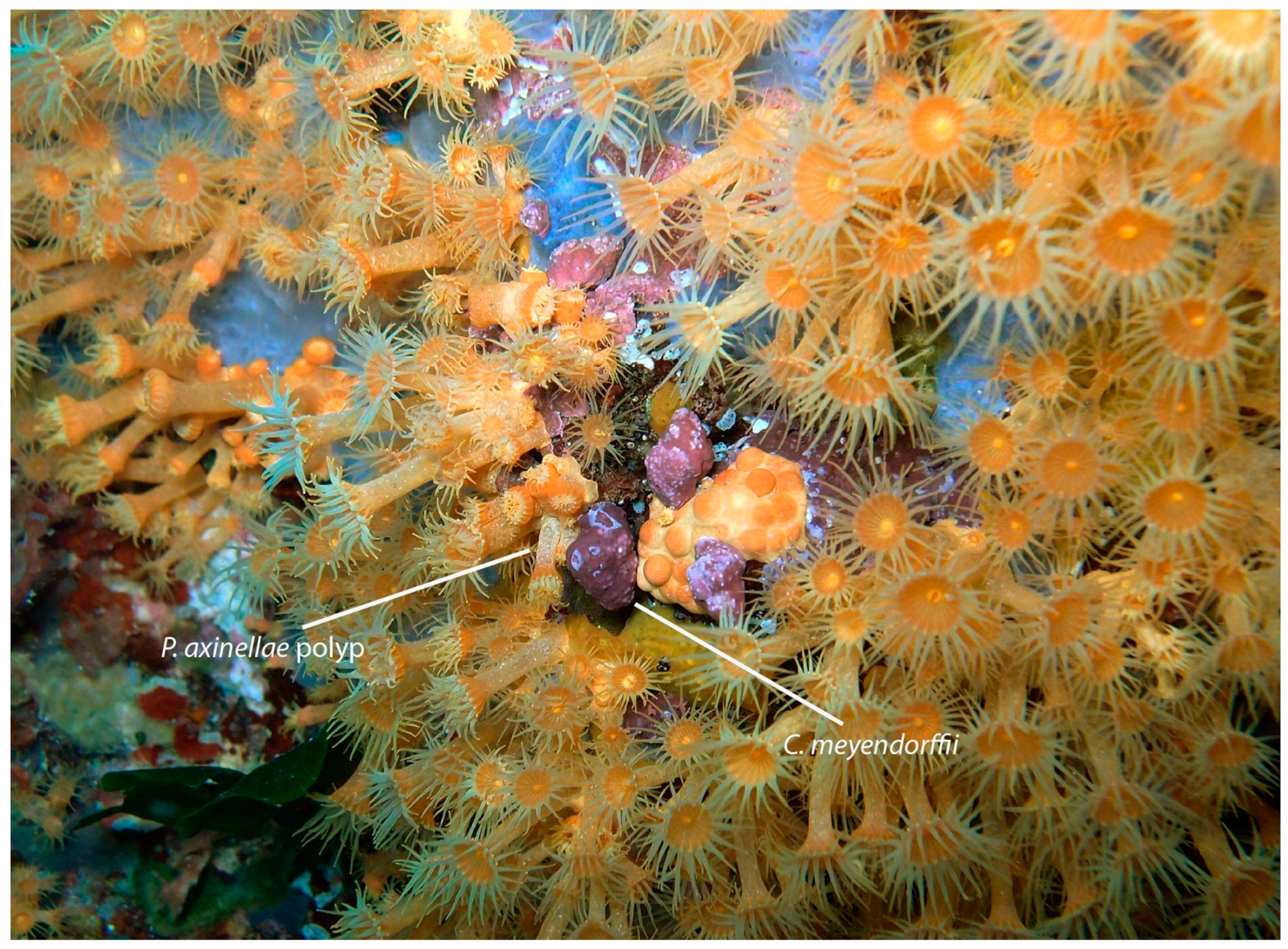
Figure 1. Coralliophila meyendorffii, feeding on a Parazoanthus axinellae colony collected for this study in Ponza Island, Italy; 9 metres depth. Photo: Paolo Mariottini.
Microbiolres 15 00157 g001
Figure 3. Sampling localities 1 = Ponza Is., Italy; 2 = Giannutri Is., Italy. Map created using QGIS version 3.28.11.
Microbiolres 15 00157 g003
Figure 4. Taxonomic composition based on relative abundances of ASVs at the family level from six stomachs of Coralliophila meyendorffii (BAU-3165.1S, BAU-3165.2S, BAU-3165.5S, BAU-3165.7S, BAU-3165.8S, BAU-3165.10S), three polyps of Parazoanthus axinellae (BAU-3166.1, BAU-3166.2 and BAU-3166.3) and a stomach of Babelomurex cariniferus (BAU-3180.S). All ASVss with a relative count below 1% in each sample were removed.
Microbiolres 15 00157 g004
Figure 5. Taxonomic composition based on relative abundances of ASVs at the genus level from six stomachs of Coralliophila meyendorffii (BAU-3165.1S, BAU-3165.2S, BAU-3165.5S, BAU-3165.7S, BAU-3165.8S, BAU-3165.10S), three polyps of Parazoanthus axinellae (BAU-3166.1, BAU-3166.2 and BAU-3166.3) and a stomach of Babelomurex cariniferus (BAU-3180.S). All ASVs with a relative count below 1% in each sample were removed.
Microbiolres 15 00157 g005
Figure 6. Frequency heatmap depicting the differences in the relative abundance of the 34 common bacterial genera among samples. The colour intensity in each cell of the matrix reflects the frequency of the feature in the corresponding sample, scaled logarithmically to visualise a wide range of abundance values. Black = not present.
Microbiolres 15 00157 g006
Figure 7. Beta diversity of the microbiota found in the polyps of Parazoanthus (orange), in the stomachs of Coralliophila (blue) and in the stomach of Babelomurex (green). Bray-Curtis distances were calculated based on the genus composition and relative abundance and then visualised through Principal Coordinates Analysis (PCoA). The percentages of variance for the first three principal components (PC1, PC2, PC3) are shown.
Microbiolres 15 00157 g007
Table 1. Comparisons of beta-diversity measures between selected groups and family and genus taxonomic levels using the PERMANOVA and ANOSIM methods.
FAMILYGENUS
Testp-ValueTestp-Value
PERMANOVAPSEUDO-F =
1.527733
0.038PSEUDO-F =
1.520639
0.039
ANOSIMR = 0.0144030.416R = 0.0246910.403
Disclaimer/Publisher’s Note: The statements, opinions and data contained in all publications are solely those of the individual author(s) and contributor(s) and not of MDPI and/or the editor(s). MDPI and/or the editor(s) disclaim responsibility for any injury to people or property resulting from any ideas, methods, instructions or products referred to in the content.