Article

A Comprehensive Methodology for Identifying Pseudomonas aeruginosa Strains Exhibiting Biofilm and Virulence Factor Traits and Assessment of Biofilm Resistance Against Commercial Disinfectant

1
Laboratory of Molecular and Cellular Screening Processes, Centre of Biotechnology of Sfax, Sfax 3018, Tunisia
2
Faculty of Sciences of Gafsa, University of Gafsa, Gafsa 2112, Tunisia
3
Department of Nephrology, Hedi Chaker University Hospital, Sfax 3029, Tunisia
4
Research Laboratory of Renal Pathology LR19ES11, Faculty of Medicine, University of Sfax, Sfax 3029, Tunisia
5
Regional Hygiene Laboratory, Hedi Chaker University Hospital, Sfax 3029, Tunisia
6
Faculty of Pharmacy, University of Monastir, Monastir 5000, Tunisia
7
Unity of Analysis and Processes Applied to the Environment UR17ES32, Higher Institute of Applied Sciences and Technology Mahdia, Mahdia 5100, Tunisia
8
Research Laboratory of Chronic Pulmonary Pathologies: From Genome to Management, LR 19SP02, Abderrahman Mami U. Hospital, Ariana 2080, Tunisia
*
Author to whom correspondence should be addressed.
Microbiol. Res. 2025, 16(3), 62; https://doi.org/10.3390/microbiolres16030062
Submission received: 9 February 2025 / Revised: 4 March 2025 / Accepted: 6 March 2025 / Published: 9 March 2025

Abstract

:
In this study, biofilm formation and the production of key virulence factors were systematically evaluated across 33 strains of Pseudomonas aeruginosa isolated from diverse hospital environments in Tunisia. Among these, 13 strains demonstrated strong biofilm-forming capacities. Adding glucose (9%, w/v) to the culture medium generally enhanced biofilm development, indicating that glucose supplementation may promote biofilm formation in clinical isolates. The 13 selected biofilm-forming strains exhibited a consistent production of critical virulence factors, including pyocyanin, rhamnolipid, and LasA protease, known for its staphylolytic activity. However, profiles of extracellular polysaccharide (EPS) production and motility showed considerable heterogeneity among the strains, suggesting a strain-specific regulation of these traits. Based on a comprehensive analysis of biofilm formation and virulence expression, strain 2629 was chosen as a model organism due to its robust biofilm-producing ability and high virulence factor output. This strain was used in subsequent in vitro assays to evaluate the anti-biofilm potential of a commercial disinfectant containing peracetic acid and other active agents. Results indicated that a 3% (v/v) concentration of the disinfectant, applied for 5 min, was nearly sufficient to eradicate the biofilm formed by the model strain. These findings underscore the importance of selecting a representative biofilm-forming strain for accurate in vitro assessments of disinfectant activity.

1. Introduction

Pseudomonas aeruginosa (P. aeruginosa) is a paradigmatic model microorganism for elucidating the intricate mechanisms of microbial adaptability and persistence across various ecological niches. Its exceptional capacity to form robust biofilms, in conjunction with highly regulated and sophisticated quorum sensing networks, exemplifies its profound resilience [1]. This multifaceted adaptability poses significant challenges in both clinical and industrial contexts, where biofilm-associated resistance and virulence factors contribute to persistent infections and biofouling. Given these complexities, the development of innovative antimicrobial strategies is urgently needed to effectively manage the impact of this versatile pathogen and facilitate eradication efforts [2,3]. Quorum sensing, a highly sophisticated cell-to-cell communication system in P. aeruginosa, orchestrates population-wide behaviors critical for biofilm maturation and virulence factor expression [4]. This regulatory mechanism enables the synchronized response to environmental signals, facilitating the coordinated expression of genes involved in biofilm development and antibiotic resistance [5]. The intricate relationship between quorum sensing and biofilm formation underscores the necessity of deciphering these signaling pathways to inform the development of novel therapeutic strategies. The inherent ability of P. aeruginosa to resist a wide range of antimicrobials and disinfectants poses a formidable challenge in clinical settings. Mechanisms such as efflux pumps, the enzymatic degradation of antibiotics, alterations in target sites, and biofilm-mediated tolerance collectively drive its multidrug-resistant phenotype, further complicating treatment efforts [6]. The rise in antimicrobial resistance presents an alarming global health threat, severely restricting therapeutic options and amplifying the healthcare burden. As an opportunistic pathogen, P. aeruginosa poses a particular threat to immunocompromised individuals, especially those with cystic fibrosis, chronic kidney disease, or those receiving immunosuppressant treatment. Its capacity to form biofilms on various surfaces in clinical settings exacerbates the challenge, as these biofilm-associated infections are notoriously difficult to eradicate due to their intrinsic resistance to antimicrobial agents [7].
Within biofilms, microbial communities are embedded in a self-produced extracellular polymeric substance (EPS) matrix, which shields bacterial cells from environmental stresses, including antibiotics and host immune responses. Biofilm development in P. aeruginosa is tightly regulated by a multitude of environmental cues and regulatory networks that modulate the expression of genes governing EPS synthesis, cell adhesion, and motility. This highly coordinated process facilitates both the structural integrity of the biofilm and the pathogen’s ability to persist in hostile environments, complicating clinical treatment efforts [8].
The virulence of P. aeruginosa is multifaceted, involving an array of factors such as exotoxins, flagella, pili, and lipopolysaccharides (LPSs), which collectively facilitate bacterial adhesion, colonization, and the invasion of host tissues. A key element of its pathogenic arsenal is the secretion system, which plays a critical role in delivering toxins and effector proteins directly into host cells, while proteases and other enzymes induce direct tissue damage. Furthermore, P. aeruginosa’s ability to communicate via quorum sensing and regulate biofilm formation significantly amplifies its virulence, enabling the bacterium to establish persistent infections [9].
Recent advances in molecular microbiology have unveiled the complex regulatory networks governing biofilm formation and virulence in P. aeruginosa, emphasizing the intricate interplay between bacterial signaling systems. Notably, the two-component system (TCS) FleS/FleR has been identified as a key positive regulator of virulence-related traits, including motility and biofilm development, both of which are crucial during acute and chronic infections. Studies have shown that FleS/FleR controls biofilm formation through a mechanism dependent on cyclic di-GMP and FleQ, providing valuable insights into potential therapeutic targets aimed at disrupting biofilm integrity and attenuating virulence [7].
Furthermore, the Las and Rhl systems in P aeruginosa regulate the expression of various virulence factors. The Las system includes LasR, a transcriptional activator, and LasI, which synthesizes the autoinducer N-(3-oxododecanoyl) homoserine lactone. Similarly, the Rhl system comprises RhlR, another transcriptional activator, and RhlI, which directs the production of N-butyryl homoserine lactone (C4-HSL). Both systems play crucial roles in quorum sensing, coordinating the bacterial response and virulence factor expression based on cell population density [10]. In summary, the FleS/FleR system and QS are interconnected in regulating the pathogenic traits of P. aeruginosa, including biofilm formation and the expression of virulence factors [11]. Understanding this relationship is crucial for developing strategies to combat infections caused by this bacterium. Moreover, the emergence of antibiotic-resistant strains of P. aeruginosa due to the misuse and overuse of antibiotics has exacerbated the challenge of treating infections. This resistance, coupled with the bacterium’s biofilm-forming capability, underscores the urgent need for novel antimicrobial strategies. Research has been directed towards identifying natural antimicrobial agents that can be used in various applications, including food processing, to combat the spoilage and pathogenicity of P. aeruginosa. For that, the need for identifying a good Pseudomonas biofilm-forming and virulence factor producer strain is of great interest [12].
In the present work, biofilm production and several main virulence factor productions of 33 P. aeruginosa strains isolated from different hospital settings were evaluated. Moreover, the virulence factor production was also investigated, showing consistent results with the biofilm-forming strains identified; these exhibited high levels of pyocyanin, rhamnolipids, and LasA (staphylolytic activity). In the second part of the study, the anti-biofilm activity of a commercial disinfectant was evaluated, and P. aeruginosa strain 2629 was used as a biofilm model.

2. Materials and Methods

Pseudomonas strains were collected from different water samples collected from hospital departments (dialysate from hemodialysis unit at nephrology department, endoscopy unit of the gastrological department, dental chair of the stomatology department, reanimation, etc.) in various hospitals in Tunisia and they were isolated on selective cetrimide agar medium following the standard ISO 16266:2006 [13] on the Regional Laboratory of Hygiene in Sfax during the period from 23 June 2022 to 2 August 2022.

2.1. Evaluation of Biofilm Formation

Quantification Method Using Crystal Violet Microplate Assay

Biofilm formation was conducted in sterile 96-well flat-bottomed polystyrene plates with lids. Cultures were diluted in LB medium (for P. aeruginosa) until reaching a final OD of 0.1 at 600 nm, with a final volume of 200 μL in each well. Each inoculated plate was covered and incubated in the incubator under static aerobic conditions for 24 h or more at 37 °C. After incubation, the contents of each well were emptied into a waste container. Then, a double wash with 200 μL of sterile phosphate-buffered saline (1 × PBS; pH 7.2) using an appropriate micropipette was added to remove all planktonic (non-adherent) bacteria while preserving biofilm integrity. Following this wash, the attached bacteria were left to dry at 60 °C for 1 h to facilitate biofilm fixation. The formed biofilm layer was stained with 150 μL of 0.2% (v/v) crystal violet for 15 min at room temperature. After staining, the excess crystal violet was removed, and the remaining dye was rinsed thrice with 200 μL of distilled water. Finally, 200 μL of 33% glacial acetic acid was added to each well, and incubation at room temperature for 60 min was necessary to disrupt the biofilm and solubilize the crystal violet, which had already penetrated and fixed within the cells. Optical density measurement at 570 nm was conducted using a microplate reader [14].
The classification of strains based on their biofilm-forming ability was determined after calculating the cut-off OD of the negative control strain Escherichia coli (ODc = (average of the OD 570 nm values of the negative control) + (3 × standard deviation of the negative control)) following the protocol described by [15] Pseudomonas strains can thus be classified using the following formulas: non-biofilm producer: OD ≤ ODc 0; weak biofilm producer: ODc < OD ≤ 2 × ODc +; moderate biofilm producer: 2 × ODc < OD ≤ 4 × ODc ++; and strong biofilm producer: 4 × ODc < OD +++. The effect of glucose on biofilm formation was also studied with the 13 selected strains using a stock solution of glucose at a concentration of 9% (w/v) [15].

2.2. Assessment of Virulence Factor Production in Selected Biofilm-Forming Strains of Pseudomonas aeruginosa

The P. aeruginosa strains isolated from hospital environments that demonstrated strong biofilm-forming abilities were used to investigate their capacity to produce virulence factors in relation to biofilm formation and pathogenicity.

2.2.1. Evaluation of Polysaccharide Production

The Congo Red Agar (CRA) method is an effective solid medium for detecting exopolysaccharide-producing (slime-producing) bacterial strains, which appear as blackish colonies. This medium is prepared in two steps. First, a Congo Red solution is autoclaved separately to achieve a concentration of 0.8 g/L in the culture medium, composed of 37 g/L Brain Heart Infusion (BHI), 50 g/L sucrose, and 10 g/L agar in 1 liter of distilled water. The pH is adjusted to 7 before autoclaving for 20 min at 121 °C [16]. The resulting solid medium is streaked with a suspension of the bacterial strain using a platinum loop. After overnight incubation at 37 °C, the Petri dishes are assessed. A positive result was indicated by black streaks or dry crystalline colonies, while pink or red colonies signify a negative result (non-slime-producing strain) [17].

2.2.2. Bacterial Motility

The evaluation of the bacterial motility of the isolated strains was carried out by studying three tests, including “Swimming, Swarming, and Twitching”. These tests are performed in Petri dishes containing LB culture medium with 0.3%, 0.4%, and 0.15% agar, respectively. For the “Swimming and Swarming” tests, 3 μL of bacterial culture is deposited with the tip of a sterile cone on the surface of the agar. For the “Twitching” test, 3 μL of bacterial culture is injected at the bottom center of the agar with the tip of the cone. The Petri dishes are incubated for 48 h at 37 °C in an incubator, and then the diameters of the migration zones of each bacterium are measured [16].

2.2.3. Evaluation of Pyocyanin Production

Pyocyanin (PYO) is a blue-green phenazine produced by the opportunistic pathogen P. aeruginosa [16]. Initially, precultures of the 13 most biofilm-forming Pseudomonas strains isolated from hospital water samples were prepared. After 48 h of incubation, 2 mL of each cell culture was taken and centrifuged for 10 min at ~6700× g. The supernatant was used for further investigations. An equal volume of chloroform was added to each tube, and after vigorous mixing, the mixture was allowed to separate. The organic fraction was collected, and 1 mL of 0.2 N HCl was added. The resulting pink solution was centrifuged, and the amount of pyocyanin was estimated by measuring the absorbance of the supernatant at 520 nm [16].

2.2.4. Extraction and Purification of Pyocyanin

To quantify pyocyanin production, the pigment was purified from Pseudomonas strain 2955 to establish the correlation between absorbance at 520 nm and concentration. The bacterial culture was spread on LB agar plates and incubated at 37 °C for 48 h, and the agar was cut into squares and placed in a flask with chloroform. After agitation and filtration, 0.2 N HCl was added, transferring the red pigment to the aqueous phase [18]. NaOH was added dropwise until the solution turned blue, and then chloroform extracted the pigment [19]. Petroleum ether was added to precipitate blue pyocyanin crystals, which were collected, dried, and weighed. This allowed for plotting the absorbance curve at 520 nm against pyocyanin concentration (y = 0.021x).

2.2.5. Evaluation of LasA Activity (Staphylolytic)

The preparation of cell cultures of Staphylococcus aureus was performed using 50 mL of liquid LB medium. The thirteen Pseudomonas strains isolated from a hospital environment were cultured on LB medium under uniform conditions, with identical inoculum sizes and incubation parameters (37 °C for 24 h). The Staphylococcus aureus culture (≈109 CFU/mL) was boiled for 10 min and then centrifuged for 10 min at 6700× g to recover the pellet. The resulting cell pellet was diluted in a 0.02 M Tris-HCl solution (pH 8.5) to achieve an optical density (OD) of 1.2 at 600 nm. To this, 100 µL of each Pseudomonas cell culture obtained from a 24h culture of P. aeruginosa after centrifugation was added to 900 µL of the prepared Staphylococcus solution. The cell density at 600 nm was determined every 15 min over a 1 h incubation period [16].

2.2.6. Extraction and Quantification of Rhamnolipids

The first step involved preparing precultures of selected Pseudomonas strains in the LB medium. After 24 h of incubation, 2 mL of each preculture was taken, followed by centrifugation to recover the supernatant for later use. The quantification of rhamnolipid production was carried out using the oil displacement method. For each supernatant from the tested strain, the following steps were performed: first, 30 mL of water was placed in a Petri dish, and 20 µL of oil was added to the water surface. Subsequently, 20 µL of the culture supernatant from each Pseudomonas strain was deposited, and the diameter of the oil displacement observed was measured [20].

2.2.7. P. aeruginosa 2629 Biofilm Eradication by Commercial Disinfectant Based on Peracetic Acid

The biofilm-forming and virulence factor producer P. aeruginosa was selected for this study. Conventional disinfectant treatment, composed of 4.5% peracetic acid, 6.7% acetic acid, and 25.5% hydrogen peroxide, was applied to assess biofilm eradication. Biofilm formation was carried out in 90 mm Petri dishes using an LB medium enriched with glucose. After 24 h of incubation, the medium was removed, and the residual biofilm was rinsed with PBS. The biofilms were then treated with either 3% or 5% of the disinfectant solution for 5 or 20 min, after which the reaction was stopped using a lecithin-based neutralizer. The residual biofilm was quantified using the crystal violet staining protocol, and the percentage of eradication was calculated as previously described.

2.3. Statistical Analysis

The results were statistically analyzed by analysis of variance tests (one-way ANOVA: Newman–Keuls multiple comparison test) using Prism versus 5 (Graph-Pad Software, Inc., La Jolla, CA, USA). For each condition, means, with different asterisks, are considered as significantly different.
To assess the virulence factors of P. aeruginosa strains, we employed the pheatmap R package (version 1.0.12) to generate a heatmap visualizing the relative abundance of these factors. To identify potential groupings of strains based on their virulence factor profiles, we applied Ward’s method with Euclidean distance as the clustering method. This approach helps to uncover clusters of strains that share similar virulence factor patterns.

3. Results And Discussion

3.1. Pseudomonas Strains and Their Origins

Pseudomonas bacteria are Gram-negative, including P. aeruginosa, also known as the pyocyanic Bacillus. This bacterial species is now on the list of bacteria for which antibiotic resistance has become a major public health issue. In this context, a collection of P. aeruginosa bacterial strains was gathered in collaboration with the regional laboratory of hygiene and other departments of the tertiary healthcare Hédi Chaker Hospital, as well as additional hospitals in the Sfax region and hospitals located in the south of Tunisia. The table below provides a summary of the code numbers of the strains and their respective isolation locations. It is well documented that P. aeruginosa has a “ubiquitous” reservoir, often found in wastewater rich in organic matter [21].
Hospitals provide an ideal ecological niche for the development of P. aeruginosa, with common reservoirs including siphons, showers, toilets, endoscopes, nebulizers, humidifiers, and respirators. Patient contamination can occur directly from these environmental sources or indirectly through medical equipment or the hands of healthcare personnel. In rarer cases, P. aeruginosa can colonize the human digestive flora, especially when disrupted by antibiotic use (dysbiosis). The bacterium is often isolated sporadically but can also be responsible for significant outbreaks in healthcare settings, with some strains exhibiting a heightened ability to spread and persist compared to others.
The so-called “high-risk” clones are found in many healthcare facilities across different countries and sometimes even on different continents. Therefore, the study of Pseudomonas strains is of great importance in terms of assessing their ability to form biofilms and produce virulence factors. In this context, the Pseudomonas collection, including the code of strains, origin, and date of isolation, was included in Table 1.

3.2. Evaluation of Biofilm Formation in Pseudomonas Strains

Biofilm formation is evaluated through the crystal violet microplate quantification method, which detects the presence of biofilms by fixing the dye to the peptidoglycans in the cell membrane and the exopolysaccharides within the extracellular matrix. This technique involves measuring the optical density at 570 nm to quantify biofilm production. In this study, we are assessing the biofilm-forming capabilities of 33 Pseudomonas strains that were isolated in collaboration with the hygiene department from various hospital environments. Escherichia coli DH5α, a weak-biofilm-forming strain, serves as a negative control to facilitate the classification of the Pseudomonas strains based on their biofilm production after a 24 h incubation at 37 °C, as illustrated in Table 1.
The results indicate that Pseudomonas species are generally recognized as biofilm-forming bacteria, as noted by [22]. Specifically, P. aeruginosa has emerged as a key model organism for elucidating the mechanisms of biofilm formation. Concurrently, other Pseudomonas species leverage biofilm development as a strategy for plant colonization and environmental persistence. These bacteria can synthesize a diverse array of biofilm matrix components, including polysaccharides, nucleic acids, and proteins, which are crucial for both the initiation and maintenance of biofilms. Additionally, Pseudomonas cells produce accessory matrix components that significantly enhance biofilm formation and adaptability to varying environmental conditions, thereby contributing to their ecological success and pathogenicity [22].

3.3. Classification of Pseudomonas Strains Based on Biofilm Production

Comparing the OD values obtained after 24 h with the ODc allows for the classification of strains into different categories, including non-biofilm-producing bacteria (0), weak producers (+), moderate producers (++), and strong biofilm producers (+++), as indicated in the previous tables. Strong biofilm formers (e.g., 2582, 2629, 2653, 2659, 2676, 2677, 2687) are the most potent in biofilm production, indicating a higher potential for virulence and resilience against antimicrobial treatments. Moderate biofilm formers (e.g., 2560, 2712, 2778) may still demonstrate notable pathogenic potential but with less biofilm mass than the strong formers, while weak biofilm formers (e.g., 2643, 2956) exhibit limited biofilm production, suggesting reduced capacity for biofilm-associated infections. Additionally, several strains (e.g., 2501, 2654, 2888) showed no biofilm formation under the tested conditions, indicating a lack of biofilm-associated virulence. By selecting the 13 strains with “+++” ratings for further study, the focus is placed on those most likely to exhibit increased resistance to environmental stresses and antimicrobial agents, making them critical targets for investigating biofilm-associated pathogenicity and potential therapeutic interventions. These strains were selected to perform new biofilm quantification tests with crystal violet but this time to study the effects of glucose on biofilm production.
  • Selection of Highly Biofilm-Forming Strains
Thirteen of the most filmogenic strains were selected (2653, 2955, 2823, 2629, 2676, 2779, 2582, 2687, 2677, 2659, 2954, 2889, and 2947). Strains that have shown to be strong biofilm producers (+++) were eliminated (not retained), since their results showed quite high standard deviations due to the lack of reproducibility and stability in their ability to produce biofilms (five strains).

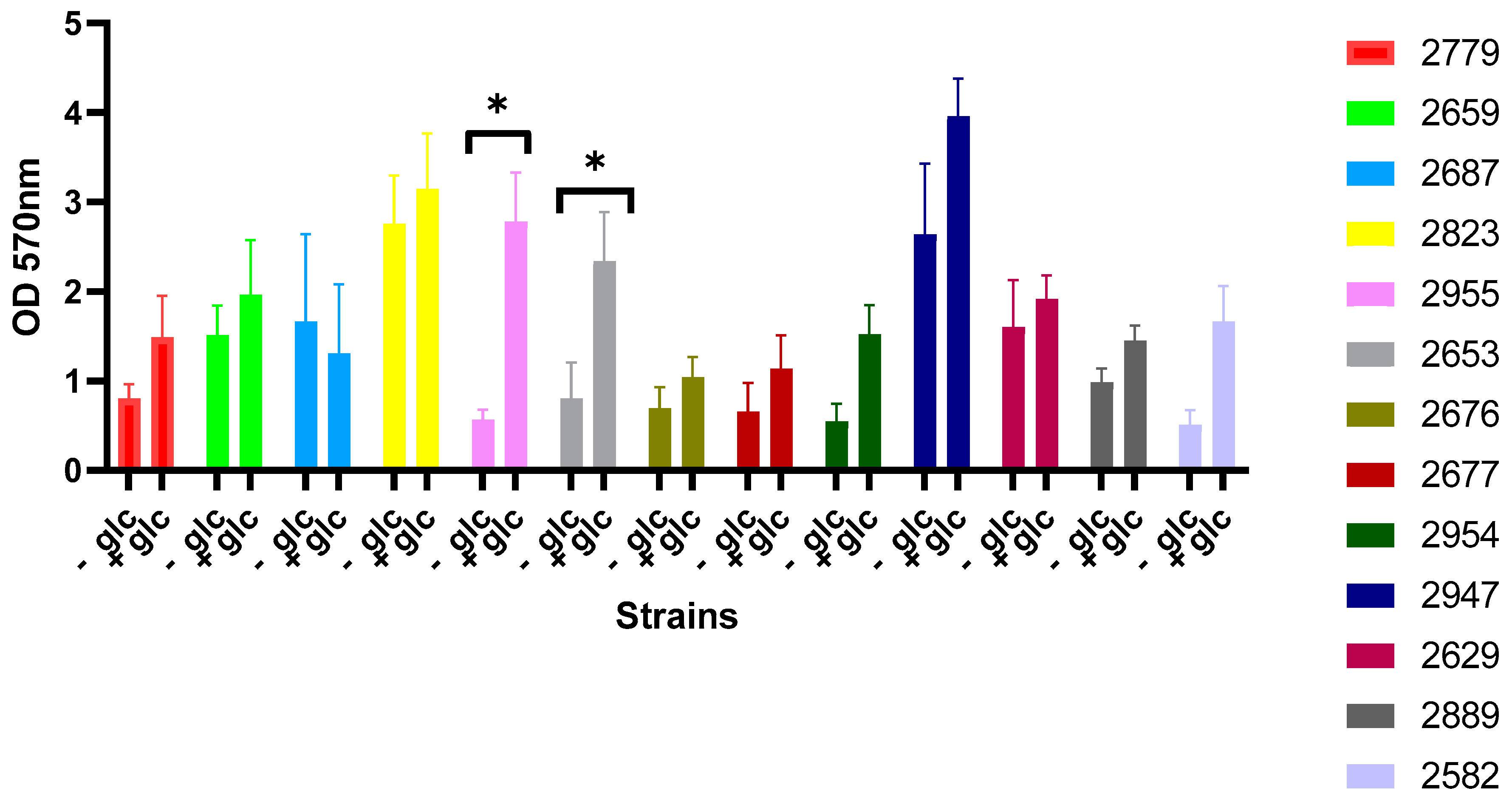
3.4. Effect of Glucose Supplementation on Biofilm Formation by Selected 13 Strains

It has been reported that glucose may enhance biofilm formation in certain bacterial species [20]. To investigate this, we tested the effect of glucose addition on biofilm formation in the 13 selected bacterial strains. The results are summarized in Figure 1.
According to the figure, it can be observed that in the presence of glucose, the majority of strains that exhibited OD values at 570 nm were higher than those found under conditions without glucose addition. In this case, glucose may enhance biofilm formation by promoting cell division or facilitating the production of polysaccharides, which are key components of the extracellular matrix [23], demonstrating that glucose-6-phosphate acts as an extracellular signal, activating the motile-to-sessile switch in P. aeruginosa, thereby enabling it to adapt to nutrient-poor environments and enhance biofilm formation. Similarly, ref. [24] found that increasing glucose concentrations accelerate biofilm maturation and thickening in P. aeruginosa. Furthermore, ref. [25] reported that glucose effectively promotes biofilm formation in P. aeruginosa by upregulating the expression of the pslA gene, which is associated with extracellular polysaccharide production.

3.5. Evaluation of the Production of Virulence Factors by Selected Pseudomonas aeruginosa Strains

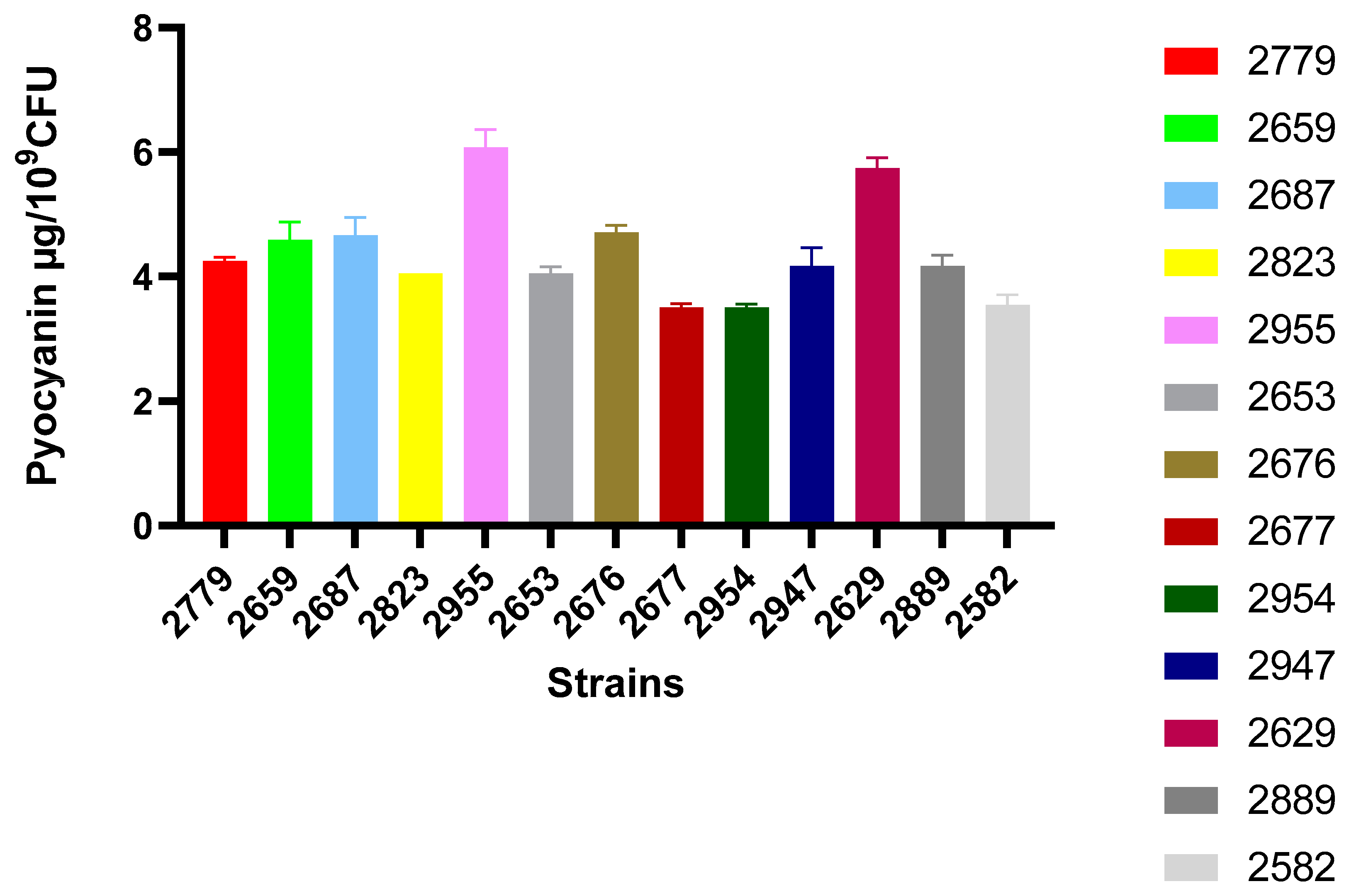
3.5.1. Pyocyanin Production

Pyocyanin, a blue-green pigment produced by P. aeruginosa, exhibits antibacterial properties and holds promise for pharmaceutical applications. It also has potential uses in biosensors and bacterial signaling processes [26]. Among its array of virulence factors, pyocyanin plays a pivotal role in enabling P. aeruginosa to exert its full pathogenic potential. Macrophages, beyond serving as primary sentinels of the innate immune system, are integral to maintaining homeostasis, facilitating tissue remodeling, and orchestrating the crosstalk between innate and adaptive immunity [27]. In significant studies utilizing vitro model systems, the impact of pyocyanin has been observed across respiratory, urological, neurological, cardiovascular, and hepatic systems. These investigations highlight pyocyanin’s influence on host pro-inflammatory responses and immune-modulating properties. The exposure of cellular systems to this compound induces increased oxidative stress, ultimately triggering progressive apoptosis [26]. Pyocyanin can also penetrate the blood–brain barrier, potentially impairing cognitive functions by elevating the production of reactive oxygen species (ROS) and influencing behavioral patterns [28]. In this study, we analyzed the production of pyocyanin, a key virulence factor of Pseudomonas aeruginosa, and its role in the pathogenicity of this bacterial species. However, one limitation of our work is its primary focus on pyocyanin without addressing other crucial metabolites that significantly contribute to P. aeruginosa virulence. Notably, pyoverdine and pseudopaline play essential roles in iron and zinc acquisition, respectively, and are integral to the overall pathogenic potential of this bacterium. Pyoverdine (PVDi) functions as both a siderophore, facilitating iron acquisition, and a signaling molecule that regulates the expression of virulence factors. As a core component of P. aeruginosa pathogenicity, pyoverdine binds ferric iron (Fe3+) in the extracellular environment and is subsequently transported into the bacterial periplasm via the outer membrane receptors FpvAi and FpvB. The release of iron from PVDi-Fe3+ within the periplasm involves multiple proteins encoded by the fpvGHJKCDEF operon and an iron-reduction mechanism. The intricate interactions between these proteins, explored through systematic bacterial two-hybrid screening, underscore the complexity of iron homeostasis in P. aeruginosa [29]. Similarly, pseudopaline biosynthesis follows a two-step enzymatic process. The first step, catalyzed by PaCntL, involves the nucleophilic attack of an α-aminobutyric acid moiety from S-adenosylmethionine (SAM) onto L-histidine, yielding the intermediate yNA. This intermediate then undergoes an NADH-dependent reductive condensation with α-ketoglutarate (αKG), mediated by PaCntM, to form pseudopaline. Pseudopaline differs from its staphylococcal counterpart, staphylopine, in its stereochemistry, incorporating L-histidine instead of D-histidine, as well as in its structural composition, where it contains an αKG moiety rather than pyruvate. These species-specific metallophores reflect an evolutionary adaptation to optimize metal acquisition within competitive microbial environments. Once secreted, metallophores become public goods within bacterial communities, with privileged access providing a selective advantage. A comparative analysis of pseudopaline and staphylopine biosynthesis highlights key differences in their metabolic pathways. Unlike Staphylococcus aureus, which utilizes an MFS family protein (SaCntE) for staphylopine export, P. aeruginosa employs a DMT family transporter (PaCntI) featuring two predicted EamA domains for pseudopaline export. While the import mechanisms for pseudopaline–metal complexes remain unidentified, the possibility of metallophore recycling in P. aeruginosa—as demonstrated for pyoverdine—warrants further investigation. Given the critical role of metallophores in bacterial growth and virulence, these molecules represent promising targets for antimicrobial development. Elucidating their biosynthetic pathways and transport mechanisms may offer novel therapeutic strategies against P. aeruginosa infections. Future studies should integrate the analysis of multiple virulence factors, including pyocyanin, pyoverdine, and pseudopaline, to provide a more comprehensive understanding of P. aeruginosa pathogenicity [30].
Figure 2 below illustrates the assessment of pyocyanin production among the 13 selected biofilm-forming strains. The results indicate that the highest producers of pyocyanin are strains 2955, 2629, and 2676, as determined by quantifying pyocyanin concentration in mg/L.

3.5.2. Production of Exopolysaccharides (EPSs)

Exopolysaccharides are components of the extracellular matrix (ECM). Indeed, these EPSs serve as a barrier and a means of protection for the biofilm against external environmental stresses. The Congo Red Agar method allows for the evaluation of slime production. On this medium, the tested bacteria exhibit three different phenotypes, including pink colonies for non-slime-producing bacteria (-) and gray and black colonies for moderately and strongly slime-producing bacteria (+) and (++), respectively. All strains exhibit black colonies with a dry consistency on Congo Red Agar, indicative of exopolysaccharide production, which reacts with Congo Red. This allows us to conclude that strains 2955, 2677, 2582, 2676, 2659, and 2629 are the most prolific slime producers. This observation aligns with the findings in [31], which reported that Pseudomonas species, particularly P. aeruginosa, are distinguished by the production of heteropolysaccharides, such as alginates, biosurfactants, and rhamnolipids. These compounds play essential roles in biofilm formation, structural integrity, and surface interactions, thereby contributing to both the pathogenicity and environmental resilience of the bacteria. The moderately slime-producing strains of P. aeruginosa are 2947, 2749, 2823, 2687, and 2889. When the coloration of the colonies and streaks remains reddish (or pinkish), this means that the production of slime is very low or absent [32].

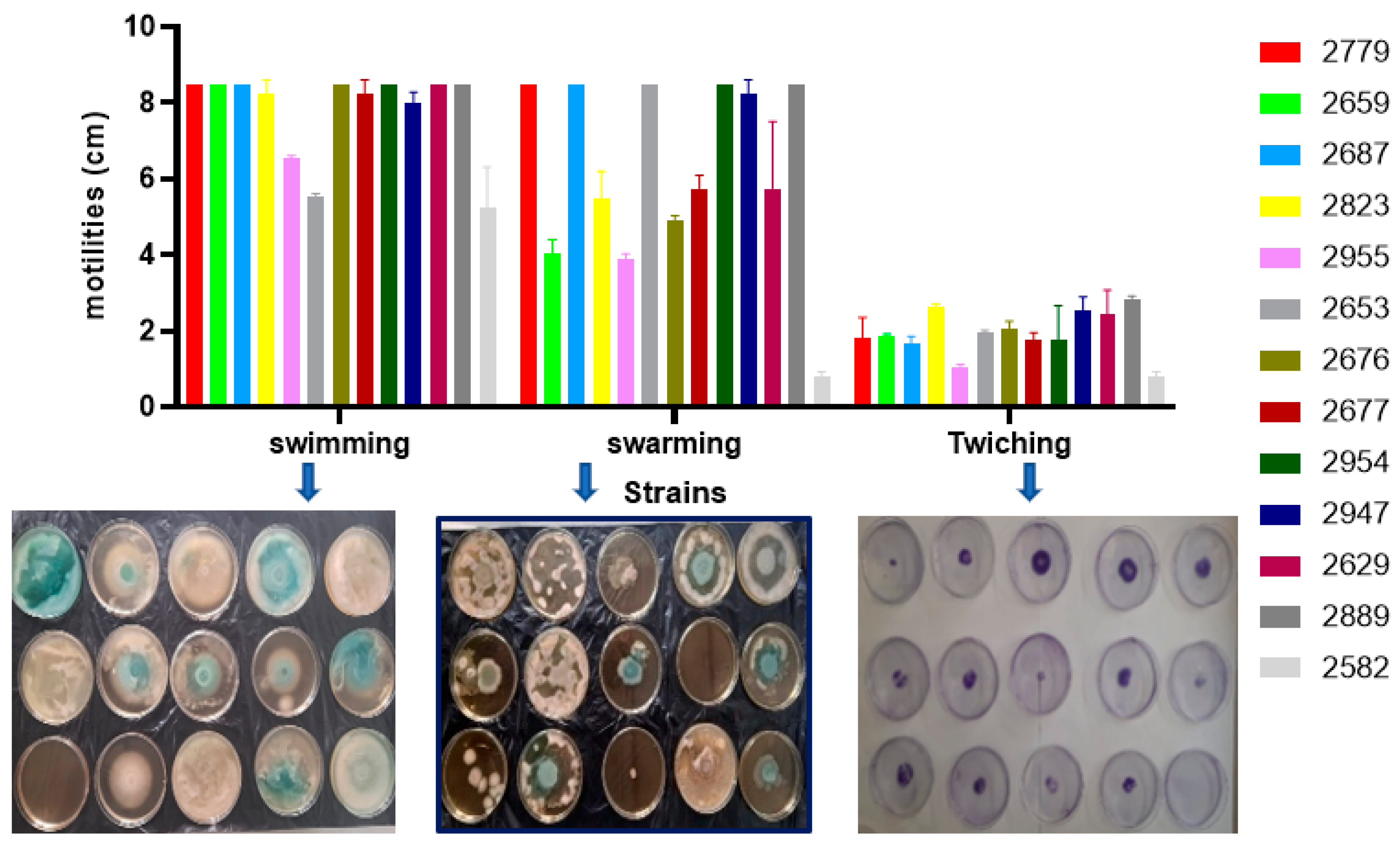
3.5.3. Motility Test

We illustrate the bacterial motility of the isolated strains by measuring the migration distances of the bacteria in Petri dishes containing agar. Photos of the results of motility by “Swimming”, “Swarming”, and “Twitching” are presented in Figure 3.
P. aeruginosa is a highly motile bacterium that employs various motility mechanisms. The most prominent and extensively studied mechanism is twitching motility, which relies on the extension and retraction of type IV pili on the bacterial cell surface. This type of motility enables P. aeruginosa to move or “crawl” along solid surfaces, including host cell surfaces. Additionally, swimming and swarming motilities are other forms of movement in P. aeruginosa, although they are less frequently observed compared to twitching motility. Each of these motility mechanisms plays a critical role in the bacterium’s ability to colonize, spread, and establish infections in various environments [33].
However, in our study, according to the histograms, it was observed that the majority of the tested bacteria migrated more by “Swimming” than by “Swarming” and “Twitching” For the “Swimming” test, most strains showed very significant migration distances, covering the entire Petri dish. Some studies have shown that swimming motility involves the movement of P. aeruginosa in a liquid environment, typically powered by flagella, which are long and thin appendages attached to the bacterial cell. Swarming motility refers to the coordinated movement of cells propelled by flagella across thin liquid films on surfaces, often accompanied by cellular differentiation into a hyper-flagellated, elongated phenotype. Additionally, twitching is another mechanism involved in surface movement. In twitching, type IV pili extend and attach to a solid surface then retract, pulling the cell forward to facilitate movement. This motility mechanism is vital for bacterial colonization, biofilm formation, and virulence [34].

3.5.4. Evaluation of LasA Activity (Staphylolytic Activity)

Proteases, also known as proteinases or peptidases, are a class of hydrolase enzymes that catalyze the cleavage of peptide bonds within proteins [35]. Indeed, the LasA gene encodes a protease with a staphylolytic activity that contributes to bacterial virulence. Although not an elastase itself, LasA enhances the elastolytic activity of elastase enzymes, such as LasB, which are responsible for lung tissue and skin damage [36]. It cleaves the peptide bonds of the peptidoglycan of Staphylococcus aureus, thus allowing P. aeruginosa to predominate during pulmonary infections in cystic fibrosis patients [37]. The Figure 4 below shows the OD 600 nm values recorded after 1 h of incubation of the Staphylococcus aureus suspension in the presence of the culture supernatant of the selected Pseudomonas strains to evaluate their staphylolytic LasA activity. All strains showed fairly similar activities, since the recorded OD values after treatment were between 0.6 and 0.8. MRSA was used as a negative control, because its modified peptidoglycan structure reduces susceptibility to LasA-mediated lysis, ensuring that any observed OD600 decrease is specifically due to LasA activity rather than general bacterial susceptibility. This allows for differentiation between true LasA-mediated staphylolytic activity and potential non-specific effects of the Pseudomonas culture supernatants on S. aureus cell viability.

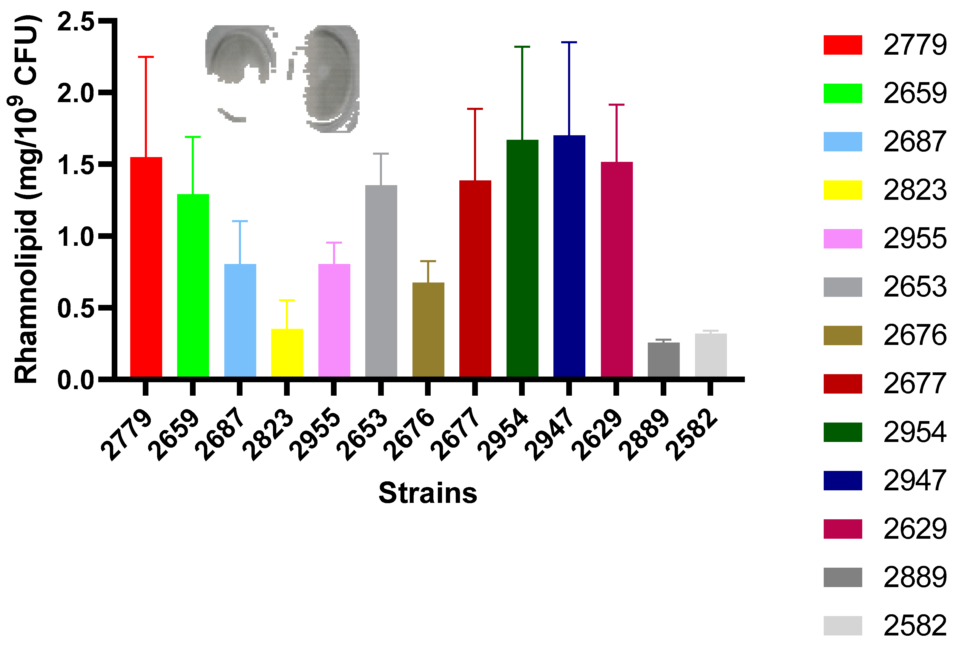
3.5.5. Quantification of Rhamnolipid Production

Rhamnolipids are biosurfactants in the form of surfactant molecules naturally produced by P. aeruginosa and certain species of Burkholderia [38].They are thermostable extracellular glycolipids that can emulsify membrane phosphates due to their detergent activity. They disrupt mucociliary transport and ciliary movements of the human respiratory epithelium and are considered virulence factors. The oil displacement test allows for the demonstration of the production of these biosurfactants in the culture supernatants of selected Pseudomonas strains [20]. The results in Figure 5 showed that 2954, 2947, 2629, and 2779 are the most common strain producers of rhamnolipids.

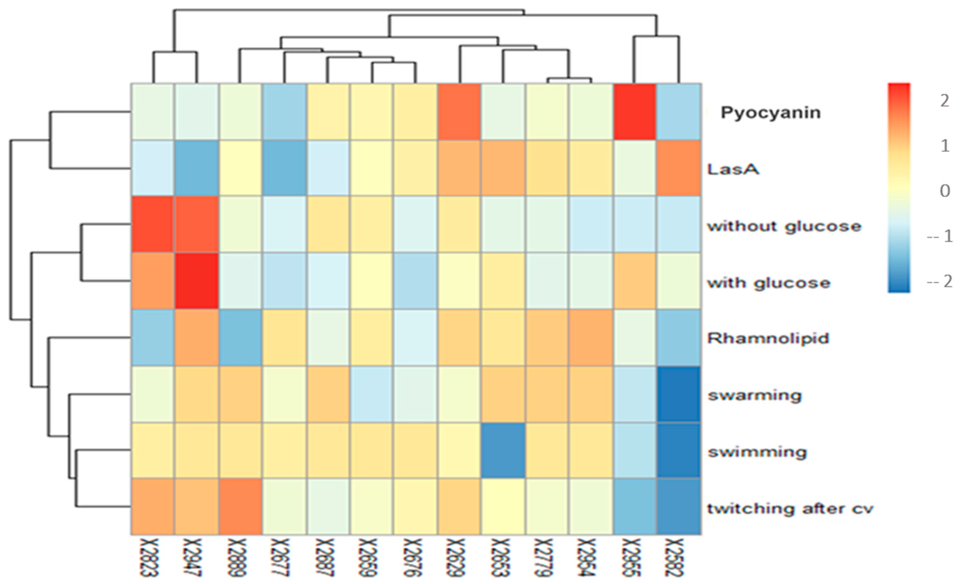
3.5.6. Heatmap: Combining Virulence Factor Production and Biofilm-Forming Capacity to Select a Model of Pseudomonas Strain

Heatmaps are a powerful tool for visualizing multi-dimensional data, displaying individual values in a two-dimensional matrix with color-coded representation. Hierarchical clustering allows rows and columns to be arranged by their similarity, with dendrograms added to the plot to aid interpretation. Additional annotations, such as colored tiles, can be applied to both rows and columns for enhanced clarity. Several R packages have been developed to facilitate the creation of heatmaps with various customization [39].
These data used to assess the virulence factors of Pseudomonas strains enabled us to create a detailed heatmap. This heatmap visually represents the differences in virulence characteristics among the strains, allowing us to identify patterns of gene expression, genetic diversity, and antimicrobial resistance. A detailed analysis of these data enables us to evaluate the virulence factor profiles of each strain, providing a foundation for predicting its pathogenicity. The heatmap shows variations between strains, often with a color gradient representing the intensity or presence/absence of specific features. The heatmap analysis reveals significant variations in virulence factors across Pseudomonas strains 2653, 2955, 2823, 2629, 2676, 2779, 2582, 2687, 2677, 2659, 2954, 2889, and 2947. Using a color scale where red represents high virulence and blue signifies low virulence, the heatmap displays distinctive profiles for each strain across the key factors of motility. Based on the heatmap analysis, we have selected strain 2629, isolated from a dental chair, for further investigation. Unlike other strains, 2629 does not exhibit any blue coloration, indicating that it consistently expresses moderate-to-high virulence across all measured factors. This lack of low-virulence markers suggests that the 2629 strain is resilient and potentially dangerous in clinical contexts. Strain 2629 stands out as the most virulent P. aeruginosa strain in the dataset, exhibiting moderate-to-elevated levels of multiple virulence factors (Figure 6). This strain demonstrates moderate-to-high motility, enabling it to efficiently spread and colonize surfaces, a crucial trait for establishing infections. Pyocyanin production, another key virulence factor, is the highest in strain 2629, contributing to oxidative stress and tissue damage in the host, further amplifying its pathogenicity. Additionally, the strain shows heightened las quorum sensing activity, essential for regulating the expression of other virulence factors, such as tissue-degrading enzymes, which increase its adaptability in infection scenarios. Rhamnolipid production is also substantial, supporting biofilm formation and surface motility, both of which are key to its survival and persistence in hostile environments. This strain’s ability to simultaneously express and regulate multiple virulence factors positions it as a highly adaptable and aggressive pathogen, making it a prime candidate for biofilm-targeted therapies or improved disinfection protocols in clinical settings. Therefore, we will focus on this strain in the remainder of our study.
The selection of motility, pyocyanin, rhamnolipid, and LasA protease for this study was based on their critical roles in P. aeruginosa virulence and their direct involvement in host–pathogen interactions. Pyocyanin contributes to oxidative stress and immune evasion, rhamnolipids facilitate biofilm formation and host cell toxicity, and the LasA protease degrades host proteins, particularly in polymicrobial infections. These factors are well characterized and have established and reproducible quantitative assays that allow for reliable comparative analysis across multiple strains.

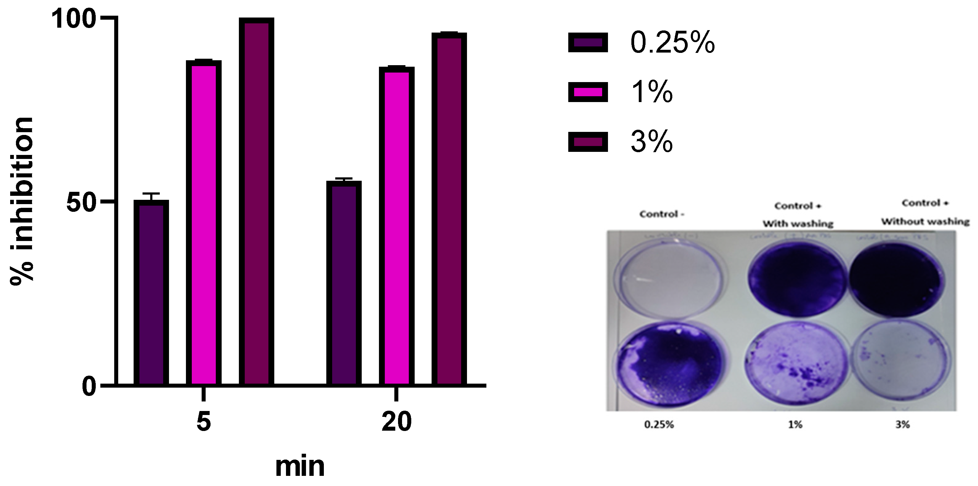
3.6. P. aeruginosa 2629 Biofilm Eradication by Commercial Disinfectant Based on Peracetic Acid

For the evaluation of the anti-biofilm properties of a commercial disinfectant, peracetic acid, P. aeruginosa strain 2629 was used as a model for biofilm formation. Biofilms are structured communities of bacteria encased in a protective matrix and pose significant challenges in clinical and industrial settings due to their resilience against conventional disinfectants. In our investigation, we demonstrated that a concentration of 3% peracetic acid was necessary to achieve effective biofilm removal (Figure 7). This concentration, combined with a contact time of just 5 min, demonstrated significant efficacy in disrupting the biofilm structure and reducing bacterial viability. These results suggest that peracetic acid can be a viable option for controlling P. aeruginosa biofilms, highlighting its potential application in infection control and sanitation protocols. Our findings align with those of Chino et al., reinforcing the effectiveness of peracetic acid in eradicating mature P. aeruginosa biofilms. They found that although mature P. aeruginosa biofilms are characterized by the production of matrix components that impede effective biofilm eradication, peracetic acid (PAA) proves to be an effective disinfectant. Their observations allowed for an analysis of PAA’s mechanism of action, revealing changes in biofilm morphology after 5 min and structural damage after 30 min of exposure. Notably, mature P. aeruginosa biofilms aged 96 h were eradicated at 3000 ppm (0.3%) PAA after just a 5 min exposure [40].
In this study, a proposed method was developed to select a model strain of P. aeruginosa for in vitro studies of anti-biofilm molecules, disinfectant formulations, and antibiotics. This model is intended to confirm the efficacy of these treatments for application at sites of contamination or infection. Additionally, attention was given to the exploration of novel alternatives to antibiotics, specifically neutralizers that can interfere with the quorum sensing regulatory system and act against the virulence and pathogenicity of P. aeruginosa strains [41]. This newly developed model will be utilized in future research to screen for anti-biofilm and anti-quorum sensing molecules effectively.

4. Conclusions

In this study, we demonstrated that P. aeruginosa strains isolated from hospital environments exhibit significant variability in biofilm formation and virulence factor production, with glucose supplementation enhancing biofilm development in the majority of strains. Notably, the strong biofilm producers consistently generated high levels of pyocyanin, rhamnolipids, and LasA, although EPS production and motility were less uniform. Our findings highlight the complex relationship between biofilm formation and virulence expression in P. aeruginosa. Furthermore, we assessed the anti-biofilm efficacy of peracetic acid, identifying that a 3% concentration with a 5 min contact time was required for effective biofilm eradication. These results underscore the importance of optimizing disinfectant use in clinical settings to mitigate biofilm-associated infections and provide a foundation for future investigations into the mechanisms driving biofilm resilience and disinfectant resistance in P. aeruginosa.

Author Contributions

Conceptualization, M.G. and S.M. (Sami Mnif); methodology, S.S., F.M.-A. and S.M. (Sami Mnif); formal analysis, M.B.H. and M.A.; investigation, M.G., S.S., S.M. (Salma Maalej) and S.T.; data curation, M.G. and S.S.; writing—original draft preparation, M.G.; writing—review and editing, S.S., S.A., M.B.H., K.K. and S.M. (Sami Mnif); supervision, S.M. (Sami Mnif). All authors have read and agreed to the published version of the manuscript.

Funding

This work was financially supported by the ministry of higher education and scientific research under a federated research project (PRF2023-D5P1), Tunisia, and from the Hedi Chaker University Hospital Research Committee under a research project program, Sfax Tunisia.

Institutional Review Board Statement

Not applicable.

Informed Consent Statement

Not applicable.

Data Availability Statement

Data may be shared and should be requested from the corresponding author.

Conflicts of Interest

The authors declare no conflicts of interest.

References

  1. Tuon, F.F.; Dantas, L.R.; Sus, P.H.; Ribeiro, V.S.T. Pathogenesis of the Pseudomonas aeruginosa Biofilm: A Review. Pathogens 2022, 11, 300. [Google Scholar] [CrossRef]
  2. Reynolds, D.; Kollef, M. The Epidemiology and Pathogenesis and Treatment of Pseudomonas aeruginosa Infections: An Update. Drugs 2021, 81, 2117–2131. [Google Scholar] [CrossRef] [PubMed]
  3. Sharma, S.; Mohler, J.; Mahajan, S.D.; Schwartz, S.A.; Bruggemann, L.; Aalinkeel, R. Microbial Biofilm: A Review on Formation, Infection, Antibiotic Resistance, Control Measures, and Innovative Treatment. Microorganisms 2023, 11, 1614. [Google Scholar] [CrossRef] [PubMed]
  4. Ghssein, G.; Ezzeddine, Z. A Review of Pseudomonas aeruginosa Metallophores: Pyoverdine, Pyochelin and Pseudopaline. Biology 2022, 11, 1711. [Google Scholar] [CrossRef]
  5. Ambreetha, S.; Singh, V. Genetic and Environmental Determinants of Surface Adaptations in Pseudomonas aeruginosa: This Article Is Part of the Environmental Sensing and Cell-Cell Communication Collection. Microbiology 2023, 169, 001335. [Google Scholar] [CrossRef]
  6. Zhang, S.; Wang, J.; Ahn, J. Advances in the Discovery of Efflux Pump Inhibitors as Novel Potentiators to Control Antimicrobial-Resistant Pathogens. Antibiotics 2023, 12, 1417. [Google Scholar] [CrossRef] [PubMed]
  7. Zhou, T.; Huang, J.; Liu, Z.; Xu, Z.; Zhang, L. Molecular Mechanisms Underlying the Regulation of Biofilm Formation and Swimming Motility by FleS/FleR in Pseudomonas aeruginosa. Front. Microbiol. 2021, 12, 707711. [Google Scholar] [CrossRef]
  8. Li, X.; Gu, N.; Huang, T.Y.; Zhong, F.; Peng, G. Pseudomonas aeruginosa: A Typical Biofilm Forming Pathogen and an Emerging but Underestimated Pathogen in Food Processing. Front. Microbiol. 2023, 13, 1114199. [Google Scholar] [CrossRef]
  9. Liao, C.; Huang, X.; Wang, Q.; Yao, D.; Lu, W. Virulence Factors of Pseudomonas aeruginosa and Antivirulence Strategies to Combat Its Drug Resistance. Front. Cell. Infect. Microbiol. 2022, 12, 926758. [Google Scholar] [CrossRef] [PubMed]
  10. Uzunbayir-Akel, N.; Tekintas, Y.; Yilmaz, F.F.; Ozturk, I.; Okeer, M.; Aydemir, S.S.; Cilli, F.F.; Hosgor-Limoncu, M. Effects of Disinfectants and Ciprofloxacin on Quorum Sensing Genes and Biofilm of Clinical Pseudomonas aeruginosa Isolates. J. Infect. Public Health 2020, 13, 1932–1938. [Google Scholar] [CrossRef]
  11. Zhou, T.; Huang, J.; Feng, Q.; Liu, Z.; Lin, Q.; Xu, Z.; Zhang, L.-H. A Two-Component System FleS/FleR Regulates Multiple Virulence-Related Traits in Pseudomonas aeruginosa. BioRxiv 2021. Available online: https://www.biorxiv.org/content/10.1101/2021.04.22.441042v1 (accessed on 23 April 2023).
  12. Botelho, J.; Grosso, F.; Peixe, L. Antibiotic Resistance in Pseudomonas aeruginosa—Mechanisms, Epidemiology and Evolution. Drug Resist. Updates 2019, 44, 100640. [Google Scholar] [CrossRef] [PubMed]
  13. ISO 16266:2006 (En); Water Quality—Detection and Enumeration of Pseudomonas aeruginosa—Method by Membrane Filtration. ISO: Geneva, Switzerland, 2006.
  14. Stepanović, S.; Vuković, D.; Hola, V.; Bonaventura, G.D.; Djukić, S.; Ćirković, I.; Ruzicka, F. Quantification of Biofilm in Microtiter Plates: Overview of Testing Conditions and Practical Recommendations for Assessment of Biofilm Production by Staphylococci. APMIS 2007, 115, 891–899. [Google Scholar] [CrossRef]
  15. Stepanovic, S.; Vukovic, D.; Dakic, I.; Savic, B.; Svabic-Vlahovic, M. A Modified Microtiter-Plate Test for Quantification of Staphylococcal Biofilm Formation. J. Microbiol. Methods 2000, 40, 175–179. [Google Scholar] [CrossRef]
  16. Rajkumari, J.; Borkotoky, S.; Murali, A.; Suchiang, K.; Mohanty, S.K.; Busi, S. Attenuation of Quorum Sensing Controlled Virulence Factors and Biofilm Formation in Pseudomonas aeruginosa by Pentacyclic Triterpenes, Betulin and Betulinic Acid. Microb. Pathog. 2018, 118, 48–60. [Google Scholar] [CrossRef] [PubMed]
  17. Freeman, D.J.; Falkiner, F.R.; Keane, C.T. New Method for Detecting Slime Production by Coagulase Negative Staphylococci. J. Clin. Pathol. 1989, 42, 872–874. [Google Scholar] [CrossRef]
  18. Barakat, R. Etude Des Propriétés Biologiques et Antimicrobiennes de la Pyocyanine, Pigment Redox-Actif Produit Par Pseudomonas aeruginosa. Ph.D. Thesis, Université de La Rochelle, La Rochelle, France, 2012. [Google Scholar]
  19. Feghali, P.; Na’was, T. Pyocyanin: A Powerful Inhibitor of Bacterial Growth and Biofilm Formation. Madridge J. Case Rep. Stud. 2018, 3, 101–107. [Google Scholar] [CrossRef]
  20. Zhao, F.; Wang, Y.; Hu, X.; Huang, X. How to Simply and Efficiently Screen Microbial Strains Capable of Anaerobic Biosynthesis of Biosurfactants: Method Establishment, Influencing Factors and Application Example Evaluation. Front. Microbiol. 2022, 13, 989998. [Google Scholar] [CrossRef]
  21. Roulová, N.; Mot’ková, P.; Brožková, I.; Pejchalová, M. Antibiotic Resistance of Pseudomonas aeruginosa Isolated from Hospital Wastewater in the Czech Republic. J. Water Health 2022, 20, 692–701. [Google Scholar] [CrossRef]
  22. Mann, E.E.; Wozniak, D.J. Pseudomonas Biofilm Matrix Composition and Niche Biology. FEMS Microbiol. Rev. 2012, 36, 893–916. [Google Scholar] [CrossRef]
  23. Park, S.; Dingemans, J.; Gowett, M.; Sauer, K. Glucose-6-Phosphate Acts as an Extracellular Signal of SagS To Modulate Pseudomonas aeruginosa c-Di-GMP Levels, Attachment, and Biofilm Formation. mSphere 2021, 6, e01231-20. [Google Scholar] [CrossRef]
  24. Li, Z.C. Study on Biofilm Formation Process Based on Image Structure Analyzer Software Analysis. Indian J. Pharm. Sci. 2021, 83, 204–212. [Google Scholar] [CrossRef]
  25. She, P.; Wang, Y.; Liu, Y.; Tan, F.; Chen, L.; Luo, Z.; Wu, Y. Effects of Exogenous Glucose on Pseudomonas aeruginosa Biofilm Formation and Antibiotic Resistance. Microbiologyopen 2019, 8, e933. [Google Scholar] [CrossRef] [PubMed]
  26. Sahoo, S.; Pradhan, A.; Das, R.; Panigrahi, L.; Arakha, M. Pyocyanin Is the Microbial Blue-Green Pigment: A Review on Its History, Virulence, and Therapeutic Use. Curr. Bioact. Compd. 2022, 19, 62–76. [Google Scholar] [CrossRef]
  27. Mahgoub, Y.; Arif, R.; Zughaier, S. Pyocyanin Pigment from Pseudomonas aeruginosa Modulates Innate Immune Defenses in Macrophages. In Proceedings of the Qatar University Annual Research Forum and Exhibition (QUARFE 2021), Doha, Qatar, 20 October 2021. [Google Scholar] [CrossRef]
  28. Rashid, M.I.; Rashid, H.; Andleeb, S.; Ali, A. Evaluation of Blood-Brain-Barrier Permeability, Neurotoxicity, and Potential Cognitive Impairment by Pseudomonas aeruginosa’s Virulence Factor Pyocyanin. Oxidative Med. Cell. Longev. 2022, 2022, 3060579. [Google Scholar] [CrossRef] [PubMed]
  29. Bonneau, A.; Roche, B.; Schalk, I.J. Iron acquisition in Pseudomonas aeruginosa by the siderophore pyoverdine: An intricate interacting network including periplasmic and membrane proteins. Sci. Rep. 2020, 10, 120. [Google Scholar] [CrossRef]
  30. Lhospice, S.; Gomez, N.O.; Ouerdane, L.; Brutesco, C.; Ghssein, G.; Hajjar, C.; Liratni, A.; Wang, S.; Richaud, P.; Bleves, S.; et al. Pseudomonas aeruginosa zinc uptake in chelating environment is primarily mediated by the metallophore pseudopaline. Sci. Rep. 2017, 7, 17132. [Google Scholar] [CrossRef]
  31. Vélez, J.M.B.; Martínez, J.G.; Ospina, J.T.; Agudelo, S.O. Bioremediation Potential of Pseudomonas Genus Isolates from Residual Water, Capable of Tolerating Lead through Mechanisms of Exopolysaccharide Production and Biosorption. Biotechnol. Rep. 2021, 32, e00685. [Google Scholar] [CrossRef]
  32. Jali, J.N.; Naik, S.S.; Mishra, R.; Soren, N.; Sardar, K.K. Study of Antimicrobial Diversity Profile of Both Planktonic and Sessile Phenotypes during Chronic Infections of Bovine. Int. J. Curr. Microbiol. Appl. Sci. 2018, 7, 452–459. [Google Scholar] [CrossRef]
  33. de Sousa, T.; Hébraud, M.; Alves, O.; Costa, E.; Maltez, L.; Pereira, J.E.; Martins, Â.; Igrejas, G.; Poeta, P. Study of Antimicrobial Resistance, Biofilm Formation, and Motility of Pseudomonas aeruginosa Derived from Urine Samples. Microorganisms 2023, 11, 1345. [Google Scholar] [CrossRef]
  34. Palma, V.; Gutiérrez, M.S.; Vargas, O.; Parthasarathy, R.; Navarrete, P. Methods to Evaluate Bacterial Motility and Its Role in Bacterial–Host Interactions. Microorganisms 2022, 10, 563. [Google Scholar] [CrossRef]
  35. Song, P.; Zhang, X.; Wang, S.; Xu, W.; Wang, F.; Fu, R.; Wei, F. Microbial Proteases and Their Applications. Front. Microbiol. 2023, 14, 1236368. [Google Scholar] [CrossRef]
  36. Islamieh, D.I.; Afshar, D.; Esmaeili, D. Effect of Satureja Khuzistanica Essential Oil (SKEO) Extract on Expression of lasA and lasB Genes in Pseudomonas aeruginosa. Iran. J. Microbiol. 2019, 11, 55–59. [Google Scholar] [CrossRef] [PubMed]
  37. Kessler, E.; Safrin, M.; Gustin, J.K.; Ohman, D.E. Elastase and the LasA Protease of Pseudomonas aeruginosa Are Secreted with Their Propeptides. J. Biol. Chem. 1998, 273, 30225–30231. [Google Scholar] [CrossRef] [PubMed]
  38. Soberón-Chávez, G.; González-Valdez, A.; Soto-Aceves, M.P.; Cocotl-Yañez, M. Rhamnolipids Produced by Pseudomonas: From Molecular Genetics to the Market. Microb. Biotechnol. 2021, 14, 136–146. [Google Scholar] [CrossRef] [PubMed]
  39. Mangiola, S.; Papenfuss, A. tidyHeatmap: An R Package for Modular Heatmap Production Based on Tidy Principles. J. Open Source Softw. 2020, 5, 2472. [Google Scholar] [CrossRef]
  40. Chino, T.; Nukui, Y.; Morishita, Y.; Moriya, K. Morphological Bactericidal Fast-Acting Effects of Peracetic Acid, a High-Level Disinfectant, against Staphylococcus Aureus and Pseudomonas aeruginosa Biofilms in Tubing. Antimicrob. Resist. Infect. Control 2017, 6, 122. [Google Scholar] [CrossRef]
  41. Mudgil, U.; Khullar, L.; Chadha, J.; Prerna; Harjai, K. Beyond Antibiotics: Emerging Antivirulence Strategies to Combat Pseudomonas aeruginosa in Cystic Fibrosis. Microb. Pathog. 2024, 193, 106730. [Google Scholar] [CrossRef]
Figure 1. Influence of glucose addition on biofilm development in 13 selected strains. -glc: without glucose supplementation; +glc: with glucose supplementation. The results are presented as means ± SD of three independent experiments. * p < 0.05 between +glc versus -glc.
Microbiolres 16 00062 g001
Figure 2. Pyocyanin production of the 13 selected strains. Pyocyanin production was expressed in mg/L within 48 h of culture for each of the selected strains. The average of the cell count for each strain was 109 CFU/mL.
Microbiolres 16 00062 g002
Figure 3. The bacterial motility of isolated strains by swimming, swarming, and twitching expressed in cm. The photos are positioned below the corresponding results.
Microbiolres 16 00062 g003
Figure 4. Evaluation of staphylolytic LasA activity of Pseudomonas strains. The results are compared to the negative control. The MRSA control was composed of a buffer containing Staphylococcus lysate, combined with 100 µL of LB medium free of cells. The results are presented as means ± SD of three independent experiments. *** p < 0.001 versus control values.
Microbiolres 16 00062 g004
Figure 5. Concentration of rhamnolipids produced by the selected Pseudomonas strains expressed in mg/mL. Representative images from the oil displacement test are given on the top. The average of the cell count for each strain was 109 CFU/mL.
Microbiolres 16 00062 g005
Figure 6. Heatmap of virulence factor abundance in selected Pseudomonas strains, including pyocyanin, LasA, biofilm, rhamnolipids, and motilities.
Microbiolres 16 00062 g006
Figure 7. Temporal analysis of disinfectant-induced biofilm reduction—percentage inhibition of biofilm after use of disinfectant over two different time periods.
Microbiolres 16 00062 g007
Table 1. Accession information, origin characterization, and biofilm-forming abilities of Pseudomonas aeruginosa strains.
Isolation DateStrain CodeHospitalServiceOriginBiofilm-Forming Capacities
23 June 20222501Hédi Chaker University Hospital SfaxGastro endoscopy unitSoftened water (filtered)0
29 June 20222560Hédi Chaker University Hospital SfaxHemodialysis: water treatment roomMachine-treated water++
29 June 20222582Military HospitalHealthcare roomTap water+++
5 July 20222629Hossine Bouzayen Regional Hospital GafsaStomatology serviceDental chair water+++
8 July 20222642Hédi Chaker University Hospital SfaxGastro endoscopy unitTap 1 filtered softened water+++
8 July 20222643Hédi Chaker University Hospital SfaxGastro endoscopy unitTap 2 filtered softened water+
12 July 20222653Hédi Chaker University Hospital SfaxGastro endoscopy unitTap 1 softened water+++
12 July 20222654Hédi Chaker University Hospital SfaxGastro endoscopy unitTap 2 softened water0
12 July 20222659Jebéniana Regional HospitalHemodialysis: water treatment roomPost-softener water+++
14 July 20222676Mahres Regional HospitalIntensive care unit Tap water+++
14 July 20222677Mahres Regional HospitalHemodialysis: water treatment roomTap water+++
14 July 20222687Military HospitalIntensive care unit Tap water +++
19 July 20222712Hédi Chaker University Hospital SfaxGastro endoscopy unitTap 1 softened water++
19 July 20222713Hédi Chaker University Hospital SfaxGastro endoscopy unitTap 2 softened water0
21 July 20222778Hédi Chaker University Hospital SfaxGastro endoscopy unitSoftened water tap 1 with thermal filter+++
21 July 20222779Hédi Chaker University Hospital SfaxGastro endoscopy unitSoftened water tap 2 with thermal filter+++
21 July 20222784Jebéniana Regional HospitalHemodialysis: water treatment roomLoop inlet water++
23 July 20222822Hédi Chaker University Hospital SfaxGastro endoscopy unitSoftened water without thermal filter (+UV) tap 2++
23 July 20222823Hédi Chaker University Hospital SfaxGastro endoscopy unitSoftened water without thermal filter (without UV) tap 2+++
28 July 20222887Hédi Chaker University Hospital SfaxGastro endoscopy unitSoftened water without thermal filter tap 1++
28 July 20222888Hédi Chaker University Hospital SfaxGastro endoscopy unitSoftened water without thermal filter tap 10
28 July 20222889Hédi Chaker University Hospital SfaxGastro endoscopy unitSoftened water without thermal tap filter 2+++
28 July 20222890Hédi Chaker University Hospital SfaxGastro endoscopy unitSoftened water without thermal tap filter 2+++
29 July 20222946Hédi Chaker University Hospital SfaxGastro endoscopy unitSoftened water tap 1+++
29 July 20222947Hédi Chaker University Hospital SfaxGastro endoscopy unitSoftened water tap 2+++
29 July 20222952Hédi Chaker University Hospital SfaxGastro endoscopy unitPost-softener water+++
29 July 20222953Hédi Chaker University Hospital SfaxGastro endoscopy unitPost-filtration water++
29 July 20222954Hédi Chaker University Hospital SfaxGastro endoscopy unitPre-UV water+++
29 July 20222955Hédi Chaker University Hospital SfaxGastro endoscopy unitPre-UV water+++
29 July 20222956Hédi Chaker University Hospital SfaxGastro endoscopy unitTap water 1 +
29 July 20222957Hédi Chaker University Hospital SfaxGastro endoscopy unitTap water 2+++
2 August 20222965Hédi Chaker University Hospital SfaxHemodialysis: water treatment roomStart of treatment cycle+
2 August 20222966Hédi Chaker University Hospital SfaxHemodialysis: water treatment roomStart of treatment cycle+
0: Non-biofilm-forming strain. +: Weak biofilm forming strain. ++: Moderate biofilm-forming strain. +++: High biofilm-forming strain.
Disclaimer/Publisher’s Note: The statements, opinions and data contained in all publications are solely those of the individual author(s) and contributor(s) and not of MDPI and/or the editor(s). MDPI and/or the editor(s) disclaim responsibility for any injury to people or property resulting from any ideas, methods, instructions or products referred to in the content.

