Communication

Multisystemic Inflammatory Syndrome Temporally Associated with COVID-19 in a Regional Pediatric Hospital from México

by
Joel Barroso-Santos
1,*,
Angelina Ingrid Robledo-Martínez
1,
Sara Elva Espinosa-Padilla
2,
Rubén Genaro Hurtado del Ángel
1,
Felipe Arteaga-García
1,
Mónica Langarica-Bulos
1,
José Antonio Madrid-Gómez-Tagle
3,
Beatriz Adriana Sánchez-Reyes
1,
Sarai Eunice Hernández-Cadena
3,
Jorge Iván Suárez-Soto
1,
Carolina Delgado-Amézquita
1,
Brenda Godínez-Hernández
1,
Octavio Otamendi-Canales
1 and
Angélica Saraí Jiménez-Osorio
4,5,*
1
Hospitalization Service, Hospital del Niño DIF Hidalgo, Pachuca 42080, Hidalgo, Mexico
2
Immunodeficiency Research Unit, National Institute of Pediatrics, Coyoacán, Mexico City 04530, Mexico
3
Immunology and Allergy Service, Club Pediatría, Pachuca 42080, Hidalgo, Mexico
4
Área Académica de Enfermería, Instituto de Ciencias de la Salud, Universidad Autónoma del Estado de Hidalgo, San Agustín Tlaxiaca 42060, Hidalgo, Mexico
5
Research Service, Hospital del Niño DIF Hidalgo, Pachuca 42080, Hidalgo, Mexico
*
Authors to whom correspondence should be addressed.
Pediatr. Rep. 2023, 15(2), 341-348; https://doi.org/10.3390/pediatric15020030
Submission received: 4 March 2023 / Revised: 1 May 2023 / Accepted: 4 May 2023 / Published: 26 May 2023

Abstract

:
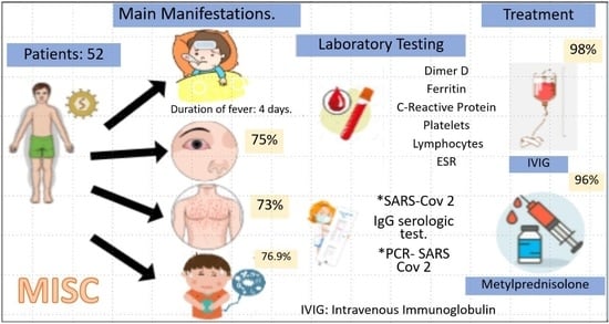
Multisystemic inflammatory syndrome (MIS-C) is an inflammatory condition temporally associated with COVID-19 in children; nevertheless, the clinical and immunologic spectrum of MIS-C is heterogeneous, and its long-term effects are unknown. During the period of August 2020 to December 2021, a total of 52 MIS-C cases were confirmed in pediatric patients from the Hospital del Niño DIF Hidalgo, diagnosed using criteria from the World Health Organization. All patients had serologic IgG confirmation of SARS-CoV2, the mean age of the patients was 7 years, and 94% of the patients did not have a previous underlying disease. In addition to the presentation of lymphopenia, neutropenia, and thrombocytopenia, elevations in D-dimer and ferritin levels were observed in all patients. There was clinical improvement with intravenous gamma globulin and corticosteroid treatment.

Graphical Abstract

1. Introduction

On 25 April 2020, the National Health Service of the United Kingdom issued a warning about a multisystemic inflammatory syndrome in children (MIS-C) that was temporally associated with SARS-CoV-2, with clinical characteristics similar to those found in Kawasaki disease (KD), toxic shock syndrome, hemophagocytic lymphohistiocytosis, and macrophage activation syndrome [1,2]. The first reports in 2020 compared MIS-C symptoms to those of KD (with fever mucocutaneous features and cardiac sequelae), but the recent Clinical Guidance established by the American College of Rheumatology (version 3) discusses differences between the MIS-C and KD phenotypes that are worth mentioning. First, this guide states that the incidence of KD is higher in Japan and East Asia, and it is common in children under 5 years of age. Additionally, the clinical presentations of left ventricular dysfunction and shock are more characteristic of patients with MIS-C than KD, and gastrointestinal and neurologic symptoms are more frequent in MIS-C patients [3].
The etiology of KD is far from fully understood, although it has been hypothesized that a respiratory infectious agent infecting the bronchial ciliated epithelium could be the causal agent of KD [4]. Luchman (2021) [5] proposed an analogy because in SARS-CoV-2 animal models, the neutralizing antibodies of SARS-CoV-2 protein S enhance the inflammatory response associated with MIS-C [6]. Therefore, a complex comprehension of this inflammatory phenomenon is required in order to understand the immune responses to SARS-CoV-2 in infants to establish an appropriate approach to MIS-C [7,8,9].
The World Health Organization (WHO) defines MIS-C as the presence of a fever lasting ≥3 days in children and teenagers from 0 to 19 years old and two of the following symptoms: rash or bilateral non-purulent conjunctivitis or signs of mucocutaneous inflammation (oral, hands, or feet), hypotension or shock, features of myocardial dysfunction, pericarditis, valvulitis or coronary anomalies (echo findings included or high troponin/NT-proBNP), evidence of coagulopathy, (prothrombin time or thromboplastin or high D-dimer), acute gastrointestinal issues (diarrhea, vomiting, or abdominal pain) and increased biomarkers of inflammation, such as the erythrocyte sedimentation rate (ESR), C-reactive protein (CRP), or procalcitonin (PCT), excluding a microbial causal agent of inflammation, such as bacterial sepsis, staphylococcus, or streptococcal shock syndrome, as well as evidence of COVID-19 (RT-PCR), such as antigen tests, positive serology, or probable contact with COVID-19 patients [10,11,12]. Even if the reports have increased during the pandemic, the description of the clinical presentations of MIS-C is imperative for the comprehension of this new syndrome and its approach in the pediatric population.

2. Materials and Methods

This was an observational, retrospective, and descriptive study including pediatric patients who attended the Hospital del Niño DIF Hidalgo (HNDH) in Mexico from August 2020 to December 2021. Since August 2020, the HNDH has been declared a COVID-19-hospital, and the HNDH–MIS-C Committee was established to identify pediatric patients with MIS-C according to the WHO criteria [13,14]. Therefore, at the operational level, this observational study includes pediatric patients with fever for more than three days, physical signs of inflammation at admission, clinical data for inflammatory biomarkers, and positivity for COVID-19 via a serologic test (IgG and IgM) or previous contact with COVID-19.
Clinical measures taken at admission included blood count, CRP, ESR, PCT, D-dimer, ferritin, creatinine, and sodium. We also registered demographic and anthropometric data upon admission. All patients who tested negative for COVID-19 or had a fever duration of less than 3 days were excluded. In order to evaluate a diagnosis of incomplete KD/KD shock syndrome, chest radiology and echocardiography were carried out at admission.
The patients’ parents or legal guardians signed the informed consent form, and all the cases were registered according to the Institutional Ethics Committee guidelines (register number CICEICB-2021-CC07).

Statistical Analysis

The results of the continuous data (age, body mass index: BMI, CRP, PCT, ESR, D–dimer, ferritin, platelets, lymphocytes, and sodium) are presented as means ± standard deviations (SDs) for variables with a Gaussian distribution. In cases of non-Gaussian distribution, the results of continuous data are presented with the median (interquartile range: IQR). Frequencies are presented in numbers (percentages) for categorical data. We analyzed associations between the variables of interest (age, sex, PICU stay, and MIS-C severity). The analysis was performed using STATA v.14.

3. Results

Fifty-two patients met the case definition of MIS-C (5), with a mean duration of fever of 4 days. More cases of males diagnosed with MIS-C were included (33 males and 19 females, 63.4% vs. 36.6%, respectively), with a mean age of 7 years (Min: 1 and Max: 14 years) and 19 children (36.6%) under 5 years of age. The median BMI was 16.6 (IQR = 15.4 − 17.6) kg/m2. Of the participants, 7.7% (n = 4) were in the obese range, 23.1% (n = 12) were overweight, and 7.7% (n = 4) were underweight. All patients were positive for SARS-CoV-2, determined via IgG serologic test and 41.4% via the epidemiologic nexus. PCR confirmation was accessible in two patients only, both of whom were positive. The most frequent symptoms identified at admission to the hospital were conjunctivitis, cutaneous rash, emesis, oral changes, abdominal pain, and diarrhea. Irritability was the most common sign of mental alteration (Table 1).
CRP, PCT, ESR, D-dimer, and ferritin concentrations were determined at admission, and the median values of the results were superior to the maximum reference limits (Table 2). The serum CRP concentrations were found to be in the normal range (<5 mg/dL) in seven patients (13.4%), and the PCT levels were under 0.1 ng/mL in only three (5.7%) patients. On the other hand, the median for platelet values was found to be in the normal range (184 × 109 cells/L), and the platelet values were below the lower limit (150 × 109 cells/L) in only 18 patients (34.6%). Lymphopenia was present in 37 patients (71.1%). In 17 patients (32.7%), serum sodium levels were found to be below the lower limit (<135 mEq/L), although the average was in the normal range.
The mean hospital stay was 6 days (min: 2, max: 25). During their stay in the hospital’s Pediatric Intensive Care Unit (PICU), 23.1% of the patients manifested shock criteria, and 1 case manifested moderate upper gastrointestinal bleeding as a complication, thus requiring aminergic management for 3 days in the PICU. Of the patients, 15.4% (n = 8) presented with criteria for acute kidney injury according to the RIFLE criteria (risk, injury, failure, loss, and end stage), defined as a 50% increase in serum creatinine at 24 h [15]. Vasopressor support was applied in nine cases (17%, Table 3).
We observed bilateral opacities in eight patients (15.4%). The echocardiogram evidenced three patients with pericarditis, and one of them demonstrated left ventricular function decline, which was recovered after treatment. Respiratory failure was observed in 11 patients (21%), and 7 patients required noninvasive mechanical ventilation, oxygen masks (n = 8), and nasal tips (n = 1) (Table 3).
Fifty-one cases (98%) were treated with gamma globulin (2 g/kg of weight/day for 12 h, with a maximum dose of 80 gm), 96.1% (n = 50) with methylprednisolone (2 mg/kg of weight/day for 14 days), and 88% with acetylsalicylic acid (50 mg/kg of weight/day for 8 weeks until a follow-up echocardiogram without alterations). Prior to hospital admission, four patients (7.7%) received antiviral treatment (two with acyclovir and two with oseltamivir), and 53.8% (n = 28) received empirical antibiotic treatment for at least 48 h (Table 3). In the correlation analysis, no significant associations were observed when analyzing the frequency of the characteristics at admission according to age, sex, admission to the PICU, or MIS-C severity due to our small sample size.
Of the patients, 88.2% (n = 46) were discharged in a mean time of 8 days without clinical data indicating complications. However, 3 patients were discharged after 11 days due to the severity of MIS-C related to the PICU stay, the use of vasopressors, and respiratory support. Three patients had the longest hospital stays (15 to 25 days), which were related to underlying diseases. A severe MIS-C case was identified in a female patient who was diagnosed with high-risk myeloid lymphoblastic leukemia in 2017, aged 10 years, with an isolated relapse in the second-line protocol who was admitted to the PICU in February 2021 for febrile neutropenia and respiratory distress (intercostal indrawing and polypnea) with severe thrombocytopenia and elevated acute phase reactants (PCR = 3.8 mg/dL). After IVIG and methylprednisolone treatment, she was discharged on day 23 with a normal echocardiogram.
Mortality (n = 2) was observed in two patients. One male child with Down Syndrome (8 years old) was diagnosed with pre-B acute lymphoblastic leukemia in April 2019. On October 2020, he was admitted due to neutropenia and cervical abscess, with a history of COVID-19 infection 4 weeks prior to admission, which was confirmed by PCR. At admission, he presented with a fever of more than 3 days of evolution and elevated D-dimer (2000 mg/mL) without data indicating bacteremia. Chest tomography reported pneumonia with focal lobar interstitial condensation lesions which was predominantly bilateral at baseline. During the hospital stay, he was clinically stable with supplemental oxygen support. Intravenous gamma globulin (1 gr/kg/dose; 20 gr total) and methylprednisolone (2 mg/kg/day for 14 days) were administered. He was discharged on day 25 with a normal echocardiogram. One week later, this patient was readmitted for septic shock and died. The other case was a one-year-old female patient with congenital cardiopathy, post-operation for patent ductus arteriosus, and a history of perinatal asphyxia that required hospitalization for one month. She had a positive antigen test for SARS-CoV-2 with torpid evolution, septic shock, and cardiogenic shock which required broad-spectrum antimicrobial and antifungal management as well as management with vasoactive amines. After 25 days, hemodynamic and respiratory deterioration was observed without improvement, presenting septic and cardiogenic shock and death.

4. Discussion

In this report, we present the clinical characteristics of children with MIS-C associated with SARS-CoV-2 and with an exacerbated inflammatory response. The patients had heterogeneous phenotypes, and the differences in the first series of MIS-C cases in 2020 were reported as positivity for SARS-CoV-2, age of presentation, proportion of males, and less cardiac involvement.
The majority of patients were found to be positive for SARS-CoV-2 via serologic testing, which has been observed with less frequency in previous studies (20–53%) [16,17]. All patients with MIS-C presented with a fever and more than three criteria for MIS-C. The majority of cases reported correspond to males, with a proportion of 1.5 to 1 (male/female), as previously reported in the general population [18,19]. A higher prevalence of MIS-C in males has been reported in the Mexican population; although the reason is not clear, it could be due to a higher risk of developing COVID-19, and it has been hypothesized that specific immune defects could predispose males to MIS-C [20,21].
It has been suggested that KD and MIS-C are two pathological entities [22]. The age of presentation with MIS-C is older than in classic KD (10 years vs. 5 years in KD). Therefore, the age of presentation has been suggested as an important characteristic of MIS-C; nevertheless, given the epidemiological curse of the pandemic, children with MIS-C under one year of age have been documented in case series [23], coinciding with this report, which includes patients from August 2020 to July 2021, and with the severity of the clinical picture. Therefore, it is imperative to study the influence of SARS-CoV-2 variants to support the understanding of the pathophysiological mechanisms of MIS-C and its differences from KD.
The mechanisms that exacerbate the inflammatory response via SARS-CoV-2 are still a matter of investigation, even though current reports highlight important clinical differences. In a meta-analysis of 969 patients with MIS-C, low levels of leukocytes, platelets, and serum sodium were observed, including higher CRP, D-dimer, ferritin, and creatinine levels than in KD or incomplete KD patients, even though the PCT and ESR levels were similar [24,25]. In this series of cases, all patients showed higher levels of CRP, PCT, and D-dimer at admission, in addition to leukopenia.
Cardiovascular complications are a prominent characteristic of KD and are compatible with MIS-C, such as ventricular and valvular dysfunction or persistent coronary aneurysm, which is described in 20–25% of patients who do not receive immunomodulatory treatment [26,27,28]. Left ventricle systolic dysfunction has been described as the most frequent cardiac affection in a large proportion of children diagnosed with MIS-C [29,30]. In the first series of MIS-C cases described in the United Kingdom, cardiac dysfunction was present in 6/8 patients (75%). A subgroup of patients with an MIS-C diagnosis may present hypotension and shock via acute myocardial dysfunction, as well as hyperinflammation/systemic vasodilation [31,32]. The late development of the aneurysm in the coronary artery highlights the necessity of the continuous follow-up of these patients. In this report, we did not observe cardiovascular warning signs; the echocardiogram evidenced three patients with pericarditis, and only one of them had a decrease in left ventricular function, which recovered after immunomodulatory treatment.
The basis of the treatment for MIS-C is focused on immunomodulatory therapy with intravenous human gamma globulin, high, anti-inflammatory doses of corticosteroids, and their intensification in refractory cases with methylprednisolone pulse [33]. All patients who did not have an underlying disease showed a fast recovery with the indicated treatment. However, the presence of underlying diagnoses of acute lymphoblastic leukemia and myeloid lymphoblastic leukemia in two patients with leukopenia and trisomy 21 could be a bad prognosis factor, as has been previously reported [12].

5. Conclusions

D-dimer and PCT were present in all cases; however, cardiovascular complications were less frequent. The continuous communication of the clinical manifestations of MIS-C is important to increase the knowledge of this pathology; therefore, it the continuous follow-up of patients is imperative in order to know the possible mid- to long-term implications.

Author Contributions

Conceptualization, J.B.-S., J.I.S.-S., C.D.-A., B.G.-H., O.O.-C., S.E.E.-P. and A.S.J.-O.; methodology, J.B.-S., B.A.S.-R., B.G.-H., O.O.-C. and J.A.M.-G.-T.; software, A.I.R.-M. and O.O.-C.; validation, A.S.J.-O. and S.E.H.-C.; formal analysis, A.S.J.-O., A.I.R.-M. and J.B.-S.; investigation, J.B.-S., S.E.H.-C., B.A.S.-R., J.I.S.-S., C.D.-A., O.O.-C. and J.A.M.-G.-T.; resources, M.L.-B. and O.O.-C.; data curation, J.B.-S., A.I.R.-M. and A.S.J.-O.; writing—original draft preparation, J.B.-S.; writing—review and editing, S.E.E.-P. and A.S.J.-O.; visualization, B.A.S.-R., J.I.S.-S., A.I.R.-M., B.G.-H. and J.A.M.-G.-T.; supervision, R.G.H.d.Á., M.L.-B. and C.D.-A.; project administration, M.L.-B., R.G.H.d.Á. and F.A.-G. All authors have read and agreed to the published version of the manuscript.

Funding

This research received no external funding.

Institutional Review Board Statement

The study was conducted in accordance with the Declaration of Helsinki and approved by the Institutional Ethics Committee of the Hospital del Niño DIF Hidalgo (registration number CICEICB-2021-CC07 approved on 21 January 2021).

Informed Consent Statement

Informed consent was obtained from all subjects involved in the study.

Data Availability Statement

The data presented in this study are available upon request from the corresponding author. The data are not publicly available due to Hospital Privacy Statements.

Conflicts of Interest

The authors declare no conflict of interest.

References

  1. Royal College of Paediatrics and Child Health. Paediatric Multisystem Inflammatory Syndrome Temporally Associated with COVID-19 (PIMS)—Guidance for Clinicians [Internet]. RCPCH. 2020. Available online: https://www.rcpch.ac.uk/resources/paediatric-multisystem-inflammatory-syndrome-temporally-associated-covid-19-pims-guidance (accessed on 3 March 2022).
  2. Soma, L.; Shust, F.J.; Ratner. Multisystem Inflammatory Syndrome in children. Infect. Dis. Immun. 2020, 33, 152–158. [Google Scholar] [CrossRef] [PubMed]
  3. Henderson, L.A.; Canna, S.W.; Friedman, K.G.; Gorelik, M.; Lapidus, S.K.; Bassiri, H.; Behrens, E.M.; Kernan, K.F.; Schulert, G.S.; Seo, P.; et al. American College of Rheumatology Clinical Guidance for Multisystem Inflammatory Syndrome in Children Associated With SARS-CoV-2 and Hyperinflammation in Pediatric COVID-19: Version 3. Arthritis Rheumatol. 2022, 74, e1–e20. [Google Scholar] [CrossRef] [PubMed]
  4. Rowley, A.H.; Baker, S.C.; Shulman, S.T.; Garcia, F.L.; Guzman-Cottrill, J.A.; Chou, P.; Terai, M.; Kawasaki, T.; Kalelkar, M.B.; Crawford, S.E. Detection of Antigen in Bronchial Epithelium and Macrophages in Acute Kawasaki Disease by Use of Synthetic Antibody. J. Infect. Dis. 2004, 190, 856–865. [Google Scholar] [CrossRef]
  5. Lutchman, D. PIMS-TS and Kawasaki Disease: The Mystery Deepens. Pediatr. Infect. Dis. 2020, 39, e215–e216. [Google Scholar] [CrossRef] [PubMed]
  6. Tay, M.Z.; Poh, C.M.; Rénia, L.; Macary, P.A.; Ng, L.F.P. The trinity of COVID-19: Immunity, inflammation, and intervention. Nat. Rev. Immunol. 2020, 20, 363–374. [Google Scholar] [CrossRef]
  7. Gustine, J.; Jones, D. Immunopathology of Hyperinflammation in COVID-19. Am. J. Pathol. 2021, 191, 4–17. [Google Scholar] [CrossRef]
  8. Rosat, C.; Cotugno, N.; Sardh, F.; Pou, C.; Amodio, D.; Rodriguez, L.; Tan, Z.; Zicari, S.; Ruggiero, A.; Rubens, G.; et al. The Immunology of Multisystem Inflammatory Syndrome in Children with COVID-19. Cell 2020, 183, 968–981. [Google Scholar]
  9. Kaklamanos, A.; Belogiannis, K.; Skendros, P.; Gorgoulis, V.; Vlachoyiannopoulos, P.; Tzioufas, A. COVID-19 Immunobiology: Lessons Learned, New Questions Arise. Front. Immunol. 2021, 12, 719023. [Google Scholar] [CrossRef]
  10. World Health Organization. Síndrome Inflamatorio Multisistémico en Niños y Adolescentes con COVID-19. Informe Científico 15 de Mayo de 2020. Available online: https://apps.who.int/iris/bitstream/handle/10665/332191/WHO-2019-nCoV-Sci_Brief-Multisystem_Syndrome_Children-2020.1-spa.pdf?sequence=1&isAllowed=y (accessed on 2 March 2022).
  11. Nakra, N.; Blumberg, D.; Herrera, A.; Lakshminrusimha, S. Multi-system inflammatory Syndrome in Children (MIS-C) Following SARS-CoV-2 Infection: Review of Clinial Presentation, Hypothetical Pathogenesis, and Proposed Management. Children 2020, 7, 69. [Google Scholar] [CrossRef]
  12. Henderson, L.A.; Canna, S.W.; Friedman, K.G.; Gorelik, M.; Lapidus, S.K.; Bassiri, H.; Behrens, E.M.; Ferris, A.; Kernan, K.F.; Schulert, G.S.; et al. American College of Rheumatology Clinical Guidance for Multisystem Inflammatory Syndrome in Children Associated With SARS-CoV-2 and Hyperinflammation in Pediatric COVID-19: Version 2. Arthritis Rheumatol. 2021, 73, e13–e29. [Google Scholar] [CrossRef]
  13. Sperotto, F.; Friedman, K.G.; Son, M.; Vanderpluym, C.; Newburger, J.; Dionne, A. Cardiac manifestations in SARS-CoV-2-associated multisystem inflammatory syndrome in children: A comprehensive review and proposed clinical approach. Eur. J. Pediatr. 2021, 180, 307–322. [Google Scholar] [CrossRef] [PubMed]
  14. Gottlieb, M.; Bridwell, R.; Ravera, J.; Long, B. Multisystem inflammatory syndrome in children with COVID-19. Am. J. Emerg. Med. 2021, 49, 148–152. [Google Scholar] [CrossRef] [PubMed]
  15. Ricci, Z.; Cruz, D.N.; Ronco, C. Classification and staging of acute kidney injury: Beyond the RIFLE and AKIN criteria. Nat. Rev. Nephrol. 2011, 7, 201–208. [Google Scholar] [CrossRef] [PubMed]
  16. Abrams, J.Y.; Godfred-Cato, S.E.; Oster, M.E.; Chow, E.J.; Koumans, E.H.; Bryant, B.; Leung, J.W.; Belay, E.D. Multisystem Inflammatory Syndrome in Children Associated with Severe Acute Respiratory Syndrome Coronavirus 2: A Systematic Review. J. Pediatr. 2020, 226, 45–54.e1. [Google Scholar] [CrossRef] [PubMed]
  17. Radia, T.; Williams, N.; Agrawal, P.; Harman, K.; Weale, J.; Cook, J.; Gupta, A. Multisystem inflammatory syndrome in children and adolescents (MIS-C): A systematic review of clinical features and presentation. Paediatr. Respir. Rev. 2021, 38, 51–57. [Google Scholar] [PubMed]
  18. Kaushik, S.; Aydin, S.I.; Derespina, K.R.; Bansal, P.B.; Kowalsky, S.; Trachtman, R.; Gillen, J.K.; Perez, M.M.; Soshnick, S.H.; Conway, E.E., Jr.; et al. Multisystem Inflammatory Syndrome in Children Associated with Severe Acute Respiratory Syndrome Coronavirus 2 Infection (MIS-C): A Multi-institutional Study from New York City. J. Pediatr. 2020, 224, 24–29. [Google Scholar] [CrossRef]
  19. Zhang, Q.Y.; Xu, B.W.; Du, J.B. Similarities and differences between multiple inflammatory syndrome in children associated with COVID-19 and Kawasaki disease: Clinical presentations, diagnosis, and treatment. World J. Pediatr. 2021, 17, 335–340. [Google Scholar] [CrossRef]
  20. Castano-Jaramillo, L.M.; Yamazaki-Nakashimada, M.A.; O’Farrill-Romanillos, P.M.; Muzquiz Zermeño, D.; Scheffler Mendoza, S.C.; Venegas Montoya, E.; García Campos, J.A.; Sánchez-Sánchez, L.M.; Gámez González, L.B.; Ramírez López, J.M.; et al. COVID-19 in the Context of Inborn Errors of Immunity: A Case Series of 31 Patients from Mexico. J. Clin. Immunol. 2020, 41, 1463–1478. [Google Scholar] [CrossRef]
  21. Bucciol, G.; COVID Human Genetic Effort; Meyts, I. Inherited and acquired errors of type I interferon immunity govern susceptibility to COVID-19 and multisystem inflammatory syndrome in children. J. Allergy Clin. Immunol. 2023, 151, 832–840. [Google Scholar] [CrossRef]
  22. Sharma, C.; Ganigara, M.; Galeotti, C.; Burns, J.; Berganza, F.; Hayes, D.; Grewal, D.; Bharath, S.; Sajjan, S.; Bayry, J. Multisystem inflammatory syndrome in children and Kawasaki disease: A critical comparison. Nat. Rev. 2021, 17, 731–748. [Google Scholar] [CrossRef]
  23. Zhou, C.; Zhao, Y.; Wang, X.; Huang, Y.; Tang, X.; Tang, L. Laboratory parameters between multisystem inflammatory syndrome in children and Kawasaki disease. Pediatr. Pulmonol. 2021, 56, 3688–3698. [Google Scholar] [CrossRef]
  24. Alsaied, T.; Tremoulet, A.H.; Burns, J.C.; Saidi, A.; Dionne, A.; Lang, S.M.; Newburger, J.W.; de Ferranti, S.; Friedman, K.G. Review of Cardiac Involvement in Multisystem Inflammatory Syndrome in Children. Circulation 2021, 143, 78–88. [Google Scholar] [CrossRef] [PubMed]
  25. Stankovic, C.; DeLaroche, A.; Arora, R.; Ehrman, R. Laboratory trends in severe MIS-C. Acad. Emerg. Med. 2022, 29, 1258–1260. [Google Scholar] [CrossRef] [PubMed]
  26. Adeyinka, A.; Bailey, K.; Pierre, L.; Kondamudi, N. COVID-19 infection: Pediatric perspectives. J. Am. Coll. Emerg. Physicians Open 2021, 2, e12375. [Google Scholar]
  27. Chen, M.; Kuo, H.; Lee, Y.; Chi, H.; Li, S.; Lee, H.; Yang, K. Phenotype, Susceptibility, Autoimmunity, and Immunotherapy between Kawasaki Disease and Coronarivurs Disease-19 Associated Multisystem Inflammatory Syndrome in Children. Front. Immunol. 2021, 12, 632890. [Google Scholar] [CrossRef] [PubMed]
  28. Wessels, P.A.; Bingler, M.A. A comparison of Kawasaki Disease and multisystem inflammatory syndrome in children. Prog. Pediatr. Cardiol. 2022, 65, 101516. [Google Scholar] [CrossRef]
  29. Jiang, L.; Tang, K.; Irfan, O.; Li, X.; Zhang, E.; Bhutta, Z. Epidemiology, Clinical Features, and Outcomes of multisystem inflammatory syndrome in children (MIS-C) and adolescents- a Live Systematic Review and Metanalysis. Curr. Pediatr. Rep. 2022, 10, 19–30. [Google Scholar] [CrossRef] [PubMed]
  30. Feldstein, L.R.; Tenforde, M.W.; Friedman, K.G.; Newhams, M.; Billing, E.; Dapul, H.; Soma, V.; Maddux, A.; Mourani, P.; Bowens, C.; et al. Characteristics and Outcomes of US Children and Adolescents with Multisystem Inflammatory Syndrome in Children (MIS-C) Compared with Severe Acute COVID-19. JAMA 2021, 325, 1074–1087. [Google Scholar] [CrossRef] [PubMed]
  31. Haslak, F.; Barut, K.; Durak, C.; Aliyeva, A.; Yildiz, M.; Guliyeva, V.; Varol, S.E.; Cebeci, S.O.; Aygun, F.; Varli, Y.Z.; et al. Clinical features and outcomes of 76 patients with COVID-19-related multi-system inflammatory syndrome in children. Clin. Rheumatol. 2021, 40, 4167–4178. [Google Scholar] [CrossRef]
  32. Shimizu, V.; Brodin, P.; Cobat, A.; Biggs, C.; Toubiana, J.; Lucas, C.; Henrickson, S.; Belot, A.; Tangye, S.; Milner, J.; et al. SARS-Cov-2 related MIS-C: A Key to the viral and genetic causes of kawasawki disease. J. Exp. Med. 2021, 218, e20210446. [Google Scholar] [CrossRef]
  33. Giacalone, M.; Scheier, E.; Shavit, I. Multisystem inflammatory syndrome in children (MIS-C): A mini-review. Int. J. Emerg. Med. 2021, 14, 50. [Google Scholar] [CrossRef]
Table 1. Summary of symptoms at hospital admission.
SymptomsFrequency (%)
Duration of fever (days)4 (min. 3, max. 5)
Diarrhea30 (57.7)
Abdominal pain40 (76.9)
Emesis36 (69.2)
Cutaneous rash38 (73)
Conjunctivitis39 (75)
Cleft lips/strawberry tongue35 (67.3)
Lymphadenopathy26 (50)
Limb edema28 (54.9)
Headache15 (28.8)
Mental alteration/Irritability15 (28.8)
Somnolence8 (15.4)
Table 2. Laboratory test results from admission.
TestValueReference Range
C-Reactive protein (mg/dL)18.1 (6.3–26.5)0–5 mg/L
Procalcitonin (ng/mL)2.7 (0.8–9)<0.1 ng/mL
Erythrocyte sedimentation rate (mm/h)40 ± 15.30–10 mm/h
D-dimer2974 (1457.7–6074) <0.5 mg/mL
Ferritin245.7 (175.7–463)10–200 ng/mL
Platelets184 (104.5–303)150–400 × 109 cells/L
Lymphocytes1230 (907–2943)3–9.5 × 10³ cells/μL
Sodium135 ± 4.9(135–145 mEq/L)
Table 3. Clinical characteristics of MIS-C patients during hospital stay.
OutcomesFrequency (%)
General findings in PICU
Shock12 (23.1)
Gastrointestinal Bleeding1 (1.9)
Acute Kidney Injury8 (15.4)
Vasopressor support
Norepinephrine4 (7.7)
Epinephrine1 (1.9)
Dobutamine1 (1.9)
Norepinephrine + Epinephrine2 (3.8)
Norepinefrine + Dobutamine1 (1.9)
Cardiovascular findings
Bilateral opacities8 (15.4)
Acute pericarditis4 (7.7)
Pericardia Effusion3 (5.8)
LV ejection fraction <50%1 (1.9)
Respiratory support
Mechanic ventilation7 (13.46)
Oxygen3 (5.8)
Nasal tips1 (1.92)
Pharmacologic therapy
Empirical antibiotic treatment28 (53.8)
Ceftriaxone26 (50)
Clindamycin8 (15.4)
Cefepime2 (3.85)
Intravenous immunoglobulin51 (98)
Methyl prednisolone50 (96.15)
Acetylsalicylic acid46 (88.4)
Antiviral treatment4 (7.7)
Oseltamivir2 (3.9)
Acyclovir2 (3.9)
Mortality2 (3.8)
Disclaimer/Publisher’s Note: The statements, opinions and data contained in all publications are solely those of the individual author(s) and contributor(s) and not of MDPI and/or the editor(s). MDPI and/or the editor(s) disclaim responsibility for any injury to people or property resulting from any ideas, methods, instructions or products referred to in the content.