Next Issue
Volume 12, September
Previous Issue
Volume 12, March


J. Xenobiot., Volume 12, Issue 2 (June 2022) – 6 articles

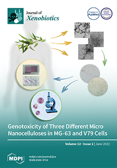
Cover Story (view full-size image): Nanocellulose is an innovative nanomaterial with enormous potential for use in a wide array of industrial and biomedical applications, and with fast-growing economic value. However, its expanding production is raising concerns about the potential health effects of unintentional human exposure. This study assessed the potential cytotoxic and genotoxic effects of two fibrillar and one crystalline micro-/nanocellulose in two mammalian cell lines (MG-63 and V79 cells). Cytotoxicity was analyzed via MTT and clonogenic assays, and genotoxicity via micronucleus assay. All nanocelluloses revealed cytotoxicity and genotoxicity, although at different concentrations, which may be related to their physicochemical differences, to differences in their cellular uptake, and to the DNA damage response of cell models. View this paper
  • Issues are regarded as officially published after their release is announced to the table of contents alert mailing list.
  • You may sign up for e-mail alerts to receive table of contents of newly released issues.
  • PDF is the official format for papers published in both, html and pdf forms. To view the papers in pdf format, click on the "PDF Full-text" link, and use the free Adobe Reader to open them.
Order results
Result details
Section
Select all
Export citation of selected articles as:
14 pages, 1496 KiB  
Article
Comparative Study of Δ9-Tetrahydrocannabinol and Cannabidiol on Melanogenesis in Human Epidermal Melanocytes from Different Pigmentation Phototypes: A Pilot Study
by Shilpi Goenka
J. Xenobiot. 2022, 12(2), 131-144; https://doi.org/10.3390/jox12020012 - 10 Jun 2022
Cited by 7 | Viewed by 2895
Abstract
Δ9-tetrahydrocannabinol (THC) is one of the primary ingredients of cannabis plants and is responsible for the psychoactive properties of cannabis. While cannabidiol (CBD), the non-psychoactive compound from cannabis, has been shown to stimulate human epidermal melanogenesis, the effects of THC have not been [...] Read more.
Δ9-tetrahydrocannabinol (THC) is one of the primary ingredients of cannabis plants and is responsible for the psychoactive properties of cannabis. While cannabidiol (CBD), the non-psychoactive compound from cannabis, has been shown to stimulate human epidermal melanogenesis, the effects of THC have not been addressed in human epidermal melanocytes. Moreover, to date, no study has tested the effects of these compounds on melanocytes differing in pigmentation, representative of different skin phototypes, which would be significant as different ethnicities are known to differentially metabolize these xenobiotics. Herein, the effects of THC were studied and compared alongside CBD in human epidermal melanocytes derived from lightly-pigmented (HEMn-LP; Caucasian) and darkly-pigmented (HEMn-DP; African-American) cells over a chronic exposure of 6 d. Results demonstrated that both compounds displayed cytotoxicity at 4 µM but stimulated melanin synthesis and tyrosinase activity in a similar manner in LP and DP cells at nontoxic concentrations of 1–2 µM. However, THC and CBD showed a differential effect on dendricity in both cells; THC and CBD reversibly increased dendricity in LP cells while there was no significant change in DP cells. THC and CBD induced higher levels of reactive oxygen species (ROS) in LP cells while there was no change in the ROS levels in DP cells. In summary, although THC was relatively less cytotoxic as compared to CBD to both LP and DP cells, it exhibited a similar capacity as CBD to stimulate melanin synthesis and export in LP cells which was accompanied by a significant oxidative stress. DP cells were relatively resistant to the effects of both THC and CBD which might implicate the protective effects conferred by melanin in dark-skinned individuals. Full article
Show Figures

Graphical abstract

9 pages, 449 KiB  
Article
Vitamin B12 Deficiency and Clinical Neuropathy with Metformin Use in Type 2 Diabetes
by Malik Dilaver Farooq, Farooq Ahmad Tak, Fauzia Ara, Samia Rashid and Irfan Ahmad Mir
J. Xenobiot. 2022, 12(2), 122-130; https://doi.org/10.3390/jox12020011 - 31 May 2022
Cited by 8 | Viewed by 4449
Abstract
Introduction: Type 2 diabetes (T2DM), which is more prevalent (more than 90% of all diabetes cases) and the main driver of the diabetes epidemic, now affects 5.9% of the world’s adult population, with almost 80% of the total in developing countries. At [...] Read more.
Introduction: Type 2 diabetes (T2DM), which is more prevalent (more than 90% of all diabetes cases) and the main driver of the diabetes epidemic, now affects 5.9% of the world’s adult population, with almost 80% of the total in developing countries. At present, 537 million adults (20–79 years) are living with diabetes—1 in 10. This number is predicted to rise to 643 million by 2030 and 783 million by 2045. In India, reports show that 69.2 million people are living with diabetes (8.7%) as per 2015 data. Long-term metformin treatment is a known pharmacological cause of vitamin B12 (Vit B12) deficiency, as was evident within the first 10–12 years after it started to be used. Methods: This was a cross-sectional study conducted in the Postgraduate Department of Medicine in one of the tertiary hospitals in Kashmir. A total of 1600 consecutive patients with T2DM were taken for the study. Out of which 700 patients met the inclusion criteria. These 700 patients were divided into two groups: those taking metformin, and those who were not on metformin. Cumulative metformin doses were recorded in patients taking metformin, using history of dose and duration of treatment. Serum Vit B12 levels were taken for all patients. Based on the results of Vit B12 levels, patients were classified into normal levels (20 pmol/L), possible B12 deficiency (150–220 pmol/l), and definite deficiency (<150 pmol/L). Results: Our results depicted that patients on prolonged metformin therapy showed an increase in Vit B12 deficiency by 11.16%. The prevalence of clinical neuropathy in the metformin-exposed group was 45%, whereas, a prevalence of 31.8% was found in the non-metformin group. The mean age of patients with neuropathy was higher than those without neuropathy (59.01 ± 7.14 vs. 49.95 ± 7.47) (p-value < 0.514, statistically insignificant). Conclusions: In our study, we found that metformin use is associated with Vit B12 deficiency, which is dependent upon the cumulative dose of metformin. Importantly, prolonged metformin use is also associated with an increase in the prevalence of clinical neuropathy. Full article
Show Figures

Figure 1

13 pages, 1722 KiB  
Article
Isolation and Quantification of Polystyrene Nanoplastics in Tissues by Low Pressure Size Exclusion Chromatography
by François Gagné
J. Xenobiot. 2022, 12(2), 109-121; https://doi.org/10.3390/jox12020010 - 9 May 2022
Cited by 12 | Viewed by 3073
Abstract
Ecotoxicity investigations of plastic nanoparticles (NPs) should pay more attention to their ability to pass barriers, accumulate, and initiate toxicity in cells. The purpose of this study was to develop a simple size exclusion chromatography (SEC) methodology to measure plastic NPs in biological [...] Read more.
Ecotoxicity investigations of plastic nanoparticles (NPs) should pay more attention to their ability to pass barriers, accumulate, and initiate toxicity in cells. The purpose of this study was to develop a simple size exclusion chromatography (SEC) methodology to measure plastic NPs in biological tissues. A SEC column was prepared using a high-resolution gel for large macromolecules to separate plastic NPs from the protein/lipid pools in tissues. It was necessary to prepare the samples in high salt and non-ionic detergent (0.5 M NaCl and 0.2% Tween-20) and apply 0.2% Tween-20 containing 14 mM NaCl for the elution buffer to limit proteins adsorption to NPs. This methodology was able to resolve 50 and 100 nm polystyrene NPs from the protein/lipid pools in tissue homogenates. The fluorescent dye neutral red (NR) was also used for transparent NPs. Moreover, a sample fractionation step was also proposed for plastic NPs concentration using a salting-out methodology with saturated NaCl (5 M) and acetonitrile. Polystyrene NPs partition in acetonitrile, which were further analyzed by SEC. This methodology was tested in two case studies with clams collected in a high boat traffic (harbor) area and with caged freshwater mussels downstream of a large urban area. Although the present methodology was developed with polystyrene NPs it should be amenable to other plastic polymers that react with the NR fluorescent probe. Full article
Show Figures

Figure 1

18 pages, 5679 KiB  
Article
Genotoxicity of Three Micro/Nanocelluloses with Different Physicochemical Characteristics in MG-63 and V79 Cells
by Célia Ventura, Catarina Marques, João Cadete, Madalena Vilar, Jorge F. S. Pedrosa, Fátima Pinto, Susete Nogueira Fernandes, Rafaela Raupp da Rosa, Maria Helena Godinho, Paulo J. T. Ferreira, Henriqueta Louro and Maria João Silva
J. Xenobiot. 2022, 12(2), 91-108; https://doi.org/10.3390/jox12020009 - 21 Apr 2022
Cited by 5 | Viewed by 3403
Abstract
(1) Background: Nanocellulose is an innovative engineered nanomaterial with an enormous potential for use in a wide array of industrial and biomedical applications and with fast growing economic value. The expanding production of nanocellulose is leading to an increased human exposure, raising concerns [...] Read more.
(1) Background: Nanocellulose is an innovative engineered nanomaterial with an enormous potential for use in a wide array of industrial and biomedical applications and with fast growing economic value. The expanding production of nanocellulose is leading to an increased human exposure, raising concerns about their potential health effects. This study was aimed at assessing the potential toxic and genotoxic effects of different nanocelluloses in two mammalian cell lines; (2) Methods: Two micro/nanocelluloses, produced with a TEMPO oxidation pre-treatment (CNFs) and an enzymatic pre-treatment (CMFs), and cellulose nanocrystals (CNCs) were tested in osteoblastic-like human cells (MG-63) and Chinese hamster lung fibroblasts (V79) using the MTT and clonogenic assays to analyse cytotoxicity, and the micronucleus assay to test genotoxicity; (3) Results: cytotoxicity was observed by the clonogenic assay in V79 cells, particularly for CNCs, but not by the MTT assay; CNF induced micronuclei in both cell lines and nucleoplasmic bridges in MG-63 cells; CMF and CNC induced micronuclei and nucleoplasmic bridges in MG-63 cells, but not in V79 cells; (4) Conclusions: All nanocelluloses revealed cytotoxicity and genotoxicity, although at different concentrations, that may be related to their physicochemical differences and availability for cell uptake, and to differences in the DNA damage response of the cell model. Full article
(This article belongs to the Section Nanotoxicology and Nanopharmacology)
Show Figures

Figure 1

17 pages, 3655 KiB  
Article
Toxicity Assessment and Control of Early Blight and Stem Rot of Solanum tuberosum L. by Mancozeb-Loaded Chitosan–Gum Acacia Nanocomposites
by Ravinder Kumar, Joginder Singh Duhan, Anju Manuja, Pawan Kaur, Balvinder Kumar and Pardeep Kumar Sadh
J. Xenobiot. 2022, 12(2), 74-90; https://doi.org/10.3390/jox12020008 - 14 Apr 2022
Cited by 17 | Viewed by 3041
Abstract
Biopolymers such as chitosan and gum acacia are used for nanotechnological applications due to their biosafety and ecofriendly nature. The commercial fungicide mancozeb (M) was loaded into chitosan–gum acacia (CSGA) polymers to form nanocomposite (NC) CSGA-M (mancozeb-loaded) measuring 363.6 nm via the ionic [...] Read more.
Biopolymers such as chitosan and gum acacia are used for nanotechnological applications due to their biosafety and ecofriendly nature. The commercial fungicide mancozeb (M) was loaded into chitosan–gum acacia (CSGA) polymers to form nanocomposite (NC) CSGA-M (mancozeb-loaded) measuring 363.6 nm via the ionic gelation and polyelectrolyte complexation method. The physico-chemical study of nano CSGA-M was accomplished using dynamic light scattering (DLS), scanning electron microscope (SEM), transmission electron microscope (TEM), X-ray diffraction (XRD), and thermogravimetric analysis (TGA). Nano CSGA-M-1.0 (containing 1.0 mg/mL mancozeb) at 1.5 ppm demonstrated a maximum inhibition (83.8 ± 0.7%) against Alternaria solani, while Sclerotinia sclerotiorum exhibited a 100% inhibition at 1.0 and 1.5 ppm through the mycelium inhibition method. Commercial mancozeb showed an inhibition of 84.6 ± 0% and 100%, respectively, for both fungi. In pot house conditions, NCs were found to exhibit good antimicrobial activity. Disease control efficiency (DCE, in %) in pathogen-treated plants for CSGA-M-1.0 was 64.6 ± 5.0 and 60.2 ± 1.4% against early blight and stem rot diseases, respectively. NCs showed lower cytotoxicity than commercial mancozeb at the given concentration. In conclusion, both in vitro and in vivo antifungal efficacy for nano CSGA-M was found to be quite comparable but less toxic than mancozeb to Vero cell lines; thus, in the future, this formulation may be used for sustainable agriculture. Full article
Show Figures

Figure 1

10 pages, 519 KiB  
Review
Membrane Hormone Receptors and Their Signaling Pathways as Targets for Endocrine Disruptors
by Yves Combarnous and Thi Mong Diep Nguyen
J. Xenobiot. 2022, 12(2), 64-73; https://doi.org/10.3390/jox12020007 - 25 Mar 2022
Cited by 2 | Viewed by 3027
Abstract
The endocrine disruptors are mostly small organic molecules developed for numerous and very diverse industrial applications. They essentially act through nuclear receptors with small and hydrophobic endogenous ligands. Nevertheless, potential adverse effects through membrane hormone receptors cannot be ruled out, and have indeed [...] Read more.
The endocrine disruptors are mostly small organic molecules developed for numerous and very diverse industrial applications. They essentially act through nuclear receptors with small and hydrophobic endogenous ligands. Nevertheless, potential adverse effects through membrane hormone receptors cannot be ruled out, and have indeed been observed. The present paper reviews how orthosteric and allosteric binding sites of the different families of membrane receptors can be targets for man-made hydrophobic molecules (components of plastics, paints, flame retardants, herbicides, pesticides, etc.). We also review potential target proteins for such small hydrophobic molecules downstream of membrane receptors at the level of their intracellular signaling pathways. From the currently available information, although endocrine disruptors primarily affect nuclear receptors’ signaling, membrane receptors for hormones, cytokines, neuro-mediators, and growth factors can be affected as well and deserve attention. Full article
Show Figures

Figure 1

Previous Issue
Back to TopTop