Article

Multifunctional and Durable Coatings for Stone Protection Based on Gd-Doped Nanocomposites

1
Laboratory of Advanced Materials, National School of Engineering, University of Sfax, Sfax 3038, Tunisia
2
Department of Chemistry, University of Pavia, Via Taramelli 12, 27100 Pavia, Italy
3
Department of Chemical, Biological, Pharmaceutical and Environmental Sciences, University of Messina, Viale F. Stagno d’Alcontres 31, 98122 Messina, Italy
*
Authors to whom correspondence should be addressed.
Sustainability 2021, 13(19), 11033; https://doi.org/10.3390/su131911033
Submission received: 10 August 2021 / Revised: 23 September 2021 / Accepted: 29 September 2021 / Published: 5 October 2021
(This article belongs to the Special Issue Toward Sustainable Multifunctional Coatings)

Abstract

:
The development of nanocomposite materials with multifunctional protective features is an urgent need in many fields. However, few works have studied the durability of these materials. Even though TiO2 nanoparticles have been extensively applied for self-cleaning effect, it displays a weak activity under visible light. Hence, in this study, pure and Gd-doped TiO2 nanoparticles (molar ratios of doping ions/Ti are 0.1 and 1) were synthesised, characterised, and then mixed with polydimethylsiloxane (PDMS), used as a binder, in order to produce a homogenised thin film on a very porous stone substrate. To our knowledge, Gd-doped TiO2/PDMS protective coatings are studied for the first time for application on historic structures. The protective coatings developed in this work are intended to reduce the surface wettability of the stone and protect the historic stones from dye pollution and microorganism colonisation. Moreover, in this study, the durability of the developed nanocomposite was deeply studied to evaluate the stability of the coatings. Results confirmed that samples treated with the lowest concentrations of Gd ions (0.1 mol%) showed acceptable chromatic variations, a good repellent feature, acceptable water vapour permeability, good durability, the highest self-cleaning activity, and good inhibitory behaviour against microbial colonisation.

1. Introduction

Heritage sites are the symbol of history. In addition to their intrinsic value, their conservation and maintenance provide economic benefits. However, historic building materials, in particular when exposed outdoors, are subject to many phenomena such as microorganisms colonisation which consequently cause a transformation and deterioration of art surfaces [1]. On the other hand, water, which is one of the most deteriorating factors especially for porous materials, air pollution, and dyes, which strongly adhere to the stone substrate [2,3,4,5], contribute to the decay of external historic buildings. Thus, conservation and protection of cultural heritage materials become a necessity in countries with tourism potential [4].
Several methodologies have been developed such as cleaning (e.g., by organic solvents, chelating agents), biocleaning, and laser methods [3,6,7,8,9]. Despite the efficiency of these methods, the use of these techniques has declined due to different problems (irreversibility, toxicity, difficulty to manipulate, high cost, etc.) [9].
Recently, interest concerning the preservation of monuments from microorganisms colonisation and pollution is shifting towards the application of nanomaterials displaying self-cleaning properties [10] in order to reduce maintenance costs. In particular, nanosized titanium dioxide (TiO2) has been studied and tested for the restoration and preservation of cultural heritage materials [11,12,13,14,15] due to its high chemical stability and non-toxicity [16]. The self-cleaning and antibacterial activity of TiO2 nanoparticles (NPs), in addition to their easy and non-expensive application procedures, makes them of particular interest. Nevertheless, its technological application seems limited due to some factors, including the easy recombination of charge carriers and the need for ultraviolet radiation as an excitation source, because of its broad bandgap (3.2 eV for anatase). This limitation is particularly restrictive, as UV radiation corresponds to only 3% of the solar irradiance at the surface of Earth [17]. Many scientists have focused their research on enhancing the photocatalytic and antimicrobial activity of pure TiO2 NPs by doping with different materials; transition-metal ions, and other metals and ions of non-metals [18,19,20]. However, the application of visible light-active TiO2 NPs is limited to some fields such as water treatment, photovoltaic cells, etc. [21]. Indeed, few works have been published with the aim to elaborate enhanced photoresponse doped TiO2 for protecting the historic structures from biodeterioration [22,23,24].
Taking this matter into consideration, several research studies have been conducted for improving the photocatalytic activity of pure TiO2 NPs by doping, in particular, with Lanthanide ions [25,26,27]. A comparative study was performed by Xu et al. [17] in order to evaluate the photocatalytic activity of pure TiO2 and Ln3+-doped Titania (Ln3+ = La3+, Er3+, Ce3+, Pr3+, Gd3+, Nd3+, Sm3+) for inorganic water-pollutant degradation. Results reported that Gd3+-doped TiO2 showed the highest photocatalytic efficiency among all tested Ln3+-doped samples owing to its distinct properties such as having a half-filled electronic configuration, the largest amount of adsorption, the largest redshift of the Gd3+-doped materials, and the f state of Gd [17]. Moreover, many studies have proved that Gd-ion-doped TiO2 NPs would improve the photocatalytic activity of pure titania under UV and visible light irradiation [28,29,30,31,32]. Additionally, many studies have been performed in order to study the physiological toxicity of these elements [33,34], in particular Gadolinium [35]. The outcomes showed that a low dose of these elements does not present an evident risk to the environment, while a high dose of them revealed negative effects, displaying an evident harmless phenomenon [33,34,35]. In addition to its beneficial characteristics, the inert colour of Gd ions, which does not affect the original colour of pure TiO2 NPs, could ensure an acceptable chromatic variation when applied to the heritage materials. To our knowledge, no study has been published with the aim to elaborate a protective coating with enhanced photoresponse TiO2 NPs by doping with rare-earth ions for application on the historic structures.
In addition, the binder material is very important to ensure the efficacy of conservative treatments [36]. In fact, a good binder should (i) have a good interaction with the stone substrate, (ii) have a high water-repellent character, (iii) displays a good resistance against ageing induced by UV–Vis radiation (e.g., after long exposition under solar light), and (iv) ensure a homogeneous dispersion with TiO2 NPs. Recently, polydimethylsiloxane (PDMS) has been extensively used as a binder material, because of its beneficial characteristics such as a good water repellent feature, resistance to photodegradation, as well as to heat, water, or oxidising agents [37,38]. The interesting characteristics of PDMS make it compatible with the chosen application. Recently, nanocomposites have been elaborated by mixing PDMS with nanoparticles in order to prepare thin films displaying self-cleaning properties on buildings surfaces [38], even on heritage structures [16,39,40].
In our previous work [41], the photocatalytic and the antibacterial activity of Gd-doped TiO2 NPs contained different doping amounts (0, 0.1, 1, and 5 mol%) were tested. Results confirmed that doping with the proper amount of Gd would enhance the photoreactivity of TiO2 NPs by inhibiting the recombination of the photogenerated electron–hole pairs and shift the absorbance of TiO2 NPs to a higher wavelength (visible). It was found that 0.1 and 1 mol% Gd-ion-doped TiO2 NPs improve the photo-induced activity of pure titania.
Hence, in this work, pure and Gd-ion-doped TiO2 NPs with two different doping amounts (0.1 and 1 mol%) were synthesised by sol–gel method in the laboratory and investigated by DLS, XRD, SEM-EDS, and UV–Vis analysis. In the second step, as-prepared NPs were mixed with polydimethylsiloxane. The resulting homogeneous materials were applied on the Lecce stone (LS) surface, aiming to obtain thin films with a protective function. LS is a biocalcarenite stone with a mean open porosity usually higher than 30%, with calcite as the main material [42]. This stone has been used for a long time to erect churches and monuments and to create precious sculptures and bas reliefs in the south of Italy, particularly during the Baroque period [43]. This study aims to exploit the interesting results achieved in our previous work [41] based on the synthesis of Gd-doped TiO2 NPs with low doping amounts, controllable nanosize particles, and photodegradation activity under visible light to elaborate Gd/PDMS nanocomposites as protective coatings with multifunctional features in cultural heritage applications. The protective coatings developed in this work are expected to have self-cleaning activity by protecting the historic stones from pollution and microorganism colonisation, in addition to ensuring good durability.
For this purpose, LS specimens were treated with the as-prepared nanocomposite materials, and the protecting effectiveness was evaluated by performing biological experiments, along with capillary suction, vapour permeability, contact angle, and chromatic and hardness measurements. In addition, a methylene blue (MB) degradation test was performed in order to examine the self-cleaning performances of the coatings. The distribution of the material on the stone surface was assessed by means of SEM–EDS and optical microscope analyses. An ageing test was also performed to assess the durability of the coating films.

2. Materials and Methods

2.1. Materials

Titanium (IV) isopropoxide: Ti[OCH(CH3)2]4 (98%, Sigma-Aldrich) was used as precursors for Ti. Acetic acid glacial (CH3COOH, ≥99.5%, CARLO-ERBA), hydrochloric acid (HCl, ACS reagent, 37%, Sigma-Aldrich), ethanol (C2H5OH, absolute, ≥99.8%, Sigma-Aldrich), methanol (CH3OH, Fisher chemical), tert-Butyl alcohol (TBA) (C4H10O, ≥99.7%, Riedel-de Haen) were used as solvents. Gadolinium (III) nitrate (Gd(NO3)3) was used as dopant precursors. Polydimethylsiloxane (PDMS) (HO[-Si(CH3)2O-]n H, M.W.4200, Alfa Aesar) was used as a binder. MB dye (Dye content, ≥82%, Sigma Aldrich) was used as a model of polluting agent for the self-cleaning test.
According to the standard procedure (UNI 10921 Protocol) [44], before treatment, Lecce stone (squared 5 × 5 × 1 and 5 × 5 × 2 cm3) specimens were cleaned, and their dry weight was measured [44].

2.2. NPs Synthesis

The typical sol–gel synthesis process of pure and doped TiO2 NPs with different Gadolinium amounts (0.1 and 1 mol%) is detailed in our previous work [41]. The same procedure was used for preparing all the NPs and the obtained nanopowder underwent heat-treatment at 500 °C for 2 h [45].

2.3. Characterisation of Gd-TiO2 Nanoparticles

Crystalline structures were analysed by XRD diffractometer BRUKER-AXS-D8-Advance equipped with a Cu Kα1 radiation (Kα1 = 1.54056 Å) as an X-ray source. X-ray tube working conditions were set at 40 kV voltages, 40 mA current source in the range (2θ) between 20° to 80°, and 0.04° 2 step size. The parameters (a and c) of the elementary cell of the quadratic system of TiO2 phases (anatase and rutile) were calculated using the following Equation (1),
d h k l = 1 h 2   + k 2 a 2 + l 2 c 2  
where dhkl is the inter-reticular distances extracted from the XRD graph. The volume (V) of the unit cell is calculated based on the following equation:
V = a2c
Scanning electron microscopy (SEM), backscattered electron (BSE), and EDS spectrums were collected by using a Tescan FE-SEM, MIRA XMU series (located at Arvedi Laboratory, CISRiC, University of Pavia), equipped with a Schottky field emission source, operating in both a low and high vacuum system. Samples were platinum sputtered using a Cressington sputter coater 208HR. The particle size distribution of NPs suspensions was studied using a zeta potential analyzer DTS 1060 C model clear disposable zeta cell (Malvern Instruments). The interparticle interactions of different NPs dispersions were examined by using the Hamaker 2.2 software. The optical properties of different nanocomposites were investigated using UV–Vis spectroscopy (Optima SP-3000 plus) within the wavelength range of 200 to 800 nm. The optical bandgap energy of synthesised NPs was calculated through the Tauc plots of (α·hv)1/2 versus E allowing indirect transition [46].

2.4. Preparation of Nanocomposites and Application Procedures

PDMS was used as a binder in order to have homogeneous NPs dispersion on the stone surface. Preliminary experiments showed that the application of plain commercial PDMS on LS specimens significantly affected the chromatic properties of the stone substrate, with an overall variation corresponding to chromatic changes that can be detected by the naked eye (ΔE* ≥ 5) [47]. For this reason, the present study was conducted after diluting PDMS with TBA, which has been reported as an appropriate solvent to dilute this polymer [48]. After the addition of TBA to PDMS (1:1 PDMS/TBA), the resulting mixture was kept for 15 min in an ultrasonic bath to allow the solvent to be well mixed with PDMS. Then, 1% (w/v) of pure or Gd-ion-doped TiO2 NPs (contained different doping amounts) were added. Mixtures were homogenised by using an ultrasonic homogeniser, as reported in the literature (Bandelin SONOPULS HD 2070 ultrasonic homogeniser probes for which the dispersion time was 5 min and power was 50%) [49].
The prepared coatings were applied to the Lecce stone specimens. Then, 1.5 g (±0.02) of each formulation was treated on 25 cm2 of each specimen, which corresponds to an amount of nanoparticles equal to 6 g/m2. In order to compare the results, a series of samples was treated only with PDMS:TBA (1:1), and another series of samples were kept untreated. The applied amount was chosen based on a previously reported work by Crupi et al. [16]. They reported that a concentration of TiO2 NPs higher than ≈7 g/m2 would cause unacceptable colour variation to the tested stones by altering their original colour (ΔE* > 5). This result is suitable for lithotypes whose colour is creamy white, i.e., very similar to that of LS.
All coatings were applied by brushing method using a small paintbrush (width: 1 cm) because it has been reported that this application method provides a good homogeneous layer on the stone surface, and also because it is the method commonly used in real conservation treatments [42]. For each specimen, only one surface with 5 × 5 cm2 was coated. After applying considered coatings, all the LS specimens were kept to dry at room temperature for at least 21 days. After drying, the absorbed amount was calculated in order to figure out the quantity of the product remaining on the surface of LS samples.

2.5. Testing Methods

Several experiments were performed on the stone samples in order to assess the characteristics of the coating as well as their interaction with the stone substrate.
Chromatic variations of treated LS specimens were measured by a Konica Minolta CM-2600D spectrophotometer, and L*, a*, and b* coordinates of the CIELAB space were determined according to the UNI EN 15886 Protocol [50]. Five different measurements on each specimen (three different specimens of each kind of coating) were made, and all the reported results were average values from 15 measurements.
The wettability of the surfaces was examined by using the contact angle values. This analysis was handled according to the Italian protocol UNI EN 15802 [51] by a Lorentzen and Wettre instrument. The same test was repeated on three different samples with the same treatment to have greater precision, and the resulting contact angles were averaged values.
Optical microscopy observations of the treated specimens were evaluated by a light polarised microscope Olympus BX51TF, equipped with the Olympus TH4-200 lamp (visible light) in order to examine the morphological properties and the distribution of newly synthesised coatings on the stone surfaces, with respect to the plain PDMS. For the same purpose, SEM–EDS analysis was also performed, as explained earlier (Section 2.3). Moreover, for measuring the thickness of the coatings, both SEM–EDS and optical microscope analyses were involved, as mentioned in our previous paper [49] and also in the literature [12]. This analysis was carried out on cross sections of the treated stone specimens.
The capillary absorption test was performed to examine the water suction through the treated LS substrate. The amount of absorbed water as a function of time was set on the 5 × 5 × 2 cm LS specimens (three specimens of each kind of treatment were used to obtain the average value) according to the UNI EN 15801 Protocol [52].
The water vapour transmission characteristics of the treated and untreated stone material were evaluated at 20 °C by performing a permeability test on the 5 × 5 × 1 cm stone specimens according to the UNI EN 15803:2010 protocol [53]. The results reported in this experiment were average values obtained from three different samples.
Mechanical properties of the developed nanocomposite thin films were assessed through the pencil hardness test. The test was carried out on all the treated specimens following the guidance of the International standard (ISO15184:1998 Standard) [54]. Here, the scale ranging between “9B (softer) -9H (harder)” and the intermediate is “F”. The obtained results are an average of three measurements. Vickers hardness measurements were made using the following conditions: loading of 500 g using a microhardness tester INNOVATEST falcon-500 according to the ASTM C1327-96, 2003 [55]. Five indents were made for each measurement, and the average values were calculated.
The self-cleaning analysis (self-cleaning efficiency of investigated coatings) was performed by a MULTIRAYS photochemical reactor, composed of a UV chamber equipped with 8 UV lamps which produce 120 W total power. The reactor is equipped with a rotating disc in order to ensure homogenised irradiation on all stained samples. A thin layer of MB dye (0.1% (w/v) in ethanol solution) was applied on the surface of untreated/treated LS specimens using a small paintbrush (width: 1 cm). The discolouration of MB dye was determined by chromatic variations before and after applying the dye, after 48 and 96 h of UV exposure. The discolouration factor of stain (D*) over time was assessed by considering just b* coordinate, because this parameter is corresponding to blue colour. According to Quagliarini et al. [56], D* is defined as
D * = b *   t b * M B b *   M B b *   0 × 100  
where b* (0) is the value of chromatic coordinate b* before staining, while b*(MB) and b*(t) are the mean values after staining the MB over the surfaces and after t hours of UV-A light exposure, respectively.
Moreover, this phenomenon (photocatalytic discolouration) of the untreated/treated stones was evaluated by measuring the overall chromatic variation using the ratio of ΔE*/ΔE*0 at different irradiation time intervals, where ΔE*0 expresses the total colour difference between treated MB stained and treated unstained surface at a time equal to zero minutes. ΔE*corresponds to the same measurements recorded at each irradiation time [39].
With
Δ E * t = ( L * t L * 0 ) 2 + ( a * t a * 0 ) 2 + ( b * t b * 0 ) 2
and
Δ E * 0 h = ( L * M B L * 0 ) 2 + ( a * M B a * 0 ) 2 + ( b * M B b * 0 ) 2
-
L*, a*, b*(MB) and L*, a*, b*(t): the mean values after the application of methylene blue over surfaces and after t hours of UV-A light exposure, respectively.
-
L*(0), a*(0), and b*(0): three different chromatic coordinates of treated LS before staining with MB dye (the difference in between treated and untreated LS).
The biocidal efficiency of treatments on the surfaces was studied following the protocol described in detail by De Leo et al. [57]. To summarise, stabilised microbial suspensions were inoculated on the surface of stone probes. The microbial suspension was composed of one Gram-positive strain (Micrococcus luteus BC657), one Gram-negative strain named Stenotrophomonas maltophila BC656, one hyphomycete (Cladosporium sp. (MC853), one yeast (Aureobasidium pullulans MC852), and one eukaryotic unicellular alga (Chlorella-like Erc4). All strains were isolated from biodegraded surfaces and preserved in the fungal and bacterial collections in the Department of CHIBIOFARAM at the University of Messina. The suspension was composed of five microbial strains and stabilised for one month at room temperature in the light; the suspension was maintained under continuous stirring. After sterilisation of stone probes under UV light for around 4 h, 300 µL of stabilised microbial suspension was inoculated on the surfaces of LS. The incubation for 90 days was performed at room temperature (about 22 °C at 1000–1400 lux). Constant humidity was ensured via a vermiculite wetted layer. During the incubation time, a microscopic overview of untreated/treated LS surfaces was obtained by photographic documentation using the stereomicroscope (Leica WILD M10). At the end of the experiments (i.e., three months), adhesive tapes were utilised for all specimen surfaces to acquire a mirror image of the surficial colonisation that had occurred on the tested stone, as described by Urzì and De Leo [58]. Each and every adhesive tape was placed in a cover glass (12 × 66 mm) with 30 µL of sterile 0.1% Acridine Orange (AO): distilled H2O (1:2). The AO was used in this study as a fluorochrome to stain non-self-fluorescent cells; thus, bacteria, in addition to yeasts, were coloured in green, and the algae were coloured in red (self-fluorescence). The black fungi were non-stained and hence, they were black. Then, the slides were observed by confocal microscopy (Zeiss LSM 700, Zeiss, Jena, Germany) equipped with argon and helium–neon lasers for the excitation of GFP, A488 green, and FITC, A594 red.
Accelerated ageing tests were carried out to investigate the durability of the nanocomposite coatings by exposing the specimens to a 300-W OSRAM Ultravitalux light with a UV-A component (λ = 315–400 nm, – power = 13.6 W) and UV-B component (λ = 280–315 nm, power = 3.0 W). Stone specimens were irradiated for a period up to 1000 h. The test was aimed at detecting any variations related to the colour induced by solar light and the stability of the hydrophobic, hardness, and photodegradation properties of the coatings due to ageing, which were evaluated by chromatic analysis, contact angle, pencil hardness measurements, and through the degradation of MB dye, respectively. Moreover, the durability of the coatings was investigated after one year of incubation with microorganisms.

3. Results and Discussion

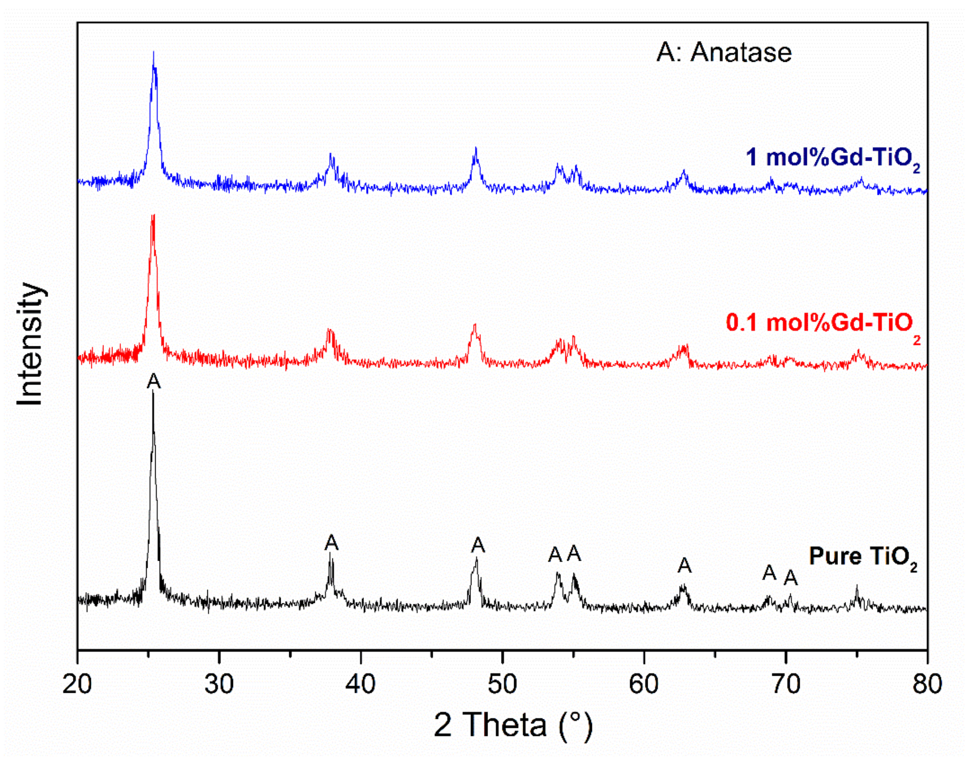
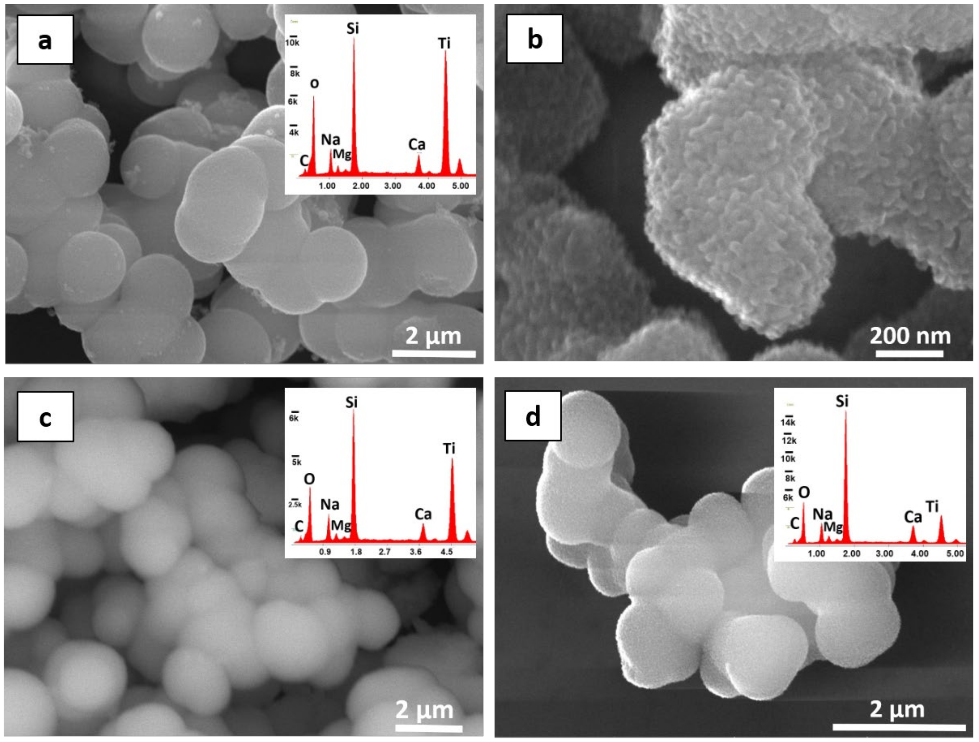
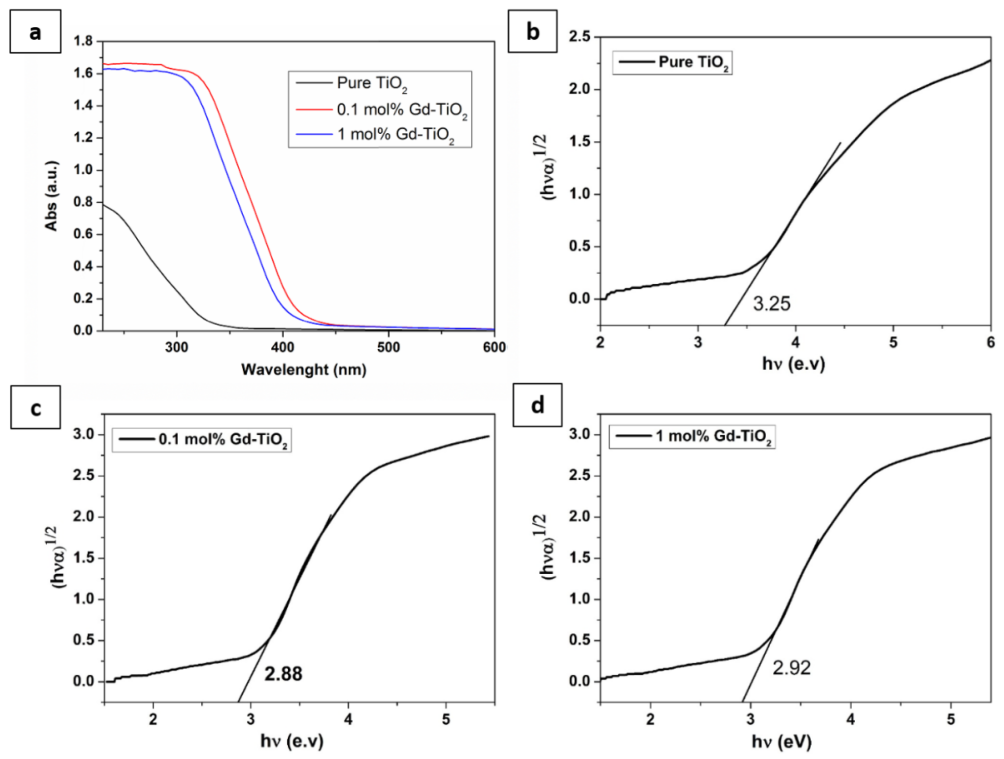
3.1. Characterisation of As-Prepared Pure and Doped TiO2 Nanoparticles

Figure 1 shows the XRD pattern of pure and Gd-doped TiO2 nanoparticles. Undoped and Gd-doped TiO2 samples exhibit anatase phase (JCPDS 21-1272). In this study, no peaks corresponding to rutile and brookite phases are detected in the XRD patterns.
Table 1 illustrates all the results related to the lattice parameters (a, b, and c) and lattice volume of all the samples. The average crystallite sizes were calculated based on the XRD data. It can be assumed that doping by Gd ions alters slightly lattice parameters and lattice volume of pure TiO2 NPs. This reduction cannot be considered significant and could be also attributed to instrumental and/or experimental errors. These results dismiss the hypothesis that Gd3+ may be inserted inside the TiO2 lattice. In fact, if this was the case, a distortion and volume inflation would be observed in the TiO2 lattice. It seems that gadolinium ions are impossible to enter the lattice of the TiO2 structure to replace Ti4+ ions due to the mismatch of the ionic radii of Ti4+ (0.94 Å) and Gd3+ (0.68 Å). Thus, in the Gd3+–TiO2 interface, Gd3+ ion may substitute for Ti ions in the lattice of TiO2, and a Ti–O–Gd bond might be created [59].
The morphology, size, and shape of pure and doped NPs were investigated by using SEM analysis (Figure 2). It can be observed from the micrographs that all the NPs are composed of agglomerated spheres of about 1–2 μm. These spheres consist of a considerable number of monodispersed crystallites with the size varying from 10 to 30 nm (Figure 2b) which is in agreement with the XRD result. Moreover, EDS analysis confirmed the chemical composition of those particles (Figure 2a,c,d). However, it was impossible to see the peak of ‘Gd’ due to the lower amount of doping which could not be detected by the instrument.
The distribution of particle size of pure and 0.1–1 mol% Gd-TiO2 NPs suspensions was measured by the Zeta Sizer. The obtained result displays a monomodal curve revealing that the NPs agglomerate in size of about 700 nm and 1000 nm (Figure S1). This result is in agreement with SEM micrographs.
The UV–Vis spectra of different NPs are plotted in Figure 3a. The absorption spectrum of TiO2 NPs consists of a single broad intense absorption around 300 nm attributed to the charge transfer from the valence band to the corresponding conduction band [60]. It is possible to infer that there is an important shift in the onset absorption towards the higher wavelength for all doped samples, compared with pure TiO2. In fact, doped samples exhibit enhanced absorption from λ = 380 nm, the onset of the visible region, compared with undoped samples. It was reported that the light absorption behaviour of TiO2 generally changes after doping [60]. Xu et al. suggested also that the appearance of a new electronic state in the middle of the TiO2 bandgap is behind this redshift [28]. It was also stated that the distance of charge transfer between f electrons of the gadolinium ions and the valence and conduction band of TiO2 is narrowed which consequently leads to an absorption response in the visible region [28]. Furthermore, a significant increase in the intensity of Gd-doped samples is observed in the UV region. Particularly, the sample doped with 0.1 mol% Gd3+ nanoparticles exhibited the highest visible-light absorption. A larger redshift might reveal that the sample absorbs more photons, and photoactivity is consequently enhanced [28].
Figure 3c–d exhibits the variation of the bandgap energy with the increase in dopant concentration. This is a key indicator of the visible light effectiveness of doped samples. Comparing the bandgap energy of neat TiO2 (3.2 eV) and the Gd3+-doped samples, doped samples showed a significant reduction in the bandgap values. It can be also seen (Figure 3c) that the bandgap of Gd3+–TiO2 NPs decreases to a minimum value of 2.88 eV at a Gd3+ concentration of 0.1 mol%; then, the optical bandgap starts to increase with sample doped with 1 mol% Gd (Figure 3d). Agorku et al. reported that the reduction in the bandgap with gadolinium doping might lead to the dominance of the d-f transitions over the sp-d transitions [60]. Xu et al. have assigned this amelioration to the expansion of excitation energy from UV to visible light due to the introduction of structural defects in the lattice of TiO2, which leads to changes in the bandgap energy [28]. These structural defects are established due to the partial substitution of Ti4+ by lanthanide ions to constitute the Ti-O-Gd structure. The formed compound displays two characteristic properties—the bandgap is reduced compared with that of pure TiO2 (the absorption spectra extend to longer wavelengths; consequently, photons with lower energy can be absorbed for photoreactions), and the positively charged Gd3+ may operate as an electron scavenger which affects the photocatalytic properties.

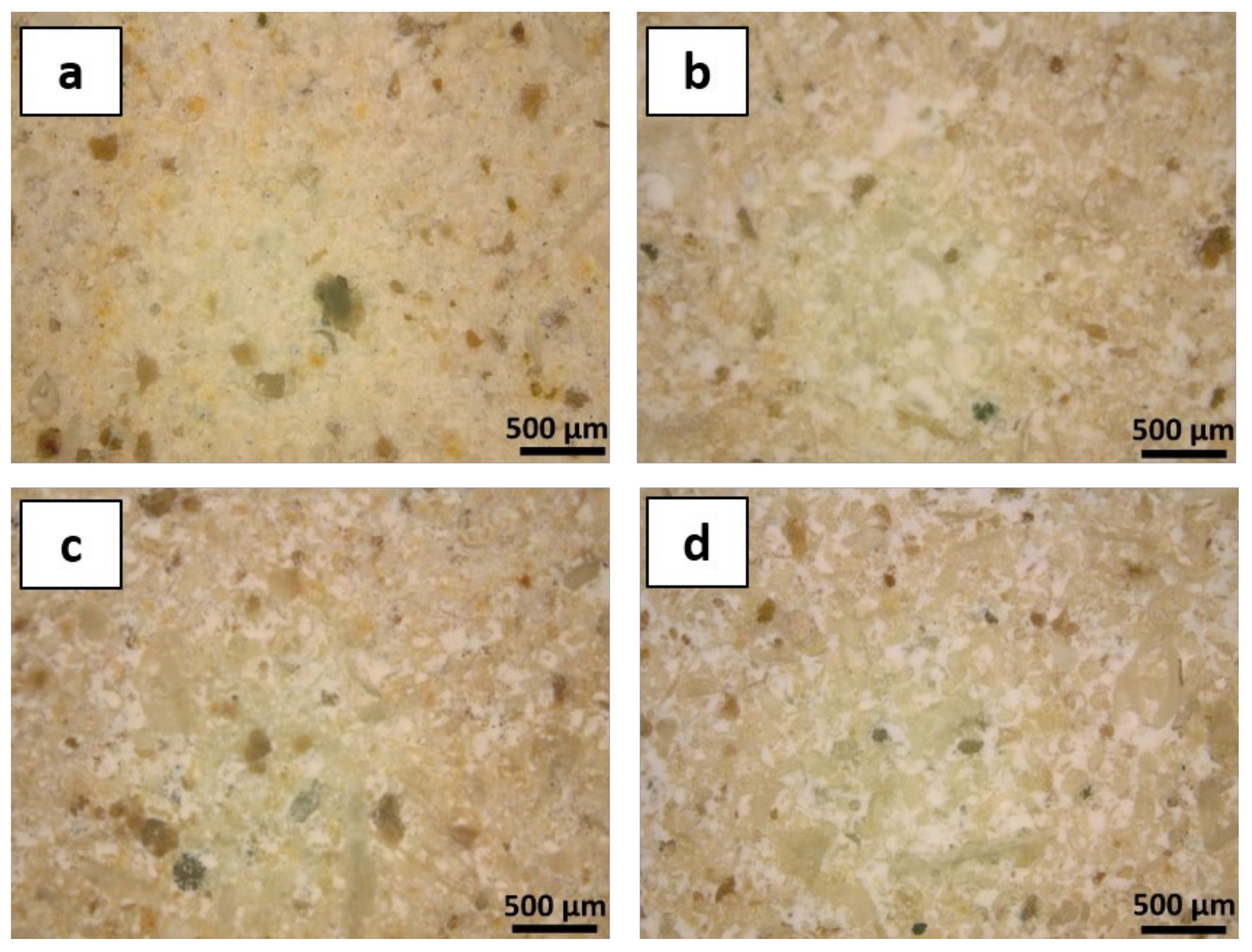
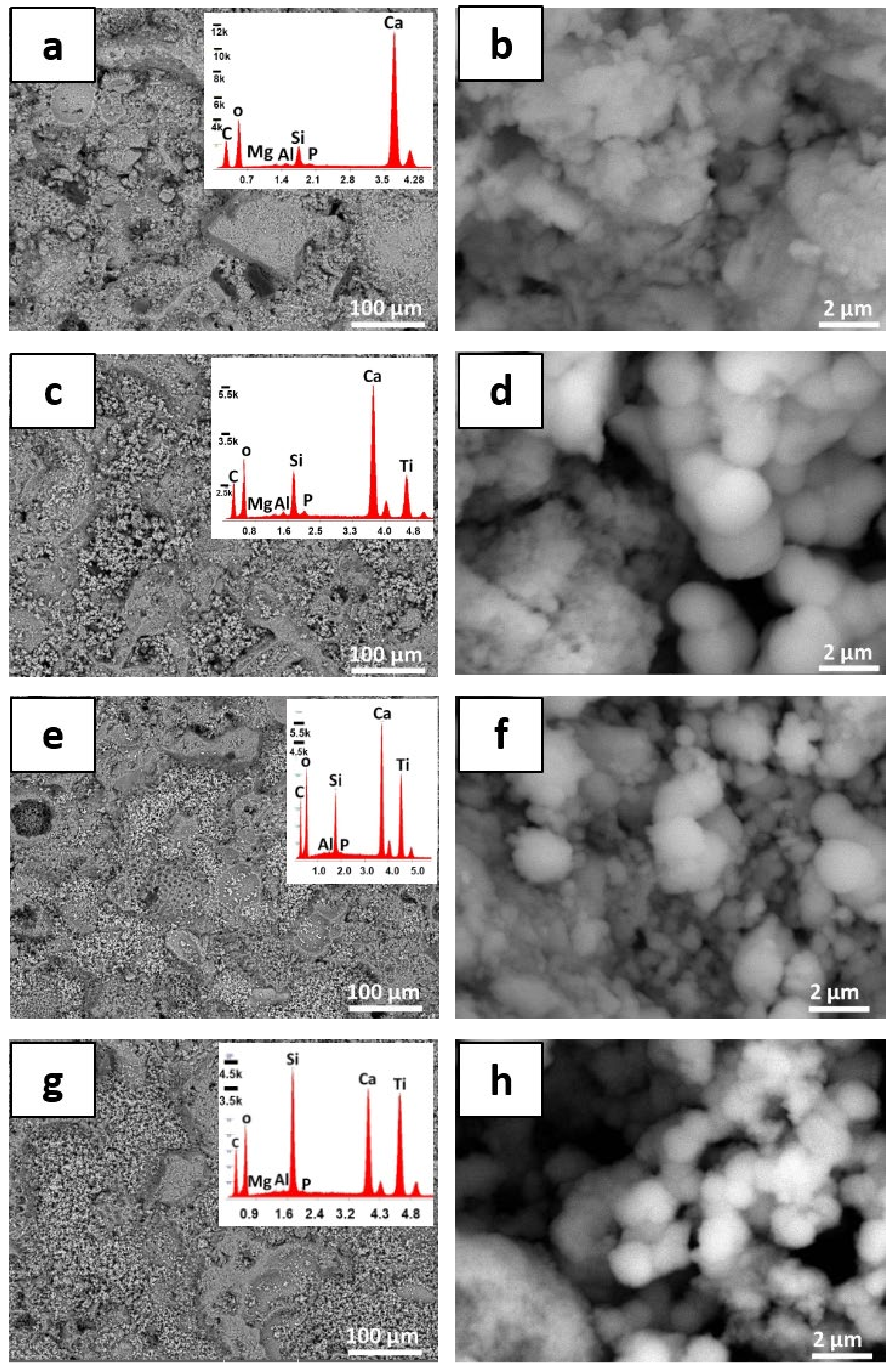
3.2. Modifications of the Stone Surface after Coatings Application

The thickness of the obtained film coatings was determined first because it is an essential and important result to understand the performance of the protective coatings, and it was estimated to be 20 ± 2 µm. Optical microscopy experiments were carried out under visible light, in order to have a clearer vision of the stone morphology after the application of each treatment (Figure 4). Observations exhibited that PDMS-treated specimens did not noticeably modify the morphology of the LS surface. Moreover, coatings made by both plain polymer and nanocomposites are well distributed on the LS surface and particularly localised in correspondence of the large pores or other less uniform areas (e.g., microfossils).
SEM observations were considered to evaluate the homogeneity of NPs dispersions on the LS surface and to obtain a clearer insight on the general microscopic features of the coating layer and the modifications induced to the surface morphology of the tested stone after application of different coatings. The micrographs were taken at two different magnifications for all the samples. Stone structural details such as large pores and microfossil shells can be clearly detected in the case of samples treated with plain PDMS (Figure 5a,b), while particles displaying the size varying between 1 and 2 µm (Figure 5c–h) and spherical shape are observed on the stone surface after application of the polymer containing both pure and doped TiO2 NPs. These large spheres were composed of a large number of nanosize agglomerated nanoparticles, which is in accordance with SEM micrographs previously taken on similar NPs (Figure 2). According to SEM observations, nanoparticles are quite homogeneously dispersed on the LS surface, although more localised in the pores of the stone surface. This is also in accordance with the optical microscopic observations. However, some microfossil shells and pores can be still observed on the surface of treated specimens, probably due to the poorly regular surface of Lecce stone, which may cause some occasional imperfections of the polymer thin films [5].
EDS analyses were also performed in order to gain information about the elemental composition of the coatings deposited on the stone surface. Most peaks observed in EDS spectra can be ascribed to the component of the stone substrate, Ca, O, and C, due to the main calcite (CaCO3) phase, while less intense peaks with, e.g., Si, Al, Mg, and P, can be due to the minor components of LS such as different aluminium–silicates and even phosphates deriving from microfossils. However, it should be considered that PDMS is a polymeric organosilicon compound; therefore, peaks corresponding to Si, C, and O can be ascribed also to the coating organic matrix. Analyses performed on LS specimens treated with NPs-containing polymer clearly indicate the presence of Ti, in addition to the previously mentioned elements, confirming the existence of dispersed inorganic NPs on the entire surface of LS.
EDS elemental mapping was carried out on the different treated stones to study the distribution of NPs in the polymeric matrix of the LS surface. Silicon and titanium distribution maps obtained from specimens treated with plain PDMS and with a polymer containing pure TiO2 NPs and Gd-doped NPs (1 mol% Gd) are reported in Figure 6. Elemental mapping experiments concerning silicon suggested that PDMS is fairly well distributed on the LS surface of all the examined samples. Ti elemental maps showed that this element, as expected, is not present in the stone substrate and that the TiO2 NPs are dispersed on the surface, similar to what was observed in the PDMS matrix. Some areas seem to be not effectively covered by both binder and nanoparticles. This can be explained by the poorly regular surface of Lecce stone that prevents a perfect distribution of coatings.
The overall chromatic variation ΔE* induced by treatment is a primary concern for aesthetic reasons in the field of heritage science. The colourimetric variation induced by the investigated nanocomposites on LS was determined by measuring the chromatic coordinates of treated and untreated specimens (CIELAB space) and expressed as ΔE* values. Results showed that overall chromatic variations for all treated samples are lower than 5, suggesting that all applied coatings do not alter, or alter to an acceptable extent, the original chromatic properties of heritage surfaces (Figure 7). It is worth noting that this result is valid for stone with creamy white colour. The quite high standard deviation values, which are observed in colourimetric measurements carried out on LS, are probably attributed to the surface heterogeneity of the substrate [43].
Colourimetric measurements also suggest that treatments affect the chromatic coordinates (L*, a*, and b*) of the LS surface to a different extent (Table 2). In fact, it could be observed that a* (green-red colour) is almost unaffected by treatments, while L* (lightness/darkness) and b* (blue/yellow) undergo more considerable variations. All treatments showed negative Δb* variations (an increase in blue colour) except the case of specimens treated with plain polymer (an increase in yellow colour). Moreover, all the specimens treated with polymer coatings underwent a deviation of the L* coordinate to negative values (lower lightness).
Before ageing, in accordance with the pencil test, the hardness of stone treated with pure PDMS exhibited the lowest level (H) with regard to all the other coatings (Table 3). Moreover, the surface hardness was improved by incorporating NPs on the LS surface. It is noteworthy that the PDMS coating containing 1 mol% Gd-doped TiO2 NPs was considerably harder, compared with undoped NPs. These findings are confirmed by the Vickers microhardness test. Indeed, an increase in hardness level can be showed after introducing TiO2 NPs on the LS surface, compared with their analogues treated by only PDMS (71 ± 2 Hv and 39 ± 3 Hv, respectively). In particular, Vickers hardness attained its maximum value (75 ± 2 Hv and 76 ± 3 Hv) with specimens treated with 0.1 and 1 Gd-doped NPs, respectively. These outcomes are probably attributed to the deposition of the NPs in the first mm of the stone matrix, therefore achieving a remarkable cohesion with the grains that are close to the stone surface [46].

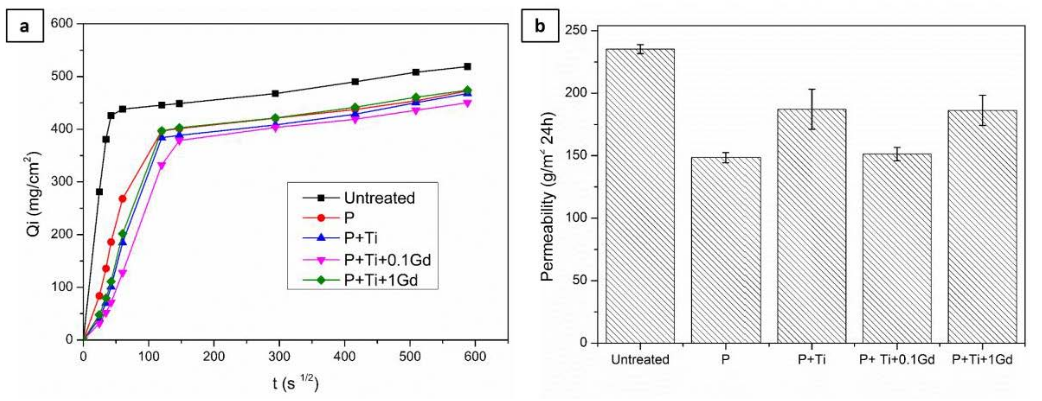
3.3. Evaluation of Hydrophobic Features of Different Treatments

The contact angle values were involved to examine the hydrophobic properties of LS due to the treatments. All the obtained results were resumed in Table 4. As can be seen, all the samples showed contact angle values higher than 90°, and even larger than 120°, ensuring the hydrophobic property of the protective films. As can be observed, the NPs enhance the water repellent features induced by PDMS on the LS surface. Interestingly, titanium dioxide NPs do not decrease the hydrophobic property of PDMS despite the super-hydrophilic character of nanosized titania; this fact may be reflected in the compatibility of NPs with the binder or might be due to the low concentration of NPs, compared with the binder material (PDMS). However, it could be also explained on the basis of the well-known lotus effect [61]. Hence, the surfaces of many plants display a high water repellence behaviour due to particular hierarchical micro- and nanoscale structures of the hydrophobic surface components [61,62]. Several works have been performed to reproduce similar structures by mixing hydrophobic polymers with nanoparticles. The resulting composite materials have been also applied to protect stony archaeological surfaces and, in some cases, even superhydrophobic behaviour has been observed [63,64]. It has also been reported that NPs create a roughened surface, while the siloxane polymers minimise contact with the water, resulting in what is known as the ‘Cassie–Baxter scenario’. Here, the air is trapped between the surface of the treated stone and the droplet of water inhibiting direct contact [65].
The capillary water absorption test was performed to gain a deeper insight into the water repellence behaviour of each treatment. Absorption curves (adsorbed water vs. square root of time) of different tested specimens are displayed in Figure 8a. In the case of untreated stone, saturation was obtained after 1 h of contact with water, while the treated stone samples show a considerable reduction in the water absorption rate, and saturation is reached after about 6 h. However, at the equilibrium, only a slight reduction in adsorbed water (about 10%) can be observed by comparing coated and uncoated specimens. These results do not appear in agreement with the high static angle measurements. Water capillary absorption diagrams (Figure 8a) indicate that the protective action of treatment improved the stone hydrophobicity during the first few hours after contacting the specimens with water.
It has been reported that the results of capillary water suction are often in disagreement with the hydrophobic properties carried out on the same stone samples [12]. This incongruity can be explained taking into account that the contact angle value is related to an instantaneous water-repellent behaviour (contact angle is usually measured after a very short contact time [51]), while the long-term resistance to water penetration can be better assessed by the capillary absorption test (which usually lasts several hours [52]). Thus, the results of contact angle measurements should be considered with caution when studying the real hydrophobic features of a coating [42].
The results of the water vapour permeability test are graphically summarised in Figure 8b. The permeability of all treated specimens decreased (by about 20–35%) when compared to untreated LS. It is due to the superficial thin films formed after treatments, which may act as a hydrophobic barrier against not only liquid water but also water vapour [5]. Moreover, treatments involving nanoparticles may induce, in general, a reduction in vapour permeability [42]. A weak rate of vapour diffusion could cause a local increase in the vapour pressure at each stone–coating interface and may lead consequently to stress and damage of the substrate [5]. In addition, inhibition of vapour transport from inside the stone to the environment can lead to a gradual waste of the protective coating performance [66]. Indeed, water could partially condense under the polymer film which can weaken the adhesion of the coating. Thus, a substantial reduction in vapour diffusion can be a drawback for the protecting materials [2]. In the present work, the investigated coatings did not dramatically alter the ‘breath function’ of the original stone, since the permeability reduction levels are lower than 40%. Thus, residual permeability is still considered acceptable. Results of vapour permeability test, in agreement with capillary absorption data, suggest that coatings induce only slight variations in the porous structure of the tested stone, as both suction rate and vapour permeability undergo moderate and acceptable reduction.
Generally, a hybrid coating applied to enhance the stone hydrophobicity of artwork materials is intended to certainly improve the stone hydrophobicity and, at the same time, do not significantly alter the colour parameters and do not dramatically affect the ‘breath function’ of the original material. All these basic requirements were obtained after the application of the investigated nanocomposites.

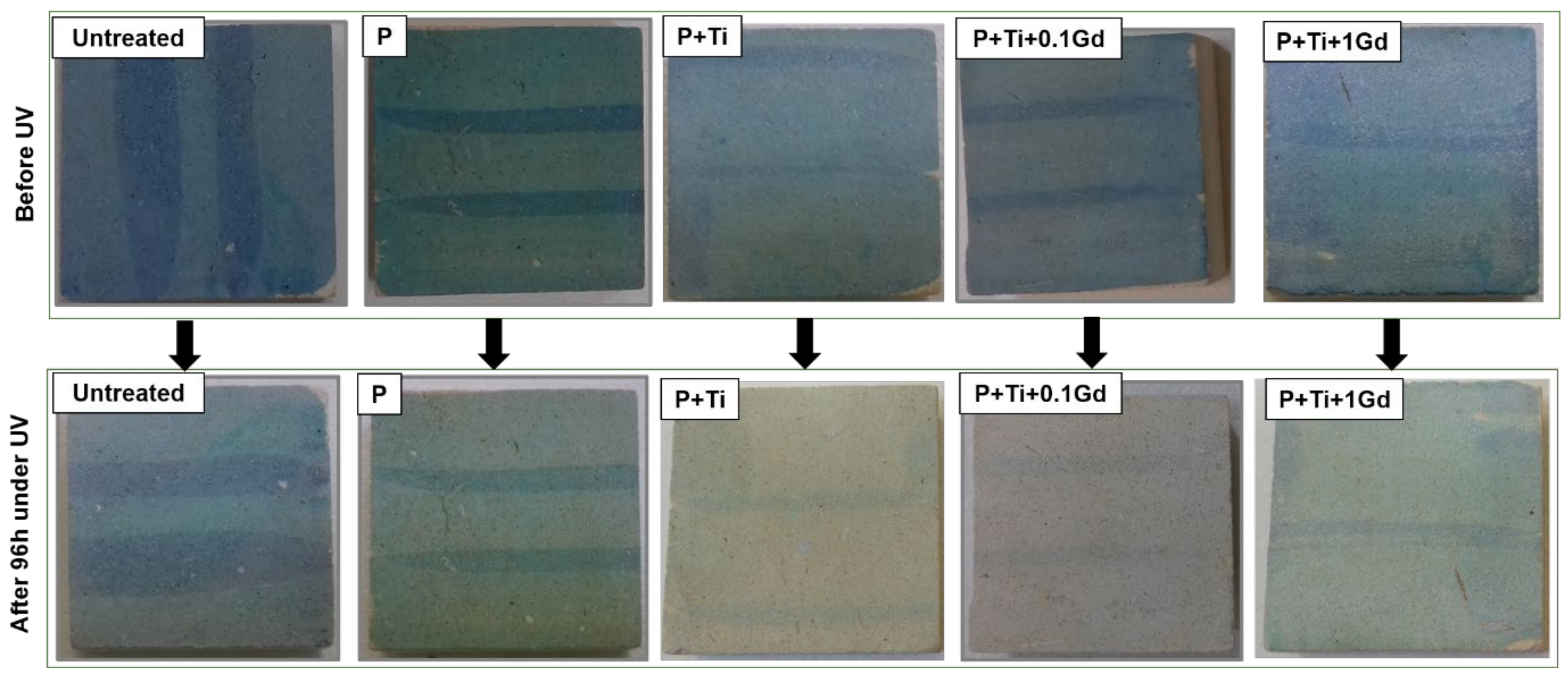
3.4. Self-Cleaning Test

In order to investigate the photodegradation performance of applied coatings, untreated and treated samples, stained by MB dye, were exposed to UV irradiation and monitored via chromatic changes. The photocatalytic discolouration of dye over time was performed by two methods—namely, (i) evaluating the chromatic coordinate b* attributed to methylene blue colour and (ii) evaluating the overall colour variation after applying the methylene blue dye and at the end of UV exposition.
The results obtained from the photocatalytic analysis on LS samples are presented in Table 5, where the obtained discolouration factor D* (%) at different times is reported. Untreated samples indicated a slight MB degradation due to the photochemical mechanism (i.e., photolysis) [67]. The discolouration rate showed a 15% increase after applying plain PDMS. This fact may due to the water repellent behaviour of the surface, which may prevent the penetration of the dye into the stone pores, and consequently, the stain remains on the surface of LS. Thus, the photodecomposition of the dye becomes more feasible. As reported previously and confirmed by this study, the direct photolysis of the organic dyes is slower than the photodegradation induced by TiO2 [68]. Better performances are obtained on samples treated with NPs-containing polymer. The increased oxidation rates of methylene blue dye can be attributed to the photocatalytic activity of pure or doped TiO2 NPs on the stone surface. Faster degradation of organic dyestuff was observed for LS treated with PDMS containing Gd–TiO2 NPs with the lowest doping concentration (0.1 mol%). This material showed an enhanced degradation profile, compared with pure TiO2 NPs with a discolouration rate of about 71% and 49% after 96 h, respectively. On the contrary, 1 mol% Gd-TiO2 showed an insignificant increase in the oxidation rate, compared with pure TiO2 NPs (D* of about 53% after 96 h). It is also worth noting that the photodegradation activity mainly occurs in the first 48 h; after that, its kinetics becomes slower.
The effect of the self-cleaning behaviour was also evaluated by considering the overall chromatic changes. The results of the photocatalytic test performed on LS samples are presented in Figure 9, in which ΔE*/ΔE0* (%) ratios obtained at different times are graphically illustrated. Results showed that treatment with plain PDMS enhanced the degradation ratio to double (in comparison with plain LS) at the end of the experiment. However, a faster degradation of organic dyestuff was observed for LS treated with doped materials. The coating containing the lowest dopant (Gd) concentration (0.1 mol%) showed enhanced degradation effectiveness, compared with pure TiO2 NPs. Furthermore, the measurements confirmed that TiO2 NPs doped with 1% Gd were less efficient than 0.1% analogues, and its degradation efficiency was comparable to (or even lower than) the plain TiO2 NPs. This result can be explained by the bandgap calculations that were performed on both pure and Gd-doped TiO2 NPs, indicating that samples doped with 0.1 mol% Gd ions revealed the narrowest bandgap and, consequently, the higher photocatalytic activity (Figure 3).
Figure 10 indicates the photographs of the Lecce stone specimens before and after the accomplishment of the analysis. After finishing, UV illumination (96 h), MB stains applied on the bare stone surface are quite similar to those before irradiation. Specimens treated with only polymer showed a slight discolouration of the MB dye after 96 h of irradiation. However, samples treated with polymer-containing NPs showed a significant discolouration of the blue stains. In particular, the coating containing TiO2 NPs doped with 0.1 mol% Gd displayed the highest activity. This result may be explained on the basis of the higher e/h+ separation and the higher absorption of light of the doped titania, even if compared with NPs containing a larger amount of Gd (1%), as shown in our previous work [41].

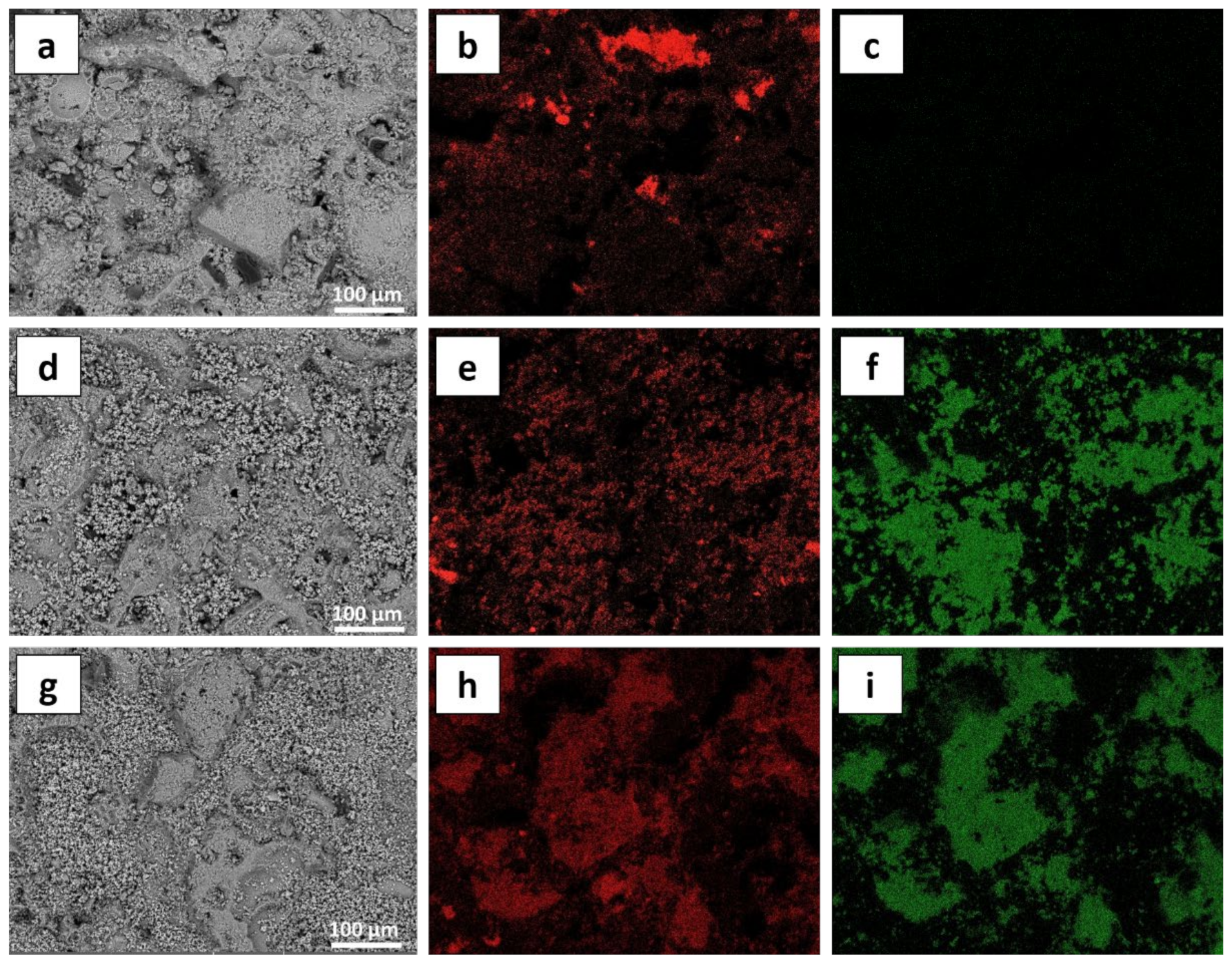
3.5. Antimicrobial Activity

Antimicrobial tests were carried out on the treated LS to test the inhibition efficacy of doped coating (Figure 11). In this research work, untreated (as a control) and specimens treated with P + Ti + 0.1Gd were tested. Only samples doped with 0.1 mol% Gd were tested in this section because they showed the best photodegradation activity, compared with other tested NPs. A stabilised mixture containing five microbial strains was used.
After three months of incubation of microorganisms on untreated/treated specimens, surface colonisation was evaluated. Results showed that, contrary to the untreated stone, colonisation occurs over all the surface, and microorganisms strictly adhere to the stone (Figure 11b). For samples treated with polymer mixed with doped TiO2 NPs, microbial suspensions are in the form of droplets on the LS surface, and biofilm is formed only where the drops stand (Figure 11f). Moreover, a dried and very thin biofilm, compared with the biofilm formed on untreated stone, is obtained. This result reveals that microbial colonisation is not able to propagate on the surface of LS when they are in contact with NPs which means that treatment with P + Ti + 0.1Gd inhibits microbial colonisation, even after three months of incubation. These interesting results are probably related to the synergistic hydrophobic features of thin-film coatings which hinder the biofilm from strongly adhering to the studied stone specimens and the photokilling action of doped NPs due to their enhanced photocatalytic activity under visible light. In fact, doping TiO2 NPs with gadolinium ions afford a simultaneous generation of reactive oxygen species (ROS) and visible photoresponse, as already in our previous work [41]. After three months of incubation, stereo microscope observations showed a diffuse growth of fungal colonies, represented as black spots, on the surface of untreated specimens (Figure 11c), while a scarce presence of fungal spores can be detected on treated probes (Figure 11g). Observations under the confocal laser-scanning microscope (CLSM) were carried out at the end of the experiment (i.e., after 90 days). CLSM images show colonisation of all the inoculated microorganisms with a wide presence of bacteria, algae, and yeasts in addition to black fungi (black colour) in untreated specimens (Figure 11d). In the case of specimens coated with doped NPs (Figure 11h), algae, yeasts, and bacteria are visible. Dark spots indicate the presence of black fungi. These observations reveal that treatment with P + Ti + 0.1Gd did not inactivate microorganisms; however, it was able to prevent biofilms from spreading out over all the surface, i.e., biofilms were formed only in the spots with drops.

3.6. Durability of Nanocomposite Protective Coatings

The durability of nanocomposite protective thin films was evaluated through accelerated artificial solar ageing cycles (exposed to both UVA and UVB irradiation around 1000 h). After the ageing test, chromatic variation, static contact angle, pencil hardness measurements were performed. Figure 7 illustrates the effect of solar light on the chromatic properties of LS surfaces by representing the overall chromatic variations. The ΔE* values suggested acceptable variations after ageing of the TiO2-doped materials. It should be noted that the conditions to accept a conservative treatment include the following [69]: the applied material should not cause a visible alteration and must be stable over time. Moreover, it is worth noting that chromatic variations are higher than the lower limit for human eye perception, also for samples treated with only polymer and PDMS mixed with pure TiO2 NPs.
In order to deeply examine the light-induced effect on each sample’s chromatic coordinates (L*, a*, and b*), values of the observed variations are presented in Table 2. According to the outcomes, for samples treated with only polymer, L* and b* are more affected; it seems that samples assume a more white-blueish colour. For specimens treated by PDMS and plain TiO2 NPs, only L* is significantly altered. The positive value of ΔL* indicates the colour of the LS surface is shifted to the white colour.
Additionally, all the examined coatings underwent a slight decrease in water repellent behaviour after the ageing test (Table 4). Nevertheless, contact angle values remained distinctly higher than 100°, confirming that the hydrophobic character displayed by the PDMS-based treatments was not or was poorly affected by the prolonged light irradiation. This suggested that the polymer coating is still present on the LS surface and keeps its protecting features even after 1000 h of exposition.
It can be also noted that solar ageing considerably reduced the hardness of surfaces treated by polymer and undoped NPs mixed with polymer (F and H, respectively), while doped NPs mixed with PDMS displayed quite similar behaviour as before (Table 3).
The self-cleaning performance of the protective coatings which underwent an accelerated ageing test was investigated by the degradation of MB dye to understand better the durability of newly synthesised coatings. The oxidation rates (D* (%)) of the MB dye are illustrated in Table 5. Results revealed only a small diminution in the effectiveness of photodegradation of all the considered coatings after the accelerated ageing test, which revealed the resistance of the protective coatings against ageing. Particularly, specimens treated with P + Ti + 0.1Gd showed the highest photodegradation performance, compared with the other coatings. These findings were confirmed by the overall chromatic variation ratios (ΔE*/ΔE0* (%)) measured at different times (Figure 9b). The results showed that samples treated with the lowest doping concentration (0.1 mol%) exhibited the highest degradation profile, compared with pure TiO2 NPs. These outcomes reveal the good durability of the newly developed protective coating composed of Gd doping ions (0.1 mol%).
Moreover, in order to analyse the durability of coatings after long-term microbial contact, the test was continued by verifying the ability of treated probes to inhibit microbial colonisation after one year of incubation (Figure 12). Untreated probes were tested as a control. Observations showed that treated samples revealed a good long-term microbial growth inhibition by preventing microorganisms to populate overall the LS surface (Figure 12c), with microbial colonisation still occurring only where there were drops (Figure S2c,d). On the contrary, a diffuse growth of fungal colonies can be detected on untreated LS, as shown by stereo microscope observations (Figure 12b). Fungal colonisation (black spots) can be clearly seen even at the edge of stony probes which confirms that colonisation occurs on the whole surface (Figure S2a,b). Inhibition in fungal cell growth induced by surface treatments can be observed by microphotography (Figure 12d). It is worth noting that only a few biocides have been reported to be active against fungal strain [70] which indicates the efficiency of the newly developed protective coating to inhibit microbial and particularly fungal colonisation.

4. Conclusions

In this work, thin films of different materials composed principally of PDMS as a binder and pure/Gd-doped TiO2 NPs were applied on the Lecce stone as protective coatings. The XRD analysis confirmed the anatase phase of as-prepared NPs. SEM analysis showed TiO2 sphere in the range of 1 µm consists of an enormous number of finer particles (10–30 nm) which is confirmed by Zeta Sizer measurements. UV–Vis analysis revealed that the optical properties of doped materials were found to be significantly enhanced by incorporating Gd ions due to the narrowed bandgap (2.88–2.92 e.V), compared with pure NPs (3.2 e.V).
All the applied coatings showed acceptable chromatic variations: ΔE* < 5. The contact angle test indicated that all the coated surfaces showed hydrophobic behaviour, while the capillary absorption test demonstrated that the kinetics of water capillary absorption is affected by treatments only during the first few hours. Moreover, treatments do not radically disturb the ‘breath function’ of the original material. Pencil and Vickers hardness test showed that surface hardness was significantly enhanced after introducing NPs in the organic polymer. Furthermore, morphological analyses suggested that NPs were well mixed in the organic polymer and distributed on practically the whole surface of LS. However, EDS map observations showed that some areas are not filled with the NPs because of the imperfectly regular substrate of LS. This fact may also explain the moderate alteration from a water transport perspective in both liquid and gas forms within LS specimens.
For the photodegradation test, samples doped with 0.1 mol% Gd showed the highest self-cleaning activity under UV light, as the discolouration of MB dye at the end of the test was about 70%. In addition, the same samples showed an inhibitory effect against microbial colonisation after 3 months of incubation. In addition, ageing test results showed that only specimens treated by doped materials showed tolerable chromatic variations over time. On the other hand, the results point out that solar irradiation did not considerably affect the water repellent behaviour of applied coatings. Moreover, the surface hardness of LS treated with doped materials was insignificantly affected by solar irradiation. Finally, the photodegradation performance of protective coatings was slightly affected and P + Ti + 0.1Gd coated specimens showed the highest self-cleaning activity (both photodegradation and antimicrobial). Hence, it can be considered a durable coating.
In conclusion, we can deduce that PDMS mixed with 0.1 mol% Gd-TiO2 materials can be considered a good durable candidate to protect highly porous heritage building surfaces. However, more work is in progress to investigate the performances of these thin-film coatings against other stains and under real-life conditions.

Supplementary Materials

The following are available online at https://www.mdpi.com/article/10.3390/su131911033/s1, Figure S1: DLS analysis of synthesized (a) pure TiO2 NPs and (b) 0.1; (c) 1 mol% Gd-doped TiO2 NPs; . Figure S2: Microphotography of the colonized LS surface of (a,b) untreated and (c,d) samples treated with P+Ti+0.1Gd after incubation for one year. The objective has a magnification of x80.

Author Contributions

Conceptualization, M.B.C., M.L.W. and M.L.; methodology, M.B.C., M.L.W., F.D.L. and C.U.; validation, M.L.W., C.U., M.M. and J.B.; formal analysis, C.U., F.D.L. and D.S.; investigation, M.B.C. and F.D.L.; resources, M.L., C.U. and M.M.; data curation, M.L.W.; supervision, M.M., M.L. and C.U.; writing—original draft preparation, M.B.C.; writing—review and editing, M.L.W., M.L. and C.U. All authors have read and agreed to the published version of the manuscript.

Funding

This research received no external funding.

Institutional Review Board Statement

Not applicable.

Informed Consent Statement

Not applicable.

Data Availability Statement

The data presented in this research study are available in the present article and in the related Supplementary Information.

Acknowledgments

The authors gratefully acknowledge Ilenia Tredici, CISRiC, the University of Pavia, for handling the SEM–EDS analysis. Gioele Capillo of the Department of Veterinary Sciences, the University of Messina, for his kind guidance with Confocal Microscopy observations and images.

Conflicts of Interest

The authors declare no conflict of interest.

References

  1. Urzì, C. Microbial Deterioration of Rocks and Marble Monuments of the Mediterranean Basin: A Review, Corrosion Reviews. Corros. Rev. 2004, 22, 441–458. [Google Scholar] [CrossRef]
  2. Licchelli, M.; Malagodi, M.; Weththimuni, M.; Zanchi, C. Anti-graffiti nanocomposite materials for surface protection of a very porous stone. Appl. Phys. A 2014, 116, 1525–1539. [Google Scholar] [CrossRef]
  3. Belfiore, C.M.; Fichera, G.V.; la Russa, M.F.; Pezzino, A.; Ruffolo, S.A. The Baroque architecture of Scicli (south-eastern Sicily): Characterization of degradation materials and testing of protective products. Period. Mineral. 2012, 81, 19–33. [Google Scholar] [CrossRef]
  4. Ricca, M.; le Pera, E.; Licchelli, M.; Macchia, A.; Malagodi, M.; Randazzo, L.; Rovella, N.; Ruffolo, S.A.; Weththimuni, M.L.; la Russa, M.F. The CRATI Project: New Insights on the Consolidation of Salt Weathered Stone and the Case Study of San Domenico Church in Cosenza (South Calabria, Italy). Coatings 2019, 9, 330. [Google Scholar] [CrossRef] [Green Version]
  5. Licchelli, M.; Malagodi, M.; Weththimuni, M.L.; Zanchi, C. Water-repellent properties of fluoroelastomers on a very porous stone: Effect of the application procedure. Prog. Org. Coat. 2013, 76, 495–503. [Google Scholar] [CrossRef]
  6. Roig, P.B.; Ros, J.L.R.; Estellés, R.M. Biocleaning of nitrate alterations on wall paintings by Pseudomonas stutzeri. Int. Biodeterior. Biodegrad. 2013, 84, 266–274. [Google Scholar] [CrossRef] [Green Version]
  7. Sanmartín, P.; DeAraujo, A.; Vasanthakumar, A.; Mitchell, R. Feasibility study involving the search for natural strains of microorganisms capable of degrading graffiti from heritage materials. Int. Biodeterior. Biodegrad. 2015, 103, 186–190. [Google Scholar] [CrossRef]
  8. Staicu, A.; Apostol, I.; Pascu, A.; Urzica, I.; Pascu, M.L.; Damian, V. Minimal invasive control of paintings cleaning by LIBS. Opt. Laser Technol. 2016, 77, 187–192. [Google Scholar] [CrossRef]
  9. Balliana, E.; Ricci, G.; Pesce, C.; Zendri, E. Assessing the value of green conservation for cultural heritage: Positive and critical aspects of already available methodologies. Int. J. Conserv. Sci. 2016, 7, 185–202. [Google Scholar]
  10. Sierra-Fernandez, A.; Gomez-Villalba, L.S.; Rabanal, M.E.; Fort, R. New nanomaterials for applications in conservation and restoration of stony materials: A review. Mater. Construcción 2017, 67, 107. [Google Scholar] [CrossRef]
  11. D’Orazio, L.; Grippo, A. A water dispersed Titanium dioxide/poly(carbonate urethane) nanocomposite for protecting cultural heritage: Preparation and properties. Prog. Org. Coat. 2015, 79, 1–7. [Google Scholar] [CrossRef]
  12. la Russa, M.F.; Rovella, N.; de Buergo, M.A.; Belfiore, C.M.; Pezzino, A.; Crisci, G.M.; Ruffolo, S.A. Nano-TiO2 coatings for cultural heritage protection: The role of the binder on hydrophobic and self-cleaning efficacy. Prog. Org. Coat. 2016, 91, 1–8. [Google Scholar] [CrossRef]
  13. Quagliarini, E.; Bondioli, F.; Goffredo, G.B.; Cordoni, C.; Munafò, P. Self-cleaning and de-polluting stone surfaces: TiO2 nanoparticles for limestone. Constr. Build. Mater. 2012, 37, 51–57. [Google Scholar] [CrossRef]
  14. Munafò, P.; Goffredo, G.B.; Quagliarini, E. TiO2-based nanocoatings for preserving architectural stone surfaces: An overview. Constr. Build. Mater. 2015, 84, 201–218. [Google Scholar] [CrossRef]
  15. Graziani, L.; Quagliarini, E.; Bondioli, F.; D’Orazio, M. Durability of self-cleaning TiO2 coatings on fired clay brick façades: Effects of UV exposure and wet & dry cycles. Build. Environ. 2014, 71, 193–203. [Google Scholar] [CrossRef]
  16. Crupi, V.; Fazio, B.; Gessini, A.; Kis, Z.; la Russa, M.F.; Majolino, D.; Masciovecchio, C.; Ricca, M.; Rossi, B.; Ruffolo, S.A.; et al. TiO2–SiO2–PDMS nanocomposite coating with self-cleaning effect for stone material: Finding the optimal amount of TiO2. Constr. Build. Mater. 2018, 166, 464–471. [Google Scholar] [CrossRef]
  17. Xu, A.W.; Gao, Y.; Liu, H.Q. The preparation, characterization, and their photocatalytic activities of rare-earth-doped TiO2 nanoparticles. J. Catal. 2002, 207, 151–157. [Google Scholar] [CrossRef]
  18. Yadav, S.; Jaiswar, G. Review on Undoped/Doped TiO2 Nanomaterial; Synthesis and Photocatalytic and Antimicrobial Activity: Review on Undoped/Doped TiO2 Nanomaterial. J. Chin. Chem. Soc. 2017, 64, 103–116. [Google Scholar] [CrossRef]
  19. Chobba, M.B.; Weththimuni, M.L.; Messaoud, M.; Urzi, C.; Bouaziz, J.; de Leo, F.; Licchelli, M. Ag-TiO2/PDMS nanocomposite protective coatings: Synthesis, characterization, and use as a self-cleaning and antimicrobial agent. Prog. Org. Coat. 2021, 158, 106342. [Google Scholar] [CrossRef]
  20. Merenda, A.; Rana, A.; Guirguis, A.; Zhu, D.M.; Kong, L.; Dumée, L.F. Enhanced Visible Light Sensitization of N-Doped TiO2 Nanotubes Containing Ti-Oxynitride Species Fabricated via Electrochemical Anodization of Titanium Nitride. J. Phys. Chem. C 2019, 123, 2189–2201. [Google Scholar] [CrossRef]
  21. Langlet, M.; Coutier, C.; Fick, J.; Audier, M.; Meffre, W.; Jacquier, B.; Rimet, R. Sol–gel thin film deposition and characterization of a new optically active compound: Er2Ti2O7. Opt. Mater. 2001, 16, 463–473. [Google Scholar] [CrossRef]
  22. Fonseca, A.J.; Pina, F.; Macedo, M.F.; Leal, N.; Romanowska-Deskins, A.; Laiz, L.; Gómez-Bolea, A.; Saiz-Jimenez, C. Anatase as an alternative application for preventing biodeterioration of mortars: Evaluation and comparison with other biocides. Int. Biodeterior. Biodegrad. 2010, 64, 388–396. [Google Scholar] [CrossRef] [Green Version]
  23. Ruffolo, S.A.; Macchia, A.; la Russa, M.F.; Mazza, L.; Urzì, C.; de Leo, F.; Barberio, M.; Crisci, G.M. Marine Antifouling for Underwater Archaeological Sites: TiO2 and Ag-Doped TiO2. Int. J. Photoenergy 2013, 2013, 251647. [Google Scholar] [CrossRef] [Green Version]
  24. la Russa, M.F.; Macchia, A.; Ruffolo, S.A.; de Leo, F.; Barberio, M.; Barone, P.; Crisci, G.M.; Urzì, C. Testing the antibacterial activity of doped TiO2 for preventing biodeterioration of cultural heritage building materials. Int. Biodeterior. Biodegrad. 2014, 96, 87–96. [Google Scholar] [CrossRef]
  25. Luo, W.; Hou, J.; Zou, D.; Cui, L.; Zhu, Q.; Dai, J. Lanthanide-titanium-oxalate clusters and their degradation products, photocurrent response and photocatalytic behaviours. New J. Chem. 2018, 42, 11629–11634. [Google Scholar] [CrossRef]
  26. Saqib, N.U.; Adnan, R.; Shah, I. A mini-review on rare earth metal-doped TiO2 for photocatalytic remediation of wastewater. Environ. Sci. Pollut. Res. 2016, 23, 15941–15951. [Google Scholar] [CrossRef] [PubMed]
  27. Mazierski, P.; Mikolajczyk, A.; Bajorowicz, B.; Malankowska, A.; Zaleska-Medynska, A.; Nadolna, J. The role of lanthanides in TiO2-based photocatalysis: A review. Appl. Catal. B Environ. 2018, 233, 301–317. [Google Scholar] [CrossRef]
  28. Xu, J.; Ao, Y.; Fu, D.; Yuan, C. Synthesis of Gd-doped TiO2 nanoparticles under mild condition and their photocatalytic activity. Colloids Surf. A Physicochem. Eng. Asp. 2009, 334, 107–111. [Google Scholar] [CrossRef]
  29. Paul, S.; Chetri, P.; Choudhury, B.; Ahmed, G.A.; Choudhury, A. Enhanced visible light photocatalytic activity of Gadolinium doped nanocrystalline titania: An experimental and theoretical study. J. Colloid Interface Sci. 2015, 439, 54–61. [Google Scholar] [CrossRef] [PubMed]
  30. Saif, M.; El-Molla, S.A.; Aboul-Fotouh, S.M.K.; Ibrahim, M.M.; Ismail, L.F.M.; Dahn, D.C. Nanostructured Gd3+-TiO2 surfaces for self-cleaning application. J. Mol. Struct. 2014, 1067, 120–126. [Google Scholar] [CrossRef]
  31. Mahalakshmi, M.; Arabindoo, B.; Palanichamy, M.; Murugesan, V. Preparation, Characterization, and Photocatalytic Activity of Gd3+ Doped TiO2 Nanoparticles. J. Nanosci. Nanotechnol. 2007, 7, 3277–3285. [Google Scholar] [CrossRef] [PubMed]
  32. Cheng, X.Q.; Ma, C.Y.; Yi, X.Y.; Yuan, F.; Xie, Y.; Hu, J.M.; Hu, B.C.; Zhang, Q.Y. Structural, morphological, optical and photocatalytic properties of Gd-doped TiO2 films. Thin Solid Films 2016, 615, 13–18. [Google Scholar] [CrossRef]
  33. Thomas, P.J.; Carpenter, D.; Boutin, C.; Allison, J.E. Rare earth elements (REEs): Effects on germination and growth of selected crop and native plant species. Chemosphere 2014, 96, 57–66. [Google Scholar] [CrossRef] [Green Version]
  34. Pagano, G.; Guida, M.; Tommasi, F.; Oral, R. Health effects and toxicity mechanisms of rare earth elements—Knowledge gaps and research prospects. Ecotoxicol. Environ. Saf. 2015, 115, 40–48. [Google Scholar] [CrossRef]
  35. Liu, Z.; Guo, C.; Tai, P.; Sun, L.; Chen, Z. The exposure of gadolinium at environmental relevant levels induced genotoxic effects in Arabidopsis thaliana (L.). Ecotoxicol. Environ. Saf. 2021, 215, 112138. [Google Scholar] [CrossRef]
  36. Weththimuni, M.L.; Capsoni, D.; Malagodi, M.; Licchelli, M. Improving Wood Resistance to Decay by Nanostructured ZnO-Based Treatments. J. Nanomater. 2019, 2019, 6715756. [Google Scholar] [CrossRef]
  37. Alexandru, M.; Cazacu, M.; Doroftei, F.; Ignat, M.; Timpu, D.; Grigoras, C.V.; Simionescu, B.C. On the morphology and potential application of polydimethylsiloxane-silica-titania composites. Express Polym. Lett. 2011, 5, 188–196. [Google Scholar] [CrossRef]
  38. Tavares, M.T.S.; Santos, A.S.F.; Santos, I.M.G.; Silva, M.R.S.; Bomio, M.R.D.; Longo, E.; Paskocimas, C.A.; Motta, F.V. TiO2/PDMS nanocomposites for use on self-cleaning surfaces. Surf. Coat. Technol. 2014, 239, 16–19. [Google Scholar] [CrossRef] [Green Version]
  39. Kapridaki, C.; Pinho, L.; Mosquera, M.J.; Maravelaki-kalaitzaki, P. Producing photoactive, transparent and hydrophobic SiO2-crystalline TiO2 nanocomposites at ambient conditions with application as self-cleaning coatings. Appl. Catal. B Environ. 2014, 156–157, 416–427. [Google Scholar] [CrossRef]
  40. Kapridaki, C.; Maravelaki-Kalaitzaki, P. TiO2-SiO2-PDMS nano-composite hydrophobic coating with self-cleaning properties for marble protection. Prog. Org. Coat. 2013, 76, 400–410. [Google Scholar] [CrossRef]
  41. Chobba, M.B.; Messaoud, M.; Weththimuni, M.L.; Bouaziz, J.; Licchelli, M.; de Leo, F.; Urzì, C. Preparation and characterization of photocatalytic Gd-doped TiO2 nanoparticles for water treatment. Environ. Sci. Pollut. Res. 2019, 26, 32734–32745. [Google Scholar] [CrossRef]
  42. Licchelli, M.; Malagodi, M.; Weththimuni, M.; Zanchi, C. Nanoparticles for conservation of bio-calcarenite stone. Appl. Phys. A 2014, 114, 673–683. [Google Scholar] [CrossRef]
  43. Weththimuni, M.L.; Licchelli, M.; Malagodi, M.; Rovella, N.; Russa, M.L. Consolidation of bio-calcarenite stone by treatment based on diammonium hydrogenphosphate and calcium hydroxide nanoparticles. Measurement 2018, 127, 396–405. [Google Scholar] [CrossRef]
  44. UNI 10921:2001, Beni Culturali—Materiali Lapidei Naturali ed Artificiali—Prodotti Idrorepellenti—Applicazione su Provini e Determinazione in Laboratorio delle loro Caratteristiche; Milan, Italy, 2001; Available online: https://www.biblio.units.it/SebinaOpac/resource/beni-culturali-materiali-lapidei-naturali-ed-artificiali-prodotti-idrorepellenti-applicazione-su-pro/TSA1388731 (accessed on 30 September 2021).
  45. Chobba, M.B.; Messaoud, M.; Bouaziz, J.; de Leo, F.; Urzì, C. The Effect of Heat Treatment on Photocatalytic Performance and Antibacterial Activity of TiO2 Nanoparticles Prepared by Sol-Gel Method. In Advances in Materials, Mechanics and Manufacturing; Chaari, F., Barkallah, M., Bouguecha, A., Zouari, B., Khabou, M.T., Kchaou, M., Haddar, M., Eds.; Springer International Publishing: Cham, Switzerland, 2020; pp. 71–79. [Google Scholar] [CrossRef]
  46. Kapridaki, C.; Verganelaki, A.; Dimitriadou, P.; Maravelaki-Kalaitzaki, P. Conservation of Monuments by a Three-Layered Compatible Treatment of TEOS-Nano-Calcium Oxalate Consolidant and TEOS-PDMS-TiO2 Hydrophobic/Photoactive Hybrid Nanomaterials. Materials 2018, 11, 684. [Google Scholar] [CrossRef] [PubMed] [Green Version]
  47. Weththimuni, M.; Crivelli, F.; Galimberti, C.; Malagodi, M.; Licchelli, M. Evaluation of commercial consolidating agents on very porous biocalcarenite. Int. J. Conserv. Sci. 2020, 11, 251–260. [Google Scholar]
  48. Koschwanez, J.H.; Carlson, R.H.; Meldrum, D.R. Thin PDMS films using long spin times or tert-butyl alcohol as a solvent. PLoS ONE 2009, 4, e4572. [Google Scholar] [CrossRef]
  49. Weththimuni, M.L.; Capsoni, D.; Malagodi, M.; Milanese, C.; Licchelli, M. Shellac/nanoparticles dispersions as protective materials for wood. Appl. Phys. A 2016, 122, 1058. [Google Scholar] [CrossRef]
  50. UNI EN 15886:2010, Conservazione dei Beni Culturali, Metodi di prova, Misura del Colore Delle Superfici; UNI Ente Italiano di Normazione: Milan, Italy, 2010.
  51. UNI EN 15802:2010, Conservazione dei Beni Culturali—Metodi di Prova—Determinazione Dell’angolo di Contatto Statico; UNI: Milan, Italy, 2010.
  52. UNI EN 15801:2010, Conservazione dei Beni Culturali, Metodi di Prova, Determinazione Dell’assorbimento Dell’acqua per Capillarità; UNI: Milan, Italy, 2010.
  53. UNI EN 15803:2010, Conservazione dei Beni Culturali, Metodi di Prova, Determinazione Della Permeabilità al Vapore D’acqua; UNI: Milan, Italy, 2010.
  54. ISO 15184:1998, Paints and Varnishes—Determination of Film Hardness by Pencil Test; International Organization for Standardization: Genève, Switzerland, 1998.
  55. ASTM C1327-03, Standard Test Method for Vickers Indentation Hardness of Advanced Ceramics; ASTM International: West Conshohocken, PA, USA, 2003.
  56. Quagliarini, E.; Bondioli, F.; Goffredo, G.B.; Licciulli, A.; Munafò, P. Self-cleaning materials on Architectural Heritage: Compatibility of photo-induced hydrophilicity of TiO2 coatings on stone surfaces. J. Cult. Herit. 2013, 14, 1–7. [Google Scholar] [CrossRef]
  57. de Leo, F.; Marchetta, A.; Capillo, G.; Germanà, A.; Primerano, P.; Schiavo, S.L.; Urzì, C. Surface Active Ionic Liquids Based Coatings as Subaerial Anti-Biofilms for Stone Built Cultural Heritage. Coatings 2021, 11, 26. [Google Scholar] [CrossRef]
  58. Urzì, C.; de Leo, F. Sampling with adhesive tape strips: An easy and rapid method to monitor microbial colonization on monument surfaces. J. Microbiol. Methods 2001, 44, 1–11. [Google Scholar] [CrossRef]
  59. Li, J.; Yang, X.; Yu, X.; Xu, L.; Kang, W.; Yan, W.; Gao, H.; Liu, Z.; Guo, Y. Rare earth oxide-doped titania nanocomposites with enhanced photocatalytic activity towards the degradation of partially hydrolysis polyacrylamide. Appl. Surf. Sci. 2009, 255, 3731–3738. [Google Scholar] [CrossRef]
  60. Agorku, E.S.; Mamba, B.B.; Pandey, A.C.; Mishra, A.K. Sulfur/Gadolinium-Codoped TiO2 Nanoparticles for Enhanced Visible-Light Photocatalytic Performance. J. Nanomater. 2014, 2014, 289150. [Google Scholar] [CrossRef] [Green Version]
  61. Barthlott, W.; Neinhuis, C. Purity of the sacred lotus, or escape from contamination in biological surfaces. Planta 1997, 202, 1–8. [Google Scholar] [CrossRef]
  62. Wagner, P.; Fürstner, R.; Barthlott, W.; Neinhuis, C. Quantitative assessment to the structural basis of water repellency in natural and technical surfaces. J. Exp. Bot. 2003, 54, 1295–1303. [Google Scholar] [CrossRef] [Green Version]
  63. Manoudis, P.N.; Karapanagiotis, I.; Tsakalof, A.; Zuburtikudis, I.; Kolinkeová, B.; Panayiotou, C. Superhydrophobic films for the protection of outdoor cultural heritage assets. Appl. Phys. A 2009, 97, 351–360. [Google Scholar] [CrossRef]
  64. Cappelletti, G.; Fermo, P.; Camiloni, M. Smart hybrid coatings for natural stones conservation. Prog. Org. Coat. 2015, 78, 511–516. [Google Scholar] [CrossRef]
  65. Facio, D.S.; Mosquera, M.J. Simple Strategy for Producing Superhydrophobic Nanocomposite Coatings In Situ on a Building Substrate. ACS Appl. Mater. Interfaces 2013, 5, 7517–7526. [Google Scholar] [CrossRef]
  66. Toniolo, L.; Poli, T.; Castelvetro, V.; Manariti, A.; Chiantore, O.; Lazzari, M. Tailoring new fluorinated acrylic copolymers as protective coatings for marble. J. Cult. Herit. 2002, 3, 309–316. [Google Scholar] [CrossRef]
  67. Mills, A.; Wang, J. Photobleaching of methylene blue sensitised by TiO2: An ambiguous system? J. Photochem. Photobiol. A Chem. 1999, 127, 123–134. [Google Scholar] [CrossRef]
  68. Ruot, B.; Plassais, A.; Olive, F.; Guillot, L.; Bonafous, L. TiO2-containing cement pastes and mortars: Measurements of the photocatalytic efficiency using a rhodamine B-based colourimetric test. Sol. Energy 2009, 83, 1794–1801. [Google Scholar] [CrossRef]
  69. NORMAL 20/85, Interventi Conservativi: Progettazione Esecuzione e Valutazione Preventiva; Milan, Italy, 1996; Available online: https://scoprirete.bibliotecheromagna.it/SebinaOpac/resource/normal-2085-interventi-conservativi-progettazione-esecuzione-e-valutazione-preventiva/RAV1021964 (accessed on 30 September 2021).
  70. Smaoui, S.; Elleuch, L.; Bejar, W.; Karray-Rebai, I.; Ayadi, I.; Jaouadi, B.; Mathieu, F.; Chouayekh, H.; Bejar, S.; Mellouli, L. Inhibition of Fungi and Gram-Negative Bacteria by Bacteriocin BacTN635 Produced by Lactobacillus plantarum sp. TN635. Appl. Biochem. Biotechnol. 2010, 162, 1132–1146. [Google Scholar] [CrossRef] [PubMed] [Green Version]
Figure 1. X-ray diffraction pattern of pure TiO2 and Gd-doped TiO2.
Sustainability 13 11033 g001
Figure 2. SEM–EDS micrographs: (a,b) pure TiO2 NPs with two different magnifications; (c) 0.1 mol%; (d) 1 mol% Gd-doped TiO2 NPs. EDS spectrums are in the inset.
Sustainability 13 11033 g002
Figure 3. (a) UV–Vis spectra of pure TiO2 and doped samples; (bd) Tauc plots for Gd3+-doped TiO2 with different concentrations of Gd3+, bandgaps Eg are obtained by extrapolation to α = 0.
Sustainability 13 11033 g003
Figure 4. Optical microscope images of samples treated with plain PDMS (a) and with PDMS containing: TiO2 (b); 0.1% Gd-TiO2 (c); 1% Gd-TiO2 NPs (d). Images taken in visible light.
Sustainability 13 11033 g004
Figure 5. SEM–EDS micrographs of the surfaces coated with (a,b) PDMS; (c,d) PDMS mixed with TiO2 NPs; (e,f) PDMS mixed with 0.1 mol% Gd-TiO2 NPs; (g,h) PDMS mixed with 1 mol% Gd-TiO2 NPs at different magnification; EDS spectra are in the inset.
Sustainability 13 11033 g005
Figure 6. SEM micrographs of LS treated with plain PDMS (a), PDMS + TiO2 NPs (d), and PDMS + Gd-doped (1%) TiO2 NPs (g), and the corresponding EDS mapping of silicon (b,e,h), respectively, and Titanium (c,f,i), respectively.
Sustainability 13 11033 g006
Figure 7. Overall chromatic variations (ΔE*) determined on untreated and treated LS before and after ageing.
Sustainability 13 11033 g007
Figure 8. (a) Water suction profiles and (b) Water vapour permeability of LS specimens.
Sustainability 13 11033 g008
Figure 9. The photocatalytic effect of MB on stained LS specimens (a) before and (b) after the ageing test.
Sustainability 13 11033 g009
Figure 10. Photographs of the untreated and treated LS before and after the self-cleaning analysis.
Sustainability 13 11033 g010
Figure 11. Overview of the considered stone specimens (a,e) before and (b,f) after incubation for 90 days of untreated and samples treated with P + Ti + 0.1Gd, respectively; microphotography (magnification of ×80) and CLSM images (Bar is 10 μm) of the colonised untreated stone (c,d) and (g,h) samples coated with P + Ti + 0.1Gd after incubation for 90 days, respectively.
Sustainability 13 11033 g011
Figure 12. Overview and stereo microscope images of (a,b) untreated and (c,d) coated stone with P + Ti + 0.1Gd after incubation for one year, respectively; microphotography of each stony probe was inserted according to its corresponding probes. The objective has a magnification of ×400.
Sustainability 13 11033 g012
Table 1. Average crystallite sizes and lattice parameters of Gd-doped TiO2.
SamplesD (nm)a = b(Å)c(Å)c/aV(Å3)
Pure TiO220 ± 23.7939.5182.509136.93
0.1 mol% Gd-TiO216 ± 33.7995232.501136.8
1 mol% Gd-TiO215 ± 23.7979.4752.495136.6
Table 2. The variation of color coordinates (Δ L*, Δ a*, and Δ b*) of untreated and treated LS specimens before and after solar ageing (1000 h).
Samples Before Ageing TestAfter Ageing Test
ΔL*Δa*Δb*ΔL*Δa*Δb*
P −2.22 ± 1.18 0.31 ± 0.15 1.33 ± 0.08 5.33 ± 0.39−1.24 ± 0.14 −4.08 ± 0.18
P + Ti−0.90 ± 1.34 −0.32 ± 0.15 −1.80 ± 1.50 4.68 ± 0.54−0.45 ± 0.42 −1.65± 0.08
P + Ti + 0.1Gd−1.22 ± 2.41 0.24 ± 0.69 −0.88 ± 0.50 2.14 ± 0.68−0.63 ± 0.33 −2.02 ± 0.37
P + Ti + 1Gd−0.03 ± 0.46 0.05 ± 0.05 −2.44 ± 0.73 0.95 ± 0.88−0.27 ± 0.01 −0.08 ± 0.90
Table 3. The pencil hardness test results obtained before and after solar ageing (1000 h).
Samples Before AgeingAfter Ageing
PHF
P + Ti 2HH
P + Ti + 0.1Gd 2H2H
P + Ti + 1Gd 3H3H
Table 4. Contact angle values (α) obtained on treated samples before and after the ageing test.
Samples α (°) Before Ageingα (°) After Ageing
P 122 ± 2 114 ± 8
P + Ti133 ± 5 113 ± 7
P + Ti + 0.1Gd135 ± 5 113 ± 4
P + Ti + 1Gd132 ± 3 110 ± 1
Table 5. Discolouration D*(%) of MB stain during a time of UV exposure (48 and 96 h) on untreated and treated samples before and after ageing.
SamplesD*(%)
Before AgeingAfter Ageing
48 h96 h48 h96 h
Untreated13.74 ± 3.1420.47 ± 3.95--
P33.74 ± 4.7538.85 ± 3.1627.7 ± 1.133.6 ± 3
P + Ti38.4 ± 3.648.27 ± 3.331.9 ± 1.1240.46 ± 0.7
P + Ti + 0.1Gd54.90 ± 2.9670.54 ± 2.244.4 ± 2.163.42 ± 1
P + Ti + 1Gd42.02 ± 1.4853.01 ± 3.1436.4 ± 1.349.79 ± 0.8
Publisher’s Note: MDPI stays neutral with regard to jurisdictional claims in published maps and institutional affiliations.

