Abstract
The scientifically backed conclusion that pollution with particulate matter presents an important negative effect on human health is the driver of the present study. Not only are the results presented herein a completion, and to some small extent a confirmation, of a previous study, but these findings are also a confirmation of the need to further investigate the best way for monitoring particulate matter pollution in agglomerated areas throughout the world. This need is emphasized by the moderately positive results obtained in this measuring campaign that was carried out in an indoor location of an industrial city and near a heavily circulated road. The results presented in this study were obtained by utilizing advanced methods such as optical microscopy, scanning electron microscopy (SEM), energy dispersive X-ray microanalysis (EDX), and X-ray diffraction (XRD).
1. Introduction
The relatively recent history of air pollution monitoring contains enough examples that can relate high levels of pollution from anthropogenic sources to a severe impact on a population’s health and immense economic costs due to disruptions in daily operations in polluted regions [1]. As a result of the research over the years, the conclusions drawn by the World Health Organization regarding the pollutants with the most negative effects on human health, identified particulate matter (PM) as being of great importance and included this type of pollutant in the list of pollutants that should be continuously monitored in large cities [2,3]. PM is characterized by its potentially toxic and harmful chemical composition that can include sulfates, trace elements, or nitrates, potentially contributing to severe haze formation and a negative impact on human health, overall air quality, and climate change [4].
The effect of PM on health is an intensely researched subject since the PM-generating sources are important and highly present mostly in big cities. The most important sources of PM generation are considered to be represented by all types of industries, biomass burning, road traffic, and construction projects [5,6], with different shares depending on various factors. Moreover, the effect on health is considered to differ with the diameter of the PM: smaller ones are a greater health hazard since they can reach deeper into the human respiratory system, carrying toxic substances [7,8]. Among all health hazards that PM exposure can create, some serious diseases have been reported in the literature such as lung cancer, cardiovascular diseases, chronic obstructive pulmonary diseases [9,10,11], and, more recently, an increased risk of diabetes [12,13]. Results suggest that the highest probability of death related to high PM exposure is among construction workers [14,15], this domain being the primary source of exposure to PM [6].
The problems that PM presence in the breathable air can create are correlated to some extent to the small diameter of these particles. Moreover, other important aspects are the morphology, state of aggregation, and chemical composition, all of which can greatly vary due to numerous factors. The most important of these aspects and factors were researched in different studies and already reveal some key notions such as: (i) notable differences in PM chemical composition from the roadside compared to a semirural site, highlighted with data obtained by using SEM-EDX (scanning electron microscopy with energy dispersive X-ray microanalysis) methodology [16]; (ii) different PM chemical compositions and morphologies in each season for the same location [17]; and (iii) PM morphology is more diverse in urban areas compared to rural areas, this fact being easily demonstrated by using data obtained through SEM methodology [18] and more recently by optical microscopy [19,20].
The abovementioned conclusions were obtained thanks to specialized measuring techniques that are commonly used to determine chemical composition and morphology in great detail, even for extremely small samples. These techniques predominantly aim to identify and quantify chemical elements or components that represent threats to a human population’s health. The most used methodology in the literature is SEM-EDX, since it can retrieve information for both chemical composition and morphology, although other means are also utilized such as: (i) inductively coupled plasma mass spectrometry (ICP-MS) [18], ion chromatography (IC) [18,21], a total organic carbon analyzer (TOC) system [21], and X-ray photoelectron spectroscopy (XPS) [22] for chemical composition, and (ii) X-ray diffraction (XRD) for mineralogical characterization and the microstructural state of PM [22].
Mostly, the chemical composition determination in the literature dealing with PM analysis is concentrated on the qualitative and quantitative detection of heavy metals. This is due to the negative impact of these elements on human health but also, to some extent, due to the easier detection than other lighter elements. Thus, chemical elements such as As, Ba, Cd, Cr, Cu, Fe, Mn, Pb, Sb, Sr, Ti, V, Ni, and Zn are usually part of the chemical analyses in the available literature, and, although not all of them are legislated, from the point of view of their maximum admissible values their presence in PM from air samples can offer some indications regarding the air quality. Moreover, these elements are indicative of some natural or anthropogenic activities and sometimes have to be complemented by other, more in-depth analyses, such as a determination of NO3−, SO42−, Na+ and SO42− [18], SO2 and NH4+ [23], or Cl−, Mg2+, K+, and Ca2+ [21,24,25].
Another important aspect of PM analysis is determining the degree of aggregation state for these pollutants, which might influence, to some extent, the chosen procedure for chemical composition analysis. Furthermore, PM morphology is an essential characteristic that can influence the impact of airborne particles on human health. Different shapes and sizes of PM have been identified in the literature, and, as mentioned in [26], in some situations the chemical composition might be influenced by the morphology such that the reaction of sulphur compounds with some mineral aerosols can create the basis of the formation of sulphuric acid coating on the mineral surfaces of PM. Moreover, by using morphological characterization, some light can be shed on the radiative and chemical properties of these pollutants, and possible sources of pollution can be pinpointed [16] or local atmospheric processes highlighted [27]. Almost all these purposes can be achieved by using SEM-EDX methodology; although, optical microscopy along with optical image recognition and machine learning has been reported in the literature as a powerful, faster, and cheaper tool in characterizing PM [19,20].
Considering the importance of PM characterization with advanced methods towards understanding the potential impact of these pollutants, the present paper’s intention is to complement a former study dealing with the quantitative and qualitative characterization of PM2.5 in an indoor location by using optical microscopy, SEM, EDX, and XRD methodology. The results obtained in this study present a novel way of PM characterization in the city where the measurement took place, offer confirmation of part of the results from a previous study, and establish the premises for further in-depth characterization of these pollutants.
2. Materials and Methods
The particulate matter analyzed in this study was collected on two glass macrofibre filters (made of borosilicate glass microfibers without a binder)—used in a set-up described in previous studies [28,29]—during an indoor measuring campaign [29]. For the same location, half of the measurements were taken in a living room (minimally furnished, wooden floor without carpets, and with a 16.27 m2 surface and a 42.3 m3 volume) and the other half on a balcony (without furniture, tiled floor without carpets, and with a 5.42 m2 surface and a 14.09 m3 volume) next to the living room. The concentrations of PM2.5 for the filters used in the present work were as follows: balcony—34 µg/m3 and living room—16 µg/m3. At all times, a tilted-open window separated the balcony from the outside and another tilted-open window separated the living room from the balcony. As for the methods utilized to analyze the collected particles, presented further on in this chapter, these were used to obtain information regarding the morphology, dimensions, and chemical composition.
Optical microscopy was used to identify clusters of particulate matter deposited on the microfiber filter, as well as individual particulates, in order to confirm the dimensional range of the particulates that we aimed to collect, using the specialized apparatus during the measuring campaign. The apparatus used in capturing the images was a Kern OLM 171 Inverted Trinocular Metallurgical Microscope, equipped with an ODC 825 Microscope Digital Camera, with a resolution of 5.1 MP and a 1/2.5″ Aptina CMOS sensor. The images were captured (using 5, 10, and 25× magnifications) and processed using Microscope-VIS software, also from KERN Optics.
The morphology of the investigated particulate matter was further addressed using the scanning electron microscopy (SEM) technique. The apparatus used for this was a Quanta 200 FEI SEM microscope, operating at 17 kV, with an electron beam current of 110 μA, using a second electron detector. The samples were analyzed using 2000 and 50,000× magnifications, the most relevant images being further presented and discussed in this paper. The analysis was performed without increasing the samples’ conductivity; thus, no further preparation was needed prior to insertion into the SEM vacuum chamber. The same apparatus was also used to determine the elemental composition of the samples, and this was realized by using the energy dispersive X-ray microanalysis (EDX) integrated system with a sapphire detector with an ultra-thin window (UTW) attached to the SEM apparatus. Data processing and quantification of chemical composition was completed using the GENESIS Spectrum software; the chemical composition was determined using a ZAF (Z—atomic number, A—absorption, F—fluorescence) correction algorithm.
The investigation of the samples continued with a Dron-3 type of X-ray diffraction (XRD) equipment. The experimental conditions for each sample were similar, using a Molybdenum anode λKa = 0.71073 Å radiation generator with an acceleration tension of 30 kV, an intensity of 20 mA, and a scanning range of 15–90° 2θ, with a 0.05° 2θ step and 3 s exposure per step. The obtained diffractograms were analyzed using MATCH 3 software connected to the Free Crystallography Open Database (COD) in order to identify constituent chemical elements. The semiquantitative analysis was realized using the reference intensity ratio method, thus obtaining the percentage ratio for each identified chemical element. Some appreciations were also possible towards crystallites’ dimensions, offering the possibility to obtain some values regarding the particulate matter dimensional measurements. For this, the Debye–Scherrer method was used, based on the following equation:
where: d—the mean size of crystallites, measured in Å (ångström); k—Scherrer constant equal to 0.94; λ—wavelength of the X-ray beam used (0.7108 Å); β—the full width of the peak at half its maximum intensity (FWHM) measured in radians; θ—Bragg angle.
3. Results and Discussion
3.1. Optical Microscopy
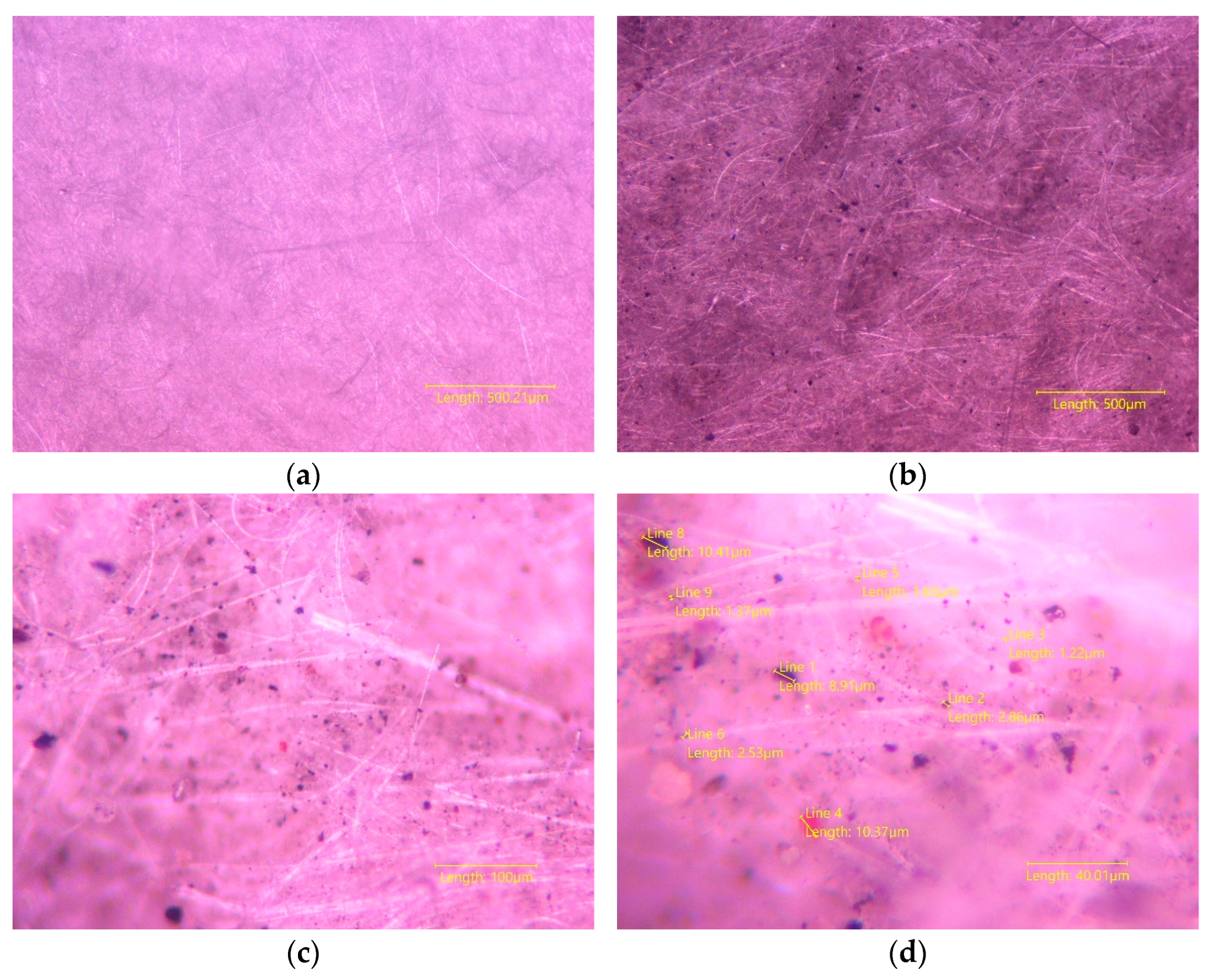
Although a clear difference is observed with the naked eye between a used and a clean filter, by using the optical microscopy methodology a clearer perspective is created on the identified particulate matter deposited on the fiber-glass filter. Figure 1 presents four captures of these types of filters; the first one presents a 2.5× magnification of a clean filter (Figure 1a), and the next three present a filter used in collecting PM2.5 on the balcony of the indoor location, a higher amount of these particulates already being demonstrated [29,30] to be present here compared to the living room of the same location. The microscopic characteristic of the PM2.5 material creates the resemblance of more-or-less dark grey regions on the analyzed filter when the 2.5× magnification (Figure 1b) was used, with individual particles beginning to become more clearly visible when 10× magnification (Figure 1c) was chosen. The last magnification available was 25×, this magnification being able to offer clearer images of some individual particulates (Figure 1d), and this made possible the evaluation from the dimensional point of view. This was done by using the Microscope-VIS type software that, after calibration, offered the possibility to visually confirm the dimensional average of these particulates by measuring lines with automatically calculated values.

Figure 1.
Magnification of a clean fiber-glass filter: (a) 2.5× magnification, and one used for PM2.5 analysis in an indoor location (filter used on a balcony): (b) 2.5× magnification, (c) 10× magnification, and (d) 25× magnification.
Besides measuring the capability of PM, the optical microscopy is a cheaper method that can offer some rough estimates regarding the morphological characterization of these particulates. Moreover, although clear images are relatively hard to obtain, an interesting option, which SEM cannot offer, is to differentiate particulates from one another by their color. This option could be integrated in a process using machine learning, similar to that mentioned in [19,20], thus creating the possibility to rapidly identify the soluble particulates fraction from various samples and, as a result, increase knowledge of the provenience and composition of PM in general for a particular location with a reduced cost.
3.2. SEM and EDX Analysis
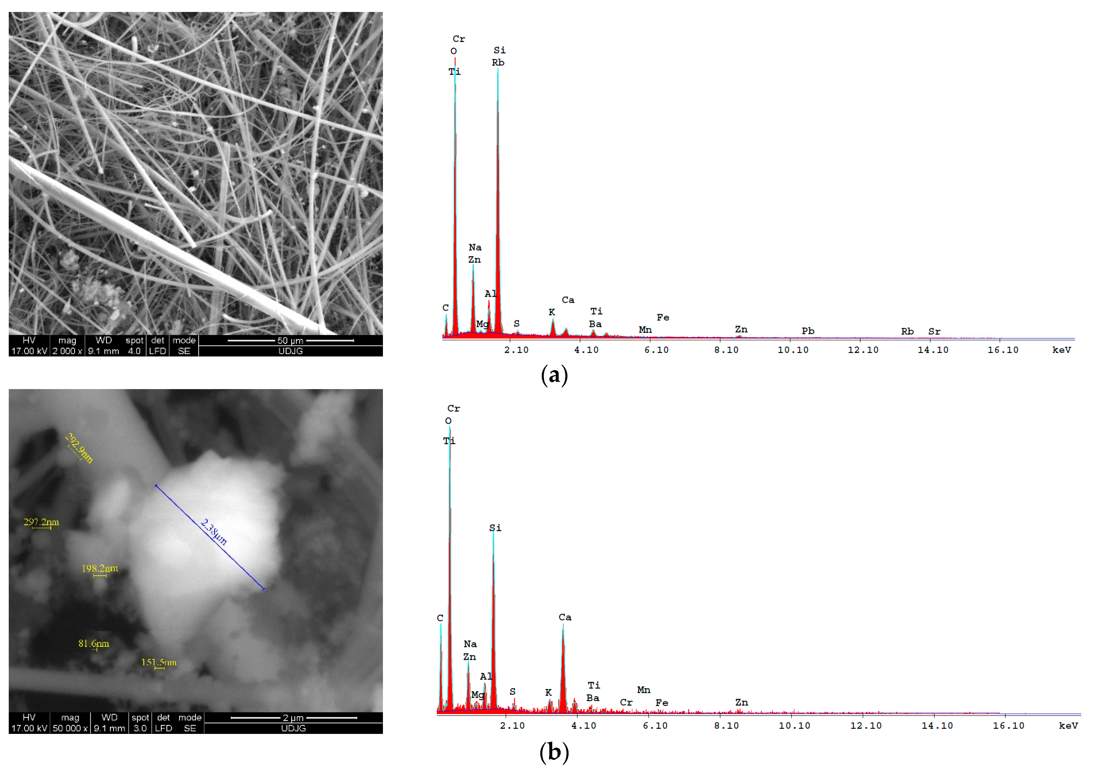
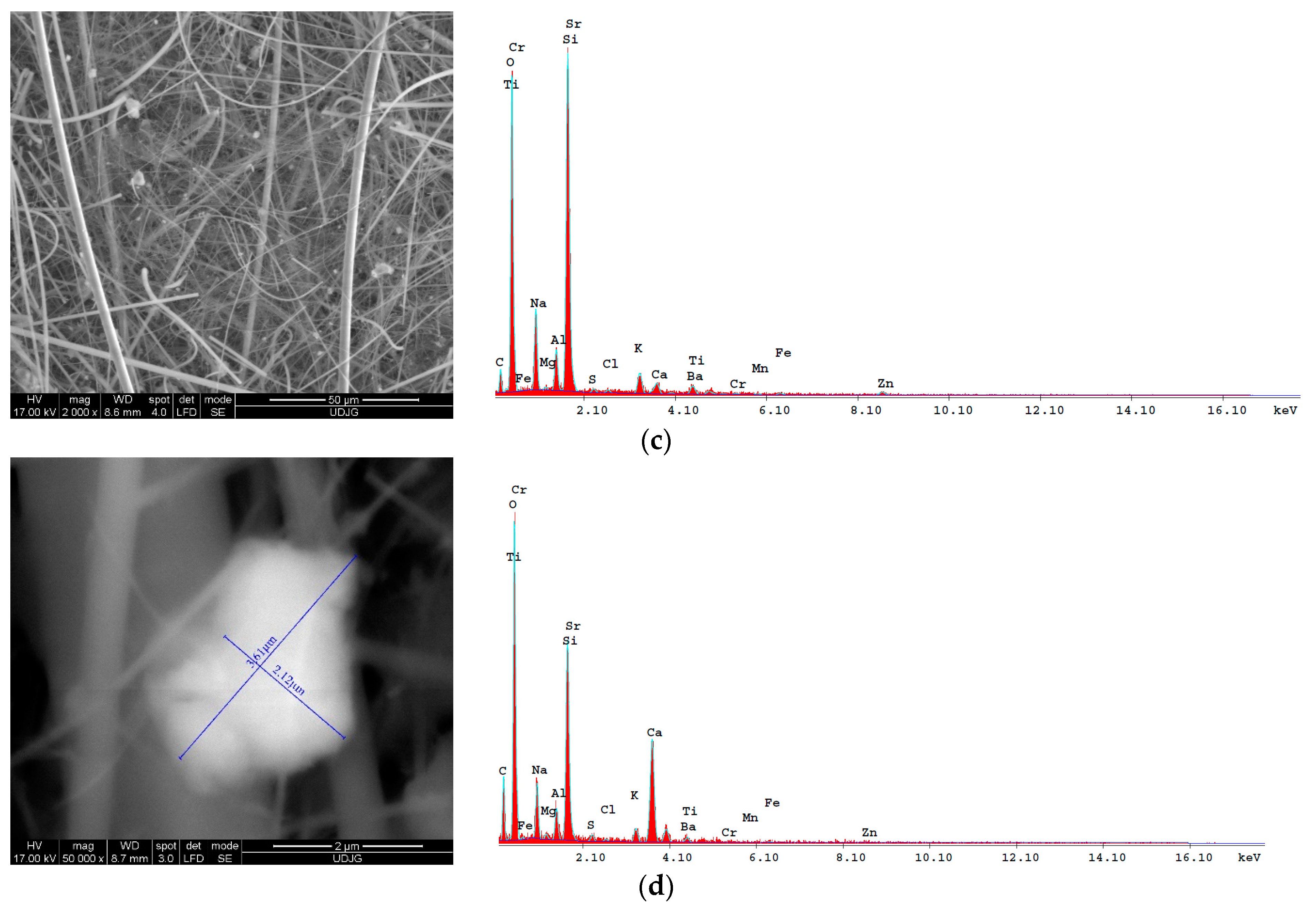
The morphology of PM is more clearly observed by using the SEM technique, as can be seen in Figure 2. Since greater magnifications are permitted using this method, identifying and measuring individual particulate matter are also more easily performed compared to using optical microscopy. Figure 2 presents in the first two coupled images (a and b) two captures of a portion of a filter used in living room measurements, each with EDX results alongside it, which offers information regarding the chemical composition of the scanned area. This scanned area is the entire image for Figure 2a and only the surface of the identified particle for Figure 2b. Similarly, the next two coupled images ((c) and (d)) were obtained for a filter used in balcony measurements. Again, as in the optical microscopy methodology, measuring single particles confirmed their expected median dimension of approximately 2.5 µm (measured as 2.38 µm and 2.12 µm, respectively) in diameter, and similar aspects were observed in both locations used in the measuring campaign. This aspect of small porous aggregates of microcrystals identified in the majority of PM is often encountered in the literature [26,27,31] and is most probably the result of the agglomeration of smaller particulates due to favorable conditions in the atmosphere.
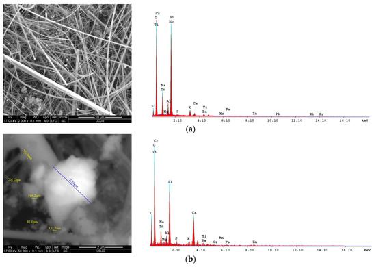
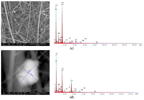
Figure 2.
Scanning electron microscopy images and the afferent EDX results for two different filters used for measuring PM2.5 in an indoor location in different positions: (a,b) morphologies at 2000× and 50,000× magnification, respectively, of a filter used in a living room; (c,d) morphologies at 2000× and 50,000× magnification, respectively, of a filter used on a balcony.
The difference in chemical composition of these aerosols is proof of different activities in the area near the sampling sites and can sometimes demonstrate the existence of abnormal natural or anthropogenic activities with various possible impacts on human health. For this, the EDX technique was used, and the results are somewhat in line with data obtained in [29], since heavy elements such as Ti, Ba, Cr, Mn, Fe, Zn, Rb, and Sr were detected, but, also, other lighter elements such as Na, Ca, Mg, Al, K, S, and C were shown to be present. The advantage of the EDX method, used in this study, in determining the lighter elements could, in some situations, be viewed as minor compared to the possibility of making more precise quantitative determinations of the chemical composition of PM collected on the fiber-glass filters used in the former study. These collective analytical possibilities are important for a complete analysis, which is preferred for an in-depth study of PM and should be available for responsible institutions in sensitive areas such as industrialized and/or highly populous cities.
For the results in Figure 2 regarding the chemical composition, it can be said that a novelty, compared to the former study, is proof of the existence of organic matter. This is envisioned here as a confirmation, not a surprise, and again is a demonstration of the need to use several techniques to obtain a comprehensive view on the nature and composition of these pollutants. In the present case, the chemical analyses proving the presence of carbon, and thus of organic matter in PM composition, were those from Figure 2b,d, since for these two cases the scanning was performed on the surface of the identified particulates, and not on an area including the filter.
3.3. X-ray Diffraction Analysis
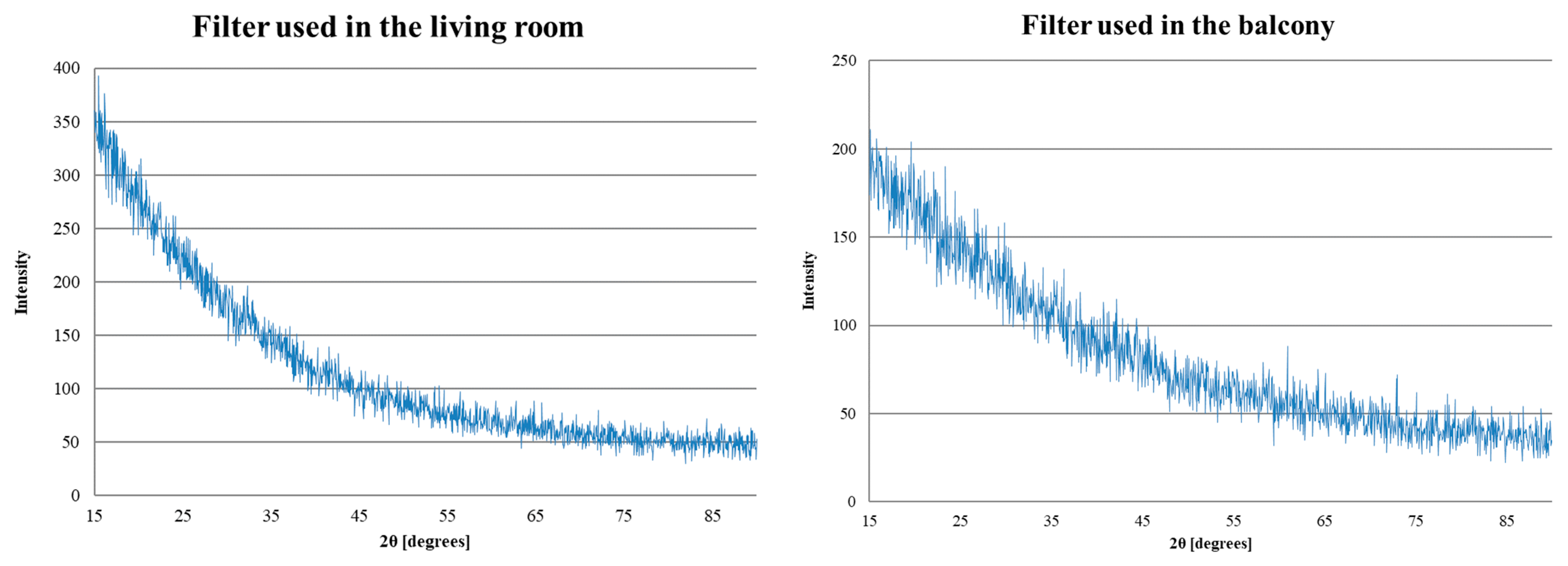
Another option to characterize the PM composition is by using X-ray diffraction. This technique was used in this study, and, although no mineralogical phases were clearly detected, as seen in Figure 3, the differences observed between the two diffractograms can provide some hints regarding the structural composition of PM from the two filters used in the sampling campaign and analyzed here. Thus, the first diffractogram, obtained after scanning the filter used in the living room, presents higher intensity values compared to the second diffractogram, obtained after scanning the filter used on the balcony. This could be an indicator of the variation in PM sizes, regardless of the same-sized fraction that we aimed to collect on the fiber-glass filters, meaning that the mean value of the PM diameters for one filter was slightly smaller than the other. Taking into account that coarse particles are usually known for having higher intensities of mineral components [32], the results presented in Figure 3 could mean that the filter used in the living room contains PM with a larger mean diameter than that of the filter used on the balcony. This result could be counterintuitive, since the air in the living room was repeatedly demonstrated to contain smaller quantities of PM, with two tilted-open windows to promote a lower PM mean diameter compared to the balcony, but the explanation could lie in the fact that different days were chosen for these particular measurements, and the variation in different factors, like weather and fluctuating traffic, could influence the obtained data.
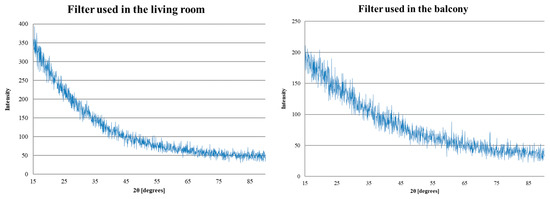
Figure 3.
Diffractograms of two filters used in the PM2.5 measuring campaign.
Clearer results could be obtained in the future by separating the particles from the filter, as in [22], during the sample preparation. In this way, the possibility to obtain individual peaks increases, although the precision could decrease since the adhesion of PM to the filter could be hard to overcome, thus some mineralogical phases could be missed entirely. Nevertheless, this technique allowed the identification of PM chemical composition to some extent, providing further proof of the presence of some heavy metals (Ti, Ba, Cr, Mn, Fe, Zn, Rb, and Sr). Moreover, the percentage of each element was provided by the software used in this study, but the accuracy was not reliable since the calculation was performed only for the mentioned elements without taking into account the lighter elements. Crystallite mean sizes were also calculated using the Debye–Scherrer equation, which ranged from 9635 Å for the living room filter to 19,770 Å for the balcony filter. These later results should not be mistaken with the PM mean diameter from the filters since these particles are most probably polycrystalline. If measured in µm, the values obtained for the crystallite mean size are 0.97 µm for the living room and 1.98 µm for the balcony, meaning that in the case of the living room a larger PM mean size (comparing the marked values in Figure 2b,d) is obtained for polycrystalline particles composed of smaller crystallites trapped on the filter containing almost half the PM2.5 quantity compared to the balcony (16 µg/m3 in the living room and 34 µg/m3 in the balcony [29]).
4. Conclusions
The modern analyzing techniques used in the present paper enabled the obtaining of data to continue the characterization of PM2.5 in an industrialized city, which was started in a previous study. Other purposes were to confirm some already obtained results and to demonstrate the utility of other, cheaper methods to morphologically characterize these pollutants, but also to compare the obtained results to the available literature that present similar results throughout the world. In this respect, especially in terms of morphology, the results from the present paper are in line with other studies, and, to some extent, the chemical composition follows the same trend, especially compared with other industrialized cities, as to some extent already demonstrated in a former study. Moreover, the morphological analysis in this study hints to more in-depth capabilities of optical microscopy that might be realized with machine learning based on the dimensions, shape, and color of particles trapped on filters, making it cheaper and providing some extra possibilities compared to the SEM technique. The PM chemical composition was confirmed using the EDX and XRD methods, although neither were able to present a quantitative aspect as precisely as the method used in the former study (X-ray fluorescence, fit mostly for heavy metals detection). XRD was also used to determine certain aspects regarding the PM dimensions and the crystallite dimensions forming these particles.
The novelty of the present work lies in its detailed analysis of PM2.5 for a city that could be better monitored in terms of the presence of particulate matter. This could be realized with some of the methods presented here and in some other studies, but future methodologies could also be put in place using adaptations of existing methodologies, as already suggested for morphology by using machine learning, for example. This suggestion is also part of the future research of this paper’s authors, along with improved sample preparation for XRD analysis and more extensive sampling campaigns for the same city of Galati, since the surroundings and the meteorological conditions are known to greatly affect the particulate matter concentration and composition.
Author Contributions
Conceptualization, M.B.; methodology, M.B., A.C. and V.B.; formal analysis, M.B., A.C. and V.B.; investigation, M.B., A.C. and V.B.; resources, M.B., A.C. and V.B.; data curation, M.B., A.C. and V.B.; writing—original draft preparation, M.B.; writing—review and editing, M.B.; visualization, M.B., A.C. and V.B.; supervision, M.B. All authors have read and agreed to the published version of the manuscript.
Funding
This research received no external funding.
Institutional Review Board Statement
Not applicable.
Data Availability Statement
Not applicable.
Conflicts of Interest
The authors declare no conflict of interest.
References
- Morawska, L.; Zhu, T.; Liu, N.; Torkmahalleh, M.A.; de Fatima Andrade, M.; Barratt, B.; Broomandi, P.; Buonanno, G.; Belalcazar Ceron, L.C.; Chen, J.; et al. The state of science on severe air pollution episodes: Quantitative and qualitative analysis. Environ. Int. 2021, 156, 106732. [Google Scholar] [CrossRef] [PubMed]
- World Health Organization; Regional Office for Europe. Health Risks of Air Pollution in Europe—HRAPIE Project, Recommendations for Concentration–Response Functions for Cost–Benefit Analysis of Particulate Matter, Ozone and Nitrogen Dioxide. Available online: https://www.euro.who.int/__data/assets/pdf_file/0006/238956/Health_risks_air_pollution_HRAPIE_project.pdf (accessed on 13 July 2021).
- World Health Organization; Regional Office for Europe. Review of Evidence on Health Aspects of Air Pollution-REVIHAAP Project Technical Report. Available online: https://www.euro.who.int/__data/assets/pdf_file/0004/193108/REVIHAAP-Final-technical-report-final-version.pdf (accessed on 13 July 2021).
- Wu, B.; Bai, X.; Liu, W.; Zhu, C.; Hao, Y.; Lin, S.; Luo, L.; Liu, X.; Zhao, S.; Hao, J.; et al. Variation characteristics of final size-segregated PM emissions from ultralow emission coal-fired power plants in China. Environ. Pollut. 2020, 259, 113886. [Google Scholar] [CrossRef]
- Samek, L.; Stegowski, Z.; Furman, L.; Styszko, K.; Szramowiat, K.; Fiedor, J. Quantitative Assessment of PM 2.5 Sources and Their Seasonal Variation in Krakow. Water Air Soil Pollut. 2017, 228, 1–11. [Google Scholar] [CrossRef]
- Khamraev, K.; Cheriyan, D.; ho Choi, J. A review on health risk assessment of PM in the construction industry—Current situation and future directions. Sci. Total Environ. 2021, 758, 143716. [Google Scholar] [CrossRef]
- Tian, G.; Wang, J.; Lu, Z.; Wang, H.; Zhang, W.; Ding, W.; Zhang, F. Indirect effect of PM1 on endothelial cells via inducing the release of respiratory inflammatory cytokines. Toxicol. Vitr. 2019, 57, 203–210. [Google Scholar] [CrossRef] [PubMed]
- Apte, J.S.; Brauer, M.; Cohen, A.J.; Ezzati, M.; Arden Pope, I.C. Ambient PM2.5 Reduces Global and Regional Life Expectancy. Environ. Sci. Technol. Lett. 2018, 5, 546–551. [Google Scholar] [CrossRef]
- Hsieh, N.-H.; Liao, C.-M.; Hsieh, N.-H.; Liao, C.-M. Assessing exposure risk for dust storm events-associated lung function decrement in asthmatics and implications for control. Atmos. Environ. 2013, 68, 256–264. [Google Scholar] [CrossRef]
- Wang, N.; Mengersen, K.; Tong, S.; Kimlin, M.; Zhou, M.; Liu, Y.; Hu, W. County-level variation in the long-term association between PM2.5 and lung cancer mortality in China. Sci. Total Environ. 2020, 738, 140195. [Google Scholar] [CrossRef]
- Feng, L.; Xu, D.; Cheng, Y.; Dong, S.; Guo, C.; Jiang, X.; Zheng, X. Systematic review and meta-analysis of the adverse health effects of ambient PM2.5 and PM10 pollution in the Chinese population. Environ. Res. 2015, 136, 196–204. [Google Scholar] [CrossRef]
- Franzin, B.T.; Guizellini, F.C.; de Babos, D.V.; Hojo, O.; Pastre, I.A.; Marchi, M.R.; Fertonani, F.L.; Oliveira, C.M.R.R. Characterization of atmospheric aerosol (PM10 and PM2.5) from a medium sized city in São Paulo state, Brazil. J. Environ. Sci. 2020, 89, 238–251. [Google Scholar] [CrossRef]
- Bowe, B.; Xie, Y.; Li, T.; Yan, Y.; Xian, H.; Al-Aly, Z. The 2016 global and national burden of diabetes mellitus attributable to PM 2·5 air pollution. Lancet Planet. Health 2018, 2, e301–e312. [Google Scholar] [CrossRef]
- Borup, H.; Kirkeskov, L.; Hanskov, D.J.A.; Brauer, C. Systematic review: Chronic obstructive pulmonary disease and construction workers. Occup. Med. 2017, 67, 199–204. [Google Scholar] [CrossRef]
- Ringen, K.; Dement, J.; Welch, L.; Dong, X.S.; Bingham, E.; Quinn, P.S. Risks of a lifetime in construction. Part II: Chronic occupational diseases. Am. J. Ind. Med. 2014, 57, 1235–1245. [Google Scholar] [CrossRef]
- Pipal, A.S.; Kulshrestha, A.; Taneja, A. Characterization and morphological analysis of airborne PM2.5 and PM10 in Agra located in north central India. Atmos. Environ. 2011, 45, 3621–3630. [Google Scholar] [CrossRef]
- Gao, Y.; Ji, H. Microscopic morphology and seasonal variation of health effect arising from heavy metals in PM2.5 and PM10: One-year measurement in a densely populated area of urban Beijing. Atmos. Res. 2018, 212, 213–226. [Google Scholar] [CrossRef]
- Quijano, M.F.C.; Mateus, V.L.; Saint’Pierre, T.D.; Bott, I.S.; Gioda, A. Exploratory and comparative analysis of the morphology and chemical composition of PM 2.5 from regions with different socioeconomic characteristics. Microchem. J. 2019, 147, 507–515. [Google Scholar] [CrossRef]
- Sarda-Esteve, R.; Baisnee, D.; Guinot, B.; Filippi, D.; O’connor, D.; Sciare, J. A new instrument for realtime detection of aeroallergens and particulate matter by optical microscopy. Rev. Française D’allergologie 2022, 62, 329. [Google Scholar] [CrossRef]
- Koval, S.; Krahenbuhl, G.; Warren, K.; O’Brien, G. Optical microscopy as a new approach for characterising dust particulates in urban environment. J. Environ. Manag. 2018, 223, 196–202. [Google Scholar] [CrossRef]
- Mateus, V.L.; Gioda, A. A candidate framework for PM2.5 source identification in highly industrialized urban-coastal areas. Atmos. Environ. 2017, 164, 147–164. [Google Scholar] [CrossRef]
- González, L.T.; Rodríguez, F.L.; Sánchez-Domínguez, M.; Leyva-Porras, C.; Silva-Vidaurri, L.G.; Acuna-Askar, K.; Kharisov, B.I.; Chiu, J.F.V.; Barbosa, J.M.V. Chemical and morphological characterization of TSP and PM2.5 by SEM-EDS, XPS and XRD collected in the metropolitan area of Monterrey, Mexico. Atmos. Environ. 2016, 143, 249–260. [Google Scholar] [CrossRef]
- Yao, X.; Chan, C.K.; Fang, M.; Cadle, S.; Chan, T.; Mulawa, P.; He, K.; Ye, B. The water-soluble ionic composition of PM2.5 in Shanghai and Beijing, China. Atmos. Environ. 2002, 36, 4223–4234. [Google Scholar] [CrossRef]
- Mateus, V.L.; Monteiro, I.L.G.; Rocha, R.C.C.; Saint’Pierre, T.D.; Gioda, A. Study of the chemical composition of particulate matter from the Rio de Janeiro metropolitan region, Brazil, by inductively coupled plasma-mass spectrometry and optical emission spectrometry. Spectrochim. Acta-Part B At. Spectrosc. 2013, 86, 131–136. [Google Scholar] [CrossRef]
- Mariani, R.L.; de Mello, W.Z. PM2.5-10, PM2.5 and associated water-soluble inorganic species at a coastal urban site in the metropolitan region of Rio de Janeiro. Atmos. Environ. 2007, 41, 2887–2892. [Google Scholar] [CrossRef]
- Alastuey, A.; Querol, X.; Castillo, S.; Escudero, M.; Avila, A.; Cuevas, E.; Torres, C.; Romero, P.-M.; Exposito, F.; García, O.; et al. Characterisation of TSP and PM2.5 at Izaña and Sta. Cruz de Tenerife (Canary Islands, Spain) during a Saharan Dust Episode (July 2002). Atmos. Environ. 2005, 39, 4715–4728. [Google Scholar] [CrossRef]
- Maia, P.D.; Vieira-Filho, M.; Prado, L.F.; da Silva, L.C.M.; Sodré, F.F.; Ribeiro, H.D.S.V.; Ventura, R.S. Assessment of atmospheric particulate matter (PM10) in Central Brazil: Chemical and morphological aspects. Atmos. Pollut. Res. 2022, 13, 101362. [Google Scholar] [CrossRef]
- Bodor, M.; Baltă, Ș.; Lazăr, L.; Pintilie, Ș.; Buruiana, D.L. Studies Regarding the Distribution and Composition of Particulate Matters in the Air of an Industrialized City. In Proceedings of the 16th International Multidisciplinary Scientific GeoConference SGEM, Albena, Bulgaria, 30 June–6 July 2016; Volume 2, pp. 587–591. [Google Scholar]
- Bodor, M. Indoor Concentration and Composition of Fine Particulate Matter (PM2.5) Near an Intense Circulated Road in an Industrial City. Ann. “Dunarea De Jos” Univ. Galati. Fascicle IX Metall. Mater. Sci. 2020, 43, 5–10. [Google Scholar] [CrossRef]
- Bodor, M. A study on indoor particulate matter variation in time based on count and sizes and in relation to meteorological conditions. Sustainability 2021, 13, 8263. [Google Scholar] [CrossRef]
- Siciliano, T.; Giua, R.; Siciliano, M.; di Giulio, S.; Genga, A. The morphology and chemical composition of the urban PM10 near a steel plant in Apulia determined by scanning electron microscopy. Source Apportionment. Atmos. Res. 2021, 251, 105416. [Google Scholar] [CrossRef]
- Choung, S.; Oh, J.; Han, W.S.; Chon, C.M.; Kwon, Y.; Shin, W. Comparison of physicochemical properties between fine (PM2.5) and coarse airborne particles at cold season in Korea. Sci. Total Environ. 2016, 541, 1132–1138. [Google Scholar] [CrossRef] [PubMed]
Publisher’s Note: MDPI stays neutral with regard to jurisdictional claims in published maps and institutional affiliations. |
© 2022 by the authors. Licensee MDPI, Basel, Switzerland. This article is an open access article distributed under the terms and conditions of the Creative Commons Attribution (CC BY) license (https://creativecommons.org/licenses/by/4.0/).